Back to Journals » Advances in Medical Education and Practice » Volume 14
Utility of the Anatomage Virtual Dissection Table in Creating Clinical Anatomy and Radiology Learning Modules
Authors Chaudhry H, Rana S, Bhatti MI, Al-Ansari N, Al Theyab A ![]() , Almutairi T, Kazani B, Almasri M, Sadiq Z
, Almutairi T, Kazani B, Almasri M, Sadiq Z ![]() , Hussein R, Kim D, Chung D, Khalil O, Alroobi H, Aly A, Raoof A
, Hussein R, Kim D, Chung D, Khalil O, Alroobi H, Aly A, Raoof A ![]()
Received 31 May 2023
Accepted for publication 20 August 2023
Published 6 September 2023 Volume 2023:14 Pages 973—981
DOI https://doi.org/10.2147/AMEP.S417831
Checked for plagiarism Yes
Review by Single anonymous peer review
Peer reviewer comments 2
Editor who approved publication: Prof. Dr. Balakrishnan Nair
Hamza Chaudhry,1 Shehroz Rana,1 Mohammad Ibraheem Bhatti,1 Nojoud Al-Ansari,1 Ahmad Al Theyab,1 Turki Almutairi,1 Bahram Kazani,2 Muna Almasri,2 Zuhair Sadiq,2 Rawan Hussein,2 Daniel Kim,2 Dabin Chung,2 Omar Khalil,2 Hasan Alroobi,2 Ahmed Aly,2 Ameed Raoof3
1Class 2024, Weill Cornell Medicine-Qatar, Doha, Qatar; 2Class 2023, Weill Cornell Medicine-Qatar, Doha, Qatar; 3Department of Medical Education, Weill Cornell Medicine-Qatar, Doha, Qatar
Correspondence: Ameed Raoof, Email [email protected]
Purpose: During the COVID-19 pandemic, teaching has required online-learning modalities to facilitate easily accessible yet high-quality education. However, since the nature of anatomy requires hands-on experience in laboratories with cadavers, teaching anatomy in an online setting has proven especially difficult. This matter may be resolved with the Anatomage Table, an advanced anatomy visualization tool, which several studies have suggested can augment learning experiences for students in anatomy courses. Our objective was to provide accessible online modules, through utilization of the Anatomage Table, for medical students to facilitate their learning and enhance online learning experience.
Materials and Methods: Ten modules were designed, consisting of a presentation, a pre- and post-self-assessment, as well as anatomical images and radiographs taken from Anatomage Table. The modules were based on a single organ system, and a clinical case pertaining to that organ system was presented. Weill Cornell Medicine-Qatar (WCM-Q) second-year medical students contributed 102 responses in total throughout the 10 modules. Using a paired t-test, the study compared the students’ pre- and post-assessment scores to determine how beneficial the modules were.
Results: A significant difference in scores on the pre- and post-assessments was found for all 10 modules using a paired t-test. At the end of the modules, the students completed a feedback survey to assess the quality and convenience. Most of the students agreed or strongly agreed that the modules were beneficial to their online anatomy learning and wanted to see similar anatomical modules in the future.
Conclusion: The Anatomage Table is an innovative virtual resource that can significantly contribute to a more engaging and productive experience for medical students.
Keywords: medical education, Anatomage table, anatomy, COVID-19
Introduction
The COVID-19 pandemic resulted in an abrupt transformation that led to the widespread utilization of all-online learning sessions.1 Traditional teaching methods, which included lecture halls, seminar rooms, and in-person didactics, became more susceptible to the risks of infectious transmission. This necessitated the utilization of technology to help provide education safely and conveniently and created a paradigm that emphasizes virtual learning. The forced lockdown anatomy education during the COVID-19 epidemic access to effective educational tools is essential for virtual teaching, where new concepts can be introduced to foster critical thinking for students. Traditional methods in teaching anatomy are limited in accommodating various learning styles.2 Despite this, anatomical education encompasses many approaches to learning that include dissection, pro-section, medical imaging, computer software, and models.3
The Anatomage Virtual Dissection Table is a computerized educational tool that enables users to explore structures in the human body by reconstructing CT scans and sectional images.4 This allows students to look across various planes and manipulate cross-sections to identify structures and correlate their anatomical positions. Thus, students can gain an enhanced and interactive learning experience as Anatomage enables them to virtually dissect cadavers with realistic functional features and details.5 Advantages include the attention to detail, ability to view any section, and magnification.6 Drawbacks of the Anatomage table include licensing and cost.7
The Anatomage table was evaluated as a learning modality at Case Western University in 2019. The Case Western Reserve University School of Medicine is shifting to a new facility that will not have “an in-house dissection laboratory” and will focus on “virtual anatomy learning technologies” including the Anatomage table.8 Early preliminary data from their institution that the Anatomage table is an effective teaching modality.8 Furthermore, studies done regarding the Anatomage table at other universities have found that it has allowed medical students to have a greater understanding of spatial anatomy while also giving students a higher degree of flexibility by allowing them to study independently.9,10 A cross-sectional national survey involving 2721 medical students from the UK on perception of online teaching during the pandemic found that the greatest perceived benefit of online instruction was flexibility.11
Our study expands current literature exploring online learning methods, particularly using the Anatomage Table, by developing and testing virtual modules tailored to the anatomy curriculum. This study aims to create learning modules using Anatomage and examine their effectiveness in teaching anatomy and medical imaging.
Materials and Methods
Designing the Online Modules
The Anatomage Table provided a diverse selection of clinical cases accompanied with relevant images. Cases were individually chosen by student researchers as part of their summer Advanced Biomedical Scientific Research (ABSR) program mentored by anatomy faculty. Microsoft PowerPoint was used to adapt each case into modules that incorporate anatomical images and radiographs into real-world clinical contexts. The difficulty of the modules was geared towards medical students with a background in clinical anatomy; the anticipated duration for module completion was approximately 30 minutes. The modules were published on Canvas; second-year medical students at Weill Cornell Medicine-Qatar were recruited. The students had completed their course in anatomy the previous year and thus had a baseline knowledge in anatomy. Each module began with a set of learning objectives and an introduction to the clinical case that relates heavily to a particular anatomic area (ie, vascular supply of the lower limbs relates to case VI. “Atherosclerosis in the Lower Limb”). Medical jargon was clarified to reduce confounding effects on module performance. This was followed by a discussion of the relevant anatomic area, beginning with gross anatomic identification of major organs and their arrangement within the body, progressing to the identification of the various features of individual organs. To assist in student understanding of spatial relationships between organs, animated diagrams, labeled Anatomage cadaver section images, and videos sourced from the built-in Visible Body Human Anatomy Atlas App were used. Ten online modules were individually designed for each of the following clinical topics:
- Crohn’s Disease
- Abdominal Aortic Aneurysm
- Abdominal Ectopic Pregnancy
- Vertebral Fractures
- Occluded Iliac Arteries
- Atherosclerosis in the Lower Limb
- Potential Ascending Aortic Dissection
- Hypertrophic Cardiomyopathy
- Normal Heart Anatomy
- Appendicitis
In addition to diagrams and videos, a wide selection of radiographic images, primarily CT, MRI, and ultrasound scans, were included to contextualize learning for the clinical setting. The radiographs were labeled to enable students to identify organs more efficiently and to enhance students’ spatial reasoning of various orientations and sections. Figures 1 and 2 from the abdominal ectopic pregnancy module are included below.
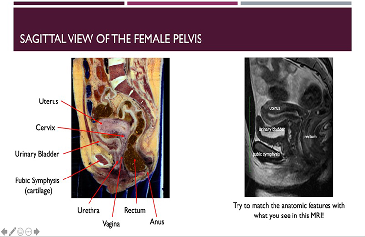 |
Figure 1 Examples of labeled Anatomage cadaver sections (sagittal and coronal), juxtaposed with a radiographic image to help students with orientation and identification of major organs and anatomical landmarks. |
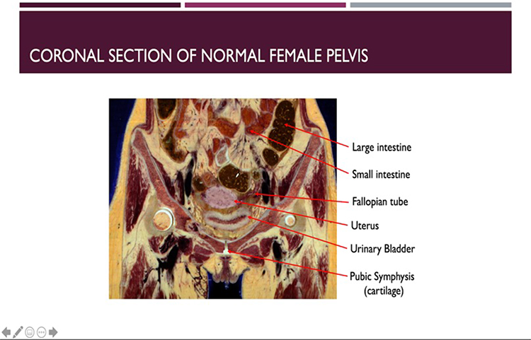 |
Figure 2 An additional example of labeled Anatomage cadaver sections (coronal). |
After the relevant anatomy and radiological imaging was depicted, the pathology of the disease was explained. In addition, images of diseased organs were contrasted with healthy organs. Each clinical case was designed to emphasize the impact of the condition on the intricate anatomy and structure of the involved organs. Relevant patient diagnostic images were utilized and thoroughly explained at a beginner’s level, including CT, MRI, and angiograms. Furthermore, several pathology photos were integrated across the module subjects to demonstrate presenting signs and symptoms of diseases. Patient symptoms, diagnostic images, and exam findings were thoroughly illustrated in the modules, along with differential diagnosis to provide a comprehensive picture of patient presentations and their potential causes. Several simple recall questions were scattered throughout each module to reinforce the new information introduced to the student.
Student Participation
Our target population is students who have obtained the same level of anatomy education. At Weill Cornell Medicine-Qatar, a course in clinical anatomy is taught during the first semester of the first year of medical school. Rising second-year medical students at Weill Cornell Medicine-Qatar were recruited to utilize the modules of their choice. The student’s completed the modules in the summer between their first and second year. Twelve students participated in the study. Seven of the twelve completed all 10 modules, while the other five completed a variable number. In total, each module had 10 participants except for the module on Abdominal Ectopic Pregnancy which had 12. Figure 3 represents the flowchart of our study. The modules were published and accessed on Canvas. Students were asked to take a pre- and post-module quiz in which scores were recorded for research purposes. Finally, students were requested to fill out a feedback survey regarding their experience. This study was approved by the Institutional Review Board, WCM-Q (IRB-20-00009).
 |
Figure 3 Flowchart of the study design. |
Statistical Analysis
For each module, the students took a pre-module assessment in which the correct answers were not shown after submission. Upon completing the module, the students completed the post-module assessment, which consisted of the same questions as the pre-module assessment. The mean, median, and standard deviation were computed for the pre- and post-assessment for each module. The pre- and post-module assessment scores were compared using a paired t-test.
Feedback Survey
At the end of each module, the students filled out an anonymous feedback survey that was prepared using Qualtrics Survey Software. It consisted of five questions, the first four using the Likert scale, and the last one using an open-ended format. The survey is displayed in Table 1. The data was examined by tabulating the percentages of student responses to questions 1 to 4. The percentages for each survey response these four questions across all the modules were determined and represented using bar charts. Responses to the fifth survey question were used as detailed feedback and suggestions for improvement.
 |
Table 1 Feedback Survey Taken at the End of Each Module by the Students |
Results
In total, there were 102 quiz responses from the second-year medical students. Using the paired data of pre- and post-module quiz scores, each module’s efficacy was assessed. The data illustrated that 91.2% (93/102) of responses demonstrated a positive increase in grade from the pre- to post-module quiz. 4.9% (5/102) showed no change in grade and 3.9% (4/102) exhibited a decrease in score.
The average score on pre- and post-module quizzes along with standard deviations and medians were computed (Table 2). Furthermore, these results are depicted in Figures 4 and 5.
 |
Table 2 Descriptive Statistics Showing the Mean, SD, Median for the Pre-Module and Post-Module Assessments for Each Module |
 |
Figure 4 Bar graph illustrating the mean scores on the pre- and post-module assessment with the standard deviations for each module. |
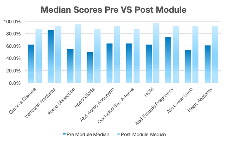 |
Figure 5 Bar graph illustrating the median scores on the pre- and post-module assessments for every module. |
For each module, we compared the grade on the pre-module quiz to the grade on the post-quiz using a paired t-test (if data was deemed normal using the Shapiro Wilks test) or the Wilcoxon signed rank test (if data was deemed not normal using the Shapiro Wilks test). A p-value was computed to determine if there was a significant difference in the scores on the pre-module quiz compared to the post-module quiz. For all ten of the modules, there was a statistically significant increase in score in the post-module assessment compared to the pre-module assessment (p≤ 0.05). Moreover, the difference in means was also calculated. These results are demonstrated in Table 3.
 |
Table 3 Advanced Statistics Showing the Difference in Means and p-values for Pre-Module vs Post-Module Assessment Scores |
Data were also collected from the feedback survey that was done after the students completed each module. Forty-one percent of the students “agreed” and 50% “strongly agreed” that the modules were easy to access and navigate. The enthusiasm of the students was further reflected in the comments as one student wrote, “very simple and easy to navigate, easy to understand!” and another commented, “loved the casualness of the module! It really helps make learning feel like a conversation rather than a lesson plan”.
Additionally, the majority of the students (90%) conveyed that they had a positive experience with the modules, particularly in helping them grasp the fundamental principles of anatomy which was one of our core objectives that was thoroughly considered during the modules’ design phase.
Moreover, the majority agreed that their understanding of anatomical clinical correlations had significantly increased after completing the modules. Students expressed their fascination with the Anatomage table images, which they mentioned allowed them to gain a more immersive experience and easily distinguish between normal vs abnormal anatomical structures. The question was posed, “Would you like to see similar modules in the future”, to which 78.4% agreed or strongly agreed that they would. One volunteer commented, “I was very happy using this as supplementary material to my learning”, and another remarked, “These modules should be a regular part of anatomy training”.
The students’ feedback also allowed us to note areas that needed improvement. Despite efforts to avoid or clarify jargon and uncommon medical terminology, a couple comments pointed out the use of undefined medical jargon or indicated preference for simpler terminology. Students also preferred having important information highlighted and having relevant labels clearly pointed out.
Discussion
Anatomical knowledge is an essential component in medical learning as well as in the clinical setting as proper understanding of normal anatomy allows physicians to accurately identify abnormalities. Cadaveric training has long served as the principal method that anatomy was taught in medical settings. However, with the advent of the COVID pandemic, the incorporation of interactive technology within the medical curriculum has increased. Moreover, due to a multitude of reasons including costs, accessibility, and time consumption, access to cadavers can be limited.12
A key aspect in the utilization of the online modules was to assess learning in a virtual environment. The COVID pandemic greatly impacted students all across the world and required educational institutions to adapt to the new circumstances so students could still receive quality learning. One study assessing COVID’s impact at five allopathic medical schools found that 54.7% of clinical-level students felt they were receiving less training.13 To help combat such feelings among WCMQ students, virtual modules fulfilled the role of an anatomical learning supplement for the medical students, and the results showed substantial promise for the incorporation of future online teaching tools.
A statistically significant difference was appreciated for all the modules, which conveys that the modules were effective in their tasks of teaching the various anatomical concepts. In addition, the vast majority of the students “agreed” or “strongly agreed” that the modules proved beneficial to their anatomical learning and clinical correlations. Furthermore, the feedback was overwhelmingly positive regarding navigating within the module and the smoothness of the overall modality. Finally, students agreed that they “would like to see similar anatomical modules in the future”. These responses support the idea of integrating the Anatomage table and similar virtual software within the anatomical curriculum.
Learning anatomy is often made difficult by the lack of access to real models/cadavers along with guided instruction on the details of the anatomy.14 Lack of access can be due to several factors: students typically need to be physically present in order to conduct dissections or to analyze live models, COVID-19 has established limitations to the number of students that can view or conduct dissections at one time, live models and dissections are often difficult to label and/color code to make distinct structures more visible, and identifying pathology catered to specific clinical cases on models and cadavers is unrealistic due to limited resources.15 The Anatomage table circumvents many of these constraints as it can be used to make presentations of clinical cases with relevant images and radiographs of the pathology along with normal structures, where the structures can be color-coded and labeled with ease.16 These can then be administered online for anyone to access and receive immediate assessment and feedback. Objective and subjective measures, through positive survey results and improved assessment scores, respectively, indicate a beneficial impact on students during the pandemic and beyond.
Our study also highlights the potential to help students in areas other than anatomy test scores. The decreased reliance on scheduled, in-person cadaveric sessions bring with it the possibility of making student learning more unstructured, potentially leading to studying for the short period of time before examinations, or “cramming”. This may be further exacerbated by a lack of immediate feedback on students’ understanding before an examination, which may cause some students to overestimate their level of knowledge. Online modules and assessments combat this possibility as it allows students to gauge their conceptual understanding, where they can adjust their individual study accordingly. Furthermore, online platforms may help the students gain new skills not only in using technology for learning but also in developing improved independent study habits, guided by module content and feedback from assessment scores.
The problem of delivering traditional anatomical medical education exists from the reliance on human donor resources, involving laboratory access, which clearly became disrupted in the wake of the pandemic.17 We compared our findings to those produced by other accredited medical institutions around the world to understand their challenges and approach to them. A study conducted in Australia and New Zealand18 analyzed the effect of online learning for anatomy from a social constructivist perspective. The study found increased interest in forming a viable assessment option that could be accessed remotely to be able to continue the course “without compromising academic integrity”.18 Another study done in Australia compared blended learning to traditional in-person teaching in gross anatomy courses. Their results showed that online learning modules made a significant contribution to students’ learning.19 Additionally, a study involving first year medical students in Saudi Arabia found that students had a significantly more positive attitude and experience when the teaching included the Anatomage table with plastinates compared to either entity alone.20 Therefore, generally, the Anatomage table is an effective online resource that is valuable to medical students in their anatomical courses.
Finally, the study and individual modules had aspects that could be improved. The sample size was relatively small as volunteers were gathered online, where communication was limited to online means without physical interaction due to the COVID-19 situation. Moreover, clinical case studies corresponding to anatomy were incorporated in the modules; thus, volunteers may have had difficulty comprehending the clinical cases rather than not understanding the actual concepts. Another drawback is that every module had a different style as each was made by a different student, so volunteers doing multiple modules may have found different styles hard to follow. Post-module quiz questions also differed in style, so performance data may have been altered due to question design.
Conclusion
The objective of this study was to evaluate the efficacy of anatomy and radiological imaging on medical education via online modules that were created through the Anatomage Virtual Dissection Table. We created and tested these virtual modules focused on anatomy using the Anatomage clinical cases in correlation with anatomical and radiographic images. Overall, our findings were supportive of the utilization of advanced technological instruments and virtual tools such as the Anatomage table in teaching anatomy to medical students. To further analyze the validity of such learning methods and explore the impact of virtual tools such as the Anatomage on anatomical education, future studies with larger sample sizes are needed that have a longitudinal component and follow students’ progression through their anatomy courses.
Disclosure
The authors report no conflicts of interest in this work.
References
1. Hilburg R, Patel N, Ambruso S, et al. Medical education during the coronavirus disease 2019 pandemic: learning from a distance. Adv Chronic Kidney Dis. 2020;27(5):412–417. doi:10.1053/j.ackd.2020.05.017
2. Afsharpour S, Gonsalves A, Hosek R, Partin E. Analysis of immediate student outcomes following a change in gross anatomy laboratory teaching methodology. J Chiropr Educ. 2018;32(2):98–106. doi:10.7899/JCE-17-7
3. O’Byrne PJ, Patry A, Carnegie JA. The development of interactive online learning tools for the study of anatomy. Med Teach. 2008;30(8):e260–e271. doi:10.1080/01421590802232818
4. Petriceks AH, Peterson AS, Angeles M, et al. Photogrammetry of human specimens: an innovation in anatomy education. J Med Educ Curric Dev. 2018;5:1–10. doi:10.1177/2382120518799356
5. Chan I, D’Eon M, Haggag H, et al. The effectiveness of learning anatomy and medical imaging using the anatomage table compared with prosections.
6. Brucoli M, Boffano P, Pezzana A, et al. The potentialities of the Anatomage Table for head and neck pathology: medical education and informed consent. Oral Maxillofac Surg. 2020;24(2):229–234. doi:10.1007/s10006-019-00821-x
7. Bartoletti-Stella A, Gatta V, Mariani GA, et al. Three-dimensional virtual anatomy as a new approach for medical student’s Learning. Int J Environ Res Public Health. 2021;18(24):13247–13260. doi:10.3390/ijerph182413247
8. Baratz G, Wilson-Delfosse AL, Singelyn BM, et al. Evaluating the Anatomage Table Compared to Cadaveric Dissection as a learning modality for gross anatomy. Med Sci Educ. 2019;29(2):499–506. doi:10.1007/s40670-019-00719-z
9. Custer TM, Michael K. “The utilization of the anatomage virtual dissection table in the education of imaging science students”. J Tomogr Simul. 2015;1:1–5.
10. Bharati AS, Kumari NSK, Rani VS. A study on student perception of virtual dissection table (Anatomage) at GSL Medical College, Rajahmundry. Acad Anatom Int. 2018;4(2):28–31. doi:10.21276/aanat.2018.4.2.8
11. Dost S, Hossain A, Shehab M, et al. Perceptions of medical students towards online teaching during the COVID-19 pandemic: a national cross-sectional survey of 2721 UK medical students. BMJ open. 2020;10(11):e042378. doi:10.1136/bmjopen-2020-042378
12. Alasmari WA. Medical students’ feedback of applying the virtual dissection table (Anatomage) in learning anatomy: a cross-sectional descriptive study. Adv Med Educ Pract. 2021;12:1303–1307. doi:10.2147/AMEP.S324520
13. Chakladar J, Diomino A, Li WT, et al. Medical student’s perception of the COVID-19 pandemic effect on their education and well-being: a cross-sectional survey in the United States. BMC Med Educ. 2020;22(1):149–159. doi:10.1186/s12909-022-03197-x
14. Dissabandara LO, Nirthanan SN, Khoo TK, et al. Role of cadaveric dissections in modern medical curricula: a study on student perceptions. Anat Cell Biol. 2015;48(3):205–212. doi:10.5115/acb.2015.48.3.205
15. Mulu A, Tegabu D. Medical students’ attitudinal changes towards cadaver dissection: a longitudinal study. Ethiop J Health Sci. 2012;22(1):51–58.
16. Fyfe S, Fyfe G, Dye D, et al. The Anatomage table: differences in student ratings between initial implementation and established use. Focus Health Profess Educ. 2018;19(2):41–52. doi:10.11157/fohpe.v19i2.215
17. Boscolo-Berto R, Tortorella C, Porzionato A, et al. The additional role of virtual to traditional dissection in teaching anatomy: a randomised controlled trial. Surg Radiol Anat. 2020;43(4):469–479. doi:10.1007/s00276-020-02551-2
18. Pather N, Blyth P, Chapman JA, et al. Forced disruption of anatomy education in Australia and New Zealand: an acute response to the Covid-19 pandemic. Anat Sci Educ. 2020;13(3):284–300. doi:10.1002/ase.1968
19. Green RA, Whitburn LY, Zacharias A, et al. The relationship between student engagement with online content and achievement in a blended learning anatomy course. Anat Sci Educ. 2018;11(5):471–477. doi:10.1002/ase.1761
20. Bin Abdulrahman KA, Jumaa MI, Hanafy SM, et al. Students’ perceptions and attitudes after exposure to three different instructional strategies in applied anatomy. Adv Med Educ Pract. 2021;12:607–612. doi:10.2147/AMEP.S310147
 © 2023 The Author(s). This work is published and licensed by Dove Medical Press Limited. The
full terms of this license are available at https://www.dovepress.com/terms
and incorporate the Creative Commons Attribution
- Non Commercial (unported, 3.0) License.
By accessing the work you hereby accept the Terms. Non-commercial uses of the work are permitted
without any further permission from Dove Medical Press Limited, provided the work is properly
attributed. For permission for commercial use of this work, please see paragraphs 4.2 and 5 of our Terms.
© 2023 The Author(s). This work is published and licensed by Dove Medical Press Limited. The
full terms of this license are available at https://www.dovepress.com/terms
and incorporate the Creative Commons Attribution
- Non Commercial (unported, 3.0) License.
By accessing the work you hereby accept the Terms. Non-commercial uses of the work are permitted
without any further permission from Dove Medical Press Limited, provided the work is properly
attributed. For permission for commercial use of this work, please see paragraphs 4.2 and 5 of our Terms.