Back to Journals » Journal of Pain Research » Volume 16
The Value of Percutaneous Ultrasound-Guided Subacromial Bursography for Rotator Cuff Tear in Elderly Patients with Shoulder Pain
Authors Liu B ![]() , Ge D, Shan Y, Li Y, Lv J, Gan S
, Ge D, Shan Y, Li Y, Lv J, Gan S
Received 9 October 2022
Accepted for publication 3 April 2023
Published 2 June 2023 Volume 2023:16 Pages 1895—1906
DOI https://doi.org/10.2147/JPR.S392833
Checked for plagiarism Yes
Review by Single anonymous peer review
Peer reviewer comments 2
Editor who approved publication: Professor Michael Überall
Bili Liu, Dan Ge, Yue Shan, Yanping Li, Juan Lv, Shuzhi Gan
Department of Ultrasound, Hangzhou TCM Hospital Affiliated to Zhejiang Chinese Medical University, Hangzhou, Zhejiang Province, 310007, People’s Republic of China
Correspondence: Bili Liu, Department of Ultrasound, Hangzhou TCM Hospital Affiliated to Zhejiang Chinese Medical University, No. 453 Stadium Road, Xihu District, Hangzhou, Zhejiang Province, 310007, People’s Republic of China, Tel +86-057185827841, Email [email protected]
Objective: Based on the analysis of the images of acromial slide, we explored the application of percutaneous ultrasound-guided subacromial bursography (PUSB) on rotator cuff tear (RCT) in diagnosing elderly patients with shoulder pain.
Methods: Eighty-five patients who were clinically diagnosed with RCT and underwent PUSB examination in the department of ultrasound in our hospital were enrolled as the subjects. Independent samples t-test was used to analyze the general characteristics. Based on the gold standard of shoulder arthroscopy, the diagnostic efficacy of ultrasound, magnetic resonance imaging (MRI), and PUSB was evaluated. The sensitivity, specificity, positive and negative predictive values, and accuracy were calculated as well. The consistency of these techniques with shoulder arthroscopy in diagnosing the RCT stage was additionally compared using Kappa test.
Results: In patients with large full-thickness RCT, the 100% detection rate was achieved by the techniques of ultrasound, MRI, and PUSB. For patients with small full-thickness RCT, the detection rate of PUSB (100%) was evidently higher than those of ultrasound and MRI. Similar results were shown in the detection rates of patients with bursal-side partial-thickness RCT (90.5%) and articular-side partial-thickness RCT (86.9%). More importantly, the sensitivity, specificity, and accuracy of PUSB in patients with both full-thickness RCT and partial-thickness RCT were significantly better than those of ultrasound and MRI.
Conclusion: PUSB has a better efficacy in the detection of RCT than ultrasound and MRI, showing its feasibility as an important imaging method to evaluate the degree of RCT.
Keywords: rotator cuff tear, percutaneous ultrasound-guided subacromial bursography, shoulder pain, acromial slide cystography, diagnosis
Introduction
Currently, shoulder pain ranks third among the most common symptoms for the diagnosis of orthopedic patients, with rotator cuff tear (RCT) as the most prevalent cause.1 Rotator cuff is a cup-like structure consisting of subscapularis, supraspinatus, and teres minor which help lift and rotate the arm, covering the head of the humerus and keeping the humerus in place.2 RCT has been identified as one of the major causes of shoulder pain and dysfunction, which seriously affects the quality of life of patients.3
RCT is a common shoulder joint disease in the elderly as well, affecting approximately 30% of asymptomatic patients over 60 years old and 65% of those over 70 years old. Supraspinatus tendon tears have been identified to be the most common tendon tear in the shoulder region.4 As a part of the four rotator cuff tendons acting to stabilize the shoulder, the supraspinatus tendon is the most frequently injured tendon of the rotator cuff. RCT is divided into two groups, namely, partial-thickness RCT and full-thickness RCT. If improperly handled or left unrepaired, partial-thickness RCT can often develop into full-thickness RCT. Surgery is the best therapeutic option for partial-thickness RCT.5 Herein, it is of high clinical value to determine the classification of RCT in order to select the corresponding treatment plans and surgical methods. With the continuous development of ultrasound technology, thanks to its non-invasive, cheap, dynamic, and other advantages, high-frequency ultrasound has been applied for the diagnosis of the location and extent of RCT. With the obvious increase in the application frequency of ultrasound, the images of ultrasound examination will become clearer, and the moving tendon can be detected dynamically in real time. In the meantime, RCT can be observed in real-time dynamic ultrasound, and its degree can be further evaluated based on the morphological changes of the tendon and the echo condition at the laceration.6 Moreover, the application of dynamic ultrasound makes it possible to push the bursal (or articular) effusion inside the tendon injury for the better visualization results (ie, effusion as a “natural” contrast agent).7 At present, percutaneous contrast-enhanced ultrasonography (CEUS) has been recognized as a reliable modality widely applied in clinical practice; however, it is rarely adopted in diseases associated with musculoskeletal systems. In light of this, our current study intends to examine elderly patients with RCT using the technique of percutaneous ultrasound-guided subacromial bursography (PUSB), along with the purposes to discover and discuss the diagnostic value of PUSB in RCT.
Methods
Ethics Statement
The ethics committee of Hangzhou TCM Hospital Affiliated to Zhejiang Chinese Medical University has reviewed and approved our present study (endorse no. 2020KY120) and all participants enrolled have also provided written informed consent. The current research was carried out in strict accordance with the Helsinki declaration.
Subjects
A total of 85 patients with clinical diagnosis of RCT who were admitted to and underwent PUSB examination in our hospital between 2018 June and 2021 October were enrolled as the candidates, including 36 males and 49 females, aging from 60 to 77 years old (average: 67.4 ± 4.0).
Of all these enrolled participants, 58 have been diagnosed as RCT on the right shoulder and the remaining 27 were confirmed to have RCT on the left shoulder. The disease duration varied from 0.13 to 42 months (average: 10.2 ± 9.7 months).
The inclusion criteria were as follows: (1) patients were ≥60 years old; (2) those with shoulder joint pain, abnormal physical signs, or impaired movement based on physical examination; (3) those agreeing to receive shoulder arthroscopy and showing willingness to follow-up.
The exclusion criteria were as follows: (1) patients with a previous history of shoulder joint surgery; (2) those with low blood coagulation and with severe hepatic and renal insufficiency; (3) those who were unconscious and had difficulty in cooperation with the examination; (4) those who were allergic to ropivacaine and ultrasound contrast agent.
Study Design
The ultrasound apparatus applied in our study was the Logiq E9 Ultrasound Machine (GE Healthcare, Chicago, Illinois, USA) with a linear phased array transducer. SonoVue (specification: 59 mg, Bracco, Milan, Italy) was used as the ultrasonic contrast agent (main gradient: six fluorinated sulfur (SF6) microbubble) and was added with 5 mL 0.9% saline solution prior to use. The mechanical index (MI) of contrast imaging in our study was set to <0.05.
The general data of these enrolled patients were recorded, including medical history, gender, age, disease duration, presence/absence of diabetes mellitus, hyperlipidemia, and history of trauma.
RCT Classification
All classification criteria for RCT were based on a previous study.8 Based on the degree of tendon tear, RCT was divided into two main categories: full-thickness RCT and partial-thickness RCT. Then, based on the avulsion sites, the partial-thickness RCT was additionally subdivided into the bursal-side partial-thickness RCT (bp), articular-side partial-thickness RCT (ap), and intratendinous partial-thickness RCT (Figure 1A). The full-thickness RCT includes large full-thickness RCT and small full-thickness RCT. In accordance with the arthroscopy, the avulsion with width >1 cm was defined as the large full-thickness RCT, and the avulsion with the width ≤1 cm was regarded as the small full-thickness RCT in our study.
 |
Figure 1 Visualization of RCT subtype and PUSB examination scheme. (A) Representative figure of RCT subtype. (B) The symbolized operational scheme of PUSB examination. Abbreviations: ap, articular-side RCT; bp, bursal-side RCT; FTT, full-thickness tear; AR, acromion; HH, humeral head; SUP, supraspinatus; PUSB, percutaneous ultrasound-guided subacromial bursography. Note: *Represents subacromial-subdeltoid bursa. |
MRI Protocols
The MRI was performed using a 1.5T MR scanner (Signa HDx 1.5T, GE Healthcare), using an eight-channel array coil. All subjects should lie on their back, head forward, in a lateral oblique position, and ensure their shoulder joint to be scanned close to examination bed. The scans ranged from the major humerus tubercle to the scapular body, including the furthest muscle fibers of infraspinatus and supraspinatus. The MRI protocol included T1-weighted imaging (including axial and coronal cuts), T2-weighted imaging with fat suppression (including axial and sagittal cuts), and coronal Short Tau Inversion Recovery (STIR) imaging with fat suppression. The repetition time (TR)/echo time (TE) of magnetic resonance sequences at the 1.5 T GE MR system were as follows: (1) STIR (TR/TE=2600/42 ms), (2) spin-echo T1-weighted imaging (SE-TIWL, TE=680/14ms), (3) fast spin echo T2 (TSE-T2WI, TR/TE=3000/68ms). The scanning parameters were 5 mm thickness, 1mm interslice interval, and field of view (FOV)=16×16 cm. Images from all subjects were transferred to a picture archiving and communication system (PACS) (Centricity; GE Healthcare) and were interpreted at PACS workstations.
Acquisition of PUSB Image
The severity of RCT was evaluated based on the acquired PUSB image as appropriate: (1) intact rotator cuff, the contrast agent was distributed within the bursa instead of the rotator cuff or the joint cavity; (2) full-thickness RCT, the contrast agent was observed to enter the rotator cuff tendon from the bursa through the avulsion sites in the tendon and to be distributed in the whole layer of the tendon up to the surface of the humeral head (HH) and in the joint cavity; and (3) partial-thickness RCT, the contrast agent could be seen entering the rotator cuff tendon from the bursa without presence on the surface of the HH and in the joint cavity yet with the strong echo in the tendon.
Ultrasound Imaging Technique Procedure
(1) Ultrasound examination: Ultrasound examination on the shoulder was implemented as per the guidelines published by the European Society of Musculoskeletal Radiology.9 The long head of musculus biceps brachii, rotator cuff (including the subscapularis, supraspinatus, infraspinatus, and teres minor), posterior glenoid labrum, coracoacromial ligament, and subacromial-subdeltoid bursa (SASD) were sequentially inspected. The type of RCT was subsequently examined and determined.
(2) CEUS examination: 0.5 mL SonoVue solution, 0.5 mL ropivacaine, and 5 mL saline were pre-mixed in the 10-mL empty puncture needle. Then, the thickest and thinnest areas of SASD were selected as the best injection site, and the corresponding images were adjusted based on the real-time dual-frame CEUS examination. The skin of the target area was disinfected and covered with surgical drape. Meanwhile, the probe surface was coated with an appropriate amount of coupling agent and wrapped with a disposable sterile probe cover. The tip of the puncture needle was guided into the SASD, and the mixture of contrast agent was slowly injected (Figure 1B). PUSB was conducted shortly after, and the real-time distribution of contrast agent in the bursa was observed. Whether the contrast agent was present in the rotator cuff and joint cavity was further determined (Figure 2). The multi-sectional observation was repeated for 5 minutes (min) in total and the dynamic images were correspondingly stored in the affiliated hard disk of the apparatus. The whole examination was strictly operated in an aseptic manner, and the enrolled participants were observed and inquired if they experienced pain or discomfort during the whole procedure. Ultimately, all enrolled participants were instructed to keep the puncture site sanitized for at least 24 hours (h).
 |
Figure 2 Detailed procedure of PUSB examination. (A) In-plane approach during PUSB examination. (B) The ultrasound guidance of the puncture needle (↓↓↓) into the SASD. (C) Strong echogenicity of the contrast agent in the SASD (↓↓). Abbreviations: SUP, supraspinatus; HH, humeral head. |
Image Analysis
All the captured images of the examination procedures above were evaluated by 2 physicians with more than 5 years of experience in musculoskeletal ultrasound diagnosis. In case of dispute, a consensus was reached after the discussion.
Statistical Analyses
All data retrieved in the study were processed with SPSS software 23.0 (SPSS, Inc., Chicago, Illinois, USA). The measurement data which were expressed as mean ± standard deviation ( ) were analyzed with independent samples t-test, whilst the count data were presented as the composition ratio of cases and compared using the chi-square test. With shoulder arthroscopy as the “gold standard”, the sensitivity, specificity, positive and negative predictive values, and accuracy were calculated to evaluate the diagnostic efficacy of ultrasonography for RCT. The Kappa test was adopted to compare the consistency of these three techniques (ultrasound, magnetic resonance imaging (MRI), and PUSB) in diagnosing the type of RCT.
Results
All 85 patients have completed PUSB examination in our current study, with no presentation of adverse reactions during the whole examination, such as dizziness, palpitations, or hypotension. No residue of ultrasound contrast was observed based on the additional ultrasound examination of the bursa, tendon, and glenohumeral cavity 30 min after the PUSB examination. Moreover, in accordance with the collected general data, 16 of the 85 patients had a history of trauma, 7 had diabetes mellitus and 29 had hyperlipidemia.
The Characterization of RCT via Shoulder Arthroscopy
Data retrieved from shoulder arthroscopy are provided in Table 1. There were 36 cases of full-thickness RCT (42.4%), including 20 of large full-thickness RCT (23.5%) and 16 of small full-thickness RCT (18.9%). Besides, 44 cases were confirmed to have partial-thickness RCT (51.8%), including 21 of bp (24.7%) and 23 of ap (27.1%). Additionally, five cases did not present RCT (5.9%).
 |
Table 1 Efficacy of US, MRI, and PUSB on Patients with RCT (Cases) |
The Efficacy of Ultrasound, MRI, and PUSB on the Diagnosis of RCT
Detailed data are provided in Table 2. Based on shoulder arthroscopy, of all 20 cases of large full-thickness RCT, the detection rates achieved by ultrasound, MRI, and PUSB were all 100%. However, in patients of small full-thickness RCT (n = 16), the detection rates of ultrasound, MRI, and PUSB were 62.5% (10 of 16), 68.8% (11 of 16), and 100% (16 of 16), respectively.
 |
Table 2 Detection Rate of RCT Subtype Using US, MRI, and PUSB |
Meanwhile, the detection rates achieved by ultrasound, MRI, and PUSB in 21 patients with bp were 38.1% (8 of 21), 42.9% (9 of 21), and 90.5% (19 of 21), while those in patients with aP (n = 23) were 73.9% (17 of 23), 78.3% (18 of 23), and 86.9% (20 of 23).
The data concerning the detection rates of these three examination techniques on small full-thickness RCT and bp were statistically significant, with the corresponding p values of 0.026 and 0.001.
As for the diagnostic efficacy of these three examination techniques (Table 3 and Table 4), the relevant data on patients with full-thickness RCT suggested that the sensitivity, specificity, and accuracy of ultrasound were 77.5%, 87.0%, and 83.5%. Those of MRI were 91.2%, 90.2%, and 90.6%. More importantly, the sensitivity, specificity, and accuracy of PUSB in patients with full-thickness RCT were 100%.
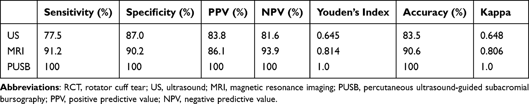 |
Table 3 Diagnostic Efficacy of Full-Thickness RCT via US, MRI, and PUSB |
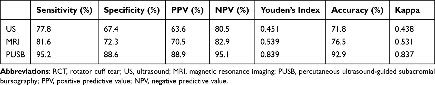 |
Table 4 Diagnostic Efficacy of Partial-Thickness RCT via US, MRI, and PUSB |
Besides, in patients with partial-thickness RCT, the sensitivity, specificity, and accuracy of ultrasound were 77.8%, 67.4%, and 71.8%, whilst those of MRI were 81.6%, 72.3%, and 76.5%. The sensitivity, specificity, and accuracy of PUSB in patients with partial-thickness RCT were 95.2%, 88.6%, and 92.9%. The analysis of Kappa test suggested a relatively consistent detection rate on the subtypes of RCT using PUSB and shoulder arthroscopy.
Discussion
Shoulder pain in the elderly is a common and frequently occurring disease, which is often misdiagnosed as periarthritis of shoulder.10 With the update and progress of medical concepts, it is currently believed that periarthritis of shoulder is actually a type of adhesive bursitis around the shoulder joint and is a self-limited disease.11 Shoulder pain in the elderly often originates from RCT (chronic RCT mainly), and its incidence increases year by year with the growing number of the elderly.
According to an existing report, when bursion-related shoulder pain is clinically suspected, the decisive step in the diagnosis of adhesive bursopathy is ultrasound-guided hydrodilation of the SASD bursa.12 Subacromial bursography was initially performed based on the fluoroscopically guided injection of iodine contrast agent into the subacromial bursa and the subsequent acquisition of shoulder joint radiographs. This technique has already been employed to evaluate RCT.13 However, due to the thickness of the bursa, it is difficult for radiologists to inject the contrast agent into the bursa accurately based on the anatomical structure, which hinders its promotion. With the help of PUSB, the contrast agent can be accurately injected into the bursa and its distribution can be dynamically traced in real time. Accordingly, the severity of RCT damage can be successfully evaluated and the application of CEUS in the musculoskeletal system can be promoted. All participants enrolled in our study successfully received PUSB, with no obvious discomfort or aggravated pain reported. It is thus indicated that PUSB is a safe operation technique, which deserves to be clinically applied and promoted, along with some other extra-vascular techniques like hysterosalpingo-contrast sonography of fallopian tubes and percutaneous transhepatic cholangiography.14,15
Under normal circumstances, SASD is a closed structure that does not connect with the shoulder joint cavity. When inflammation and fibrosis of the bursa and rotator cuff develop into RCT, the fluid (also including contrast agent) in the bursa can enter the avulsion sites of the rotator cuff or even the joint cavity. The scarce fluid in the bursa suggests that ultrasound or MRI may not be the best option under the following conditions: the severity of tear is mild, there is no retraction in the severed end of the tendon, and the bursal tissue is stuck.16 More importantly, the echogenicity of ultrasound is devoid of specificity and MRI technique may not be so effective in examining full-thickness RCT and partial-thickness RCT. PUSB is an innovative application of extravascular CEUS during the evaluation of RCT, which can clearly depict the contour and location of RCT and can provide better visualization results to improve the diagnosis of RCT. Besides, although SASD is usually separated from the superior subscapular recess by a thin fibrous septum, the thin canaliculus supports their communication. During excessive intra-bursal effusion, the canaliculus may dilate progressively to decrease the pressure in either bursa.17 As such, given the complex anatomy of the rotator interval, it is crucial to bear in mind that the bursal effusion may also flow in the superior subcoracoid space of the shoulder in patients with RCT. Based on the different dynamic ultrasound patterns, SASD and superior subscapular recess, as well as complex subcoracoid effusion could be identified in a timely manner.17 In our current study, the total accuracy (%) of ultrasound and 1.5-T MRI in the detection of RCT was 67.1% and 71.8%, respectively. Meanwhile, a relatively higher accuracy in the diagnosis of full-thickness RCT (83.3% of ultrasound and 86.1% of MRI) was also seen. The main manifestation of ultrasound examination includes the interrupted echo within the supraspinatus tendon, which affects the full-thickness tendon and the direct attachment of deltoid muscle to the HH, with cortical thickening of greater tuberosity and the presence of subacromial effusion. Also, the accuracy (%) of ultrasound and MRI in the detection of partial-thickness RCT in our study was not satisfactory either (56.8% of ultrasound and 61.4% of MRI), lowering than the existing research.18 It can thus be assumed that these results may be related to RCT classification scheme of the research, which included full-thickness RCT and partial-thickness RCT only, with further subtype classification. More importantly, it is worth noting that the overall accuracy (%) of PUSB in diagnosing RCT was 91.8%, which was consistent with the arthroscopic findings. It was observable from PUSB examination that the contrast agent could enter the rotator cuff tendon from the bursa and that a strong echo is distributed over the whole layer of the tendon and up to the surface of the HH. The articular cavity, in the meantime, was visualized as well (Figure 3). The accuracy (%) of PUSB on diagnosing partial-thickness RCT was 88.6%. These data collectively indicated that the correct diagnostic rate of PUSB for RCT was profoundly higher than those of ultrasound and MRI, partial-thickness RCT in particular. Therefore, PUSB can be used to pinpoint the accurate location of the lesion and clearly outline the morphology and size of the surrounding around the tear site, with an excellent image quality, which is more beneficial for the diagnosis of RCT and the selection of surgical options.
 |
Figure 3 Representative figure of full-thickness RCT on the SUP. (A) The oblique coronal T2-weighted image showing the high signal with the interrupted continuity within the SUP (↓↓). (B) The demonstration of two-dimensional ultrasound examination on the continuity of SUP in the middle segment and the extension from bursal surface to the articular surface (↓↓↓). (C) The mixture of contrast agent enters the SUP from the bursa and reaches the surface of the HH, along with a strong echogenicity (↓↓↓). Abbreviations: SUP, supraspinatus; HH, humeral head. |
The detection rates of ultrasound and MRI in patients with bp were 38.1% and 42.9% only. The analysis on the image retrieved from ultrasound examination indicated that thinning supraspinatus tendon and limited echogenic interruption were evident, while the whole tendon was not affected. The relevant image of PUSB showed the infiltration of the contrast agent into the tendon from the bursa, causing the strong echogenicity in the tendon but not reaching the surface of the HH, with the invisible joint cavity (Figure 4). The detection rate of PUSB on bp was 90.5% (19 of 21), which was slightly lower than a previous study (94.4%), presumably due to the relatively small sample size (17 cases of bp).19 In our study, based on the detection of ultrasound, two cases of tendinitis were misdiagnosed as partial-thickness RCT, eight cases of partial-thickness RCT as tendinitis, and six cases of small full-thickness RCT as partial-thickness RCT. The underlying reason may be related to the sensitivity of the ultrasound apparatus and operator experience. Also, only focal echogenic abnormality of the tendon is manifested in the detection of patients with small full-thickness RCT, which lacks the specificity. Moreover, in patients with partial-thickness RCT, tendonitis, surgical scars, and degenerated rotator cuffs, uneven echogenicity may exist within the tendon and local hypoechogenicity, which affects the accurate determination of the RCT type. During the examination via PUSB, two cases of bursal-side partial-thickness RCT were misdiagnosed as tendonitis, which might possibly result from the completely adherent bursal wall and the incapability of contrast agent to enter the bursa through the avulsion sites (which leads to their aggregation in the proximal end of the adherence). Besides, three cases of ap were misdiagnosed as tendonitis, and two cases of tendonitis as ap. PUSB can present the characteristics of bp clearly and objectively, the efficacy of which is evidently better than ultrasound and MRI. However, as to those partial-thickness RCT occurring far away from the bursal surface, PUSB is not superior and its detection accuracy is similar to that of ultrasound. One possible reason is that PUSB may not be employed for the diagnosis of ap based on the direct observation of the contrast agent. Instead, PUSB needs to be combined with two-dimensional acoustic images in the dual-display mode for co-diagnosis, which thus leads to some limitation of PUSB on the detection of ap. Our data here suggest that PUSB can help improve the diagnostic efficacy on the subtype of bp, similar to the research of Tang et al.20
 |
Figure 4 Symbolic figure of partial-thickness RCT on the SUP. (A) The oblique coronal T2-weighed image showing the high signal in the avulsion site on the bursal surface of the SUP (↓↓). (B) The demonstration of two-dimensional ultrasound examination on the continuity of SUP in the middle segment (↓↓↓). (C) The mixture of contrast media diffuses within the bursa and enters the tendon with a strong echogenicity (↓↓↓↓), whilst not entering the surface of HH. Abbreviations: SUP, supraspinatus; HH, humeral head. |
Of all 85 participants in our study, 7 have diabetes mellitus and 29 have hyperlipidemia. Existing reports have already recognized that both diabetes mellitus and hyperlipidemia are among the independent risk factors for RCT.21,22 The excessive accumulation of late glycosylated metabolic end products can weaken the tendons, alter the physiochemical properties of proteins, and make the tendons less elastic and probe to be torn.21 Hyperlipidemia can trigger fatty infiltrative effect due to lipid deposition in the tendon, leading to tearing in the tendon and difficulties in its repair.22 Considering these, prior to the randomized-controlled trial, in addition to the consideration on the patient's age and history of injury, the attention should be also paid to the present/absent history of diabetes mellitus and hyperlipidemia in these eligible patients.
Nonetheless, there are some limitations in this study that should be aware as follows: (1) the sample size is small and is required to be expanded for further in-depth research; (2) despite no complication reported in this research, the technique of PUSB is invasive in essence and patients may have the risk of infection; (3) inter-observer agreement test has not been performed for PUSB, and the results are affected by the operators’ experience; (4) the comparative analysis with the surgical tear area is not conducted in this study, which will become a direction for our future research to evaluate the diagnostic value in predicting the degree of RCT, thus laying great significance to the selection of therapeutic options for patients with RCT; (5) in some patients, adhesions can be located in between the synovial layers of the SASD bursa (ie, adhesive bursopathy), in this sense, the flow of contrast agents inside the bursal cavity can be limited by the aforementioned adhesions;12 and (6) taking sample size limitations into consideration, 1.5 and 3-T MRI on the knee have comparable accuracy in the diagnosis of meniscal tears;23 however, another report proved that 3-T MRI is superior to 1.5-T MRI in diagnosing scapholunate interosseous ligament (SLIL) injuries; besides, 1.5-T MRI has been reported to accurately diagnose anterior cruciate ligament (ACL) and medial meniscal tears,24 while 3-T MRI seems to be equivalent to 3-T magnetic resonance arthrogram (MRA) in the diagnosis of full- and partial-thickness tears.25 Further exploration, herein, might be performed to consider the accuracy of arthro-magnetic resonance imaging.
Conclusion
PUSB is a novel, safe, and effective examination method for the diagnosis of elderly patients with shoulder pain. The sensitivity and specificity for the subtype of RCT detected by PUSB are evidently higher than those detected by ultrasound and MRI, showing its value in the diagnosis of RCT and its feasibility in clinical application.
Data Sharing Statement
The analyzed data sets generated during the study are available from the corresponding author on reasonable request.
Author Contributions
All authors made a significant contribution to the work reported, whether that is in the conception, study design, execution, acquisition of data, analysis and interpretation, or in all these areas; took part in drafting, revising, or critically reviewing the article; gave final approval of the version to be published; have agreed on the journal to which the article has been submitted; and agree to be accountable for all aspects of the work.
Funding
This study was supported by the Zhejiang Provincial Public Welfare Technology Application Research Program Project [LGF21H180006]; the Zhejiang Provincial Medical and Health Science and Technology Program Project [2021KY257].
Disclosure
The authors declare no conflicts of interest.
References
1. Burnier M, Elhassan BT, Sanchez-Sotelo J. Surgical management of irreparable rotator cuff tears: what works, what does not, and what is coming. J Bone Joint Surg Am. 2019;101(17):1603–1612. doi:10.2106/JBJS.18.01392
2. Maruvada S, Madrazo-Ibarra A, Varacallo M. Anatomy, rotator cuff. In: StatPearls. Treasure Island (FL): StatPearls PublishingCopyright © 2022, StatPearls Publishing LLC; 2022.
3. Keener JD, Patterson BM, Orvets N, Chamberlain AM. Degenerative rotator cuff tears: refining surgical indications based on natural history data. J Am Acad Orthop Surg. 2019;27(5):156–165. doi:10.5435/JAAOS-D-17-00480
4. Matsen FA
5. Plancher KD, Shanmugam J, Briggs K, Petterson SC. Diagnosis and management of partial thickness rotator cuff tears: a comprehensive review. J Am Acad Orthop Surg. 2021;29(24):1031–1043. doi:10.5435/JAAOS-D-20-01092
6. Zhang X, Gu X, Zhao L. Comparative analysis of real-time dynamic ultrasound and magnetic resonance imaging in the diagnosis of rotator cuff tear injury. Evid Based Complementary Altern Med. 2021;2021:2107693. doi:10.1155/2021/2107693
7. Ricci V, Chang KV, Güvener O, et al. EURO-MUSCULUS/USPRM dynamic ultrasound protocols for shoulder. Am J Phys Med Rehabil. 2022;101(3):e29–e36. doi:10.1097/PHM.0000000000001833
8. Tsuchiya S, Davison EM, Rashid MS, et al. Determining the rate of full-thickness progression in partial-thickness rotator cuff tears: a systematic review. J Shoulder Elbow Surg. 2021;30(2):449–455. doi:10.1016/j.jse.2020.08.022
9. Martinoli C. Musculoskeletal ultrasound: technical guidelines. Insights Imaging. 2010;1(3):99–141. doi:10.1007/s13244-010-0032-9
10. Shi WJ, Mao BY, Wang FQ, Mi YF, Zhu YC. 老年肩袖撕裂关节镜下修复方式再探讨 [Re discussion on arthroscopic repair of rotator cuff tear in aged patients]. Zhongguo Gu Shang. 2021;34(11):1040–1043. Chinese. doi:10.12200/j.issn.1003-0034.2021.11.011
11. Pandey V, Madi S. Clinical guidelines in the management of frozen shoulder: an update! Indian J Orthop. 2021;55(2):299–309. doi:10.1007/s43465-021-00351-3
12. Ricci V, Galletti S, Chang KV, Özçakar L. Ultrasound imaging and guidance in the management of adhesive bursopathy of the shoulder: a video demonstration. J Med Ultrasound. 2020;39(3):633–635. doi:10.1002/jum.15117
13. Lie S, Mast WA. Subacromial bursography. Radiology. 1982;144(3):626–630. doi:10.1148/radiology.144.3.7100481
14. Luciano DE, Exacoustos C, Luciano AA. Contrast ultrasonography for tubal patency. J Minim Invasive Gynecol. 2014;21(6):994–998. doi:10.1016/j.jmig.2014.05.017
15. Chang HY, Liu B, Wang YZ, et al. Percutaneous transhepatic cholangiography versus endoscopic retrograde cholangiography for the pathological diagnosis of suspected malignant bile duct strictures. Medicine. 2020;99(11):e19545. doi:10.1097/MD.0000000000019545
16. Brockmeyer M, Schmitt C, Haupert A, Kohn D, Lorbach O. Limited diagnostic accuracy of magnetic resonance imaging and clinical tests for detecting partial-thickness tears of the rotator cuff. Arch Orthop Trauma Surg. 2017;137(12):1719–1724. doi:10.1007/s00402-017-2799-3
17. Ricci V, Mezian K, Naňka O, Özçakar L. Assessing/imaging the subcoracoid space: from anatomy to dynamic sonography. J Med Ultrasound. 2022;41(9):2149–2155. doi:10.1002/jum.15898
18. Lenza M, Buchbinder R, Takwoingi Y, Johnston RV, Hanchard NC, Faloppa F. Magnetic resonance imaging, magnetic resonance arthrography and ultrasonography for assessing rotator cuff tears in people with shoulder pain for whom surgery is being considered. Cochrane Database Syst Rev. 2013;2013(9):Cd009020. doi:10.1002/14651858.CD009020.pub2
19. Zhu Y, Qiang H, Gao Y, Wang Z, Guo L. 经皮超声引导肩峰下滑囊造影对肩袖损伤的诊断价值 [Diagnosis of rotator cuff injury by dynamic assessment of acromiocystography guided by percutaneous ultrasound]. Zhong Guo Yi xue Ying Xiang Xue Za Zhi. 2020;28(6):461–464. Chinese doi:10.3969/j.issn.1005-5185.2020.06.014
20. Tang YQ, Zeng C, Su XT, et al. The value of percutaneous shoulder puncture with contrast-enhanced ultrasound in differentiation of rotator cuff tear subtypes: a preliminary prospective study. Ultrasound Med Biol. 2019;45(3):660–671. doi:10.1016/j.ultrasmedbio.2018.10.012
21. Yoshikawa T, Mifune Y, Inui A, et al. Influence of diabetes-induced glycation and oxidative stress on the human rotator cuff. Antioxidants. 2022;11(4):743. doi:10.3390/antiox11040743
22. Hebert-Davies J, Teefey SA, Steger-May K, et al. Progression of fatty muscle degeneration in atraumatic rotator cuff tears. J Bone Joint Surg Am. 2017;99(10):832–839. doi:10.2106/JBJS.16.00030
23. Grossman JW, De Smet AA, Shinki K. Comparison of the accuracy rates of 3-T and 1.5-T MRI of the knee in the diagnosis of meniscal tear. AJR Am J Roentgenol. 2009;193(2):509–514. doi:10.2214/AJR.08.2101
24. Koch JEJ, Ben-Elyahu R, Khateeb B, et al. Accuracy measures of 1.5-tesla MRI for the diagnosis of ACL, meniscus and articular knee cartilage damage and characteristics of false negative lesions: a level III prognostic study. BMC Musculoskelet Disord. 2021;22(1):124. doi:10.1186/s12891-021-04011-3
25. McGarvey C, Harb Z, Smith C, Houghton R, Corbett S, Ajuied A. Diagnosis of rotator cuff tears using 3-Tesla MRI versus 3-Tesla MRA: a systematic review and meta-analysis. Skeletal Radiol. 2016;45(2):251–261. doi:10.1007/s00256-015-2299-x
 © 2023 The Author(s). This work is published and licensed by Dove Medical Press Limited. The
full terms of this license are available at https://www.dovepress.com/terms
and incorporate the Creative Commons Attribution
- Non Commercial (unported, 3.0) License.
By accessing the work you hereby accept the Terms. Non-commercial uses of the work are permitted
without any further permission from Dove Medical Press Limited, provided the work is properly
attributed. For permission for commercial use of this work, please see paragraphs 4.2 and 5 of our Terms.
© 2023 The Author(s). This work is published and licensed by Dove Medical Press Limited. The
full terms of this license are available at https://www.dovepress.com/terms
and incorporate the Creative Commons Attribution
- Non Commercial (unported, 3.0) License.
By accessing the work you hereby accept the Terms. Non-commercial uses of the work are permitted
without any further permission from Dove Medical Press Limited, provided the work is properly
attributed. For permission for commercial use of this work, please see paragraphs 4.2 and 5 of our Terms.