
Back to Journals » Drug Design, Development and Therapy » Volume 20
Stachydrine: A Systematic Review of Its Multi-Targeted Therapeutic Potential in Cardiovascular, Oncology, Renal, Gynecological, and Inflammatory Disorders
Received 1 November 2025
Accepted for publication 5 March 2026
Published 12 March 2026 Volume 2026:20 578362
DOI https://doi.org/10.2147/DDDT.S578362
Checked for plagiarism Yes
Review by Single anonymous peer review
Peer reviewer comments 3
Editor who approved publication: Prof. Dr. Georgios Panos
Songlin Tang,1 Yongpan Huang2
1Department of Neurology, The First Affiliated Hospital of Shaoyang College, Shaoyang, Hunan, 422039, People’s Republic of China; 2School of Medicine, Changsha Social Work College, Changsha, Hunan, 410004, People’s Republic of China
Correspondence: Yongpan Huang, School of Medicine, Changsha Social Work College, Changsha, Hunan, 410004, People’s Republic of China, Email [email protected]
Abstract:
Background: Stachydrine, a principal bioactive alkaloid derived from Leonurus japonicus (motherwort), has attracted significant interest due to its diverse pharmacological activities and nutritional relevance. This systematic review synthesizes current evidence on its therapeutic potential across multiple organ systems. Stachydrine core pharmacological activities are: Cardiovascular protection: Stachydrine mitigates myocardial ischemia/reperfusion injury by scavenging free radicals, reducing myocardial biomarkers (CK, LDH, cTnT), and enhancing nitric oxide (NO) production. It attenuates pathological ventricular remodeling by suppressing ROS-mediated activation of NF-κB and improves cardiac calcium handling by protecting sarcoplasmic reticulum function. Antitumor effects: In cancers (e.g. hepatocellular carcinoma, breast cancer, colorectal cancer), stachydrine inhibits tumor proliferation, metastasis, and chemoresistance by targeting pathways such as TGF-β/Smad, PI3K/Akt/mTOR, and JAK2/STAT3. It also modulates the tumor microenvironment by reprogramming tumor-associated macrophages. Renoprotective actions: It ameliorates drug-induced renal fibrosis by suppressing tubular cell apoptosis via downregulation of caspase-9/caspase-12 and inhibiting inflammatory cytokine release.
Uteroprotective benefits: Stachydrine regulates uterine hemorrhage by balancing Th1/Th2/Th17/Treg immune homeostasis and modulating endothelial function (e.g. NO and endothelin-1 levels), while enhancing uterine smooth muscle contractility. Antioxidant mechanisms: It reduces oxidative stress via ROS scavenging and NOX2 pathway inhibition, thereby protecting cardiovascular and neuronal tissues. Anti-inflammatory properties: Through modulation of NF-κB, JAK2/STAT3, and AMPK/SIRT1 pathways, stachydrine alleviates acute and chronic inflammation in models ranging from arthritis to neuroinflammation.
Conclusion: This review comprehensively documents stachydrine’s multi-targeted and multi-organ therapeutic potential, driven by its pleiotropic mechanisms. It provides a robust foundation for clinical translation in cardiovascular diseases, cancer, renal disorders, gynecological conditions, and inflammation-associated pathologies. Future research should prioritize high-quality clinical trials and synergistic drug-combination strategies to harness its therapeutic efficacy fully.
Keywords: stachydrine, cardioprotection, antitumor, renal protection, uterine protection, antioxidant, anti-inflammatory
Introduction
Motherwort (Leonurus japonicus), a flowering herbaceous plant from the Lamiaceae family, is highly valued for both medicinal and ornamental purposes.1–4 For centuries, it has been a staple in traditional medicine, particularly for managing gynecological conditions such as menstrual irregularities, dysmenorrhea, amenorrhea, edema, and ulcers with its therapeutic efficacy widely recognized.2,5 Modern pharmacological studies have identified its active constituents as demonstrating diverse bioactivities, including cardioprotective, antioxidant, anticancer, analgesic, anti-inflammatory, antibacterial, and neuroprotective effects.6,7 Among these, alkaloids, flavonoids, and terpenoids have emerged as key research targets due to their significant contributions to these pharmacological properties. Stachydrine, the principal bioactive component of motherwort (Leonurus japonicus), has been widely investigated for its diverse biological activities.8,9 Notably, it also occurs in substantial concentrations in citrus juices, endowing it with both pharmacological and nutritional significance. Emerging research highlights its beneficial effects on multiple physiological systems-including cardiovascular protection, nephroprotection, uterine tissue repair, and anti-inflammatory modulation, thereby expanding its therapeutic potential.10
This study presents a comprehensive review of global research on stachydrine’s pharmacological properties, synthesizing key advancements from both domestic and international studies. Through systematic analysis of existing literature, it aims to provide critical insights for future research directions and clinical applications, ultimately facilitating the broader utilization of stachydrine in therapeutic contexts.
Methods
Search Strategy
By utilizing an “advanced search” strategy within the Web of Science Core Collection (WOSCC), we systematically searched for publications relevant to stachydrine, as outlined in Figure 1. Only articles and reviews published from May 1, 2006 to November 5, 2025, were considered. The data collection was executed on November 5, 2025. The compilation of this review was conducted based on a thorough examination of research articles obtained from various databases, including PubMed, Scopus, and ScienceDirect. Article searches were carried out using the keywords: “(stachydrine)”. The obtained articles were subsequently managed based on the PRISMA (Preferred Reporting Items for Systematic Reviews) guideline, starting with the removal of duplicate articles, screening for article relevance based on titles and abstracts, screening of full-text articles, and eligibility screening based on data conformity with inclusion criteria. The inclusion criteria encompassed articles in the English language, computational studies in whole or in part of the research, as well as providing detailed information on the execution of molecular docking studies, such as the software used. The workflow for the search and inclusion screening process is depicted in Figure 1. Based on the information obtained from each included article, the pharmacological activities of stachydrine and its molecular mechanisms, as revealed by studies, are discussed in detail.
 |
Figure 1 Flowchart of literature screening based on PRISMA guideline. |
Stachydrine is one of the main active components of motherwort (Leonurus japonicus) and possesses multiple pharmacological effects. It promotes blood circulation to regulate menstruation, promotes diuresis to reduce edema, and induces uterine contraction.2,5 It can increase coronary blood flow, reduce myocardial cell necrosis, and exhibits antiplatelet aggregation activity.7,8,10 Studies have shown that it improves atherosclerosis by activating the AMPK/SIRT1 signaling pathway, exerts neuroprotective effects by modulating Notch1 protein expression, and demonstrates bronchial smooth muscle relaxant activity in the respiratory system.6
Protective Effects of Stachydrine on Cardiovascular Health
Cardiovascular diseases, particularly coronary heart disease (CHD) and stroke, have become the foremost global health threats. According to recent statistics, cardiovascular diseases account for approximately 17.9 million deaths annually, representing 31% of total global deaths.11,12 These conditions are not only associated with high mortality rates, particularly due to sudden death risks in acute myocardial infarction (AMI) or stroke patients, but also lead to severe disability. Approximately 75% of stroke survivors experience, varying degrees of long-term sequelae, imposing substantial economic burdens on both patients’ families and society. Among cardiovascular diseases, ischemic heart disease, especially acute myocardial infarction, stands as the primary cause of mortality. Myocardial ischemia typically arises from coronary artery atherosclerosis, creating an imbalance between myocardial oxygen demand and coronary oxygen supply, ultimately leading to myocardial injury and the development of coronary heart disease.13 In recent years, the increasing prevalence of coronary heart disease driven by shifting lifestyles and aging populations has highlighted the urgent need for research into novel drugs and therapies for myocardial ischemia. Myocardial ischemia/reperfusion (I/R) injury is the central pathophysiological mechanism underlying myocardial ischemia-related diseases, and its effective management critically determines the therapeutic outcomes for myocardial ischemia. A rat acute myocardial I/R model was established using the coronary artery ligation method, followed by intravenous administration of varying doses of stachydrine.14 The experimental results demonstrated that stachydrine exerts cardioprotective effects by: (1) scavenging oxygen free radicals, (2) stabilizing cell membranes, and (3) suppressing the release of myocardial injury biomarkers (CK, LDH, and cTnT). Additionally, it enhances nitric oxide (NO) production, further contributing to its protective mechanisms against I/R injury. These findings suggest that stachydrine exerts a notable protective effect against myocardial I/R injury in rats, offering novel insights for clinical treatment of myocardial ischemia.
Ventricular remodeling, induced by pressure overload, metabolic dysfunction, and other pathological stimuli, plays a central role in heart failure development. This process features pathological hypertrophy, excessive non-myocyte proliferation, and metabolic derangements, which disrupt myocardial architecture and function. These alterations lead to progressive cardiac damage, culminating in arrhythmias and overt heart failure.15,16 Reactive oxygen species (ROS) emerge as central mediators of ventricular remodeling. When oxidative stress overwhelms antioxidant defenses, excessive ROS not only induce cellular damage but also activate oxygen-sensitive signaling cascades (eg, kinases, transcription factors).17 This dysregulation of gene expression fuels inflammation, drives pathological cardiomyocyte hypertrophy and apoptosis, and ultimately exacerbates ventricular remodeling and cardiac dysfunction.
There are studies that have investigated the anti-hypertrophic effects of stachydrine in angiotensin II-exposed neonatal rat cardiomyocytes, demonstrating its ability to attenuate hypertrophy by suppressing ROS production and inhibiting the NF-κB signaling pathway.18 These findings suggest stachydrin’s potential as a therapeutic agent for ventricular remodeling, mediated through modulation of oxidative stress-driven mechanisms, including ROS suppression and NF-κB pathway inhibition.19 These results align with broader research on ROS-targeted therapies for cardiovascular diseases, such as triptolide’s role in diabetic cardiomyopathy, reinforcing the clinical relevance of oxidative stress modulation in cardiac protection. Cardiac function relies on the sarcoplasmic reticulum (SR)’s calcium uptake capacity, which is severely disrupted in hypertrophy. Norepinephrine-induced SR structural damage, as shown in a cardiomyocyte model, compromises calcium ion sequestration.7 However, administration of different doses of stachydrine partially restored the impaired calcium uptake capacity in a dose-dependent manner.20 These findings suggest that stachydrine exerts protective effects on the SR, thereby enhancing calcium ion handling and preserving normal myocardial function. Collectively, these findings highlight stachydrine’s multifaceted cardioprotective effects, including mitigation of myocardial I/R injury, suppression of ventricular remodeling, and enhancement of SR calcium uptake. The accumulating evidence robustly supports stachydrine’s potential as a therapeutic candidate for cardiovascular and cerebrovascular diseases, warranting further clinical development. As the single-layered flat cells lining blood vessels, vascular endothelial cells are essential for maintaining vascular homeostasis. However, exposure to risk factors like hyperglycemia and hypertension induces endothelial dysfunction, a pivotal event that triggers pathological cascades, ultimately promoting atherosclerosis and cardiovascular disease development. Oxidative stress and metabolic disorders accelerate endothelial aging, impairing nitric oxide secretion and promoting procoagulant release. This leads to vascular stiffening, thrombus formation, and myocardial ischemia. An in vitro high-glucose-induced endothelial aging model was established to investigate the protective effects of stachydrine and the findings demonstrated that stachydrine significantly alleviated endothelial dysfunction and cellular senescence by activating the Sirt1 signaling pathway.21,22 This study not only elucidates novel molecular mechanisms but also proposes promising therapeutic avenues for cardiovascular disease prevention through endothelial preservation. These insights underscore the pivotal role of endothelial integrity in mitigating cardiovascular risk, offering potential translational implications for clinical interventions.
Antagonistic Effects of Stachydrine on Cancer
Malignant tumors, driven by intricate genetic alterations, continue to pose a formidable challenge in modern oncology. Despite the efficacy of current clinical interventions, their substantial adverse effects underscore the critical need for low-toxicity yet highly potent antitumor agents. Stachydrine has emerged as a breakthrough candidate in cancer therapy, leveraging its multi-target action to disrupt tumor progression.23,24 This unique profile opens avenues for designing next-generation treatments with optimized safety and potency. Stachydrine disrupts tumor growth via multi-pathway mechanisms, engaging distinct biological targets to orchestrate its anticancer activity.24 In hepatocellular carcinoma, it activates the LIF/AMPK pathway to induce autophagy and senescence, effectively suppressing tumor progression.25 In breast cancer cells, stachydrine inhibits proliferation and promotes apoptosis via Akt/ERK pathway modulation.8,26,27 Additionally, in colorectal cancer models, it attenuates liver metastasis by targeting both the JAK2/STAT3 pathway and tumor-associated macrophages.28 These findings collectively demonstrate stachydrine’s multi-targeted antitumor activity.
Regulation of Cancer Metastasis
Stachydrine exhibits distinct advantages in suppressing metastasis by targeting key molecular pathways.24,28 Recent studies reveal its ability to block TGF-β1-induced epithelial-mesenchymal transition through inhibition of the TGF-beta/Smad and PI3K/Akt/mTOR cascades, thereby disrupting a pivotal step in tumor dissemination.21 In prostate cancer, it synergistically suppresses growth, chemokine release, and drug resistance by inducing senescence/ferroptosis, offering a novel strategy to overcome therapeutic resistance.27
Modulation of the Tumor Microenvironment
Stachydrine holds significant potential in reshaping the tumor immune microenvironment.29 Emerging evidence demonstrates that it enhances anti-tumor immunity by reprogramming tumor-associated macrophages toward an immunostimulatory phenotype.27 Across diverse cancer models, stachydrine has been shown to modulate immune cell activity and function, offering novel strategies for cancer immunotherapy. Collectively, these findings highlight stachydrine’s multifaceted therapeutic effects, encompassing tumor suppression, metastasis inhibition, and immune microenvironment remodeling, thereby positioning it as a promising candidate for comprehensive cancer management. The accumulating evidence positions stachydrine as a promising candidate for further development as an antitumor agent, with potential to revolutionize cancer treatment strategies.30 These studies provide a robust theoretical foundation for the development of stachydrine, whose multi-targeted mechanisms and low toxicity profile position it as a promising candidate for antitumor therapy. Future studies will aim to harness its clinical potential, paving the way for innovative cancer therapies. The compound’s capacity to concurrently regulate tumor cell proliferation, metastasis, and the immune microenvironment highlights its distinct advantages for precision oncology. As research progresses into its synergistic potential with conventional therapies, stachydrine is poised to become a transformative therapeutic agent in cancer treatment.
Renoprotective Effects of Stachydrine: Targeting Apoptosis and Fibrosis in Kidney Injury
The increasing frequency of drug use and the widespread application of medical diagnostic procedures have contributed to a rising incidence of drug-induced and procedure-related renal injury, with renal interstitial fibrosis emerging as a central pathological hallmark that drives progressive loss of renal function through tubular structural disintegration and excessive extracellular matrix accumulation, ultimately resulting in irreversible nephron dysfunction.31 This fibrotic process is intimately linked to the apoptosis of renal tubular epithelial cells, orchestrated by dysregulated signaling pathways that converge on intrinsic apoptotic machinery, including the activation of caspase-9 and caspase-3.32,33 Oxidative stress, particularly through the salusin-β/PKC/ROS axis, induces DNA damage and stabilizes p53, while gut microbiota dysbiosis amplifies systemic inflammation and reactive oxygen species production, creating a self-sustaining loop that intensifies apoptotic signaling.34,35 In parallel, macrophage-derived pro-inflammatory mediators and innate immune sensors activated by oxidative stress or microbial triggers further engage the caspase cascade, reinforcing cell death.36 Environmental toxins such as 1-methoxypyrene, acting as aryl hydrocarbon receptor agonists, exacerbate tubulointerstitial fibrosis by promoting oxidative stress and inflammatory cascades that culminate in epithelial apoptosis. Current predictive models for apoptosis in chronic kidney disease remain inadequate due to their oversimplification of these interconnected, multi-factorial triggers, despite emerging evidence from polycystic kidney disease highlighting conserved apoptotic pathways across distinct etiologies.37 In this context, stachydrine has demonstrated potent renoprotective effects by concurrently suppressing apoptosis through downregulation of caspase-9 and caspase-12, attenuating interstitial inflammation via modulation of cytokine networks, and directly inhibiting the epithelial-to-myofibroblast transdifferentiation that underlies fibrosis progression, thereby targeting the core pathological triad of apoptosis, inflammation, and fibrosis in a unified mechanistic framework.8,38
These findings collectively highlight stachydrine’s multi-targeted action, offering a robust foundation for developing novel nephroprotective therapies. Its ability to concurrently address apoptosis, inflammation, and epithelial-mesenchymal transition positions it as a compelling candidate for preventing drug-induced renal injury in clinical settings.
Uteroprotective Effects of Stachydrine: Balancing Immune Homeostasis and Vascular Function
The uterus, a central organ of the female reproductive system, is critical for reproductive function and overall physiological health. With age, the risk of uterine disorders increases significantly, with abnormal uterine bleeding being a common gynecological condition that severely compromises quality of life.39 Excessive bleeding, whether due to miscarriage, postpartum hemorrhage, or other causes, can lead to life-threatening blood loss, while immune dysregulation is closely associated with pregnancy-related complications.40 Current surgical interventions often cause irreversible uterine damage, highlighting the urgent need for non-invasive therapeutic alternatives. Stachydrine demonstrates unique mechanisms of action in uterine protection.38,41 Studies utilizing RU486-induced abortion models have revealed that stachydrine significantly reduces abnormal uterine bleeding by modulating the Th1/Th2/Th17/Treg immune balance pathway, offering a novel therapeutic strategy for drug-induced abortion-related hemorrhage.42
As reported in dysfunctional uterine bleeding, stachydrine showed the characteristics of vasoregulatory modulation by inhibiting excessive NO release in uterine homogenates, and further alleviating vascular dysfunction and improves uterine contractility.43–45 Moreover, Recent studies indicate that stachydrine promotes endothelin-1 secretion from uterine smooth muscle cells, thereby enhancing vascular tone and reducing bleeding.45 This precise modulation of vascular active substances enables stachydrine to effectively control hemorrhage while preserving uterine physiological function. The findings highlight its potential as a targeted therapeutic agent for uterine disorders, with implications for improving reproductive outcomes and reducing treatment-related morbidity. Notably, stachydrine exhibits multi-targeted uterine protection beyond direct hemostasis.46 By regulating inflammatory cytokine expression in the uterine microenvironment and improving endometrial receptivity, it simultaneously achieves rapid bleeding control while avoiding excessive uterine contraction, a common side effect of conventional therapies. This comprehensive mechanism enables stachydrine to address both acute hemorrhage and underlying pathological processes in dysfunctional uterine bleeding. The findings provide critical insights for developing next-generation uteroprotective agents and establish a novel therapeutic paradigm for uterine hemorrhagic disorders.
Antioxidant Effects of Stachydrine
Stachydrine, a bioactive compound with notable therapeutic potential, has recently gained attention for its antioxidant properties. Numerous studies showed stachydrine mitigates oxidative stress through multi-targeted pathways, including free radical scavenging and modulation of redox-sensitive signaling cascades.31,47,48 Its ability to harmonize pro-oxidant/antioxidant balance positions it as a promising candidate for preventing oxidative-driven disorders. Future research should focus on clinical translation, particularly for high-risk conditions such as cardiovascular diseases and neurodegenerative disorders where oxidative stress plays a pivotal role.
Antioxidant Mechanisms of Stachydrine: Unique Advantages and Therapeutic Potential
Stachydrine demonstrates distinct advantages in antioxidant mechanisms. Research indicates that stachydrine significantly reduces ROS levels, thereby exerting anti-hypertrophic effects. In phenylephrine (PE)-induced cardiac hypertrophy models, stachydrine effectively suppresses oxidative stress by inhibiting NOX2 activity, ultimately improving cardiac dysfunction.49,50 This discovery offers novel insights for clinical treatment of myocardial hypertrophy-related diseases. Furthermore, studies on transverse aortic constriction models reveal that stachydrine mitigates pressure overload-induced heart failure by modulating the NOX2-ROS pathway, thereby alleviating myocardial damage.47,51 These findings provide valuable references for clinical management of heart failure. Notably, stachydrine’s multi-targeted approach, including ROS scavenging and NOX2 pathway regulation, positions it as a promising candidate for cardiovascular protection. Future research should explore its synergistic effects with other antioxidants (eg, ascorbic acid or glutathione) to enhance therapeutic efficacy. The clinical translation of stachydrine, particularly in oxidative stress-driven cardiovascular disorders, warrants further investigation through large-scale trials and mechanistic studies.
Beyond Cardiovascular Applications: The Multifaceted Antioxidant Potential of Stachydrine
While stachydrine’s antioxidant properties have been extensively studied in cardiovascular diseases, emerging evidence suggests its broader therapeutic potential. In neurodegenerative disorders, oxidative stress is a pivotal contributor to neuronal damage and cell death. By effectively reducing ROS levels, stachydrine may mitigate oxidative injury to neurons, thereby potentially slowing disease progression.23 In the realm of oncology, stachydrine’s antioxidant activity could offer novel strategies for cancer management.52 Collectively, stachydrine exerts its antioxidant effects through multiple pathways, including ROS reduction and modulation of oxidative stress-related signaling cascades.26,50,51,53,54 These mechanisms not only provide new insights for treating cardiovascular and neurodegenerative diseases but also offer valuable references for oncology research. Future research should prioritize translating stachydrine’s preclinical antioxidant insights into clinical applications through rigorous trials and interdisciplinary collaboration, enabling novel therapies for unmet medical needs.
Antagonistic Effects of Stachydrine on Inflammation
Stachydrine, a natural compound with multifaceted biological activities, has garnered increasing attention for its anti-inflammatory properties. As a defense response to injury or infection, inflammation can lead to various diseases when dysregulated. Studies demonstrate that stachydrine exerts significant protective effects in both acute and chronic inflammation models by modulating key signaling pathways.55,56 In a xylene-induced mouse ear swelling model, stachydrine significantly alleviated acute inflammation by suppressing redness, swelling, and exudation at inflammatory sites. Mechanistically, stachydrine reduced pro-inflammatory cytokine levels (eg, IL-6, TNF-α) in pleural exudate while simultaneously inhibiting PGE2 and NO production. This multi-targeted anti-inflammatory action demonstrates stachydrine’s potential as a therapeutic agent for acute inflammatory conditions.26,57 Notably, stachydrine’s ability to concurrently inhibit multiple inflammatory mediators underscores its advantages over single-target agents. Future research should explore its synergistic effects with conventional anti-inflammatory drugs to enhance clinical efficacy. The findings highlight stachydrine as a promising candidate for inflammation-related disorders, warranting further investigation into its clinical translation and safety profile. In the field of chronic inflammation, stachysine demonstrates broader regulatory capabilities. In diabetic retinopathy models, stachysine exerts protective effects on retinal cells by simultaneously suppressing inflammatory responses and promoting autophagy through activation of the AMPK/SIRT1 signaling pathway.26,58 In skeletal muscle insulin resistance models, stachysine effectively alleviates lipid-induced inflammation and endoplasmic reticulum stress while improving insulin sensitivity through the AMPK/HO-1 pathway.59 In neuroinflammatory regulation, stachysine exhibits particularly remarkable effects in cerebral I/R injury models.56 By inhibiting the P65 and JAK2/STAT3 signaling pathways, it significantly reduces inflammatory cytokine release and cell apoptosis, exerting neuroprotective effects.60,61 In carbon tetrachloride (CCl4)-induced hepatic fibrosis models, stachysine exhibits significant antifibrotic activity by alleviating inflammatory responses and oxidative stress while regulating the MMPs/TIMPs balance, thereby effectively attenuating liver fibrosis progression.62 In allergen-induced inflammatory models, stachysine significantly attenuates house dust mite-driven inflammatory responses.63 For osteoarthritis treatment, it effectively inhibits IL-1β-induced chondrocyte inflammation by blocking the NF-κB signaling pathway, providing novel therapeutic potential for this condition.64 Further studies demonstrated that stachysine significantly inhibits LPS-induced secretion of interleukin-1α (IL-1α) and nitric oxide (NO) in endothelial cells while modulating 6-keto-prostaglandin F1α levels, highlighting its potent anti-endotoxin activity.65 These findings strengthen its therapeutic potential for inflammatory diseases. Collectively, these studies demonstrate that stachysine exerts potent anti-inflammatory effects across diverse inflammatory models through modulation of key signaling pathways. Its mechanisms include suppressing pro-inflammatory cytokine production, regulating oxidative stress, and enhancing cellular function. These findings provide strong experimental support for stachysine’s therapeutic potential in inflammatory-related disorders and offer promising insights for developing next-generation anti-inflammatory therapies.
Recent Advances and Clinical Perspectives
Stachydrine, a bioactive compound derived from traditional Chinese medicine, has emerged as a promising therapeutic agent with multifaceted effects, as evidenced by recent research advancements. A study elucidated a novel mechanism by which stachydrine mitigates oxidative stress through the activation of the Nrf2/HO-1 signaling pathway.57 Under oxidative stress conditions, stachydrine facilitates the nuclear translocation of Nrf2, a pivotal transcription factor, thereby triggering the upregulation of HO-1 expression. HO-1 plays a essential role in catalyzing heme degradation, which generates biliverdin, carbon monoxide, and free iron. These products collectively enhance cellular antioxidant defenses by mitigating ROS-induced oxidative damage. This mechanism highlights stachydrine’s potential as a natural antioxidant agent in managing oxidative stress-related diseases.66 In addition to its antioxidant properties, stachydrine exhibits significant hepatoprotective effects, as demonstrated in an animal model. The research employed a high-fat diet-induced non-alcoholic fatty liver disease (NAFLD) model in mice, revealing that stachydrine administration effectively reduces hepatic lipid accumulation, improves liver function markers, and alleviates inflammation.67 These findings suggest that stachydrine protects against NAFLD progression by modulating lipid metabolism and reducing oxidative stress in hepatocytes, thereby supporting its therapeutic application in metabolic liver disorders. Furthermore, a metabolomics study explored stachydrine’s impact on the gut microbiota-bile acid metabolic axis.68 Using advanced multi-omics techniques, the research identified that stachydrine alters the composition of gut microbiota, promoting beneficial bacterial species while inhibiting harmful ones.69 These changes in microbiota composition subsequently influence bile acid metabolism, leading to improved bile acid profiles and enhanced metabolic regulation. This mechanism underscores stachydrine’s role in maintaining gut health and its potential to modulate systemic metabolism through the gut-liver axis, offering insights into its broader therapeutic effects beyond direct organ protection.
The integration of these findings from recent studies collectively demonstrates stachydrine’s diverse pharmacological actions. By activating the Nrf2/HO-1 pathway to mitigate oxidative stress, protecting against NAFLD-induced liver injury, and modulating the gut microbiota-bile acid axis, stachydrine emerges as a compound with significant therapeutic potential. These mechanisms support its application in oxidative stress-related diseases, metabolic disorders, and gut health management, bridging traditional medicine with modern pharmacological research (Table 1). Future studies should explore clinical applications and optimize therapeutic strategies based on these multi-omics insights (Figure 2).
 |
Figure 2 The diagram illustrates the pleiotropic pharmacological mechanisms of stachydrine, which underpin its therapeutic potential. These actions are categorized into four primary domains: (1) Anti-inflammatory Effects, (2) Cardio-protective Properties, (3) Renoprotective effects, (4) Uteroprotective Effects and (5) Antagonistic effects. |
Collectively, present study provides a systematic review of the pharmacological research on stachydrine, highlighting its protective effects across multiple organ systems. It focuses on six core pharmacological activities: cardiovascular protection, anti-tumor effects, renal protection, uterine protection, and antioxidative properties. Collectively, these findings offer crucial theoretical support for understanding the clinical potential of stachydrine.
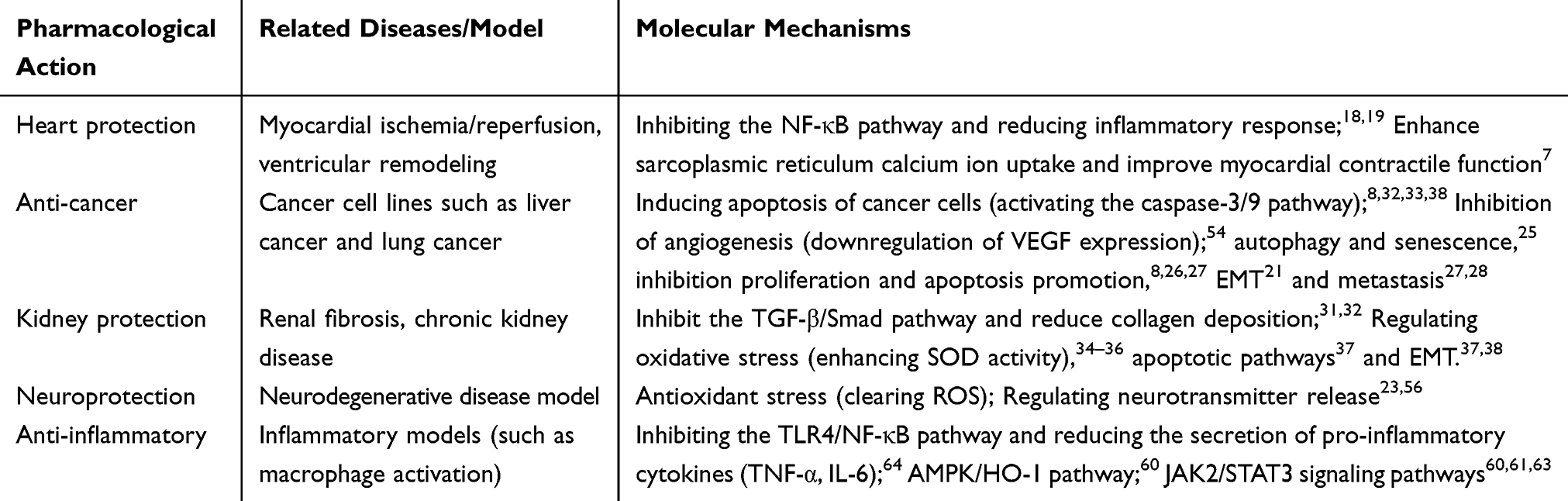 |
Table 1 Pharmacological Effects and Molecular Mechanisms of Stachydrine |
Funding
This work was supported by the funding of the Hunan Provincial Natural Science Foundation (no. 2023JJ60262, 2025JJ70217, 2026JJ80933). The funders supported study design, data collection and analysis, decision to publish, and preparation of the manuscript.
Disclosure
The authors declare that the research was conducted in the absence of any commercial or financial relationships that could be construed as a potential conflict of interest.
References
1. Miao LL, Zhou QM, Peng C, Liu ZH, Xiong L. Leonurus japonicus (Chinese motherwort), an excellent traditional medicine for obstetrical and gynecological diseases: a comprehensive overview. Biomed Pharmacother. 2019;117:109060. doi:10.1016/j.biopha.2019.109060
2. Wojtyniak K, Szymański M, Matławska I. Leonurus cardiaca L. (motherwort): a review of its phytochemistry and pharmacology. Phytother Res. 2013;27(8):1115–11. doi:10.1002/ptr.4850
3. Wang C, Lv X, Liu W, Liu S, Sun Z. Uncovering the pharmacological mechanism of motherwort (Leonurus japonicus Houtt.) for treating menstrual disorders: a systems pharmacology approach. Comput Biol Chem. 2020;89:107384. doi:10.1016/j.compbiolchem.2020.107384
4. Huang L, Xu DQ, Chen YY, Yue SJ, Tang YP. Leonurine, a potential drug for the treatment of cardiovascular system and central nervous system diseases. Brain Behav. 2021;11(2):e01995. doi:10.1002/brb3.1995
5. Liao L, Tang Y, Li B, et al. Stachydrine, a potential drug for the treatment of cardiovascular system and central nervous system diseases. Biomed Pharmacother. 2023;161:114489. doi:10.1016/j.biopha.2023.114489
6. Wang Y, Wu X, Wu R, Sun X, Kang Y, Yu Y. Pharmacological insights and therapeutic applications of leonurus in neurological disorders. J Inflamm Res. 2025;18:11437–11450. doi:10.2147/JIR.S527517
7. Witkowska A, Gryn-Rynko A, Syrkiewicz P, Kitala-Tańska K, Majewski MS. Characterizations of white mulberry, sea-buckthorn, garlic, lily of the valley, motherwort, and hawthorn as potential candidates for managing cardiovascular disease-in vitro and ex vivo animal studies. Nutrients. 2024;16(9):1313. doi:10.3390/nu16091313
8. He Z, Li P, Liu P, Xu P. Exploring stachydrine: from natural occurrence to biological activities and metabolic pathways. Front Plant Sci. 2024;15:1442879. doi:10.3389/fpls.2024.1442879
9. Cheng F, Zhou Y, Wang M, et al. A review of pharmacological and pharmacokinetic properties of stachydrine. Pharmacol Res. 2020;155:104755. doi:10.1016/j.phrs.2020.104755
10. Zhang S, Zhang Q, Lu Y, et al. Roles of integrin in cardiovascular diseases: from basic research to clinical implications. Int J Mol Sci. 2024;25(7):4096. doi:10.3390/ijms25074096
11. Chong B, Jayabaskaran J, Jauhari SM, et al. Global burden of cardiovascular diseases: projections from 2025 to 2050. Eur J Prev Cardiol. 2025;32(11):1001–1015. doi:10.1093/eurjpc/zwae281
12. Monizzi G, Di Lenarda F, Gallinoro E, Bartorelli AL. Myocardial ischemia: differentiating between epicardial coronary artery atherosclerosis, microvascular dysfunction and vasospasm in the catheterization laboratory. J Clin Med. 2024;13(14):4172. doi:10.3390/jcm13144172
13. Yuhong M, Yang J. Protective effect of stachydrine on myocardial ischemia-reperfusion in rats. Chin J Exp Traditional Med Formulae. 2006;12(5):40–42.
14. Frantz S, Hundertmark MJ, Schulz-Menger J, Bengel FM, Bauersachs J. Left ventricular remodelling post-myocardial infarction: pathophysiology, imaging, and novel therapies. Eur Heart J. 2022;43(27):2549–2561. doi:10.1093/eurheartj/ehac223
15. Jiang H, Fang T, Cheng Z. Mechanism of heart failure after myocardial infarction. J Int Med Res. 2023;51(10):3000605231202573. doi:10.1177/03000605231202573
16. Paulus WJ, Tschöpe C. A novel paradigm for heart failure with preserved ejection fraction: comorbidities drive myocardial dysfunction and remodeling through coronary microvascular endothelial inflammation. J Am Coll Cardiol. 2013;62(4):263–271. doi:10.1016/j.jacc.2013.02.092
17. Zhao L, Wu D, Sang M, Xu Y, Liu Z, Wu Q. Stachydrine ameliorates isoproterenol-induced cardiac hypertrophy and fibrosis by suppressing inflammation and oxidative stress through inhibiting NF-kappaB and JAK/STAT signaling pathways in rats. Int Immunopharmacol. 2017;48:102–109. doi:10.1016/j.intimp.2017.05.002
18. Guo W, Zhang C, Liao YL, Lv R, Wei HC. Effect of Leonurus stachydrine on myocardial cell hypertrophy. Zhong Yao Cai. 2012;35(6):940–943.
19. Zhang C, Shan XL, Liao YL, Zhao P, Guo W, Wei HC. Effects of stachydrine on norepinephrine-induced neonatal rat cardiac myocytes hypertrophy and intracellular calcium transients. BMC Complement Altern Med. 2014;14(1):474. doi:10.1186/1472-6882-14-474
20. Servillo L, D’Onofrio N, Longobardi L, et al. Stachydrine ameliorates high-glucose induced endothelial cell senescence and SIRT1 downregulation. J Cell Biochem. 2013;114(11):2522–2530. doi:10.1002/jcb.24598
21. Li S, Cao H, Sun P, Feng Y, Chen L, Wang D. Stachydrine hydrochloride induces cell senescence and ferroptosis of castration-resistant prostate cancer. Naunyn Schmiedebergs Arch Pharmacol. 2025;398(10):14165–14173. doi:10.1007/s00210-025-04182-z
22. Zhai Z, Mu T, Zhao L, et al. Stachydrine represses the proliferation and enhances cell cycle arrest and apoptosis of breast cancer cells via PLA2G2A/DCN axis. Chem Biol Drug Des. 2024;103(1):e14429. doi:10.1111/cbdd.14429
23. Zeng H, Xu D, Song Y, et al. Synthesis, characterization and anti-breast cancer activities of stachydrine derivatives. Eur J Med Chem. 2023;259:115679. doi:10.1016/j.ejmech.2023.115679
24. Chen X, Yan N. Stachydrine inhibits TGF-beta1-induced epithelial-mesenchymal transition in hepatocellular carcinoma cells through the TGF-beta/Smad and PI3K/Akt/mTOR signaling pathways. Anticancer Drugs. 2021;32(8):786–792. doi:10.1097/CAD.0000000000001066
25. Tao J, Zhang P, Liu G, et al. Cytotoxicity of Chinese motherwort (YiMuCao) aqueous ethanol extract is non-apoptotic and estrogen receptor independent on human breast cancer cells. J Ethnopharmacol. 2009;122(2):234–239. doi:10.1016/j.jep.2009.01.013
26. Wang M, Shu ZJ, Wang Y, Peng W. Stachydrine hydrochloride inhibits proliferation and induces apoptosis of breast cancer cells via inhibition of Akt and ERK pathways. Am J Transl Res. 2017;9(4):1834–1844.
27. Gui Y, Xue G, Yuan Y, et al. Stachydrine targeting tumor-associated macrophages inhibit colorectal cancer liver metastasis by regulating the JAK2/STAT3 pathway. Front Pharmacol. 2025;16:1514158. doi:10.3389/fphar.2025.1514158
28. Rathee P, Rathee D, Rathee D, Rathee S. In vitro anticancer activity of stachydrine isolated from Capparis decidua on prostate cancer cell lines. Nat Prod Res. 2012;26(18):1737–1740. doi:10.1080/14786419.2011.608673
29. Bao H, Chen Y, Meng Z, Chu Z. The causal relationship between CSF metabolites and GBM: a two-sample mendelian randomization analysis. BMC Cancer. 2024;24(1):1119. doi:10.1186/s12885-024-12901-7
30. Guo J, Jia J, Wang A, Gu Y, Xia X. Stachydrine from natural foods alleviates hyperuricemia by modulating renal urate transporters and suppressing mitochondrial oxidative stress. Foods. 2025;14(10):1718. doi:10.3390/foods14101718
31. Liu Y. Kidney fibrosis: fundamental questions, challenges, and perspectives. Integr Med Nephrol Androl. 2024;11(4):e24–00027. doi:10.1097/IMNA-D-24-00027
32. Khandpur S, Mishra P, Mishra S, Tiwari S. Challenges in predictive modelling of chronic kidney disease: a narrative review. World J Nephrol. 2024;13(3):97214. doi:10.5527/wjn.v13.i3.97214
33. Cao G, Miao H, Wang Y-N, et al. Intrarenal 1-methoxypyrene, an aryl hydrocarbon receptor agonist, mediates progressive tubulointerstitial fibrosis in mice. Acta Pharmacol Sin. 2022;43(11):2929–2945. doi:10.1038/s41401-022-00914-6
34. Chung JY-F, Zhang -Y-Y, Ji ZZ-Y, et al. Immunodynamics of macrophages in renal fibrosis. Integr Med Nephrol Androl. 2023;10(3):e00001. doi:10.1097/IMNA-D-23-00001
35. Xiao-Jun L, Shan Q-Y, Xin W, Miao H, Zhao Y-Y. Gut microbiota regulates oxidative stress and inflammation: a double-edged sword in renal fibrosis. Cell Mol Life Sci. 2024;81(1):480. doi:10.1007/s00018-024-05532-5
36. Ndongo M, Nehemie LM, Coundoul B, Diouara AAM, Seck SM. Prevalence and outcomes of polycystic kidney disease in African populations: a systematic review. World J Nephrol. 2024;13(1):90402. doi:10.5527/wjn.v13.i1.90402
37. Aly R, Darwish S, Bala N, et al. Functional and metabolomic analysis of urinary extracellular vesicles from juvenile mice with renal compensatory hypertrophy. Biochim Biophys Acta Mol Basis Dis. 2024;1870(5):167096. doi:10.1016/j.bbadis.2024.167096
38. Jewson M, Purohit P, Lumsden MA. Progesterone and abnormal uterine bleeding/menstrual disorders. Best Pract Res Clin Obstet Gynaecol. 2020;69:62–73. doi:10.1016/j.bpobgyn.2020.05.004
39. Hill S, Shetty MK. Abnormal uterine bleeding in reproductive age women: role of imaging in the diagnosis and management. Semin Ultrasound CT MR. 2023;44(6):511–518. doi:10.1053/j.sult.2023.10.002
40. Sun Y, Xia X, Yuan G, et al. Stachydrine, a bioactive equilibrist for synephrine, identified from four citrus Chinese herbs. Molecules. 2023;28(9):3813. doi:10.3390/molecules28093813
41. Li X, Wang B, Li Y, et al. The Th1/Th2/Th17/Treg paradigm induced by stachydrine hydrochloride reduces uterine bleeding in RU486-induced abortion mice. J Ethnopharmacol. 2013;145(1):241–253. doi:10.1016/j.jep.2012.10.059
42. Zhu H, Long Z, Guozhong L. Effects of synthesis stachydrine on ET and NO of dysfunctional uterine bleeding. J Jiangxi Univers TCM. 2008;20(6):66–68.
43. Zhang RH, Liu ZK, Yang DS, Zhang XJ, Sun HD, Xiao WL. Phytochemistry and pharmacology of the genus Leonurus: the herb to benefit the mothers and more. Phytochemistry. 2018;147:167–183. doi:10.1016/j.phytochem.2017.12.016
44. Chen C, Liu Q, Lu Y, et al. Motherwort synergized with oxytocin for postpartum hemorrhage prevention: integrated clinical efficacy and mechanism exploration. Phytomedicine. 2025;145:156951. doi:10.1016/j.phymed.2025.156951
45. He YL, Shi JY, Peng C, et al. Angiogenic effect of motherwort (Leonurus japonicus) alkaloids and toxicity of motherwort essential oil on zebrafish embryos. Fitoterapia. 2018;128:36–42. doi:10.1016/j.fitote.2018.05.002
46. Hiller JK, Sand ES, Rootwelt H, et al. Metabolic deficiencies in the vitreous of diabetic retinopathy: exploring the potential of dietary supplementation to address key metabolite imbalances. Clin Ophthalmol. 2025;19:2997–3007. doi:10.2147/OPTH.S531638
47. Tao L, Huang X, Zhao F, Wang X. Stachydrine alleviates sepsis-induced cardiomyopathy by inhibiting ferroptosis via regulating SIRT1/GPX4 pathway. Dose Response. 2025;23(3):15593258251377710. doi:10.1177/15593258251377710
48. Li XQ, Lu S, Xia L, et al. Stachydrine hydrochloride ameliorates cardiac hypertrophy through CaMKII/HDAC4/MEF2C signal pathway. Am J Transl Res. 2022;14(6):3840–3853.
49. Fu M, Guo S, Yang S, et al. Stachydrine hydrochloride reduces NOX2 activity to suppress oxidative stress levels to improve cardiac insufficiency. Phytomedicine. 2025;140:156621. doi:10.1016/j.phymed.2025.156621
50. Lu S, Liang Y, Yang S, et al. Stachydrine hydrochloride regulates the NOX2-ROS-signaling axis in pressure-overload-induced heart failure. Int J Mol Sci. 2023;24(18):14369. doi:10.3390/ijms241814369
51. Liu X, Shan X, Chen H, et al. Stachydrine ameliorates cardiac fibrosis through inhibition of angiotensin II/transformation growth factor beta1 fibrogenic axis. Front Pharmacol. 2019;10:538. doi:10.3389/fphar.2019.00538
52. Yu J, Ke L, Zhou J, et al. Stachydrine relieved the inflammation and promoted the autophagy in diabetes retinopathy through activating the AMPK/SIRT1 signaling pathway. Diabetes Metab Syndr Obes. 2023;16:2593–2604. doi:10.2147/DMSO.S420253
53. Zhou F, Liu F, Liu J, et al. Stachydrine promotes angiogenesis by regulating the VEGFR2/MEK/ERK and mitochondrial-mediated apoptosis signaling pathways in human umbilical vein endothelial cells. Biomed Pharmacother. 2020;131:110724. doi:10.1016/j.biopha.2020.110724
54. Shin HY, Kim SH, Kang SM, et al. Anti-inflammatory activity of Motherwort (Leonurus sibiricus L.). Immunopharmacol Immunotoxicol. 2009;31(2):209–213. doi:10.1080/08923970802135443
55. Shao Y, Luo Y, Sun Y, et al. Leonurine exerts anti-inflammatory effects in lipopolysaccharide (LPS)-induced endometritis by modulating mouse JAK-STAT/PI3K-Akt/PPAR signaling pathways. Genes (Basel). 2024;15(7):857. doi:10.3390/genes15070857
56. Li L, Sun L, Qiu Y, Zhu W, Hu K, Mao J. Protective effect of stachydrine against cerebral ischemia-reperfusion injury by reducing inflammation and apoptosis through P65 and JAK2/STAT3 signaling pathway. Front Pharmacol. 2020;11:64. doi:10.3389/fphar.2020.00064
57. Ahmed SA, Manna P, Borah JC. Stachydrine, a pyrrole alkaloid with promising therapeutic potential against metabolic syndrome and associated organ dysfunction. RSC Med Chem. 2024;15(11):3652–3673. doi:10.1039/d4md00425f
58. Jung TW, Kim H, Park SY, et al. Stachydrine alleviates lipid-induced skeletal muscle insulin resistance via AMPK/HO-1-mediated suppression of inflammation and endoplasmic reticulum stress. J Endocrinol Invest. 2022;45(11):2181–2191. doi:10.1007/s40618-022-01866-8
59. Qifa L, Ruipeng L, Biying G, et al. Anticonvulsant effect of Stachydrine on pentylenetetrazole-induced kindling seizure mouse model via Notch and NMDAR signaling pathways. J Ethnopharmacol. 2025;349:119975. doi:10.1016/j.jep.2025.119975
60. Yu N, Hu S, Hao Z. Benificial effect of stachydrine on the traumatic brain injury induced neurodegeneration by attenuating the expressions of Akt/mTOR/PI3K and TLR4/NFkappa-B pathway. Transl Neurosci. 2018;9(1):175–182. doi:10.1515/tnsci-2018-0026
61. Zhang J, Yang A, Wu Y, et al. Stachydrine ameliorates carbon tetrachloride-induced hepatic fibrosis by inhibiting inflammation, oxidative stress and regulating MMPs/TIMPs system in rats. Biomed Pharmacother. 2018;97:1586–1594. doi:10.1016/j.biopha.2017.11.117
62. Ji-Hye D, Hong JY, Jang J-H, Jeong K-T, Kim SH, Lee H-J. Stachydrine showing metabolic changes in mice exposed to house dust mites ameliorates allergen-induced inflammation. Nutrients. 2025;17(12):2015. doi:10.3390/nu17122015
63. Haojie W, Zhang M, Weihua L, Zhu S, Zhang D. Stachydrine attenuates IL-1β-induced inflammatory response in osteoarthritis chondrocytes through the NF-κB signaling pathway. Chem Biol Interact. 2020;326:109136. doi:10.1016/j.cbi.2020.109136
64. Yiyi H, Kongwang H, Wen L. The inhibitory effects of Six alkaloids for IL-1α, NO and 6-Keto-PGF1α in LPS-induced endothelial cells. J South China Africltural Univers. 2012;33(3):407–410.
65. Feng C, Jiang Y, Zhang Y, Liu Y, Zhang L. Stachydrine protects against cyclophosphamide-induced premature ovarian insufficiency in wistar rats by inhibiting oxidative stress and apoptosis via the activation of the Nrf2/HO-1 signalling pathway. Clin Exp Pharmacol Physiol. 2025;52(8):e70052. doi:10.1111/1440-1681.70052
66. Liu F-C, Huang-Ping Y, Lee H-C, Chen C-Y, Liao C-C. The modulation of phospho-extracellular signal-regulated kinase and phospho-protein kinase b signaling pathways plus activity of macrophage-stimulating protein contribute to the protective effect of stachydrine on acetaminophen-induced liver injury. Int J Mol Sci. 2024;25(3):1484. doi:10.3390/ijms25031484
67. Cheng Y, Chen S, Cao D, et al. Stachydrine ameliorates uterine hypercontractility in primary dysmenorrhea by targeting the COX-2/PGF2α pathway. Curr Issues Mol Biol. 2025;47(11):961. doi:10.3390/cimb47110961
68. Zhang X, Sheng N, Wang Z, et al. Exploring the mechanism of carbonized typhae pollen in treating blood stasis syndrome through metabolic profiling: the synergistic effect of hemostasis without blood stasis. J Ethnopharmacol. 2025;351:120124. doi:10.1016/j.jep.2025.120124
69. Cheng L, Jiang P, Fan C, et al. Characteristics of gut microbiota and metabolites in rats with ketamine-induced cystitis. Sci Rep. 2025. doi:10.1038/s41598-025-31280-y
 © 2026 The Author(s). This work is published and licensed by Dove Medical Press Limited. The
full terms of this license are available at https://www.dovepress.com/terms
and incorporate the Creative Commons Attribution
- Non Commercial (unported, 4.0) License.
By accessing the work you hereby accept the Terms. Non-commercial uses of the work are permitted
without any further permission from Dove Medical Press Limited, provided the work is properly
attributed. For permission for commercial use of this work, please see paragraphs 4.2 and 5 of our Terms.
© 2026 The Author(s). This work is published and licensed by Dove Medical Press Limited. The
full terms of this license are available at https://www.dovepress.com/terms
and incorporate the Creative Commons Attribution
- Non Commercial (unported, 4.0) License.
By accessing the work you hereby accept the Terms. Non-commercial uses of the work are permitted
without any further permission from Dove Medical Press Limited, provided the work is properly
attributed. For permission for commercial use of this work, please see paragraphs 4.2 and 5 of our Terms.
Recommended articles

Vitamin D Ameliorates Apoptosis and Inflammation by Targeting the Mitochondrial and MEK1/2-ERK1/2 Pathways in Hyperoxia-Induced Bronchopulmonary Dysplasia
Hu J, Wu Z, Wang H, Geng H, Huo J, Zhu X, Zhu X
Journal of Inflammation Research 2022, 15:4891-4906
Published Date: 25 August 2022
Eriochloa villosa Alleviates Progression of Benign Prostatic Hyperplasia in vitro and in vivo
Baek EB, Hwang YH, Park S, Hong EJ, Won YS, Kwun HJ
Research and Reports in Urology 2022, 14:313-326
Published Date: 24 September 2022
Engeletin Alleviates the Inflammation and Apoptosis in Intervertebral Disc Degeneration via Inhibiting the NF-κB and MAPK Pathways
Li B, Yang X, Zhang P, Guo J, Rong K, Wang X, Cao X, Zhou T, Zhao J
Journal of Inflammation Research 2022, 15:5767-5783
Published Date: 10 October 2022
PANoptosis: A Cell Death Characterized by Pyroptosis, Apoptosis, and Necroptosis
Shi C, Cao P, Wang Y, Zhang Q, Zhang D, Wang Y, Wang L, Gong Z
Journal of Inflammation Research 2023, 16:1523-1532
Published Date: 12 April 2023
N-Ethyl-N-Nitrosourea Induced Leukaemia in a Mouse Model: Protective Effect of Icaritin via Inhibition of IL-6/JAK2/STAT3 Pathway Causes Apoptosis
Hou X, Han Y, Hirad AH, Alarfaj AA, Liu L
Journal of Inflammation Research 2024, 17:777-790
Published Date: 7 February 2024