
Back to Journals » Cancer Management and Research » Volume 18

Six-Transmembrane Epithelial Antigen of the Prostate 1 Promotes HCC Proliferation and Metastasis in vitro and in vivo via the Wnt/β-Catenin Signaling Pathway
Authors Liu H, Gao J  , Liu X, Guo W, Zhang S
Received 17 July 2025
Accepted for publication 20 January 2026
Published 28 January 2026 Volume 2026:18 554545
DOI https://doi.org/10.2147/CMAR.S554545
Checked for plagiarism Yes
Review by Single anonymous peer review
Peer reviewer comments 3
Editor who approved publication: Professor Kattesh Katti
Haichao Liu,1– 4 Jie Gao,1– 3 Xudong Liu,1– 3 Wenzhi Guo,1– 3 Shuijun Zhang1– 3
1Department of Hepatobiliary and Pancreatic Surgery, the First Affiliated Hospital of Zhengzhou University, Zhengzhou, Henan, People’s Republic of China; 2Henan Key Laboratory for Digestive Organ Transplantation, the First Affiliated Hospital of Zhengzhou University, Zhengzhou, Henan, People’s Republic of China; 3Henan Engineering Technology Research Center for Organ Transplantation, the First Affiliated Hospital of Zhengzhou University, Zhengzhou, Henan, People’s Republic of China; 4Department of Hepatobiliary and Pancreatic Surgery, Luoyang Central Hospital Affiliated to Zhengzhou University, Luoyang, Henan, People’s Republic of China
Correspondence: Shuijun Zhang, Department of Hepatobiliary and Pancreatic Surgery, the First Affiliated Hospital of Zhengzhou University, Zhengzhou, Henan, People’s Republic of China, Email [email protected]
Objective: To investigate the role and underlying mechanism of Six-Transmembrane Epithelial Antigen of the Prostate 1 (STEAP1) in Hepatocellular carcinoma (HCC) progression, and to validate its potential as a therapeutic target.
Materials and Methods: In this study, STEAP1-knockdown models were used to evaluate cell proliferation, invasion and migration. RNA sequencing (RNA-seq) were performed to explore downstream mechanisms. Subcutaneous Xenograft Model in nude mice was used to investigate the role of STEAP1 in the proliferation of HCC in vivo.
Results: STEAP1 knockdown significantly inhibited HCC cell proliferation, invasion, and migration, downregulated the expression of EMT-related proteins, and suppressed activation of the Wnt signaling pathway. In vivo, STEAP1 silencing effectively reduced the growth of subcutaneous tumors in nude mice.
Conclusion: STEAP1 promotes HCC growth and metastasis by activating the Wnt/β-catenin signaling pathway, and may serve as a promising therapeutic target for HCC.
Keywords: HCC, STEAP1, Wnt/β-catenin, metastasis
Introduction
Liver cancer represents a major threat to global public health, accounting for a substantial number of mortalities on an annual basis.1,2 Hepatocellular carcinoma (HCC) accounts for the vast majority of primary liver cancer cases.3 Well-established risk factors for HCC encompass chronic hepatitis B virus (HBV) infection, alcoholic and non-alcoholic cirrhosis, as well as excessive exposure to aflatoxins.1,4 Current standard treatment modalities for HCC encompass surgical resection, liver transplantation, interventional procedures, targeted therapy, and immunotherapy.5,6 Despite the widespread application of multimodal therapeutic strategies for HCC, the overall prognosis of affected patients remains unfavorable.7,8 Thus, there is an urgent need to further explore the pathogenic mechanisms underlying HCC for the development of innovative therapeutic strategies.
Six-Transmembrane Epithelial Antigen of the Prostate 1 (STEAP1), a protein characterized by six transmembrane domains, has been identified as a cell surface antigen prominently localized at cell-cell junctions.9,10 Numerous preclinical and clinical studies have consistently shown elevated STEAP1 expression in multiple malignant tumor cell lines and primary tumor tissues.9,11,12 Multiple studies have shown upregulated STEAP1 expression in many tumor cells.11,13 Additionally, STEAP1 acts as a potential biomarker for prostate cancer recurrence. A recent study has demonstrated differential STEAP1 expression in HCC, which correlates with distinct prognostic outcomes in affected patients.14 Although several studies have demonstrated that STEAP1 can modulate the proliferative phenotype of HCC cell lines, these investigations were solely reliant on small interfering RNA (siRNA)-based knockdown strategies and failed to further explore the invasive and migratory capacities of HCC cells. Moreover, such studies lacked in vivo validation at the animal model level.15 Currently, the specific role of STEAP1 in HCC lacks detailed verification through both in vivo and in vitro experiments. In this study, we validated the specific function and underlying mechanism of STEAP1 in HCC by conducting comprehensive in vivo and in vitro assays. Overall, the research on STEAP1 in HCC is still not comprehensive enough. Therefore, our research is aimed at exploring the function and underlying mechanism of STEAP1 in HCC.
The Wnt/β-catenin signaling pathway includes a set of proteins that are vital for both embryonic development and the maintenance of adult tissues.16 Changes in the Wnt/β-catenin signaling route are frequently associated with a variety of cancerous conditions.17,18 In general, the Wnt signaling pathway remains conserved. Its activation is triggered when a ligand binds to the cell membrane, promoting the translocation of β-catenin from the cytoplasm into the nucleus. Within the nucleus, β-catenin interacts with the TCL gene, thereby regulating subsequent gene expression.19 Previous studies have demonstrated that the FGF21-KLB signaling pathway can regulate the metastatic capacity of HCC by modulating the β-catenin pathway,20 which shares certain similarities with our findings. Moreover, our study adopted biological methodologies analogous to those in the aforementioned research to elucidate the regulatory role of the target gene in HCC. Additionally, STEAP1 may be associated with several components of the FGF21-KLB signaling pathway, yet this potential correlation requires further experimental validation. The activation of the Wnt signaling pathway is closely linked to the progression of HCC.21,22 Several studies have confirmed a functional association between STEAP1 and the Wnt signaling pathway.23,24 Consequently, we hypothesized whether STEAP1 could promote hepatocellular carcinoma (HCC) progression via the Wnt signaling pathway.
Materials and Methods
Cell Culture
The Huh7 and Hep3B cell lines were purchased from Procell Life Science & Technology Co., Ltd. (Wuhan, China). Both cell lines were authenticated via short tandem repeat (STR) profiling, and the corresponding authentication reports are available. In routine culture, the cells were subcultured every 3 days. Cells were cultured regularly in DMEM medium with elevated glucose levels, 10% fetal bovine serum, 100 U/mL penicillin G, and 50 mg/L streptomycin. The culture environment was maintained at 37°C with 5% CO2 humidity.
Reagents and Antibodies
The details are provided in Supplementary Table 1. The specificity and sensitivity of the antibodies used in this study were validated using known positive and negative control samples. The selection criteria for these control samples were based on the recommendations provided in the respective antibody datasheets.
Colony Assays
Huh7 and Hep3B cells were seeded into six-well plates at a density of 2000 cells/well, cultured in DMEM medium containing 10% FBS for 14 days under the conditions of 37 °C, 5% CO₂. Following this incubation period, the cells were fixed, stained and photographed. The number of colonies produced, arising from more than ten cells, was quantified using ImageJ software.
EdU Cell Proliferation Assay
According to the instructions provided by the Cell Proliferation Kit (Cell-Light EdU DNA, RiboBio, Guangzhou, China), Huh7 and Hep3B cells were processed for EDU staining. Subsequently, Hoechst 33342 was used to stain the nuclei. Finally, images were captured using an inverted fluorescence microscope.
Cell Counting Kit-8 Assay
Huh7 and Hep3B cells were seeded into 96-well plates at a ratio of 5×10³ cells/well and subsequently exposed to varying doses of drugs. After 24 to 48 hours of drug exposure, the cells were extracted, with 10 μL of CCK-8 solution (C0038, Biochem, Shanghai, China) added to each well, and then incubated at 37°C for 2 hours. Cell viability was assessed by measuring the absorbance at 450 nm.
Transfection
The control group insertion sequence was TTCTCCGAACGTGTCACGT. There were 2 shRNA sequences used (shRNA1: GCACAATACACGCATTGATTT; shRNA2: GCCTGGAATAAGTGGATAGAT). To facilitate the successful transfection of plasmids, cell lines were transfected using Lipofectamine 3000 (Thermo Fisher Scientific, Waltham, MA, USA). To perform shRNA transfection, actively dividing cells were seeded into 6-well plates in serum-free DMEM medium, along with Lipofectamine 3000. After six hours, the cells were cultured in DMEM medium containing serum for two days, followed by the selection process with puromycin (2 µg/mL) added until all cells survived. Puromycin at a concentration of 2 µg/mL serves as a core reagent for positive clone selection, which eliminates untransfected cells and yields a homogeneous population of stably transfected cells.
RNA-Seq and Enrichment Analysis
RNA sequencing was performed on Huh7 cells derived from both control and sh-STEAP1 groups (n=3). Following transfection, cells were treated with Trizol and shipped on dry ice to gzsc (Guangzhou, China) for subsequent sequencing and analysis. Briefly, the process included Total RNA sample detection, RNA enrichment, double-stranded cDNA synthesis, end repair, A-addition and splicing, fragment selection and PCR amplification, library QC, and Illumina sequencing. This was followed by differential expression and enrichment analysis.
We used clusterProfiler software to perform GO functional enrichment analysis and KEGG pathway enrichment analysis on the identified differential gene sets. The enrichment analysis is based on the principle of hypergeometric distribution. In this context, the differential gene set refers to genes that are determined by the differential significant analysis and annotated to the GO or KEGG database, while the background gene set includes all genes that are subject to differential significant analysis and annotated to the GO or KEGG database. The enrichment analysis reveals the enrichment of all differential gene sets, up-regulated differential gene sets, and down-regulated differential gene sets, for each differential comparison combination.
RT-qPCR
RNA was isolated from tissues and cells using trizol, and 1 μg of this RNA was converted into cDNA according to the instructions of Reverse Transcription Kit (vazyme, Catalog No).R323-01). Subsequent amplification was then carried out following the protocol of the Amplification Reagent (vazyme, Catalog No.Q711-02). The sequences of the primers used in the study were as follows: GAPDH reverse primer 5’- ATGCCAGTGAGCTTCCCGTTCAG −3’ and forward primer 5’- CATCACTGCCACCCAGAAGACTG −3’. Primers for STEAP1, Forward primer GGCAATACTGGCTCTGTTGGCT. Reverse primer GCGTGTATTGTGCCCAGTAGAAG. Primers for Wnt1, forward CCGATGGTGGGGTATTGTGAA, reverse TCCCCG GATTTGGCGTATC.
Western Blot
Complete protein extraction was performed using with RIPA buffer, enhanced by PMSF, protease, and phosphatase inhibitor blend. The concentration of the protein was quantified using the BCA protein assay kit from Thermo Fisher Scientific. A 30 μg protein sample was run on SDS-PAGE, transferred onto a PVDF membrane, and incubated with a primary antibody followed by an HRP-conjugated secondary antibody. Detection was carried out using BeyoECL Plus (Beyotime, Jiangsu, China). Images were captured through a Fushon Fx (Vilber Lourmat) imaging system (Marne-la-Vallée, France).
Cell Migration and Invasion Test
The cells were evenly distributed in a six-well plate. Following the confirmation of adequate cell spreading, these cells were scored using a 100 µL yellow tip and then placed in an incubator for 48 hours. Cell mobility was calculated using 1 - (0h area - 48h area). Transwell assays were conducted with BD transwell chambers, each seeded with 50,000 cells. The upper compartment was supplemented with serum-free medium, while the lower compartments contained medium with 10% FBS. After 48 hours, the chambers were removed, fixed with paraformaldehyde and stained with crystal violet. The number of cells was observed under a microscope and other techniques. The distinction between the transwell test for migration and invasion was the addition of matrix gel treatment.
Animal Feeding and Treatment
Mice were cared for according to NIH guidelines for laboratory animals. The study was carried out in compliance with the ARRIVE guidelines. The Ethics Committee of the First Affiliated Hospital of Zhengzhou University approved all animal experiments (2019-KY-21), which were conducted in accordance with the animal welfare guidelines of National Institutes of Health. BALB/c nude mice are immunocompromised animals and are widely used in animal models of HCC. The Huh7 cells used in our tumor-bearing experiments are human-derived HCC cells, which are unable to form stable xenografts in immunocompetent mice. Consistent with the experimental protocols reported in most relevant studies, BALB/c nude mice were selected as the animal model in this research to ensure successful tumor formation and reliable experimental results.25 5-week-old male BALB/c nude mice were provided by Beijing Vitality River Laboratory Animal Technology Co. Ltd. (Beijing, China) and kept in a pathogen-free environment. These subjects, sourced from Beijing Dynamic River Laboratory Animal Technology Co., Ltd., were kept in a sterile environment. Every mouse was administered a subcutaneous injection of 0.1 mL of DMEM without FBS containing 1 million HuH7 cells on the right dorsal side.26,27 The tumor size in mice was measured at the respective time intervals. It was calculated by multiplying its length by its width squared and then dividing the result by two (volume = length × width^2/2). To ensure the safety and well - being of the mice, we make sure that the diameter of the tumor does not exceed 1.5 cm and the volume does not exceed 1500 mm³. Once the diameter or volume of the tumor approaches this value, we will euthanize the mice; death will be confirmed by observing the cessation of breathing and heartbeat. Following a 28-day experimental period, all mice were injected intravenously with a 200 mg/kg dose of sodium pentobarbital, euthanized. Tumor tissue was collected, and the tumors were excised, weighed, photographed and subsequently utilized for other studies.
Statistical Analysis
SPSS software (version 22.0) was used to evaluate the experimental results. The data are presented as mean ± standard deviation. An independent samples t-test was applied for comparisons between two groups. For multiple-group analyses, one-way ANOVA was performed, followed by LSD-t test. Statistical significance was defined as P < 0.05.
Result
STEAP1 Promotes the Proliferation of HCC Cells
We knocked down STEAP1 in Hep3B and Huh7 cells using shRNA, and confirmed that both sh1 and sh2 reduced STEAP1 expression in these two cell lines—with sh2 exerting a more robust knockdown effect (Figure 1A). Therefore, sh2 was selected as the preferred treatment group for STEAP1 knockdown in subsequent experiments. We assessed the effect of STEAP1 on HCC cell viability via the CCK-8 assay, which revealed a notable reduction in cell viability upon STEAP1 knockdown (Figure 1B). To further validate the role of STEAP1 in HCC proliferation, we performed a colony formation assay; this demonstrated that colony formation was also suppressed following STEAP1 knockdown (Figure 1C). Additionally, we evaluated HCC cell proliferation using EdU staining, and found that reduced STEAP1 expression significantly decreased the EdU-positive rate (Figure 1D). Collectively, these findings indicate that STEAP1 functions to promote the proliferation of HCC cells.
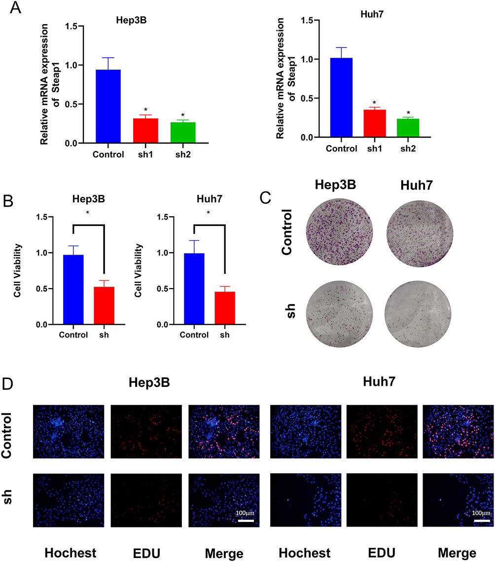 |
Figure 1 STEAP1 promotes the proliferation of HCC cells. (A) Validation of differences in STEAP1 mRNA levels between control and sh groups in Hep3B and Huh7 cell lines. Data are mean ± SD (n=3). Statistical analysis was performed using One-way ANOVA followed by LSD-t test (df=6), P<0.05*VS Control. (B) CCK-8 assay was conducted to determine changes in cell viability after knockdown of STEAP1. Data: mean ± SD (n=3). Stats: independent samples t-test (df=2), P<0.05 *. (C) The clone formation assay was used to assess alterations in the proliferation capability of cells after knockdown of STEAP1. (D) Changes in EDU staining after STEAP1 knockdown. |
STEAP1 Promotes the Migration and Invasion of HCC in vitro
To substantiate the impact of STEAP1 on the metastatic potential of HCC cells, we performed wound healing assay in Hep3B and Huh7 cells. The results showed that STEAP1 knockdown markedly impaired cell wound healing (Figure 2A–D). Additionally, we utilized Transwell assay to evaluate the invasive and migratory capacities of these cells; data revealed that reduced STEAP1 expression suppressed the migratory and invasive potential of HCC cells (Figure 2E–H). Collectively, these findings demonstrate that STEAP1 acts as a facilitator of invasion and migration in HCC cells.
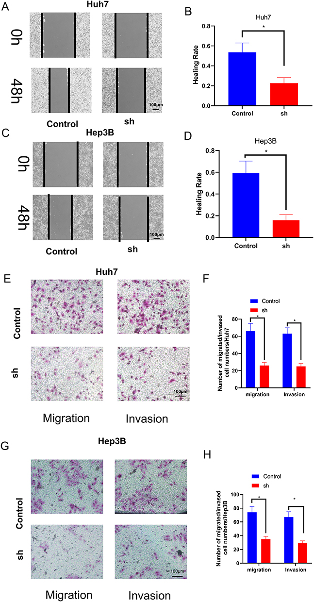 |
Figure 2 STEAP1 promotes the Invasion and migration of HCC in vitro. (A–D) Scratch wound assay to assess the migratory capability of HuH7 (A and B) and Hep-3B. Data: mean ± SD (n=3). Stats: independent samples t-test (df=2), P<0.05 *. (C and D) cells treated with sh-STEAP1 or Control for 48h. Data: mean ± SD (n=3). Stats: independent samples t-test (df=2), P<0.05 *. (E-H) Transwell assay was used to determine the invasive and migratory abilities of sh-STEAP1 group and control group in Huh7 (E and F) and Hep3B (G and H) cells. Cells were treated for 48h and photographed under a 200* microscope field of view. Data: mean ± SD (n=3). Stats: independent samples t-test (df=2), P<0.05 *. |
STEAP1 Regulates EMT and Wnt Signaling Pathways in HCC Cells
A strong association between EMT and malignant tumor metastasis has been reported in a number of papers. To delve deeper into the connection between STEAP1 and HCC metastasis, we employed Western Blot to assess the expression of EMT-associated proteins following the suppression of STEAP1. The findings indicated that reduced STEAP1 expression in Hep3B and Huh7 cell lines led to a marked decrease in N-cadherin and Vimentin levels, while concurrently enhancing E-cadherin expression (Figure 3A–D). To further explore how STEAP1 functions in hepatocellular carcinoma, we used RNA-seq technology to sequence and analyze the mRNA levels in HCC cells from STEAP1 knockdown and control groups. The findings indicated that, compared to the control group, the reduction of STEAP1 expression caused alterations in the mRNA levels of numerous downstream genes. Additionally, KEGG pathway enrichment analysis revealed significant changes in the Wnt signaling pathway (Figure 3E and F). Therefore, we decided to further investigate whether STEAP1 plays a role in HCC through the Wnt signaling pathway. Subsequently, we utilized Western Blot (WB) analysis to identify proteins associated with the Wnt signaling pathway. The findings indicated that decreased STEAP1 expression could suppress Wnt1 levels and affect β-cadherin protein expression (Figure 3G–J). These findings suggest that STEAP1 is able to regulate the alteration of Wnt signaling pathway.
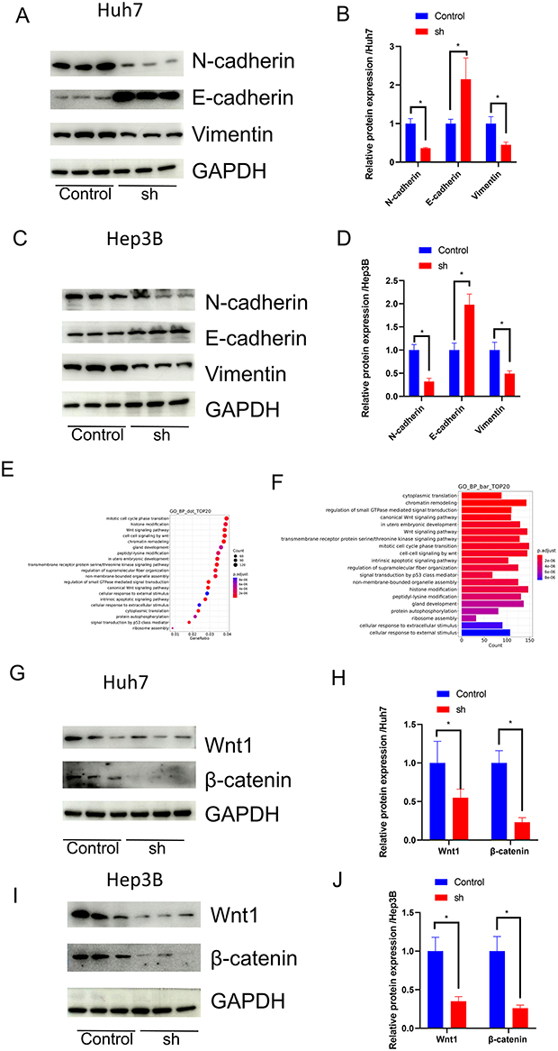 |
Figure 3 STEAP1 regulates EMT and Wnt signaling pathways in HCC cells. (A) WB was used to detect changes in EMT-associated proteins between the sh-STEAP1 group and the control group in Huh7 cells. (B) Bar graphs represent the expression differences of EMT-related proteins in Huh7 cells between the sh-STEAP1 group and the control group. Data: mean ± SD (n=3). Stats: independent samples t-test (df=2), P<0.05 *. (C) WB was used to detect changes in EMT-associated proteins between the sh-STEAP1 group and the control group in Hep3B cells. (D) Bar graphs depict the expression differences of EMT-related proteins in Hep3B cells between the sh-STEAP1 group and the control group. Data: mean ± SD (n=3). Stats: independent samples t-test (df=2), P<0.05 *. (E and F) Knockdown of STEAP1 in Huh7 cells was followed by mRNA sequencing and subsequent GO enrichment analysis. (G) WB was used to detect changes in the expression of Wnt pathway-associated proteins following knockdown of STEAP1 in Huh7 cells. (H) Quantitative analysis of Wnt-associated proteins in Huh7 cells. Data: mean ± SD (n=3). Stats: independent samples t-test (df=2), P<0.05 *. (I) WB was used to detect changes in the expression of Wnt pathway-associated proteins following knockdown of STEAP1 in Hep3B cells. (J) Quantitative analysis of Wnt-associated proteins in Hep3B cells. Data: mean ± SD (n=3). Stats: independent samples t-test (df=2), P<0.05 *. |
STEAP1 Promotes HCC Proliferation in vivo
To elucidate the role of STEAP1 in HCC progression, we performed a subcutaneous tumor xenograft assay in nude mice. Our findings showed that reduced STEAP1 expression significantly suppressed the growth of subcutaneous tumors in nude mice, as evidenced by decreased tumor size and weight (Figure 4A and B). These results indicate that STEAP1 enhances the proliferative potential of HCC in vivo.We further performed PCNA and Ki-67 immunohistochemical staining on the excised subcutaneous tumor tissues. Results demonstrated that STEAP1 knockdown notably reduced the positive rates of both PCNA and Ki-67 (Figure 4C and D), suggesting that STEAP1 silencing significantly inhibits the proliferative activity of HCC cells. PCNA and Ki-67 are well-recognized biomarkers closely associated with cell cycle progression and cell proliferation,28,29 The decreased positive rates of these two markers indicate that STEAP1 depletion impairs the ability of HCC cells to enter the cell cycle and undergo mitotic division, thereby suppressing HCC cell proliferation in vitro.
 |
Figure 4 STEAP1 promotes HCC proliferation in vivo. (A) Tumor growth curves are depicted. (B) Representative images of tumor tissues were shown (n = 6). (C) Differences in Ki-67 staining between subcutaneous tumors from various treatment groups. Data: mean ± SD (n=6). Stats: independent samples t-test (df=5), P<0.05 *. (D) PCNA staining varies between treatment groups. Data: mean ± SD (n=6). Stats: independent samples t-test (df=5), P<0.05 *. |
Collectively, these in vitro and in vivo findings confirm that STEAP1 plays a pro-proliferative role in HCC cells.
STEAP1 Promotes HCC Invasion and Migration by Activating the Wnt Signaling Pathway
To ascertain whether STEAP1 facilitates the invasion and migration capabilities of HCC through the Wnt signaling pathway, we treated Huh7 and Hep3B cells with Wnt/β-catenin agonist 2, an activator of the Wnt pathway. The findings showed that Wnt/β-catenin agonist 2 is capable to partially restore the scratch would healing ability of Huh7 and Hep3B cells impaired by STEAP1 knockdown (Figure 5A–D). This suggests that STEAP1 could enhance the migratory ability of HCC by activating the Wnt signaling pathway. Subsequently, we validated the invasive and migratory capabilities of HCC cells using the transwell assay. Consistently, the results showed that Wnt/β-catenin agonist 2 could revert the suppression of invasion and migration in STEAP1- knockdown Huh7 and Hep3B caused by (Figure 5E–H). Collectively, these results corroborate that STEAP1 enhances the invasion and migration capabilities of HCC by activating the Wnt signaling pathway.
 |
Figure 5 STEAP1 promotes HCC invasion and migration by activating the Wnt signaling pathway. (A–D) Scratch wound assay was performed to assess the migratory capacity of HuH7 (A and B) and Hep-3B (C and D) cells treated with sh-STEAP1 and Wnt/β-catenin agonist 2 for 48h. Data: mean ± SD (n=3). Stats: One-way ANOVA + LSD-t test (df=6), P<0.05*. (E–H) Transwell assay was used to determine the invasive and migratory capabilities of sh-STEAP1 group and Wnt/β-catenin agonist 2 in Huh7 (E and F) and Hep3B (G and H) cells. Cells were treated for 48h and photographed under a 200* microscope field of view. Data: mean ± SD (n=3). Stats: One-way ANOVA + LSD-t test (df=6), P<0.05*. |
Discussion
Accumulating research findings have established that selective targeting of STEAP1 not only effectively halts the malignant progression of prostate cancer but also translates into enhanced prognostic benefits for patients with this disease.30,31 Some studies have also identified the downregulation of STEAP1 expression as a predictor of good prognosis in lung cancer through bioinformatics analysis, suggesting its potential as a prognostic marker for lung cancer.32,33 Some studies have proven that STEAP1 is associated with the proliferative ability of HCC cell lines.15 However, the specific biological role and underlying regulatory mechanism of STEAP1 in malignant tumors, particularly in HCC, remain largely elusive.34,35 These investigations have inspired us to further explore the biological functions and underlying mechanisms of STEAP1 in HCC. Our study primarily focuses on the regulatory role of STEAP1 in the invasion and migration of HCC cells, with findings validated by complementary in vivo experiments and potential mechanisms proposed herein. Thus, our research possesses distinct novelty and originality. Additionally, our data indicate that high STEAP1 expression may serve as a biomarker for poor prognosis in HCC patients. Consistently, STEAP1 knockdown was shown to effectively inhibit the proliferation and migration of HCC cells, which further corroborates the conclusions of previous relevant studies.
The epithelial-mesenchymal transition (EMT) plays a crucial role in the metastasis of malignant tumor cells.36,37 To further explore the functional mechanism of STEAP1 in HCC, we focused our research on EMT. Our results confirmed that STEAP1 can inhibit EMT in HCC, thereby promoting HCC metastasis.
Through transcriptomic and bioinformatics analyses, we found that the knockdown of STEAP1 modulates downstream effects of the WNT/β-catenin pathway, a critical pathway in tumorigenesis and tumor development. The WNT/β-catenin pathway, a conserved and traditional signaling pathway, regulates cell proliferation and differentiation.21,22 The activation of the WNT signaling pathway is linked to adverse outcomes in various cancers, including hepatocellular carcinoma, cholangiocarcinoma, breast cancer, and stomach cancer. Extensive research findings indicate that inhibiting the activation of the WNT pathway significantly diminishes the proliferation and metastasis of HCC, resulting in improved outcomes.
We treated HCC cell lines with Wnt/β-catenin agonist 2, a selective activator of the canonical Wnt/β-catenin signaling pathway.38 Our results demonstrated that Wnt/β-catenin agonist 2 restored the invasive and migratory capacities of HCC cells impaired by STEAP1 knockdown. This indicates that STEAP1 promotes the malignant biological behaviors of HCC by activating the Wnt signaling pathway. Furthermore, our findings revealed a significant downregulation of the Wnt signaling pathway following STEAP1 silencing. STEAP1 may indirectly regulate the Wnt/β-catenin pathway, and there might exist an undiscovered mediator between them. To identify the direct interacting molecules of STEAP1, further screening experiments based on mass spectrometry are required.
Bispecific T-cell engagers (BiTEs) and other novel T-cell immunotherapies are emerging as promising therapeutic strategies for cancer. It has been suggested that STEAP1 may play a potential role in immunotherapy for prostate cancer.39,40 Inflammatory responses and lipid peroxidation also play a crucial role in the initiation and progression of HCC. Studies have demonstrated that modulation of the Th17/Treg axis can reduce the risk of recurrence after hepatectomy for HCC, which underscores the pivotal role of immune factors in the development and progression of HCC.41,42 A recent Raw Letter Analysis article on the prognosis of HCC based on the STEAP family suggests that the unique characteristics of STEAP may serve as a valid prognostic indicator for HCC and could be linked to tumor immune dysfunction in HCC.43 However, the specific mechanism by which STEAP1 is associated with immunotherapy for HCC awaits further investigation.
Although our data demonstrate that STEAP1 promotes HCC progression via the Wnt/β-catenin pathway, several limitations should be noted for clinical translation. First, tumor heterogeneity causes variable STEAP1 expression across HCC patients, with low-expression tumors showing intrinsic resistance to targeted therapy; dynamic STEAP1 downregulation may also induce acquired resistance. Second, our preclinical data from cell lines and xenograft models do not fully recapitulate the clinical HCC microenvironment, lacking validation in PDX or organoid models. STEAP1 targeting offers multiple clinical prospects. First, combination therapy with first-line agents (sorafenib, lenvatinib) or PD-1/PD-L1 inhibitors may synergistically enhance anti-tumor efficacy by reversing immunosuppression via Wnt/β-catenin pathway modulation. Second, theranostic integration could be achieved via STEAP1-specific probes for early screening and intraoperative localization, plus targeted nanocarriers for precise drug delivery. Third, patient stratification using STEAP1 expression combined with β-catenin biomarkers may identify the subset most likely to benefit from targeted therapy.
In conclusion, our study suggests that STEAP1 promotes HCC proliferation and metastasis in vitro and in vivo via the Wnt/β-catenin signaling pathway.
Data Sharing Statement
The datasets used and/or analyzed in the course of the present research are available from the corresponding author on reasonable request.
Ethics Approval
Informed consent was obtained for all experiments on human subjects/samples. The procurement of human tissues and mice in this study adhered to the principles outlined in the Declaration of Helsinki and received approval from the Ethics Committee of the First Affiliated Hospital of zhengzhou University (2019-KY-21).
Acknowledgments
This work was supported by the National Natural Science Foundation of China (82170670).
Author Contributions
All authors made a significant contribution to the work reported, whether that is in the conception, study design, execution, acquisition of data, analysis and interpretation, or in all these areas; took part in drafting, revising or critically reviewing the article; gave final approval of the version to be published; have agreed on the journal to which the article has been submitted; and agree to be accountable for all aspects of the work.
Disclosure
The authors declare that they have no conflict of interest.
References
1. McGlynn KA, Petrick JL, El-Serag HB. Epidemiology of hepatocellular carcinoma. Hepatology. 2021;73(Suppl 1):4–13. doi:10.1002/hep.31288
2. Yang JD, Hainaut P, Gores GJ, Amadou A, Plymoth A, Roberts LR. A global view of hepatocellular carcinoma: trends, risk, prevention and management. Nat Rev Gastroenterol Hepatol. 2019;16(10):589–604. doi:10.1038/s41575-019-0186-y
3. Donne R, Lujambio A. The liver cancer immune microenvironment: therapeutic implications for hepatocellular carcinoma. Hepatology. 2023;77(5):1773–1796. doi:10.1002/hep.32740
4. Jiang Y, Han Q, Zhao H, Zhang J. The mechanisms of HBV-induced hepatocellular carcinoma. J Hepatocell Carcinoma. 2021;8:435–450. doi:10.2147/jhc.S307962
5. Llovet JM, De Baere T, Kulik L, et al. Locoregional therapies in the era of molecular and immune treatments for hepatocellular carcinoma. Nat Rev Gastroenterol Hepatol. 2021;18(5):293–313. doi:10.1038/s41575-020-00395-0
6. Wang Y, Deng B. Hepatocellular carcinoma: molecular mechanism, targeted therapy, and biomarkers. Cancer Metastasis Rev. 2023;42(3):629–652. doi:10.1007/s10555-023-10084-4
7. Reig M, Forner A, Rimola J, et al. BCLC strategy for prognosis prediction and treatment recommendation: the 2022 update. J Hepatol. 2022;76(3):681–693. doi:10.1016/j.jhep.2021.11.018
8. Sartoris R, Gregory J, Dioguardi Burgio M, Ronot M, Vilgrain V. HCC advances in diagnosis and prognosis: digital and imaging. Liver Int. 2021;41 Suppl 1(S1):73–77. doi:10.1111/liv.14865
9. Nakamura H, Arihara Y, Takada K. Targeting STEAP1 as an anticancer strategy. Front Oncol. 2023;13:1285661. doi:10.3389/fonc.2023.1285661
10. Xu M, Evans L, Bizzaro CL, et al. STEAP1-4 (six-transmembrane epithelial antigen of the prostate 1-4) and their clinical implications for prostate cancer. Cancers. 2022;14(16):4034. doi:10.3390/cancers14164034
11. Rosellini M, Santoni M, Mollica V, et al. Treating prostate cancer by antibody-drug conjugates. Int J Mol Sci. 2021;22(4):1551. doi:10.3390/ijms22041551
12. Sardinha M, Palma Dos Reis AF, Barreira JV, Fontes Sousa M, Pacey S, Luz R. Antibody-drug conjugates in prostate cancer: a systematic review. Cureus. 2023;15(2):e34490. doi:10.7759/cureus.34490
13. Mjaess G, Aoun F, Rassy E, Diamand R, Albisinni S, Roumeguère T. Antibody-drug conjugates in prostate cancer: where are we? Clin Genitourin Cancer. 2023;21(1):171–174. doi:10.1016/j.clgc.2022.07.009
14. Gomes IM, Maia CJ, Santos CR. STEAP proteins: from structure to applications in cancer therapy. Mol Cancer Res. 2012;10(5):573–587. doi:10.1158/1541-7786.Mcr-11-0281
15. Iijima K, Nakamura H, Takada K, et al. Six-transmembrane epithelial antigen of the prostate 1 accelerates cell proliferation by targeting c-Myc in liver cancer cells. Oncol Lett. 2021;22(1):546. doi:10.3892/ol.2021.12807
16. Liu J, Xiao Q, Xiao J, et al. Wnt/β-catenin signalling: function, biological mechanisms, and therapeutic opportunities. Signal Transduct Target Ther. 2022;7(1):3. doi:10.1038/s41392-021-00762-6
17. Disoma C, Zhou Y, Li S, Peng J, Xia Z. Wnt/β-catenin signaling in colorectal cancer: is therapeutic targeting even possible? Biochimie. 2022;195:39–53. doi:10.1016/j.biochi.2022.01.009
18. Zhang Y, Wang X. Targeting the Wnt/β-catenin signaling pathway in cancer. J Hematol Oncol. 2020;13(1):165. doi:10.1186/s13045-020-00990-3
19. Wu W, Nelson GM, Koch R, et al. Overcoming IMiD resistance in T-cell lymphomas through potent degradation of ZFP91 and IKZF1. Blood. 2022;139(13):2024–2037. doi:10.1182/blood.2021014701
20. Xia J, Zhu Z, Wen G, et al. Aberrant acetylated modification of FGF21‑KLB signaling contributes to hepatocellular carcinoma metastasis through the β‑catenin pathway. Int J Oncol. 2023;63(2). doi:10.3892/ijo.2023.5539
21. He S, Tang S. WNT/β-catenin signaling in the development of liver cancers. Biomed Pharmacother. 2020;132:110851. doi:10.1016/j.biopha.2020.110851
22. Perugorria MJ, Olaizola P, Labiano I, et al. Wnt–β-catenin signalling in liver development, health and disease. Nat Rev Gastroenterol Hepatol. 2019;16(2):121–136. doi:10.1038/s41575-018-0075-9
23. Davis L, Mills KI, Orchard KH, Guinn BA. Identification of genes whose expression overlaps age boundaries and correlates with risk groups in paediatric and adult acute myeloid leukaemia. Cancers. 2020;12(10):2769. doi:10.3390/cancers12102769
24. Zhang D, Han S, Pan X, et al. EFEMP1 binds to STEAP1 to promote osteosarcoma proliferation and invasion via the Wnt/β-catenin and TGF-β/Smad2/3 signal pathways. J Bone Oncol. 2022;37:100458. doi:10.1016/j.jbo.2022.100458
25. Son G, Hirano T, Seki E, et al. Blockage of HGF/c-Met system by gene therapy (adenovirus-mediated NK4 gene) suppresses hepatocellular carcinoma in mice. J Hepatol. 2006;45(5):688–695. doi:10.1016/j.jhep.2006.04.011
26. Qian X, Zhou Q, Ouyang Y, et al. Transferrin promotes fatty acid oxidation and liver tumor growth through PHD2-mediated PPARα hydroxylation in an iron-dependent manner. Proc Natl Acad Sci USA. 2025;122(5):e2412473122. doi:10.1073/pnas.2412473122
27. Shao YY, Li YS, Hsu HW, et al. Potent activity of composite cyclin dependent kinase inhibition against hepatocellular carcinoma. Cancers. 2019;11(10):1433. doi:10.3390/cancers11101433
28. Zhang Y, Du J, Jin L, Pan L, Yan X, Lin S. Iberverin exhibits antineoplastic activities against human hepatocellular carcinoma via DNA damage-mediated cell cycle arrest and mitochondrial-related apoptosis. Front Pharmacol. 2023;14:1326346. doi:10.3389/fphar.2023.1326346
29. Kim C-W, Lee HK, Nam M-W, Choi Y, Choi K-C. Overexpression of KiSS1 induces the proliferation of hepatocarcinoma and increases metastatic potential by increasing migratory ability and angiogenic capacity. Mol Cells. 2022;45(12):935–949. doi:10.14348/molcells.2022.0105
30. Bhatia V, Kamat NV, Pariva TE, et al. Targeting advanced prostate cancer with STEAP1 chimeric antigen receptor T cell and tumor-localized IL-12 immunotherapy. Nat Commun. 2023;14(1):2041. doi:10.1038/s41467-023-37874-2
31. Nolan-Stevaux O, Li C, Liang L, et al. AMG 509 (xaluritamig), an anti-STEAP1 XmAb 2+1 T-cell redirecting immune therapy with avidity-dependent activity against prostate cancer. Cancer Discov. 2024;14(1):90–103. doi:10.1158/2159-8290.Cd-23-0984
32. Huo S-F, Shang W-L, Yu M, et al. STEAP1 facilitates metastasis and epithelial–mesenchymal transition of lung adenocarcinoma via the JAK2/STAT3 signaling pathway. Biosci Rep. 2020;40(6). doi:10.1042/bsr20193169
33. Liu T, Niu X, Li Y, Xu Z, Chen J, Xu G. Expression and prognostic analyses of the significance of STEAP1 and STEAP2 in lung cancer. World J Surg Oncol. 2022;20(1):96. doi:10.1186/s12957-022-02566-6
34. Wu -Y-Y, Jiang J-N, Fang X-D, Ji F-J. STEAP1 regulates tumorigenesis and chemoresistance during peritoneal metastasis of gastric cancer. Front Physiol. 2018;9:1132. doi:10.3389/fphys.2018.01132
35. Zhang Z, Hou W-B, Zhang C, et al. A research of STEAP1 regulated gastric cancer cell proliferation, migration and invasion in vitro and in vivos. J Cell Mol Med. 2020;24(24):14217–14230. doi:10.1111/jcmm.16038
36. Lamouille S, Xu J, Derynck R. Molecular mechanisms of epithelial-mesenchymal transition. Nat Rev Mol Cell Biol. 2014;15(3):178–196. doi:10.1038/nrm3758
37. Mittal V. Epithelial mesenchymal transition in tumor metastasis. Annu Rev Pathol. 2018;13(1):395–412. doi:10.1146/annurev-pathol-020117-043854
38. Koo JWCRCGHCCJBS, inventor; Isoxazole derivatives and use thereof. Patent WO2007078113A1. 2006.
39. Palecki J, Bhasin A, Bernstein A, et al. T-Cell redirecting bispecific antibodies: a review of a novel class of immuno-oncology for advanced prostate cancer. Cancer Biol Ther. 2024;25(1):2356820. doi:10.1080/15384047.2024.2356820
40. Zhang J, Chadha JS. Developmental therapeutics in metastatic prostate cancer: new targets and new strategies. Cancers. 2024;16(17):3098. doi:10.3390/cancers16173098
41. Xia J, Fu B, Wang Z, et al. MVP enhances FGF21-induced ferroptosis in hepatocellular carcinoma by increasing lipid peroxidation through regulation of NOX4. Clin Transl Sci. 2024;17(8):e13910. doi:10.1111/cts.13910
42. Ren H, Chen Y, Zhu Z, et al. FOXO1 regulates Th17 cell-mediated hepatocellular carcinoma recurrence after hepatic ischemia-reperfusion injury. Cell Death Dis. 2023;14(6):367. doi:10.1038/s41419-023-05879-w
43. Fu D, Zhang X, Zhou Y, Hu S. A novel prognostic signature and therapy guidance for hepatocellular carcinoma based on STEAP family. BMC Med Genomics. 2024;17(1):16. doi:10.1186/s12920-023-01789-0
 © 2026 The Author(s). This work is published and licensed by Dove Medical Press Limited. The
full terms of this license are available at https://www.dovepress.com/terms
and incorporate the Creative Commons Attribution
- Non Commercial (unported, 4.0) License.
By accessing the work you hereby accept the Terms. Non-commercial uses of the work are permitted
without any further permission from Dove Medical Press Limited, provided the work is properly
attributed. For permission for commercial use of this work, please see paragraphs 4.2 and 5 of our Terms.
© 2026 The Author(s). This work is published and licensed by Dove Medical Press Limited. The
full terms of this license are available at https://www.dovepress.com/terms
and incorporate the Creative Commons Attribution
- Non Commercial (unported, 4.0) License.
By accessing the work you hereby accept the Terms. Non-commercial uses of the work are permitted
without any further permission from Dove Medical Press Limited, provided the work is properly
attributed. For permission for commercial use of this work, please see paragraphs 4.2 and 5 of our Terms.