
Back to Journals » International Journal of General Medicine » Volume 19
Screening for Abdominal Aortic Aneurysm in Elderly Male Patients with Coronary Artery Disease During Standard Transthoracic Echocardiography
Authors Lei Y  , Zhang L, Wang M , Wang L, Su J, Zhou M, Wang X, Qi X, Xue Y
Received 14 April 2026
Accepted for publication 19 June 2026
Published 25 June 2026 Volume 2026:19 616767
DOI https://doi.org/10.2147/IJGM.S616767
Checked for plagiarism Yes
Review by Single anonymous peer review
Peer reviewer comments 3
Editor who approved publication: Dr Redoy Ranjan
Yuping Lei,1,2,* Li Zhang,1,3,4,* Mengmeng Wang,5 Lei Wang,6 Jingli Su,1 Min Zhou,1 Xiaohua Wang,1 Xuewen Qi,1 Yuzeng Xue1
1Department of Cardiology, Liaocheng People’s Hospital, Liaocheng, People’s Republic of China; 2Medical Integration and Practice Center, Shandong University, Jinan, People’s Republic of China; 3Second Clinical Medical College, Shandong University of Traditional Chinese Medicine, Jinan, People’s Republic of China; 4Department of Dermatology, Liaocheng Traditional Chinese Medicine Hospital, Liaocheng, People’s Republic of China; 5School of Clinical Medicine, Shandong Second Medical University, Weifang, People’s Republic of China; 6Department of Comprehensive Ultrasound, Jinan Central Hospital, Jinan, People’s Republic of China
*These authors contributed equally to this work
Correspondence: Xuewen Qi, Department of Cardiology, Liaocheng People’s Hospital, No. 67, Dongchang West Road, Liaocheng, People’s Republic of China, Tel: +86 13306359016, Email [email protected] Yuzeng Xue, Department of Cardiology, Liaocheng People’s Hospital, No. 67, Dongchang West Road, Liaocheng, People’s Republic of China, Tel +86 13346256908, Email [email protected]
Purpose: No studies on abdominal aortic aneurysm (AAA) screening in elderly male patients with coronary artery disease (CAD) in China have been conducted. This study aims to determine AAA prevalence, identify risk factors, and assess transthoracic echocardiography (TTE) as a screening modality to optimize screening strategies and enhance early diagnosis.
Patients and Methods: This observational study prospectively enrolled male CAD patients aged ≥ 65 who had coronary angiography at Liaocheng People’s Hospital from January to June 2024. All underwent routine TTE with abdominal aortic diameter measurements for AAA screening. AAA prevalence was calculated, and clinical characteristics were compared using appropriate tests. Univariate and multivariate logistic regression analyses were done to find independent risk factors.
Results: 384 elderly male CAD patients were included, all completing AAA screening via TTE in 2.1± 1.7 minutes on average. AAA was detected in 23 patients (6.0%), and 361 had no evidence. 4 AAA cases (17.4%) with aneurysms ≥ 50 mm were referred to vascular surgery. Multivariate analysis identified age, smoking history, hypertension, and multivessel coronary disease as independent risk factors. The overall AAA prevalence was 6.0% (23/384), and it was 16.8% (16/95) in patients with concurrent multivessel disease, hypertension, and smoking history.
Conclusion: This study shows a relatively high AAA prevalence among elderly male CAD patients in China and identifies multivessel disease as a potential independent risk factor for the first time, although this finding should be interpreted cautiously. TTE, a non - invasive and easily implementable tool, is effective in AAA detection and suitable for routine cardiac evaluations of CAD patients. These findings provide a basis for AAA screening in this high - risk population, potentially improving early detection, reducing rupture risk, and enhancing patient outcomes.
Keywords: abdominal aortic aneurysm, coronary artery disease, transthoracic echocardiography, screening, risk factors
Introduction
Abdominal aortic aneurysm (AAA) is a degenerative cardiovascular disorder characterized by permanent, localized dilation of the abdominal aorta, clinically defined as an aortic diameter exceeding 30mm.1,2 The disease progression is often insidious, with minimal clinical manifestations during its prolonged development. However, rupture of the aneurysm is associated with a mortality rate surpassing 80%.3 Early diagnosis of AAA remains challenging, as detection frequently occurs incidentally during evaluations for unrelated conditions or post-rupture. Consequently, early screening and timely intervention are essential to mitigate mortality and improve clinical outcomes.
Current screening protocols predominantly derive from studies conducted in European and American populations.4 Notably, epidemiological data reveal regional disparities in AAA prevalence, with lower incidence rates observed in Asian populations compared to their Western counterparts—a phenomenon potentially attributable to genetic, lifestyle, and environmental factors.5 Given that screening cost-effectiveness is contingent upon disease prevalence, it is imperative to delineate high-risk subgroups within the Chinese population and elucidate associated risk factors to optimize targeted screening strategies.
Emerging evidence suggests that the pathophysiological link between coronary artery disease (CAD) and AAA may involve shared mechanisms, including atherosclerosis, chronic inflammation, and vascular remodeling.6 Atherosclerotic degeneration of the aortic wall can compromise vascular elasticity, predisposing to aneurysm formation.7 Furthermore, chronic inflammatory cascades may induce endothelial dysfunction and smooth muscle cell apoptosis, exacerbating AAA pathogenesis.8 Established risk factors for AAA encompass advanced age, male sex, smoking history, and familial predisposition,9 with males aged ≥65 years representing a particularly high-risk cohort.10,11 Additional contributory factors include diabetes mellitus, dyslipidemia, and peripheral vascular disease.12–14 Given that elderly males constitute a high-risk demographic for both CAD and AAA, integrating AAA screening into the clinical management of elderly male CAD patients may enhance diagnostic yield, improve cost-efficiency, and ultimately reduce mortality.15,16
The early diagnosis of AAA predominantly relies on imaging modalities, with conventional screening approaches including abdominal ultrasound, computed tomography (CT), and magnetic resonance imaging (MRI). Among these, abdominal ultrasound is the most widely utilized screening tool owing to its non-invasive nature, cost-effectiveness, and procedural convenience.17,18 Transthoracic echocardiography (TTE), a standard cardiovascular diagnostic procedure, not only evaluates cardiac structure and function but also demonstrates utility in detecting abdominal aortic abnormalities.19 The concurrent assessment of the abdominal aorta during TTE requires no supplementary equipment or additional appointments, thereby offering advantages in efficiency, safety, and cost-effectiveness. Emerging evidence suggests that TTE may serve as a viable modality for AAA screening, with recent studies demonstrating its capacity to identify early abdominal aortic dilation in elderly high-risk populations.20
While current research has predominantly examined AAA screening in patients with CAD, investigations targeting specific high-risk subgroups—such as elderly Chinese male CAD patients—remain limited. Addressing this knowledge gap, the present study seeks to evaluate the prevalence and risk factors of AAA in this demographic while assessing the feasibility of TTE as a screening tool. The findings aim to contribute novel evidence supporting early AAA detection strategies in high-risk populations.
Materials and Methods
Study Design
This prospective observational study was designed to assess the prevalence and associated risk factors of AAA in elderly male patients with CAD. Furthermore, the study evaluated the feasibility of routine TTE as a screening modality for AAA detection in this population. The study protocol received ethical approval from the Institutional Review Board of Liaocheng People’s Hospital (protocol code: 2024390).
Study Population
As depicted in Figure 1, the study population comprised consecutive elderly male patients hospitalized in the Department of Cardiology at Liaocheng People’s Hospital between January 2024 and June 2024. All participants were diagnosed with CAD based on coronary angiography (CAG) findings.
 |
Figure 1 The flowchart of patient selection for the study. Abbreviations: CAD, coronary artery disease; CAG, coronary angiography; TTE, Transthoracic echocardiography; AAA, abdominal aortic aneurysm. |
The inclusion criteria were as follows: (1) male sex; (2) age ≥65 years; (3) confirmed CAD diagnosis by CAG (defined as ≥50% luminal stenosis in at least one major coronary artery or its major branch, or a history of coronary revascularization); and (4) provision of written informed consent.
Patients were excluded if they met any of the following criteria: (1) inadequate visualization of the abdominal aorta on ultrasound due to obesity or excessive intestinal gas; (2) acute life-threatening conditions, including cardiogenic shock or decompensated heart failure; or (3) inability or unwillingness to provide informed consent.
Collection and Definition of Clinical Parameters
Comprehensive clinical data were systematically extracted from medical records, encompassing demographic characteristics (age, body mass index [BMI]), behavioral factors (smoking status), comorbid conditions (hypertension, hyperlipidemia, diabetes mellitus, prior myocardial infarction, chronic heart failure, atrial fibrillation, prior cerebral infarction, peripheral atherosclerotic disease, chronic kidney disease, chronic obstructive pulmonary disease), laboratory parameters (white blood cell count, hemoglobin, uric acid, homocysteine levels), medication history, and CAD classification. Standardized operational definitions were applied to all clinical variables.21,22
Coronary Angiography
CAG was performed in accordance with standard procedure.23 CAD was quantitatively defined as luminal stenosis exceeding 50% in at least one major coronary vessel, as confirmed by percutaneous CAG. Left main disease was specifically characterized by >50% stenosis in the left main coronary artery. Lesion severity was classified based on the number of affected major coronary territories (left anterior descending artery, left circumflex artery, right coronary artery, and their primary branches), with categories including single-vessel, double-vessel, or triple-vessel disease. Multivessel disease was defined as involvement of two or more major coronary vessels.
TTE Assessment
Comprehensive TTE examinations were performed by cardiac sonographers following established protocols. Standard two-dimensional, M-mode, and Doppler imaging modalities were systematically acquired and analyzed, with all data recorded in a standardized electronic database. Key measurements included interventricular septal thickness at end-diastole (IVSd), left ventricular posterior wall thickness at end-diastole (LVPWd), left ventricular end-diastolic diameter (LVEDD), left ventricular ejection fraction (LVEF), left atrial anteroposterior diameter (LA), and ascending aortic diameter (AAO). All the echocardiographic parameters were assessed following established guidelines.24,25
As part of the cardiac evaluation, TTE was utilized to measure abdominal aortic dimensions for AAA screening. All TTE exams were performed by the same two sonographers, and interobserver variability was not assessed as this was a screening study. The abdominal aorta was imaged using the standard cardiac transducer without requiring additional equipment or separate patient visits.
The abdominal aortic imaging protocol was as follows: Patients were maintained in a supine position with the transducer marker aligned caudally. Following longitudinal visualization of the abdominal aorta, the transducer was rotated 90° counterclockwise to obtain transverse cross-sectional images perpendicular to the aortic axis. Systematic imaging commenced inferior to the renal artery origins, with continuous distal tracking to the maximal feasible extent. The maximum anteroposterior diameter was recorded in the short-axis plane, as this measurement demonstrates superior reproducibility compared to transverse diameter assessment. The total duration of abdominal aortic examination was documented. AAA diagnosis was established when the maximal aortic diameter measured ≥30 mm. Representative TTE findings of AAA detection are illustrated in Figure 2.
 |
Figure 2 TTE imaging findings in patients with AAA. |
Patient Grouping and Clinical Management
A total of 384 eligible elderly male patients with CAD were enrolled in this study. All participants successfully underwent abdominal aortic assessment via TTE, with a mean examination duration of 2.1±1.7 minutes. Based on screening outcomes, patients were stratified into two groups: 23 cases (6.0%) with concomitant AAA and 361 cases (94.0%) without AAA. Figure 3 presents the distribution of abdominal aortic diameters across the cohort, demonstrating a median diameter of 18 mm (range: 11–58 mm).
 |
Figure 3 Frequency distribution of abdominal aortic diameter in all patients. |
Figure 4 details the size distribution among AAA cases. Of the 23 diagnosed AAA patients, 12 (52.2%) exhibited aneurysms measuring 30–39 mm in maximal diameter, 7 (30.4%) measured 40–49 mm, and 4 (17.4%) presented with aneurysms ≥50 mm. Clinical management adhered to established guidelines: patients with aneurysms <50 mm (n=19) received scheduled ultrasonographic surveillance coupled with comprehensive lifestyle modification counseling and individualized risk factor management. Cases with aneurysms ≥50 mm (n=4) were referred for vascular surgical consultation to evaluate operative indications based on comprehensive clinical and systemic assessments.
 |
Figure 4 Frequency distribution of AAA patients based on diameter size. Abbreviations: AAA, abdominal aortic aneurysm. |
Statistical Methods
Comparative analyses of baseline characteristics were performed using independent samples t-tests for continuous variables and chi-square tests for categorical variables. The sample size was not formally justified a priori due to the observational pilot nature of this study. Univariate logistic regression was initially conducted to identify potential risk factors associated with AAA, followed by multivariate logistic regression analysis incorporating variables meeting predefined criteria (univariate P<0.1 or clinical significance). All statistical analyses were executed using SPSS version 27.0 (SPSS Inc., Chicago, IL, USA), with a two-tailed P-value <0.05 considered statistically significant.
Results
Comparative Analysis of Baseline Characteristics
Table 1 summarizes the comparative analysis of baseline clinical parameters between the two cohorts. Evaluated variables encompassed demographic characteristics (age, BMI), cardiovascular risk factors (smoking status, hypertension, hyperlipidemia, diabetes mellitus), medical history (prior myocardial infarction, cerebrovascular events, PAD, CKD, COPD), laboratory parameters (white blood cell count, hemoglobin, uric acid, homocysteine levels), medication use (aspirin, statins, ACE inhibitors/ARBs), and the number of coronary artery lesions.
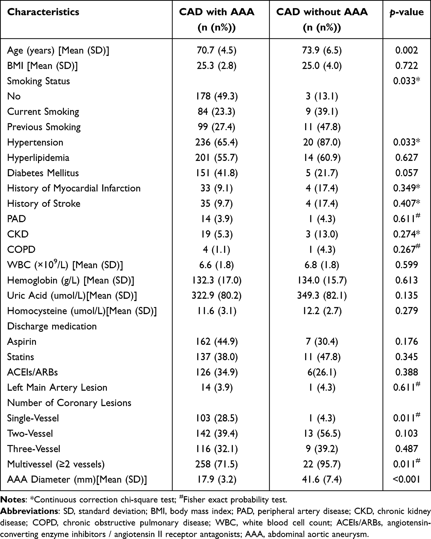 |
Table 1 Comparison of Baseline Clinical Factors Between the CAD with AAA Group and the CAD Without AAA Group |
The AAA cohort demonstrated significantly advanced mean age (73.9±6.5 years) compared to the non-AAA group (70.7±4.5 years; p=0.002). The proportion of smoking history in the AAA group was significantly higher (P=0.033). The current smoking prevalence was markedly higher in AAA patients (39.1% vs 23.3%), and similar trends were observed for former smokers (47.8% vs 27.4%). Hypertension prevalence differed significantly between groups (82.6% vs 65.9%; p=0.033). While diabetes mellitus showed lower prevalence in the AAA group (21.7% vs 41.8%), this difference approached but did not reach statistical significance (p=0.057). Notably, multivessel CAD was significantly more prevalent in AAA patients (95.7% vs 71.5%; p=0.011).
Echocardiographic Characteristics
Table 2 presents a comparative analysis of echocardiographic parameters between the two groups. Statistical analysis revealed no significant differences in parameters including IVSd, LVPWd, LVEDD, LVEF, LA, and AAO between elderly male patients with CAD, stratified by the presence or absence of abdominal aortic aneurysm (all P-values>0.05).
 |
Table 2 Comparison of Echocardiographic Parameters Between the CAD with AAA Group and the CAD Without AAA Group |
Risk Factors for AAA in Elderly Male Patients with CAD
To determine independent predictors of AAA in elderly male patients with CAD, both univariate and multivariate logistic regression analyses were performed. As presented in Table 3, univariate analysis identified age, smoking status, hypertension, diabetes mellitus, and the extent of coronary artery involvement as potential risk factors for AAA (P<0.10). Specifically, in male CAD patients aged ≥65 years, each incremental year of age was associated with a 12.2% increase in AAA risk (OR=1.122, 95% CI: 1.040–1.210, P=0.003). Current smokers (OR=6.357, 95% CI: 1.678–24.088, P=0.007) and former smokers (OR=6.593, 95% CI: 1.797–24.191, P=0.004) demonstrated significantly elevated AAA risks compared to non-smokers. Hypertensive patients exhibited a 3.5-fold higher AAA risk (OR = 3.531, 95% CI: 1.029–12.113, P=0.045), while multi-vessel CAD was associated with an 8.8-fold increased risk (OR=8.783, 95% CI: 1.169–66.010, P=0.035). Interestingly, diabetes showed a trend toward reduced AAA risk (OR=0.386, 95% CI: 0.140–1.063, P=0.066).
 |
Table 3 Univariate and Multivariate Logistic Regression Analyses of Risk Factors for AAA in Elderly Male Patients with CAD |
Significant variables from the univariate analysis were subsequently incorporated into a multivariate logistic regression model. The final analysis revealed that advancing age (OR=1.127, 95% CI: 1.034–1.224, P=0.006), smoking history (current smoking: OR=7.279, 95% CI: 1.825–29.044, P=0.005; former smoking: OR=7.611, 95% CI: 1.979–29.267, P=0.003), hypertension (OR=4.736, 95% CI: 1.290–17.389, P=0.019), and multi-vessel CAD (OR=9.979, 95% CI: 1.275–78.099, P=0.028) remained independent predictors of AAA. Notably, diabetes mellitus did not retain statistical significance in the multivariate analysis. Given the limited number of AAA events, multivariate analysis is at higher risk of overfitting. Therefore, these results should be interpreted with caution.
Prevalence Analysis of AAA in Male CAD Patients Aged ≥65 Years
As illustrated in Figure 5, multivariate regression analysis revealed that age, smoking history, hypertension, and multivessel CAD independently predicted AAA occurrence in elderly male CAD patients. Within the cohort of 384 male CAD patients aged ≥65 years, the overall AAA prevalence was 6.0% (23/384). Notably, among the subset of 183 patients with a smoking history, AAA prevalence significantly increased to 10.9% (20/183). Further stratification demonstrated that in the 137 patients with concurrent hypertension and smoking history, AAA prevalence rose to 12.4% (17/137). The highest prevalence (16.8%, 16/95) was observed in the subgroup of 95 patients exhibiting the triad of multivessel CAD, hypertension, and smoking history.
 |
Figure 5 Prevalence of AAA in elderly male patients with CAD with combined risk factors. Abbreviations: AAA, abdominal aortic aneurysm; CAD, coronary artery disease. |
Discussion
This observational study provides a preliminary investigation into the prevalence of AAA, associated risk factors, and the feasibility of TTE as a screening modality among elderly Chinese male patients with CAD. Through multivariate regression analysis, we identified that multivessel CAD is a potential independent risk factor for AAA—a novel finding—while other established risk factors including advanced age, smoking history, and hypertension showed concordance with prior epidemiological evidence.26,27 The present research not only reevaluated conventional AAA risk factors but also contributed novel strategic insights for targeted screening in high-risk populations.
Existing literature has established that AAA prevalence exhibits a strong age-dependent increase, with a particularly pronounced elevation observed in males beyond 65 years of age.28 Our findings corroborated this trend, revealing that AAA patients were significantly older than their non-AAA counterparts, thereby reinforcing the clinical rationale for prioritizing AAA screening in elderly male cohorts. Mechanistically, multiple studies have attributed this age-associated risk to progressive degradation of arterial wall elasticity and vascular smooth muscle cell apoptosis, which collectively contribute to AAA pathogenesis.29 These observations suggest that routine AAA screening may represent a critical intervention for mitigating rupture risk, particularly among elderly male CAD patients.30 Smoking has been consistently identified as an independent risk factor for AAA development. Chronic tobacco use not only promotes coronary atherosclerosis but also accelerates abdominal aortic dilation through multiple pathophysiological mechanisms.31 Our analysis confirmed that smoking history significantly elevated AAA risk in elderly male CAD patients. The deleterious effects of smoking are mediated through chronic inflammatory activation, oxidative stress augmentation, and vascular endothelial dysfunction, all of which exacerbate arterial wall degeneration and potentiate AAA progression.32 Furthermore, this study identified hypertension as a significant AAA risk determinant, with hypertensive patients exhibiting a threefold higher AAA incidence compared to normotensive individuals. This finding aligns with prior epidemiological data33 and underscores the pivotal role of hemodynamic stress in AAA pathogenesis.
This single-center study is the first to identify that multivessel CAD serves as a potential independent risk factor for AAA. Prior investigations have established a significantly higher AAA prevalence among patients with CAD relative to the general population, with a positive correlation observed between AAA incidence and CAD severity. This association likely arises from shared risk factors and common pathophysiological mechanisms, including genetic predisposition and metabolic abnormalities such as dyslipidemia and chronic inflammation.34,35 These findings underscore the clinical importance of early screening and intervention in elderly male CAD patients with multivessel involvement, which may potentially improve patient prognosis.
Current evidence predominantly supports an inverse relationship between diabetes mellitus and AAA incidence, suggesting a protective effect of diabetes against AAA development.36 The precise mechanisms underlying this phenomenon remain incompletely elucidated and may involve enhanced arterial wall matrix deposition, pharmacological effects of antidiabetic medications, or diabetes-associated metabolic alterations.37 However, certain Mendelian randomization studies have failed to confirm a causal protective effect of diabetes, suggesting that the negative correlation observed in observational studies may be attributed to confounding factors or reverse causality.38 Univariate analysis in the present study confirmed reduced AAA risk among diabetic patients compared to non-diabetic individuals. However, this association did not reach statistical significance in multivariate regression analysis, potentially attributable to limited sample size or low diabetes prevalence in our cohort, which may have attenuated the observed effect. Future investigations should employ larger sample sizes and conduct stratified analyses based on clinical characteristics of diabetic patients to elucidate potential population-specific associations.
Epidemiological data indicate an AAA prevalence of 0.5% in Asian populations, substantially lower than rates reported in Western countries (United States: 2.2%; Europe: 2.5%; Australia: 6.7%).39 Given this low baseline prevalence, population-wide AAA screening in Asia demonstrates limited cost-effectiveness. Notably, our study identified a 6.0% AAA detection rate among elderly male CAD patients, significantly exceeding the general Asian population prevalence. Furthermore, Farouk et al screened for AAA using the same 2.5 echocardiographic probe and found a prevalence of 3%.40 However, they only evaluated the suprarenal segment of the descending aorta, while we included the infrarenal aorta. Strikingly, the AAA prevalence reached 16.8% in elderly male CAD patients with concurrent multivessel CAD, hypertension, and smoking history. These findings suggest that targeted AAA screening in Chinese elderly male CAD patients—particularly those with advanced age, smoking history, hypertension, and multivessel CAD—may represent a more cost-effective screening strategy.
In recent years, TTE has emerged as a routine cardiovascular imaging modality with expanding applications in AAA screening.41 Given their clinical expertise, cardiologists are uniquely positioned to identify AAA in high-risk populations, particularly among patients with CAD. This study demonstrates that concurrent AAA screening during TTE examinations requires an average of merely 2.1 minutes, without necessitating additional equipment or dedicated appointments. Compared to CT and MRI, TTE presents advantages in terms of cost-effectiveness and procedural feasibility,42 and may be considered a feasible screening alternative. In primary healthcare settings and resource-constrained regions, the broader implementation of TTE could substantially improve AAA screening accessibility, promote early detection, and facilitate timely intervention, ultimately reducing AAA rupture-related morbidity and mortality.43,44
While this study provides preliminary insights into AAA screening among elderly male CAD patients, several limitations warrant consideration. First, the single-center design and relatively small sample size may introduce regional bias and limit the generalizability of the findings. Future investigations should incorporate multicenter, large-scale prospective clinical trials to enhance the robustness and representativeness of the results. Second, regarding interobserver variability, while all measurements were performed by the same two sonographers, we did not formally assess this variability. Additionally, the lack of direct comparisons with alternative imaging modalities (eg., CT or MRI) prevents a comprehensive evaluation of the relative efficacy of TTE in AAA screening. Subsequent studies should incorporate comparative analyses to further elucidate the diagnostic performance and limitations of TTE.
Conclusions
In summary, this study confirms a persistently high prevalence of AAA among elderly male CAD patients in China and, for the first time, identifies multivessel CAD as a potential independent risk factor. As a non-invasive and technically accessible imaging technique, TTE evaluated in this single-center study may serve as a feasible screening tool for AAA among elderly male CAD patients. Future multicenter studies with larger cohorts are warranted to further validate the clinical utility of TTE in AAA screening, before formal recommendations can be developed.
Data Sharing Statement
The datasets generated during the present study are available upon reasonable request from the first corresponding author.
Institutional Review Board Statement
The study was conducted according to the guidelines of the Declaration of Helsinki and approved by the Institutional Review Board of Liaocheng People’s Hospital (protocol code: 2024390).
Informed Consent Statement
Informed consent was obtained from all subjects involved in the study.
Funding
This study was supported by the Shandong Provincial Medical and Health Science and Technology Project (Grant No. 202503011040), the Policy-Guided Scientific and Technological Projects of the Key R&D Plan in Liaocheng City (Grant No. 2024YD22), the Affiliated Hospital (Teaching Hospital) Research and Development Foundation of Shandong Second Medical University (Grant No. 2025FYM121), and the Shandong Provincial Traditional Chinese Medicine Science and Technology Project (Grant No. M20241403).
Disclosure
The authors report no conflicts of interest in this work.
References
1. Sidloff D, Saratzis A, Thompson J, Katsogridakis E, Bown M. Infra-renal aortic diameter and cardiovascular risk: making better use of abdominal aortic aneurysm screening outcomes. J Vasc Surg. 2021;74(2):679–12. doi:10.1016/j.jvs.2021.06.014
2. Jones G, Hill B, Curtis N. Comparison of three targeted approaches to screening for abdominal aortic aneurysm based on cardiovascular risk. Brit J Surg. 2016;103(9):1139–1146. doi:10.1002/bjs.10224
3. Derubertis BG, Trocciola SM, Ryer EJ, et al. Abdominal aortic aneurysm in women: prevalence, risk factors, and implications for screening. J Vasc Surg. 2007;46(4):630–635. doi:10.1016/j.jvs.2007.06.024
4. Erbel R, Aboyans V, Boileau C, et al. ESC guidelines on the diagnosis and treatment of aortic diseases. Rev Esp Cardiol. 2015;68(3):242.
5. Salem MK, Rayt HS, Hussey G, et al. Should Asian men be included in abdominal aortic aneurysm screening programmes? Eur J Vasc Endovasc. 2009;38(6):748–749. doi:10.1016/j.ejvs.2009.07.012
6. Ricco JB. Commentary on “patients with small abdominal aortic aneurysms are at significant risk of cardiovascular events and this risk is not addressed sufficiently”. Eur J Vasc Endovasc. 2017;53(2):261. doi:10.1016/j.ejvs.2016.11.011
7. Chun KC, Teng KY, Chavez LA, et al. Risk factors associated with the diagnosis of abdominal aortic aneurysm in patients screened at a regional Veterans Affairs health care system. Ann Vacs Surg. 2013;28(1):87–92. doi:10.1016/j.avsg.2013.06.016
8. Lindholt JS, Shi GP. Chronic inflammation, immune response, and infection in abdominal aortic aneurysms. Eur J Vacs Endovasc. 2006;31(5):453–463. doi:10.1016/j.ejvs.2005.10.030
9. Johansson M, Jørgensen KJ. Should we screen women for abdominal aortic aneurysm? Lancet. 2018;392(10146):454–456. doi:10.1016/S0140-6736(18)31438-7
10. Kent KC, Zwolak RM, Egorova NN, et al. Analysis of risk factors for abdominal aortic aneurysm in a cohort of more than 3 million individuals. J Vasc Surg. 2010;52(3):539–548. doi:10.1016/j.jvs.2010.05.090
11. Withers TM, Greaves CJ, Bown MJ, Saratzis A. Improving cardiovascular health in patients with an abdominal aortic aneurysm: development of the cardiovascular risk reduction in patients with aneurysms (CRISP) behaviour change intervention. Pilot Feasibility Stud. 2024;10(1):17. doi:10.1186/s40814-024-01445-z
12. Keisler B, Carter C. Abdominal aortic aneurysm. Am Fam Physician. 2015;91(8):538–543.
13. Ruggiero M, Lenti ML, Cavallari D, et al. Screening for abdominal aortic aneurysm during transthoracic echocardiography. A prospective study in 1202 consecutive patients at high risk: incidence, correlation with risk factors, feasibility, diagnostic accuracy, and increase in echocardiography time. G Ital Cardiol. 2006;7(3):217–223.
14. Palombo D, Lucertini G, Pane B, Mazzei R, Spinella G, Brasesco PC. District-based abdominal aortic aneurysm screening in population aged 65 years and older. J Cardiovasc Surg. 2010;51(6):777–782.
15. Hafez H. Abdominal aortic aneurysm disease: health risks, management and screening. Clin Risk. 2008;14:208–210. doi:10.1258/cr.2008.080076
16. Hanly AM, Javad S, Anderson LP, Horgan J, Kelly CJ. Screening for abdominal aortic aneurysms in cardiovascular patients. J Surg Res. 2005;132(1):52–55. doi:10.1016/j.jss.2005.07.038
17. Al-Saadi N, Bown J. Abdominal aortic aneurysm: epidemiology, screening and work-up for repair. Surgery. 2021;39(5):283–288.
18. Stacey BS, Cho JS, Lanéelle D, et al. A prospective longitudinal study of risk factors for abdominal aortic aneurysm. Physiol Rep. 2024;12(13):e16130. doi:10.14814/phy2.16130
19. Eisenberg MJ, Geraci SJ, Schiller NB. Screening for abdominal aortic aneurysms during transthoracic echocardiography. Am Heart J. 1995;130(1):109–115. doi:10.1016/0002-8703(95)90244-9
20. Spittell PC, Ehrsam JE, Anderson L, Seward JB. Screening for abdominal aortic aneurysm during transthoracic echocardiography in a hypertensive patient population. J Am Soc Eehocardiog. 1997;10(7):722–727. doi:10.1016/S0894-7317(97)70115-9
21. Bozkurt B, Aguilar D, Deswal A, et al. Contributory risk and management of comorbidities of hypertension, obesity, diabetes mellitus, hyperlipidemia, and metabolic syndrome in chronic heart failure: a scientific statement from the american heart association. Circulation. 2016;134(23):e535–e578. doi:10.1161/CIR.0000000000000450
22. Fan J, Sun Z, Yu C, et al. Multimorbidity patterns and association with mortality in 0.5 million Chinese adults. Chin Med J. 2022;135(6):648–657. doi:10.1097/CM9.0000000000001985
23. Mason PJ, Shah B, Tamis-Holland JE, et al. An update on radial artery access and best practices for transradial coronary angiography and intervention in acute coronary syndrome: a scientific statement from the american heart association. Circ-Cardiovasc Inte. 2018;11(9):e000035. doi:10.1161/HCV.0000000000000035
24. Mitchell C, Rahko PS, Blauwet LA, et al. Guidelines for performing a comprehensive transthoracic echocardiographic examination in adults: recommendations from the american society of echocardiography. J Am Soc Echocardiog. 2019;32(1):1–64. doi:10.1016/j.echo.2018.06.004
25. Servato ML, La Mura L, Lopez Sainz A, et al. Are aortic root and ascending aorta echocardiographic diameters by adult vs paediatric guidelines recommendations interchangeable? Eur Heart J. 2020;41(Supple2). doi:10.1093/ehjci/ehaa946.2330.
26. Franks PJ, Edwards RJ, R.m G, et al. Risk factors for abdominal aortic aneurysms in smokers. Eur J Vacs Endovasc. 1996;11(4):487–492. doi:10.1016/S1078-5884(96)80186-9
27. Valdés F, Sepúlveda N, Krämer A, et al. Frequency of abdominal aortic aneurysms in adult population with known risk factors. Revista medica de Chile. 2003;131(7):741–747.
28. Dereziński TL, Fórmankiewicz B, Migdalski A, et al. The prevalence of abdominal aortic aneurysms in the rural/urban population in central Poland-Gniewkowo Aortic Study. Kardiologia polska. 2017;75(7):705–710. doi:10.5603/KP.a2017.0071
29. Yu Z, Wu A, Ke H, et al. Age-disturbed vascular extracellular matrix links to abdominal aortic aneurysms. J Gerontol-Biol. 2024;79(11).
30. Tkaczyk J, Przywara S, Terpiłowski M, et al. Prevalence and risk factors of abdominal aortic aneurysm among over 65 years old population in Lublin. Poland ACTA Angiol. 2019;25(1):1–6. doi:10.5603/AA.2019.0001
31. Martins JVD, Mendes R, Flumignan RLG, Nakano LCU, de Amorim JE, Guedes HJ. The relationship between abdominal aortic aneurysm diameter and its risk factors: a retrospective cohort study. J Vasc Bras. 2025;24:e20230110. doi:10.1590/1677-5449.202301102
32. Zhang Y, Xu G, Wang PS. Hypertension, and GG Genotype of the IL-6 rs1800796 polymorphism are independent risk factors for abdominal aortic aneurysm in han population. Pharmgenomics Pers Med. 2021;14:1115–1121. doi:10.2147/PGPM.S328894
33. Fedakar A. Evaluation of coronary artery disease and other risk factors in patients with abdominal aortic aneurysm. Turk Gogus Kalp Dama. 2011;545–550. doi:10.5606/tgkdc.dergisi.2011.085
34. Sule S, Aronow WS, Babu S. Prevalence of risk factors and of coronary artery disease, ischemic stroke, carotid arterial disease and lower extremity peripheral arterial disease in 96 patients undergoing elective surgery for an abdominal aortic aneurysm. Int J Angiol. 2008;17(3):141–142. doi:10.1055/s-0031-1278298
35. Reutersberg B, Salvermoser M, Haller B, et al. Screening cardiovascular patients for aortic aneurysms (SCAN) - high prevalence of abdominal aortic aneurysms in coronary heart disease patients requiring intervention. VASA. 2020;49(5):375–381. doi:10.1024/0301-1526/a000881
36. Xiong J, Wu Z, Chen C, et al. Association between diabetes and prevalence and growth rate of abdominal aortic aneurysms: a meta-analysis. Int J Cardiol. 2016;221. doi:10.1016/j.ijcard.2016.07.016
37. Cho IY, Han K, Lee KN, et al. Risk factors for abdominal aortic aneurysm in patients withdiabetes. J Vasc Surg. 2025;81(1):128–136.e4. doi:10.1016/j.jvs.2024.09.007
38. Morris DR, Jones GT, Holmes MV, et al. Genetic predisposition to diabetes and abdominal aortic aneurysm: a two stage mendelian randomisation study. Eur J Vasc Endovasc Surg. 2022;63(3):512–519. doi:10.1016/j.ejvs.2021.10.038
39. Li X, Zhao G, Zhang J, Duan Z, Xin S. Prevalence and trends of the abdominal aortic aneurysms epidemic in general population--a meta-analysis. PLoS One. 2013;8(12):e81260. doi:10.1371/journal.pone.0081260
40. Farouk H, El-Chilali K, Ronge R, et al. Opportunistic screening of extracardiac disorders during a standard transthoracic echocardiography. Int J Cardiovas Imag. 2026;42(1):139–147. doi:10.1007/s10554-025-03579-4
41. Bouferrouk A, Boutamine S, Mekarnia A. 0075: screening of the aneurysm of the abdominal aorta during the echo-cardiography: experience of an Algerian center. Arch Cardiovas Dis S. 2016;8(1):81.
42. Kato T, Ishida S, Miyamoto S, et al. A propensity score-matching analysis of transthoracic echocardiography and abdominal ultrasonography for the detection of abdominal aortic aneurysms. Clin Med Insights Cardiol. 2015;9:11–15. doi:10.4137/CMC.S23166
43. Lee SH, Chang SA, Jang SY, et al. Screening for abdominal aortic aneurysm during transthoracic echocardiography in patients with significant coronary artery disease. Yonsei Med J. 2015;56(1):38–44. doi:10.3349/ymj.2015.56.1.38
44. Vishwakarma P, Patwari P, Pradhan A, et al. Screening for prevalence of abdominal aortic aneurysm during transthoracic echocardiography in patient with significant coronary artery disease. Cardiol Res. 2021;12(5):318–323. doi:10.14740/cr1288
 © 2026 The Author(s). This work is published and licensed by Dove Medical Press Limited. The
full terms of this license are available at https://www.dovepress.com/terms
and incorporate the Creative Commons Attribution
- Non Commercial (unported, 4.0) License.
By accessing the work you hereby accept the Terms. Non-commercial uses of the work are permitted
without any further permission from Dove Medical Press Limited, provided the work is properly
attributed. For permission for commercial use of this work, please see paragraphs 4.2 and 5 of our Terms.
© 2026 The Author(s). This work is published and licensed by Dove Medical Press Limited. The
full terms of this license are available at https://www.dovepress.com/terms
and incorporate the Creative Commons Attribution
- Non Commercial (unported, 4.0) License.
By accessing the work you hereby accept the Terms. Non-commercial uses of the work are permitted
without any further permission from Dove Medical Press Limited, provided the work is properly
attributed. For permission for commercial use of this work, please see paragraphs 4.2 and 5 of our Terms.
Recommended articles

Preoperative Transthoracic Echocardiography Predicts Cardiac Complications in Elderly Patients with Coronary Artery Disease Undergoing Noncardiac Surgery
Liu Z, Xu G, Zhang Y, Duan H, Zhu Y, Xu L
Clinical Interventions in Aging 2022, 17:1151-1161
Published Date: 2 August 2022
Cardiovascular Prognosis of Subclinical Chronic Obstructive Pulmonary Disease in Patients with Suspected or Confirmed Coronary Artery Disease
Ferreira EJP, Cardoso LVSC, Matos CJO, Mota IL, Lira JMC, Lopes MEG, Santos GV, Almeida MLD, Aguiar-Oliveira MH, Sousa ACS, Melo EV, Oliveira JLM
International Journal of Chronic Obstructive Pulmonary Disease 2023, 18:1899-1908
Published Date: 29 August 2023
Low Toe-Brachial Indices are Associated with Saccular Intracranial Aneurysms
Kangas E, Rantasalo V, Korpisalo P, Kuusela AM, Hakovirta E, Korhonen PE, Rahi M, Kivelev J, Rinne J, Venermo M, Hirvonen J, Hakovirta H, Laukka D
Vascular Health and Risk Management 2026, 22:547932
Published Date: 8 February 2026