
Back to Journals » Drug Design, Development and Therapy » Volume 20
Progress in the Treatment of Ulcerative Colitis by Targeting NLRP3 Inflammasome
Authors Chen J  , Zhang E, Qian X, Shang X, Yu J, Luo J, Hao W
Received 24 March 2026
Accepted for publication 16 May 2026
Published 4 June 2026 Volume 2026:20 611506
DOI https://doi.org/10.2147/DDDT.S611506
Checked for plagiarism Yes
Review by Single anonymous peer review
Peer reviewer comments 3
Editor who approved publication: Professor Anastasios Lymperopoulos
Jiaying Chen,1 Erxin Zhang,1 Xuantao Qian,1 Xiaoqing Shang,1 Jie Yu,1 Jiheng Luo,2 Weiwei Hao1
1Department of Gastroenterology, Shuguang Hospital Affiliated to Shanghai University of Traditional Chinese Medicine, Shanghai, People’s Republic of China; 2Department of Traditional Chinese Medicine, Shanghai Pudong New District Huamu Community Health Service Center, Shanghai, People’s Republic of China
Correspondence: Weiwei Hao, Shuguang Hospital Affiliated to Shanghai University of Traditional Chinese Medicine, No. 528 Zhangheng Road, Pudong New District, Shanghai, 201203, People’s Republic of China, Email [email protected]
Abstract: Ulcerative colitis (UC) is a chronic inflammatory disease occurred in intestinal tract, characterized by a prolonged treatment course and associated with severe complications, exerting significant negative impacts on patients’ quality of life. In recent years, the incidence of UC has been increasing. Consequently, identifying novel therapeutic targets and innovative drugs is of paramount importance for UC treatment. The aberrant activation of NOD-like receptor thermal protein domain-associated protein 3 (NLRP3) inflammasome exacerbates intestinal mucosal damage through releasing inflammatory cytokines. Thus, targeting NLRP3 inflammasome to inhibit its activation has emerged as a breakthrough strategy for UC therapy. Small molecule compounds can apply inhibitory effects by binding to NLRP3-associated domains and modulating the activation phase. In this review, we describe the mechanism of NLRP3 inflammasome and systematically summarizes compounds targeting different domains of NLRP3 inflammasome, along with their core structures. Furthermore, candidate compounds and those validated in Nlrp3-knockout (Nlrp3−/−) models will also be discussed, which provides innovative methods and new insights for precision treatment of UC.
Keywords: NLRP3, NLRP3 inhibitors, ulcerative colitis, inflammatory bowel disease
Introduction
Ulcerative colitis (UC) is a prolonged and progressive inflammatory disease characterized by ulceration of colorectal mucosa. Generally, UC patients have persistent and recurrent diarrhea, accompanying abdominal pain, mucopurulent bloody stool, and tenesmus.1 Extraintestinal manifestations occur in 20–35% of patients with UC, including arthritis, erythema nodosum, and iritis. And up to 20% of cases, these manifestations may precede intestinal symptoms.2 Additionally, the risk of venous thrombosis may increase by 2- to 4-fold in patients with UC, which further elevates when patients receive glucocorticoid therapy.3,4 Persistent stimulation of colonic tissue by chronic inflammatory responses associated with ulceration may induce malignant transformation of colonic epithelial cells, leading to colorectal cancer. The risk of colorectal cancer is significantly higher in patients with UC for more than 10 years compared with the normal.5 The American College of Gastroenterology (ACG) states that the treatment of UC mainly relies on non-steroidal anti-inflammatory drugs represented by 5-aminosalicylic acid derivatives, biological agents including tumor necrosis factor (TNF)-α monoclonal antibodies, and immunosuppressants such as thiopurine.6
The pathogenesis of UC is complex and has not yet been fully elucidated. Most studies hold that the onset of UC is jointly induced by immune, genetic, infectious and other factors. Inflammatory mediators, human leukocyte antigen (HLA), interleukin 23 receptor gene (IL23R), pathogenic microorganisms, and food antigens are all triggers for UC.7,8 Its pathogenesis can be briefly summarized as genetically susceptible individuals developing an aberrant intestinal immune response in response to exogenous factors, ultimately resulting in intestinal mucosal damage.1 Excessive inflammatory response contributes a lot to the pathogenesis of UC. The NOD-like receptor (NLR) family has attracted considerable attention in recent years, owing to its pivotal role in intestinal inflammatory responses. The NOD-like receptor pyrin domain-containing 3 (NLRP3) and its assembled inflammasome are the most widely studied inflammasomes.9 After activated, NLRP3 inflammasome can release inflammatory cytokines such as interleukin-1 beta (IL-1β) and interleukin-18 (IL-18). These inflammatory cytokines are involved in UC initiation and progression.
Mesalazine is a frontline drug for acute mild-to-moderate UC. However, reports have indicated that it might induce interstitial nephritis.10 Oral corticosteroids can be used in patients with mild-to-moderate UC who do not respond to 5-aminosalicylates or experience relapse during maintenance therapy with 5-aminosalicylates. Nevertheless, glucocorticoid therapy increases the risk of infection, gastrointestinal reaction and fractures.11 UC is characterized by a protracted and refractory course. Approximately 37% of UC patients experience chronic intermittent symptoms, and 6% suffer from chronic persistent symptoms.12 Despite expanding therapeutic options, 10–20% of patients still require panproctocolectomy due to ineffective treatment. To overcome these obstacles, the key lies in combining pharmacotherapy with precision therapies.1 Biologics, represented by TNF-α antibodies, and small molecule drugs, represented by Janus kinase (JAK) inhibitors, have ushered in a new era of precision treatment for UC. Unfortunately, nearly 50% of patients exhibit primary non-response or loss response to TNF-α antibodies, and JAK inhibitors may increase the risk of major adverse cardiovascular events and cancer.13,14 Therefore, exploring safe and effective novel therapeutic strategies and drug targets has become a focal point in UC research. Aberrant overactivation of NLRP3 inflammasome has been proved to be closely associated with the onset and progression of UC.15 Upon activation, NLRP3 inflammasome triggers secretion of IL-1β and IL-18, which further induces inflammatory cell infiltration and disrupts intestinal epithelial tight junctions. Multiple NLRP3 inhibitors for UC treatment have entered clinical trials, such as selnoflast and N-acetylcysteine (NAC), a reactive oxygen species (ROS) scavenger. Based on existing scaffolds, novel targeted molecules have been synthesized to enhance the specificity and efficacy of NLRP3 inflammasome inhibition. Although previous reviews have discussed therapeutic feasibility of targeting NLRP3 inflammasome for UC treatment, they mainly summarized inhibiting NLRP3 inflammasome-related pathways in UC. Several studies have investigated and estimated new inhibition avenues for NLRP3 inflammasome in recent years, which have evolved rapidly and warrant update.16,17 Therefore, we retrospect research progress on targeting NLRP3 inflammasome for treating UC. Compounds are categorized into those that inhibit NLRP3 inflammasome activation, block assembly, and suppress related proteins. Furthermore, we discuss the structure activity relationships (SAR) and core scaffolds as well as summarize current clinical drug development progress related to targeting NLRP3 for UC therapy, aiming to provide a reference for the future clinical translation of novel NLRP3 inhibitors.
NLRP3 Inflammasome and the Pathogenesis of UC
Composition and Functions of NLRP3 Inflammasome
NLRP3 inflammasome is a polyprotein complex composed of NLRP3 as the sensor, adapter protein apoptosis-associated speck-like protein containing a caspase recruitment domain (ASC), and effector pro-caspase-1.18 NLRP3 is the core protein of NLRP3 inflammasome and consists of a central nucleotide-binding oligomerization domain (NACHT), N-terminal pyrin domain (PYD), and C-terminal leucine-rich repeat (LRR) domain. NACHT possesses adenosine triphosphate (ATPase) activity and PYD mediates its interaction with the adaptor protein ASC. LRR regulates NLRP3 activity and senses endogenous danger signals. ASC contains an N-terminal PYD, which connects to upstream components, and a caspase activation and recruitment domain (CARD), which links to caspase-1.19 NLRP3 inflammasome pathway can be categorized into canonical, non-canonical, and alternative. The structure and process are shown in Figure 1.
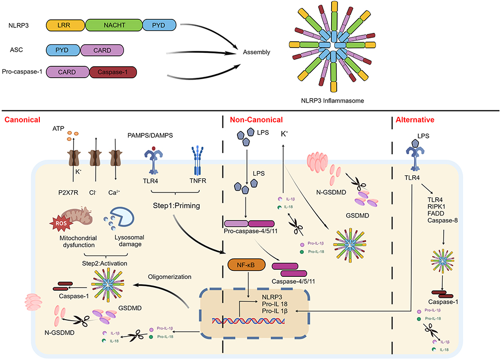 |
Figure 1 NLRP3 inflammasome structure and signaling pathway. The NLRP3 inflammasome contains NLRP3, ASC and pro-caspase-1. It will assemble after priming and activation process. NLRP3 pathway can be divided into the canonical, the non-canonical and the alternative. In the canonical pathway, priming steps involve PAMPS and DAMPS causing NF-κB upregulation, subsequently inducing NLRP3 related protein expression. ATP, K⁺, Cl− and other stimulation work as triggers for NLRP3 assembly, along with caspase-1 formation. ATP also activates the P2X7. Caspase-1 brings about GSDMD cleavage, IL-1β and IL-18 maturity, which leads pyroptosis. Different from the canonical, LPS in the non-canonical pathway directly binds to pro-caspase-4/5/11. NLRP3 is activated through K+ efflux via IL-1β and IL-18 release. The alternative, however requires TLR4-TRIF-RIPK1-FADD-CASP8 signaling to cause NLRP3 assembly. Thereafter caspase-1 forms and leads inflammatory cytokines release. Created with BioGDP.com.20 Abbreviations: NLRP3, NOD-like receptor thermal protein domain-associated protein 3; ASC, apoptosis-associated speck-like protein containing a caspase recruitment domain; LPS, lipopolysaccharide; IL-1β, interleukin-1 beta; IL-18, interleukin-18; NACHT, nucleotide-binding oligomerization domain; PYD, N-terminal pyrin domain; LRR, C-terminal leucine-rich repeat; pro-IL-1β, pro-interleukin-1β; pro-IL-18, pro-interleukin-18; PAMPs, pathogen-associated molecular patterns; DAMPs, damage-associated molecular patterns; TLR4, Toll-like receptor 4; TNF, tumor necrosis factor; ATP, adenosine triphosphate; NF-κB, nuclear factor-κB; CatB, cathepsin B; P2X7, P2X purinoceptor 7; ROS, reactive oxygen species; GSDMD, Gasdermin D; N-GSDMD, Gasdermin D N-terminal domain. |
The canonical process contains two steps: priming and activation. In the priming stage, expression of NLRP3 and pro-interleukin-1β (pro-IL-1β) is upregulated, while the activation stage triggers the assembly of NLRP3 inflammasome.21 Pathogen-associated molecular patterns (PAMPs) or damage-associated molecular patterns (DAMPs) initiate the priming stage. These signals are transduced through receptors such as Toll-like receptor 4 (TLR4) or TNF receptors, leading to the activation of nuclear factor-κB (NF-κB) signaling pathway, subsequently promoting gene transcription associated with NLRP3 inflammasome.22 A binding partner of S-phase kinase-associated protein 1 (SKP1) is suppressor of the G2 allele of SKP1 (SGT1). To form a complex with NLRP3 inflammasome, heat shock protein 90 (HSP90) synergizes with SGT1. This complex maintains NLRP3 inflammasome in an inactive, but activation-competent state prior to the reception of priming signals. If this complex fails to assemble, NLRP3 protein undergoes degradation.23
NLRP3 inflammasome activation has been the major focus of recent studies. Its signals can be triggered by a variety of molecules and cellular events, including variations in extracellular adenosine triphosphate (ATP), efflux of K⁺ and Cl−, Ca2⁺ mobilization, release of cathepsin B (CatB) resulting from lysosomal damage, and production of ROS induced by mitochondrial dysfunction.24 During the activation steps, extracellular ATP activates P2X purinoceptor 7(P2X7) and induces K⁺ efflux.25 Upon ATP stimulation, Ca2⁺ signaling triggers mitochondrial damage to activate NLRP3 inflammasome. NLRP3 inflammasome has also been reported to cause mitochondrial dysfunction. Inactive NLRP3 inflammasome localizes to the endoplasmic reticulum (ER), whereas activated NLRP3 inflammasome and its adaptor protein ASC redistribute to the perinuclear region, co-localizing with the ER and mitochondria.26 Lysosome rupture works as an endogenous danger signal and NLRP3 inflammasome can respond to the stimuli. Inhibition of CatB has been observed to reduce NLRP3 inflammasome activation.27 In the inflammatory response, accumulation of ROS leads to oxidative stress. A previous study revealed that ROS could induce Ca2⁺ influx through the transient receptor potential melastatin 2 (TRPM2) channel.28 ROS also act as a messenger to upregulate the NF-κB pathway.29 Nevertheless, NLRP3 inflammasome activators, including nigericin, ATP and ROS, were not required for NLRP3 inflammasome activation. Conversely, a decreased intracellular K⁺ concentration is necessary for activating NLRP3 inflammasome.30 After activated, the PYD of NLRP3 inflammasome interacts with the PYD of ASC. Then it recruits and oligomerizes pro-caspase-1 via CARD–CARD interactions, generating caspase-1 that amplifies inflammatory response.31,32 Caspase-1 cleaves Gasdermin D (GSDMD) into Gasdermin D N-terminal domain (N-GSDMD) to form pores in the plasma membrane, leading to pyroptosis and inflammatory cytokines secretion.33,34
Lipopolysaccharide (LPS) from gram-negative bacteria mediates the non-canonical pathway. LPS directly enters cytosol via phagocytosis, independent of TLR4, and then binds to pro-caspase-11 (caspase-11 in mice and caspase-4/5 in humans), inducing the cleavage of GSDMD.35–37 IL-1β and IL-18 are released, which causes K⁺ efflux and pyroptosis, thereby triggering NLRP3 inflammasome activation. Neuron-derived clone 77 (Nur77), a nuclear orphan receptor, is an intracellular LPS-binding protein that binds to NLRP3 inflammasome. To connect NLRP3 inflammasome, Nur77 requires LPS and dsDNA released from GSDMD pores.38 The alternative pathway involves TLR4-TRIF-RIPK1-FADD-CASP8 signaling. NLRP3 transcription is upregulated by LPS and TLR4 interactions, driving the immune response modulated by IL-1β. This pathway is independent of K⁺ efflux and pyroptosis.39
NLRP3 Inflammasome and UC
Multiple studies have demonstrated that NLRP3 inflammasome and its related signaling pathways are involved in UC. A prospective observational study indicated that NLRP3 serum levels and protein expression of NLRP3 inflammasome components in the colonic mucosa were upregulated, which was positively correlated with disease severity. Additionally, an increased number of γδT17 cells was also observed.15,40,41 In both UC patients and experimental models, the protein expression of TLR4, NF-κB, and NLRP3 was higher than the control, indicating that the TLR4/NF-κB/NLRP3 pathway contributed to UC progression through inflammatory responses and mucosal damage.42,43 In dextran sulfate sodium (DSS)-induced UC mouse models, mRNA expressions of NIMA-related kinase 7 (NEK7), caspase-1, NLRP3 and GSDMD were significantly increased. Furthermore, interactions between NEK7-NLRP3 modulate UC progression via pyroptosis.44,45 Orally administered DSS, Nlrp3-knockout (Nlrp3−/−) mice showed less severe colitis. These results emphasize that the NLRP3 inflammasome is critical for intestinal inflammation.46 At the cellular level, the imbalance between M1 and M2 macrophages during inflammation contributes to the pathogenesis of UC. M1 macrophages act as drivers of tissue damage due to their potent pro-inflammatory functions, whereas M2 macrophages facilitate tissue repair and maintain intestinal homeostasis by producing growth factors.47 NLRP3 inflammasome in M1 macrophages can release IL-18 and IL-1β after activated, which exacerbates colonic inflammation. Specifically, IL-1β induces expression of chemokines and adhesion molecules, promoting inflammatory cell infiltration and increasing intestinal epithelial permeability.48 Additionally, hyperactive IL-18 signaling hinders goblet cell maturation, thereby promoting colitis.49 Inhibiting NLRP3/caspase-1/IL-1β pathway or neutralizing IL-18 can alleviate UC symptoms and reduce gene expression of pro-inflammatory cytokines.44,50 However, under specific conditions, overexpression of NLRP3 inflammasome in phagocytes can protect the host from colitis. Nlrp3R258W mice, characterized with hyper activation of NLRP3 inflammasome, exhibited high expression of NLRP3 in lamina propria CD11b+/CD11C+ mononuclear phagocytes and were more resistant to DSS-induced colitis. The mechanism might be that NLRP3 promoted secretion of IL-1β and increases antimicrobial peptides from the intestinal epithelia, which maintained homeostasis via an alternative regulatory T cell pathway.51 The gut microbiota also influences NLRP3 inflammasome in UC. Co-culture of mouse macrophages (Raw264.7) and bone marrow-derived macrophages (BMDM) with A. muciniphila led to upregulation of NLRP3 transcription. Further experiments confirmed that A. muciniphila alleviated colitis symptoms. However, in the absence of Nlrp3, A. muciniphila failed to protect the colon from inflammatory injury.52 These findings suggest NLRP3 inflammasome is complex in regulation of intestinal homeostasis.
NLRP3 inflammasome is expressed in both colonic macrophages and colonic epithelial cells, indicating that these two cell types are both crucial in chronic colitis mediated by NLRP3 inflammasome.15
The intestinal mucosa plays a pivotal role in sensing pathogens and danger signals, and this protective mechanism mainly occurs in intestinal epithelial cells (IECs) rather than immune cells. At the early stage of infection, signaling transduction of NLRP3 and ASC in IECs can restrict pathogen colonization and prevent pathological injury.53 In vitro studies have confirmed that, compared to wild-type UC mice, Nlrp3−/− UC mice exhibited more severe pathological injury in intestinal mucosa, with significantly reduced tight junction proteins and higher release of inflammatory cytokines.54,55 In addition, Nlrp3−/− UC mice induced by oxazolone exhibited severe colitis with increased Th2 cytokines and reduced IL-1β and IL-18. This phenomenon was mitigated by exogenous IL-1β or IL-18.56 Notably, IL‑18 is mainly derived from IECs in the early stage of inflammatory bowel disease (IBD), whereas it is predominantly secreted by immune cells in the later stage of chronic inflammation, thereby driving Th1 immune response.57 Upon activation, IECs can secrete inflammatory cytokines, recruit immune cells from the underlying lamina propria, and initiate local inflammatory responses. NLRP3 inflammasome also mediates pro-inflammatory effects of IECs. Another study demonstrated that Na⁺‑K⁺‑2Cl− cotransporter knockout in IECs enhanced sensitivity of intestinal epithelial cells to alterations in intracellular K⁺ concentration and downregulated activation threshold of NLRP3 inflammasome in IECs, which facilitated secretion of IL‑1β and IL‑18.58 This provides a potential target for the future development of targeted therapeutic strategies.
Current studies suggest that NLRP3 inflammasome plays a protective role against colonic inflammation during the initial phase. But its function in different stages and microenvironments of chronic inflammation has not been fully elucidated. Although TNF‑α inhibitor infliximab has been utilized for UC, patients with poor response to monotherapy require combination therapy with TNF-α and IL-23 inhibitors to alleviate symptoms.59 However, TNF‑α is only expressed in immune cells, whereas NLRP3 inflammasome is expressed in both IECs and immune cells. Therefore, NLRP3 inflammasome inhibitors have significant advantages in UC treatment and development of drug delivery systems targeting NLRP3 is also a new direction. Considerable research has focused on inventing compounds that can selectively modulate NLRP3 inflammasome activity. In the following section, we summarize unique NLRP3 inflammasome inhibitors and highlight their potential as UC treatment options.
Small Molecule Compounds Targeting NLRP3 Inflammasome Treating UC
Inhibition of NLRP3 Inflammasome Assembly
Directly binding relevant structures of NLRP3 inflammasome can specifically inhibit its function. Designing small molecule compounds based on associated domains has become a cutting-edge area of research owing to their definite targeting properties.
The NACHT domain activates NLRP3 via ATP hydrolysis. Tetrahydroquinoline analog compound 6, triazinone derivative compound L38, non-sulfonylurea compound B6, all of them could bind to NACHT domain, thereby inhibiting the assembly NLRP3 inflammasome, and exhibited anti-inflammatory ability in DSS-induced UC models.60–62 The steric properties of R-substituent on tetrahydroquinoline derivatives significantly impacted inhibiting IL-1β triggered by NLRP3 inflammasome. Long-chain and polyaromatic substituents at R-position were conducive to improving anti-inflammatory ability of compounds with a tetrahydroquinoline core. Compound 6 with a p-acetylphenyl group at R-position showed the most potent anti-inflammatory effect.60 The thieno[2′,3′:4,5]pyrrolo[1,2-d][1,2,4]triazine scaffold possesses anti-inflammatory and antitumor properties, thus regarded as a highly promising structure.63 Nevertheless, replacement of amide moiety in side chain region A with 1,3,4-oxadiazole scaffold yielded favorable inhibitory potency. Substituted pyridine at the R group of 1,3,4-oxadiazole achieved satisfactory inhibitory effects. In contrast, modification of methylene group in region L led to compound activity or metabolic stability decline, indicating that this methylene was critical for maintaining biological activity and metabolic stability.61 Ex2AH (WO 2018167468 A1) has been identified as the first NLRP3 inhibitor without sulfonylurea scaffold. Molecular docking revealed that the nitrogen atom on the right side of the ureido group does not form any hydrogen bond with the NLRP3 protein.64 Shifting this nitrogen atom to form a new carbamate, and introducing a six-membered aromatic heterocycle at its R substituent, significantly improved the compound’s activity. The number or position of nitrogen atoms in the six‑membered aromatic heterocycle also influenced activity. While introduction of a sterically hindered methyl group or substituents on the benzene ring markedly reduced the inhibitory capacity of these compounds. Compound B6 displayed the best efficacy. Further SAR studies of B6 suggested that the hydrophobic pocket accommodating R3 was relatively small, requiring a small volume substituent.62
As a key adaptor molecule for NLRP3 inflammasome assembly, ASC participates in NLRP3 inflammasome activation via its interaction with NLRP3 and oligomerization. However, small molecules can antagonize this process by blocking NLRP3 and ASC interaction or suppressed inhibiting ASC oligomerization, which can alleviate pathological damage of UC. These compounds include (4-((E)-3,5-dimethoxy-2-((E)-2-nitrovinyl) styryl) aniline derivative compound 27, 2,3-dihydro-1H-indene-5-sulfonamide analog compound 15z, compound Z48 with arylacrylamide scaffold, quinoline derivative W16, F14 with chalcone structure and boron-containing compound 27.65–70 Pterostilbene-derived derivatives have been demonstrated to target NLRP3 inflammasome and interfere with its assembly process. But structural optimization is required due to their short half-life period.71 Nitrostyrene severed as the key structural moiety for binding to NLRP3. SAR analysis indicated appropriately sized rigid planar aromatic substituents adjacent to the carbonyl group facilitated occupation of the hydrophobic cavity. Compound 27 was then synthesized and exhibited potent anti-inflammatory activity, favorable absorption capacity, and satisfactory cell membrane permeability.65 Previous studies validated that sulfonamide derivatives inhibited NLRP3 inflammasome activity and reduced side effect of hypoglycemia.72 Based on this scaffold, introduction of propylamine at R5 position and replacement with 2-methoxyethyl at R3 position yielded optimal activity. During optimization of R2 substituent, bromoacetic acid and cinnamic acid showed high inhibitory potency. But both of them showed non-specific inhibition and possessed cytotoxicity. Compound 15z was produced by cyclization of cinnamic acid, which displayed the highest concentration in colonic tissue.66 The aryl acrylamide scaffold was proved holding potential for inhibiting NLRP3 inflammasome. Inspired by OLT1177, 3-sulfonylpropionitrile analogs were introduced to this scaffold. Halogen substitution at 4-position of the benzene ring was superior to that at 3-position, and molecular docking experiments proved that 4-bromo-substituted compound could form a halogen bond with NLRP3. However, halogen substitution on the benzene ring and ring-expansion strategies did not significantly improve compound activity. Therefore, the focus shifted to hydrophobic region at the bottom of NLRP3 protein pocket, and generated compound Z48. Notably, Z48 showed no significant therapeutic effect in Nlrp3−/− mice.67 Chloroquine and hydroxychloroquine were reported owning antagonistic effects against COVID-19. The mechanisms might lie in inhibition of NLRP3 inflammasome activity.73 Removing chlorine atom at 7-position of chloroquine and introducing 2-phenyl and 4-amide groups improved compound activity. The 2-phenyl group was identified as a key moiety, and meta-fluorophenyl substitution was conducive to potency. Introduction of nitro group at 8-position generated compound W16 illustrated the highest activity.68 Quinoline compounds are capable of binding to NACHT domain and inhibiting NLRP3-ASC interaction, making quinoline one of the most promising scaffolds. Chalcone derivatives could form hydrogen bonds and π-π stacking interactions with active sites of NLRP3. Introduction of a hydroxyl group at ortho of B ring leads to a substantial increase in compound activity. Among this series, compound F14 exhibited the best bioactivity. Molecular dynamics simulation assay demonstrated a favorable binding affinity between F14 and NLRP3, with tyrosine (Tyr)381 and phenylalanine (Phe)508 in NLRP3.69 Drug repurposing is a method for discovering new compounds and exploring novel mechanisms. Boron-containing compounds was reported inhibiting activation of NLRP3 inflammasome, with ixazomib demonstrated the most excellent capability.74 The hydrogen bond donors of boronic acid group and arylamide moiety were critical for inhibiting IL-1β release, while variations in aryl and amino acid residues affected compound bioactivity. In terms of spatial volume and polarity of the amino acid region, benzyl is the most suitable substituent. Boron-containing compound NIC-0102 induced NLRP3 polyubiquitination, thereby disrupting NLRP3 and ASC interaction.70
Caspase drives the release of inflammatory cytokines and amplifies inflammatory responses. 1-ethyl-5-methyl-2-phenyl-1H-benzo[d]imidazole, also named Fc11a-2, prevented autocleavage of caspase-1, leading to a significant reduction in caspase-1 released from the ASC/NLRP3 complex, thereby reducing intestinal damage in UC.75
Inhibition of NLRP3 Inflammasome Activation
Beyond directly targeting relevant domains of NLRP3, multi-target regulatory strategies focusing on its activation phase represent another vital area of inhibitors research. During the activation stage, P2X7 is activated by ATP and causes K⁺ efflux.25 In addition, both Ca2⁺ release from the ER and influx of extracellular Ca2⁺ are essential for NLRP3 activation.26 Taurodeoxycholate, is known as G-protein coupled receptor 19 (GPCR19) agonist, which was proved to downregulate the expression of P2X7 receptor (P2X7R) and inhibit Ca2⁺ influx, thereby suppressing NLRP3 signaling pathway.76 Brilliant blue G (BBG), a P2X7R antagonist, enhanced the inhibition of NLRP3 inflammasome combined with OLT1177.77
Acrylamide derivative INF39 targeted ATPase domain of NLRP3 and inhibited the interaction between NEK7 and NLRP3. This compound was proved to be safe and non-toxic, while its binding site has not been definitively clarified.78,79 NEK7 serves as a binding protein of NLRP3, and Cl− efflux can activate NLRP3 inflammasome as well as modulate NEK7-NLRP3 interaction.80,81 Chloroquine compounds are capable of inhibiting inflammation. 7‑position phenolic hydroxyl group and nitrogen atom of 8‑hydroxyquinoline were important and methyl group at 2‑position enhanced activity. However, introducing strong electron-withdrawing groups at 2- or 4-position, or methylating 8-position or removing hydroxyl group, led to loss of activity. Compound 10 bears a para-methoxy substituent and exhibits potent inhibitory efficacy. It disrupted interactions between key proteins such as NEK7 and NLRP3.82
In activation steps, phagocytosis of particulate leads to lysosomal damage, rupture, and leakage of contents. This process promotes CatB releasing into cytoplasm and facilitates K⁺ efflux.30,83 These mechanisms highlight significance of blocking K⁺ efflux in the activation process. Aryl hydrocarbon receptor signaling promotes polyamine biosynthesis by enhancing transcription of ornithine decarboxylase 1. Supplementation with polyamines, particularly spermine, could inhibit K⁺ efflux, block NLRP3 inflammasome assembly.84
Target NLRP3-Associated Proteins
Targeted regulation of upstream and downstream proteins related to NLRP3 provides a novel intervention strategy for specific inhibition. Nevertheless, compared to direct modulation, research on small molecule compounds targeting NLRP3-associated proteins remains exploratory. Roles of related proteins have been gradually elucidated, among which oxidative stress signaling pathway regulated by mitophagy is particularly pivotal. When mitophagy is inhibited, continuous accumulation of ROS causes dissociation of thioredoxin-interacting protein (TXNIP) from thioredoxin-1 (TRX). The C-terminus of TXNIP preferentially binds to the LRR and NACHT domains of NLRP3 inflammasome, thereby driving the activation.85–87 A synthetic flavonoid, compound VI-16, enhanced antioxidant capacity in colonic macrophages, inhibiting dissociation of TXNIP from Trx-1 and its subsequent binding to NLRP3.88 Representative structure of these compounds and their mechanism in targeting NLRP3 pathway are presented in Table 1.
 |
Table 1 Preclinical Research in Synthesized Compounds Inhibit NLRP3 Inflammasome to Treat UC |
Research on Clinical Drugs
On the basis of fundamental research, NLRP3 inflammasome inhibitors have been used in clinical trials. Cytokine release inhibitory drugs (CRIDs) were created, targeting IL-1β. CRID3, also known as MCC950 or CP-456773, binds to NLRP3 via the Walker B motif within NACHT.89–91 MCC950 is the first specific NLRP3 inflammasome inhibitor entering clinical trials and has been widely used in studies of diseases related with NLRP3 inflammasome. However, due to hepatotoxicity observed in a Phase II clinical trial for rheumatoid arthritis, the clinical development of MCC950 was forced to terminate.92 Selnoflast (formerly somalix/RG6418/IZD334) is a derivative modified from MCC950. Compared to MCC950, the sulfonyl side chain of selnoflast contains N-ethylpiperidine, replacing the furan ring and tertiary alcohol of MCC950. This modification improved water solubility and enhanced reversibility of target binding. Clinical trials have verified that selnoflast exhibits favorable safety and tolerability (No. NCT04086602). Administration of 450 mg selnoflast daily to patients with moderate-to-severe UC could maintain inhibition on IL-1β above 90% inhibitory concentration (IC90) during dosing interval, and the concentration of selnoflast in sigmoid colon tissues was higher than IC90. Blood tests revealed that IL-1β release was inhibited within 30 minutes after dosing and maintained for at least 10 hours (mean [± SD] >95% [± 4.16%]). However, no significant differences in Geboes score, Nancy index and caspase-1 expression were observed before and after treatment (data not shown). Although expression of inflammatory cytokine-related genes in colon tissue showed a slight decrease, there was no significant difference. These outcomes halted clinical development of selnoflast for UC, terminating its progress at the Phase Ib clinical trial.93
ROS is also indispensable in NLRP3 activation. NAC, a ROS scavenger, stemmed LPS-induced NLRP3 from activation.94 In the acetic acid (AA)-induced colitis model, intracolonic administration of NAC alleviated severity of colonic injury, accompanied by a reduction in myeloperoxidase.95 In a randomized, double-blind controlled clinical trial (No. IRCT20190713044185N1), UC patients in the steroid-tapering phase who took NAC demonstrated significantly lower relapse rates (P = 0.007) compared with placebo group. Taking NAC also showed lower mean fecal calprotectin and serum erythrocyte sedimentation rate (p < 0.05). However, the incidence of nausea was higher in the NAC group (19.51%) than in the placebo (3.48%) (P = 0.001).96 Although NAC has demonstrated promising efficacy in the clinical trial, further investigation is required to determine whether it specifically targets NLRP3 inflammasome in human. Based on these two clinical trials, no clinical studies have reported adverse effects or risks of targeting NLRP3 inflammasome for UC treatment. MCC950 was halted due to hepatotoxicity. Another MLRP3 inflammasome inhibitor GDC-2394, based on MCC950 structure, effectively inhibited IL-1β and IL-18 release, but exhibited potential hepatotoxicity in Phase I trial.97 This suggests that future drug development should also focus on screening for reactive metabolites in the early stages of drug discovery and rigorously monitoring liver function indicators in animal studies.
MCC950 serves as a critical structural scaffold for research and development of NLRP3 inflammasome inhibitors. While its furan ring moiety is likely to induce drug-induced liver injury. Therefore, structural optimization of MCC950, especially replacing its furan ring fragment, is particularly essential. Based on MCC950, the furan ring was replaced with a phenyl group and a dimethylaminomethyl group was introduced at the 4-position. This yielded 4-(2-(dimethylamino) ethyl)-N-((1,2,3,5,6,7-hexahydro-s-indacen-4-yl) carbamoyl) benzenesulfonamide (N14). N14 had an oral bioavailability of 85.2%. Compared with MCC950, it exhibited no significant hepatocyte toxicity at 500 μM and more effectively alleviated symptoms in UC mice.98
Expanding Therapeutic Application of NLRP3 Inflammasome Inhibitors for UC Treatment
Other drugs currently in clinical trials or already marketed also exhibit capacity to inhibit NLRP3 inflammasome.
OLT1177, a β-sulfonyl nitrile compound, is a selective NLRP3 inhibitor. It has completed Phase I (No. NCT02134964) and Phase II clinical trials for osteoarthritis (No. NCT02104050) and is currently undergoing Phase III clinical research for gout (No. NCT05658575).99,100 OLT1177 was well tolerated in humans who continuously administrated OLT1177 for 8 days at a maximum dose of 1000 mg. OLT1177 blocked interactions between NLRP3 and ASC, as well as between NLRP3 and caspase-1, showing significant efficacy in the early stage of DSS-induced UC mice.101,102 Nevertheless, a target engagement assay revealed that although OLT1177 could ameliorate inflammation, it failed to suppress NLRP3 inflammasome assembly.99 Current clinical studies have confirmed its safety. However, its ability to specifically target and inhibit NLRP3 inflammasome remains to be verified.
Transilast is an analog of tryptophan metabolites used for treating allergic diseases such as bronchial asthma.103 Its anti-allergic mechanism involves stabilizing cell membranes of mast cells and basophils, preventing their degranulation, and blocking inflammatory responses.103 Since excessive inflammation drives UC progression, exploring impacts of anti-allergic drugs on NLRP3 represents a new direction for UC treatment. Tranilast has been proved that it can covalently bind to NACHT.104 Enema administration was shown tranilast ameliorated inflammatory manifestations in UC mice.105 However, there is still a long way to go before tranilast can be truly applied to curing UC, as its pharmacological properties pose the main bottleneck, restricting its translational application. In human, the plasma concentration of tranilast peaks at 2 h after a single oral dose, with a half-life of 5 h. Oral administration of conventional therapeutic doses (600 mg/day) yields plasma concentrations ranging from 30 to 300 μM.103 Multiple hydrophobic groups (eg, cinnamoyl group) in the tranilast molecule result in extremely low water solubility (14.5 μg/mL), particularly under acidic conditions. In addition, the highly flexible acrylamide moiety in tranilast increases entropy loss and reduces binding affinity.106 Tranilast possesses reliable safety profiles, and structural optimization is required to develop its application in UC therapy.
RRx-001 (1-bromoacetyl-3,3-dinitroazetidine) is an agent for small cell lung cancer, colorectal cancer and brain metastatic tumors and has completed Phase III clinical trials for small cell lung cancer (No. NCT03699956).107,108 Its bromoacetyl group can covalently bind to cysteine (Cys) 409 of human NLRP3 and Cys405 of mouse NLRP3, alleviating clinical symptoms in UC mice.109 However, RRx-001 contains high-energy nitro functional groups, which may produce highly toxic oxides during production and processing. Compound 149–01, an RRx-001 analogue devoid of high-energy nitro groups, blocks the NLRP3–NEK7 interaction, though its efficacy has not yet been validated in UC mice.110
Chloroquinaldol, an 8-hydroxyquinoline derivative, is a topical antimicrobial agent used for skin infections. It has been proved to improve psoriasiform dermatitis by suppressing NLRP3 inflammasome activation.111,112 Chlorquinaldol blocked NLRP3-ASC interaction, and hydroxyl group on its benzene ring contributed to inhibitory effect. It could inhibit caspase-1 cleavage and IL-1β maturation and was demonstrated therapeutic effects in DSS-induced UC mice.113 These findings provide a theoretical basis for inhibition of NLRP3 inflammasome by 8-hydroxyquinoline and support the development of its derivatives.
While these drugs have not been clinically tested for UC, they have shown capacity to target and inhibit NLRP3 inflammasome. Their safety has also been confirmed through previous clinical studies. Despite remaining challenges such as unclear target specificity and suboptimal druggability, these agents, with established clinical safety evidence and confirmed NLRP3 inhibitory activity, represent one of the most promising research directions for NLRP3-targeted therapy of UC.
Drugs Dependent on NLRP3 Inflammasome
The Nlrp3−/− model enables the definitive identification of whether anti-inflammatory effects are exerted via the NLRP3 inflammasome. For example, 3-(2-Oxo-2-phenylethylidene)-2,3,6,7-tetrahydro-1H-pyrazino[2,1-a]isoquinolin-4(11bH)-one (compound 1) interfered with NLRP3 priming step to prevent activation, yet compound 1 failed to ameliorate intestinal inflammation in Nlrp3−/− mice.114
Despite suppressing the assembly and activation of NLRP3 inflammasome, promoting its degradation also represents a promising therapeutic strategy for UC. For instance, colonic clock gene Rev-erbα directly repressed Nlrp3 transcription by specifically binding to its promoter region, thereby alleviating UC symptoms in mice. However, this effect was lost in Nlrp3−/− mice.115 Therefore, concentrating on Rev-erbα may become an innovative drug target for UC. Van Gogh-like 2 (Vangl2), a planar cell polarity-related protein, was demonstrated to interact with NACHT and LRR of NLRP3 via its Prickle/cadherin-binding domain (PKBD), and recruits the E3 ubiquitin ligase MARCH8 to induce K27-linked ubiquitination of NLRP3 at lysine site K823, leading to autophagic degradation. VANGL2 is downregulated in IBD patients and DSS-induced murine colitis, which highlights its promising application of inhibiting IBD progress.116 The novel sorbicillinoid compound JNUTS013, inducing proteasomal degradation of NLRP3, and autophagy-tethering compound MC-ND-18, recruiting NLRP3 and LC3B protein, could facilitate NLRP3 degradation.117,118
Although the above compounds have been validated in Nlrp3−/− mice, to reveal their specificity for NLRP3 inflammasome, further research is required to precisely identify binding sites and underlying mechanisms of various compounds with NLRP3 inflammasome, together with its upstream and downstream signaling pathways. In addition, transcriptional regulation involving Rev-erbα and the VANGL2-mediated ubiquitination degradation pathway provides a solid foundation for NLRP3 inflammasome-specific therapeutic agents.
Others
A growing collection of miscellaneous NLRP3 inflammasome inhibition measures do not fall neatly into the mechanistic categories discussed above. Recently, these agents have emerged as an excellent therapeutic strategy.
Libertellenone M is a secondary metabolite of the endophytic fungus Phomopsis sp. Immunofluorescence assays showed that S12 blocked NLRP3 inflammasome assembly.119 Lichen metabolite, atranorin combined with ASC and suppressed secretion of cytokines.120 Beyond these, probiotics also offer a novel approach for NLRP3-targeted therapy. Roseburia intestinalis (R.I) is significantly reduced in patients with IBD. In vitro studies indicated that R.I. flagellin inhibited microRNA (miR)‑223‑3p, thereby modulating NLRP3 activation. Hence, R. I. flagellin will be a unique and promising probiotic product for UC.121
To achieve more precise regulation of the NLRP3 inflammasome, NLRP3-targeted delivery systems have been developed to improve therapeutic efficacy. Although bicalutamide promoted NLRP3 degradation, it had limitations in treating UC. Hence, a delivery system targeting NLRP3 in macrophages was invented, mediating NLRP3 protein degradation via autophagic pathway.122 Meanwhile, siNLRP3-loaded nanoparticles based on cationic liposomes were constructed, effectively smothering NLRP3 inflammasome activation.123 A novel nanosystem carrying an NLRP3 detection probe, which was cleavable by caspase-1, together with MCC950 carried out early real-time monitoring of NLRP3 activation while exerting anti-inflammatory effects on UC mice.124 Delivery systems overcome limitations of small molecule drugs and can precisely target NLRP3 inflammasome, holding promise for improving specificity and safety of NLRP3 modulation.
Conclusion and Prospects
NLRP3 inflammasome has emerged as a central regulator of intestinal inflammation, driving mucosal damage and inflammatory infiltration through inflammatory cytokines release such as, IL-1β and IL-18. Its aberrant activation is linked to the pathogenesis and progression of UC, positioning NLRP3 inflammasome as a dominant feature in the inflammatory cascade and as a revolutionary therapeutic target. Structurally, NLRP3 inflammasome consists of multiple effector proteins, providing advantages of potential targeting sites. The treatment of NLRP3 inflammasome- related diseases mainly focuses on inhibiting IL-1β at present. However, IL-1β is not merely a product of NLRP3 inflammasome activation, nor is the sole output of such activation. Therefore, direct NLRP3 inhibitors may offer advantages in terms of safety and efficacy. Nevertheless, regulatory mechanisms of NLRP3 inflammasome in different stages and microenvironments of chronic inflammation remain unclear, and relevant inhibitory mechanisms or precise targets have not been fully elucidated.60 Moreover, NLRP3 shares structural similarities with other inflammasomes, such as NOD-like receptors family CARD domain-containing protein 4 (NLRC4), absent-in-melanoma 2 (AIM2). Some inhibitors may interfere with other inflammatory pathways, potentially causing off-target effects that impairing immune defense. Furthermore, the activation mechanism of NLRP3 inflammasome is complex and involves multiple signaling pathways, which cannot be accurately recapitulated in animal models. This limits clarification of regulatory impacts of inhibition NLRP3 inflammasome on other pathways.125 Additionally, long-term safety of drugs targeting NLRP3 inflammasome remains unknown.
To date, no small molecule drug directly targeting NLRP3 inflammasome has been approved for marketing. Although selnoflast did not show significant differences in Geboes score or Nancy index after treatment (data not disclosed), leading to termination of its clinical trial in UC, the exploration of selnoflast has advanced clinical translation of NLRP3-targeting strategies. In a randomized controlled trial for UC, ROS scavenger NAC significantly reduced recurrence rates (P = 0.007), with decreases in both fecal calprotectin and serum erythrocyte sedimentation rate. But its effects on NLRP3 inflammasome in intestinal tissue of UC patients remain unclear. As a highly promising candidate, NAC is expected to proceed to further clinical trials for UC.
Drugs already on the market or in clinical trials for other diseases that have been proved to specifically inhibit NLRP3 inflammasome warrant further development due to their well-defined safety. Drugs including tranilast, RRx-001, and antibacterial agent chlorquinaldol also merit structural modification to enhance efficacy against UC. Aberrant immune responses mediate UC progression. Anti-inflammatory and anti-allergic agents are also capable of suppressing inflammatory reactions. Therefore, future research may focus on repurposing marketed anti-allergic or anti-inflammatory drugs for UC, exploring their capacity to specifically modulate NLRP3 inflammasome, and modify their structure to optimize their therapeutic effects in UC. Acrylamide moiety of tranilast contributes to inhibiting NLRP3 inflammasome, yet this structure also reduces its binding affinity to the target protein. Compound Z48, derived from structural modification of this moiety, exhibited satisfactory therapeutic efficacy in animal models, with an oral bioavailability of 16.7% in mice, warranting further investigation.
Newly synthesized small molecule compounds have achieved high affinity and specific inhibition of NLRP3 inflammasome through structural modification and optimization. Quinoline, which can bind to NACHT and ASC domains, and sulfonamide compounds, which are based on MCC950 structure, hold the greatest development promise among various compounds. Although MCC950 was halted due to hepatotoxicity, derivatives generated by modifying its furan ring structure can still be exploited for novel NLRP3 inflammasome inhibitors.
Inhibiting NLRP3 inflammasome remains a highly promising therapeutic strategy for UC that warrants deep exploration. Artificial intelligence and advanced computational technologies offer unprecedented opportunities to accelerate screening of active compounds and to guide structural optimization of next-generation NLRP3 inhibitors. Combination therapies that pair NLRP3 inhibitors with NLRP3-targeted delivery systems may enhance therapeutic efficacy and overcome drug resistance mechanisms. Inhibition of NLRP3 offers new hope for patients with UC and other inflammatory disorders related to NLRP3.
Abbreviations
UC, Ulcerative colitis; ACG, American College of Gastroenterology; TNF, Tumor necrosis factor; HLA, Human leukocyte antigen; IL23R, interleukin 23 receptor gene; NLR, NOD-like recepto; NLRP3, NOD-like receptor pyrin domain-containing 3; IL-1β, Interleukin-1 beta; IL-18, Interleukin-18; JAK, Janus kinase; NAC, N-acetylcysteine; ROS, Reactive oxygen species; SAR, Structure Activity Relationships; ASC, Apoptosis-associated speck-like protein containing a caspase recruitment domain; NACHT, Nucleotide-binding oligomerization domain; PYD, Pyrin domain; LRR, Leucine-rich repeat; ATPase, adenosine triphosphate; CARD, Caspase activation and recruitment domain; pro-IL-1β, Pro-interleukin-1β; PAMPs, pathogen-associated molecular patterns; DAMPs, damage-associated molecular patterns; TLR4, Toll-like receptor 4; NF-Κb, Nuclear factor-κB; HSP90, Heat shock protein 90; SKP1, S-phase kinase-associated protein 1; SGT1, Suppressor of G2 allele of SKP1; ATP, Adenosine triphosphate; CatB, Cathepsin B; P2X7, P2X purinoceptor 7; ER, Endoplasmic reticulum; TRPM2, Transient receptor potential melastatin 2; GSDMD, Gasdermin D; N-GSDMD, Gasdermin D N-terminal domain; LPS, Lipopolysaccharide; Nur77, Neuron-derived clone 77; DSS, Dextran sulfate sodium; NEK7, NIMA-related kinase 7; Nlrp3−/−, Nlrp3-knockout; BMDM, Bone marrow-derived macrophages; IECs, Intestinal epithelial cells; Tyr, Tyrosine; Phe, Phenylalanine; GPCR19, G-protein coupled receptor 19; P2X7R, P2X7 receptor; Cys, cysteine; KD, Equilibrium dissociation constant; IC50, Half maximal inhibitory concentration; BBG, Brilliant blue G; TXNIP, Thioredoxin-interacting protein; TRX, Thioredoxin-1; THP-1, Tohoku Hospital Pediatrics-1; CRIDs, cytokine release inhibitory drugs; AA, Acetic acid Vangl2, Van Gogh-like 2; PKBD, Prickle/cadherin-binding domain; IBD, Inflammatory bowel disease; miR, microRNA; R.I, Roseburia intestinalis; NLRC4, NOD-like receptors family CARD domain-containing protein 4; AIM2, Absent-in-melanoma 2.
Consent for Publication
All named authors agreed to submit the manuscript for publication.
Acknowledgments
The figure was created using BioGDP.com and we acknowledge their resources and support.
Author Contributions
All authors made a significant contribution to the work reported, whether that is in the conception, study design, execution, acquisition of data, analysis and interpretation, or in all these areas; took part in drafting, revising or critically reviewing the article; gave final approval of the version to be published; have agreed on the journal to which the article has been submitted; and agreed to be accountable for all aspects of the work.
Funding
This work was funded by the Pudong New Area National Traditional Chinese Medicine Inheritance and Innovation Development Pilot Zone Construction Project (PDZY⁃2025⁃0714), National Natural Science Foundation of China (81874450, 81403362).
Disclosure
The authors report no conflicts of interest in this work.
References
1. Le Berre C, Honap S, Peyrin-Biroulet L. Ulcerative colitis. Lancet. 2023;402(10401):571–17. doi:10.1016/S0140-6736(23)00966-2
2. Vavricka SR, Rogler G, Gantenbein C, et al. Chronological order of appearance of extraintestinal manifestations relative to the time of IBD diagnosis in the Swiss inflammatory bowel disease cohort. Inflammatory Bowel Dis. 2015;21(8):1794–1800. doi:10.1097/MIB.0000000000000429
3. Fumery M, Xiaocang C, Dauchet L, Gower-Rousseau C, Peyrin-Biroulet L, Colombel JF. Thromboembolic events and cardiovascular mortality in inflammatory bowel diseases: a meta-analysis of observational studies. J Crohns Colitis. 2014;8(6):469–479. doi:10.1016/j.crohns.2013.09.021
4. Sarlos P, Szemes K, Hegyi P, et al. Steroid but not biological therapy elevates the risk of venous thromboembolic events in inflammatory bowel disease: a meta-analysis. J Crohns Colitis. 2018;12(4):489–498. doi:10.1093/ecco-jcc/jjx162
5. Jess T, Rungoe C, Peyrin-Biroulet L. Risk of colorectal cancer in patients with ulcerative colitis: a meta-analysis of population-based cohort studies. Clin Gastroenterol Hepatol. 2012;10(6):639–645. doi:10.1016/j.cgh.2012.01.010
6. Rubin DT, Ananthakrishnan AN, Siegel CA, Barnes EL, Long MD. ACG clinical guideline update: ulcerative colitis in adults. Am J Gastroenterol. 2025;120(6):1187–1224. doi:10.14309/ajg.0000000000003463
7. Cleynen I, Boucher G, Jostins L, et al. Inherited determinants of crohn’s disease and ulcerative colitis phenotypes: a genetic association study. Lancet. 2016;387(10014):156–167. doi:10.1016/S0140-6736(15)00465-1
8. Liu JZ, van Sommeren S, Huang H, et al. Association analyses identify 38 susceptibility loci for inflammatory bowel disease and highlight shared genetic risk across populations. Nat Genet. 2015;47(9):979–986. doi:10.1038/ng.3359
9. Chen Y, Ye X, Escames G, et al. The NLRP3 inflammasome: contributions to inflammation-related diseases. Cell Mol Biol Lett. 2023;28:51. doi:10.1186/s11658-023-00462-9
10. Arend LJ, Springate JE. Interstitial nephritis from mesalazine: case report and literature review. Pediatr Nephrol. 2004;19(5):551–553. doi:10.1007/s00467-004-1411-6
11. Cross RK. Safety considerations with the use of corticosteroids and biologic therapies in mild-to-moderate ulcerative colitis. Inflamm Bowel Dis. 2017;23(10):1689–1701. doi:10.1097/MIB.0000000000001261
12. Fumery M, Singh S, Dulai PS, Gower-Rousseau C, Peyrin-Biroulet L, Sandborn WJ. Natural history of adult ulcerative colitis in population-based cohorts: a systematic review. Clin Gastroenterol Hepatol. 2018;16(3):343–356.e3. doi:10.1016/j.cgh.2017.06.016
13. Lei J, Lv L, Zhong L, et al. The gut microbiota affects anti-TNF responsiveness by activating the NAD+ salvage pathway in ulcerative colitis. Adv Sci. 2025;12(8):e2413128. doi:10.1002/advs.202413128
14. Ytterberg SR, Bhatt DL, Mikuls TR, et al. Cardiovascular and cancer risk with tofacitinib in rheumatoid arthritis. N Engl J Med. 2022;386(4):316–326. doi:10.1056/NEJMoa2109927
15. Liu L, Dong Y, Ye M, et al. The pathogenic role of NLRP3 inflammasome activation in inflammatory bowel diseases of both mice and humans. J Crohns Colitis. 2017;11(6):737–750. doi:10.1093/ecco-jcc/jjw219
16. Zhang J, Zeng S, Wang P, Chen Y, Zeng C. NLRP3: a promising therapeutic target for inflammatory bowel disease. Curr Drug Targets. 2023;24(14):1106–1116. doi:10.2174/0113894501255960231101105113
17. Shao BZ, Wang SL, Pan P, et al. Targeting NLRP3 inflammasome in inflammatory bowel disease: putting out the fire of inflammation. Inflammation. 2019;42(4):1147–1159. doi:10.1007/s10753-019-01008-y
18. Agostini L, Martinon F, Burns K, McDermott MF, Hawkins PN, Tschopp J. NALP3 forms an IL-1beta-processing inflammasome with increased activity in muckle-wells autoinflammatory disorder. Immunity. 2004;20(3):319–325. doi:10.1016/s1074-7613(04)00046-9
19. Lu A, Magupalli VG, Ruan J, et al. Unified polymerization mechanism for the assembly of ASC-dependent inflammasomes. Cell. 2014;156(6):1193–1206. doi:10.1016/j.cell.2014.02.008
20. Jiang S, Li H, Zhang L, et al. Generic diagramming platform (GDP): a comprehensive database of high-quality biomedical graphics. Nucleic Acids Res. 2025;53(D1):D1671–D1676. doi:10.1093/nar/gkae973
21. Jo EK, Kim JK, Shin DM, Sasakawa C. Molecular mechanisms regulating NLRP3 inflammasome activation. Cell Mol Immunol. 2016;13(2):148–159. doi:10.1038/cmi.2015.95
22. Bauernfeind F, Horvath G, Stutz A, et al. NF-kB activating pattern recognition and cytokine receptors license NLRP3 inflammasome activation by regulating NLRP3 expression. J Immunol. 2009;183(2):787–791. doi:10.4049/jimmunol.0901363
23. Mayor A, Martinon F, De Smedt T, Pétrilli V, Tschopp J. A crucial function of SGT1 and HSP90 in inflammasome activity links mammalian and plant innate immune responses. Nat Immunol. 2007;8(5):497–503. doi:10.1038/ni1459
24. Ren W, Sun Y, Zhao L, Shi X. NLRP3 inflammasome and its role in autoimmune diseases: a promising therapeutic target. Biomed Pharmacother. 2024;175:116679. doi:10.1016/j.biopha.2024.116679
25. Pétrilli V, Papin S, Dostert C, Mayor A, Martinon F, Tschopp J. Activation of the NALP3 inflammasome is triggered by low intracellular potassium concentration. Cell Death Differ. 2007;14(9):1583–1589. doi:10.1038/sj.cdd.4402195
26. Murakami T, Ockinger J, Yu J, et al. Critical role for calcium mobilization in activation of the NLRP3 inflammasome. Proc Natl Acad Sci. 2012;109(28):11282–11287. doi:10.1073/pnas.1117765109
27. Hornung V, Bauernfeind F, Halle A, et al. Silica crystals and aluminum salts activate the NALP3 inflammasome through phagosomal destabilization. Nat Immunol. 2008;9(8):847–856. doi:10.1038/ni.1631
28. Zhong Z, Zhai Y, Liang S, et al. TRPM2 links oxidative stress to NLRP3 inflammasome activation. Nat Commun. 2013;4:1611. doi:10.1038/ncomms2608
29. An Y, Zhang H, Wang C, et al. Activation of ROS/MAPKs/NF-κB/NLRP3 and inhibition of efferocytosis in osteoclast-mediated diabetic osteoporosis. FASEB J. 2019;33(11):12515–12527. doi:10.1096/fj.201802805RR
30. Muñoz-Planillo R, Kuffa P, Martínez-Colón G, Smith BL, Rajendiran TM, Núñez G. K+ efflux is the common trigger of NLRP3 inflammasome activation by bacterial toxins and particulate matter. Immunity. 2013;38(6):1142–1153. doi:10.1016/j.immuni.2013.05.016
31. Zhang X, Xu A, Lv J, et al. Development of small molecule inhibitors targeting NLRP3 inflammasome pathway for inflammatory diseases. Eur J Med Chem. 2020;185:111822. doi:10.1016/j.ejmech.2019.111822
32. Vanaja SK, Rathinam VAK, Fitzgerald KA. Mechanisms of inflammasome activation: recent advances and novel insights. Trends Cell Biol. 2015;25(5):308–315. doi:10.1016/j.tcb.2014.12.009
33. Sborgi L, Rühl S, Mulvihill E, et al. GSDMD membrane pore formation constitutes the mechanism of pyroptotic cell death. EMBO J. 2016;35(16):1766–1778. doi:10.15252/embj.201694696
34. He WT, Wan H, Hu L, et al. Gasdermin D is an executor of pyroptosis and required for interleukin-1β secretion. Cell Res. 2015;25(12):1285–1298. doi:10.1038/cr.2015.139
35. Kayagaki N, Wong MT, Stowe IB, et al. Noncanonical inflammasome activation by intracellular LPS independent of TLR4. Science. 2013;341(6151):1246–1249. doi:10.1126/science.1240248
36. Kayagaki N, Warming S, Lamkanfi M, et al. Non-canonical inflammasome activation targets caspase-11. Nature. 2011;479(7371):117–121. doi:10.1038/nature10558
37. Casson CN, Yu J, Reyes VM, et al. Human caspase-4 mediates noncanonical inflammasome activation against gram-negative bacterial pathogens. Proc Natl Acad Sci. 2015;112(21):6688–6693. doi:10.1073/pnas.1421699112
38. Zhu F, Ma J, Li W, et al. The orphan receptor Nur77 binds cytoplasmic LPS to activate the non-canonical NLRP3 inflammasome. Immunity. 2023;56(4):753–767.e8. doi:10.1016/j.immuni.2023.03.003
39. Gaidt MM, Ebert TS, Chauhan D, et al. Human monocytes engage an alternative inflammasome pathway. Immunity. 2016;44(4):833–846. doi:10.1016/j.immuni.2016.01.012
40. Ma J, Wang FY, Tang XD. Involvement of the NLRP3/IL-1β pathway in activation and effector functions of γδT17 cells in patients with ulcerative colitis. World J Gastroenterol. 2025;31(12):98174. doi:10.3748/wjg.v31.i12.98174
41. Chen Y, Wu D, Sun L. Clinical significance of high-mobility group box 1 protein (HMGB1) and nod-like receptor protein 3 (NLRP3) in patients with ulcerative colitis. Med Sci Monit. 2020;26:e919530–1. doi:10.12659/MSM.919530
42. Li Y, Yao S, Duan C, Ding Y. Analysis of the relationship between the expression of TLR4 and the NLRP3 inflammasome in peripheral blood and disease outcome in patients with ulcerative colitis. Mod Dig Interv. 2022;27(8):1022–1025.
43. Zhang W, Wang X, Xu J, et al. Study on the mechanism of Shaoyao decoction combined with mesalazine regulate TRL4-ERK1/2-NF-κB signal pathway in improving ulcerative colitis. J Qiqihar Med Univ. 2022;43(8):701–707.
44. Bian W, Wei L, Wang K. Carthamin yellow alleviates dextran sodium sulfate-induced ulcerative colitis by repairing the intestinal barrier and activating the Nrf2/GPX4 axis. Int Immunopharmacol. 2024;141:113020. doi:10.1016/j.intimp.2024.113020
45. Chen X, Liu G, Yuan Y, Wu G, Wang S, Yuan L. NEK7 interacts with NLRP3 to modulate the pyroptosis in inflammatory bowel disease via NF-κB signaling. Cell Death Dis. 2019;10(12):1–12. doi:10.1038/s41419-019-2157-1
46. Bauer C, Duewell P, Mayer C, et al. Colitis induced in mice with dextran sulfate sodium (DSS) is mediated by the NLRP3 inflammasome. Gut. 2010;59(9):1192–1199. doi:10.1136/gut.2009.197822
47. Michaeli S, Dakwar V, Weidenfeld K, et al. Soluble mediators produced by pro-resolving macrophages inhibit angiogenesis. Front Immunol. 2018;9:768. doi:10.3389/fimmu.2018.00768
48. Al-Sadi RM, Ma TY. IL-1β causes an increase in intestinal epithelial tight junction permeability. J Immunol. 2007;178(7):4641–4649. doi:10.4049/jimmunol.178.7.4641
49. Nowarski R, Jackson R, Gagliani N, et al. Epithelial IL-18 equilibrium controls barrier function in colitis. Cell. 2015;163(6):1444–1456. doi:10.1016/j.cell.2015.10.072
50. Sivakumar PV, Westrich GM, Kanaly S, et al. Interleukin 18 is a primary mediator of the inflammation associated with dextran sulphate sodium induced colitis: blocking interleukin 18 attenuates intestinal damage. Gut. 2002;50(6):812–820. doi:10.1136/gut.50.6.812
51. Yao X, Zhang C, Xing Y, et al. Remodelling of the gut microbiota by hyperactive NLRP3 induces regulatory T cells to maintain homeostasis. Nat Commun. 2017;8:1896. doi:10.1038/s41467-017-01917-2
52. Qu S, Fan L, Qi Y, et al. Akkermansia muciniphila alleviates dextran sulfate sodium (DSS)-induced acute colitis by NLRP3 activation. Microbiol Spectr. 2021;9(2):e0073021. doi:10.1128/Spectrum.00730-21
53. Song-Zhao GX, Srinivasan N, Pott J, Baban D, Frankel G, Maloy KJ. Nlrp3 activation in the intestinal epithelium protects against a mucosal pathogen. Mucosal Immunol. 2014;7(4):763–774. doi:10.1038/mi.2013.94
54. Shi Y, Zhang E, Qian X, Hao W. Effects of NLRP3 gene knockout on mucosal barrier and inflammatory factors in mice with ulcerative colitis. Acta Lab Anim Sci Sinica. 2025;33(3):399–410.
55. Zaki MDH, Boyd KL, Vogel P, Kastan MB, Lamkanfi M, Kanneganti TD. The NLRP3 inflammasome protects against loss of epithelial integrity and mortality during experimental colitis. Immunity. 2010;32(3):379–391. doi:10.1016/j.immuni.2010.03.003
56. Itani S, Watanabe T, Nadatani Y, et al. NLRP3 inflammasome has a protective effect against oxazolone-induced colitis: a possible role in ulcerative colitis. Sci Rep. 2016;6:39075. doi:10.1038/srep39075
57. Reuter BK, Pizarro TT. Commentary: the role of the IL-18 system and other members of the IL-1R/TLR superfamily in innate mucosal immunity and the pathogenesis of inflammatory bowel disease: friend or foe? Eur J Immunol. 2004;34:2347–2355. doi:10.1002/eji.200425351
58. Koumangoye RB, Ferdaus MZ, Davis X, Bohannon JK, Delpire E. Loss of NKCC1 activates the NLRP3 inflammasome in intestinal epithelia. Cell Mol Gastroenterol Hepatol. 2026;20(3):101681. doi:10.1016/j.jcmgh.2025.101681
59. Battat R, Chang JT, Loftus EV, Sands BE. IBD matchmaking: rational combination therapy. Clin Gastroenterol Hepatol. 2025;23(3):469–479. doi:10.1016/j.cgh.2024.05.051
60. Dai Z, Chen XY, An LY, et al. Development of novel tetrahydroquinoline inhibitors of NLRP3 inflammasome for potential treatment of DSS-induced mouse colitis. J Med Chem. 2021;64(1):871–889. doi:10.1021/acs.jmedchem.0c01924
61. Li N, Jiang X, Zhang R, et al. Discovery of triazinone derivatives as novel, specific, and direct NLRP3 inflammasome inhibitors for the treatment of DSS-induced ulcerative colitis. J Med Chem. 2023;66(19):13428–13451. doi:10.1021/acs.jmedchem.3c00696
62. Lv Q, Wu Y, Yan Z, et al. Discovery of novel non-sulfonylurea NLRP3 inflammasome inhibitors for the treatment of multiple inflammatory diseases. Eur J Med Chem. 2025;295:117783. doi:10.1016/j.ejmech.2025.117783
63. Hassan AY, Sarg MT, El-Sebaey SA. Synthesis and antitumor evaluation of some new derivatives and fused heterocyclic compounds derived from thieno[2,3-b]pyridine. J Heterocycl Chem. 2019;56(11):3102–3121. doi:10.1002/jhet.3709
64. Harrison D, Boutard N, Brzozka K, et al. Discovery of a series of ester-substituted NLRP3 inflammasome inhibitors. Bioorg Med Chem Lett. 2020;30(23):127560. doi:10.1016/j.bmcl.2020.127560
65. Zhang XX, Diao LZ, Chen LZ, et al. Discovery of 4-((E)-3,5-dimethoxy-2-((E)-2-nitrovinyl)styryl)aniline derivatives as potent and orally active NLRP3 inflammasome inhibitors for colitis. Eur J Med Chem. 2022;236:114357. doi:10.1016/j.ejmech.2022.114357
66. Sun S, Li Z, Huang C, et al. Discovery of novel 2,3-dihydro-1H-indene-5-sulfonamide NLRP3 inflammasome inhibitors targeting colon as a potential therapy for colitis. J Med Chem. 2023;66(23):16141–16167. doi:10.1021/acs.jmedchem.3c01511
67. Zhang Z, Wu H, Yin K, et al. Design, synthesis, and bioevaluation of novel NLRP3 inhibitor with IBD immunotherapy from the virtual screen. J Med Chem. 2024;67(18):16612–16634. doi:10.1021/acs.jmedchem.4c01445
68. Wu R, Yan Y, Liu Z, et al. Discovery, synthesis, and biological mechanism evaluation of novel quinoline derivatives as potent NLRP3 inhibitors. Eur J Med Chem. 2025;289:117466. doi:10.1016/j.ejmech.2025.117466
69. Chen L, Zheng X, Li J, et al. Discovery of (E)-1,3-diphenyl-2-propen-1-one derivatives as potent and orally active NLRP3 inflammasome inhibitors for colitis. Molecules. 2025;30(16):3340. doi:10.3390/molecules30163340
70. Wu X, Sun P, Chen X, et al. Discovery of a novel oral proteasome inhibitor to block NLRP3 inflammasome activation with anti-inflammation activity. J Med Chem. 2022;65(18):11985–12001. doi:10.1021/acs.jmedchem.2c00523
71. Chen LZ, Zhang XX, Liu MM, et al. Discovery of novel pterostilbene-based derivatives as potent and orally active NLRP3 inflammasome inhibitors with inflammatory activity for colitis. J Med Chem. 2021;64(18):13633–13657. doi:10.1021/acs.jmedchem.1c01007
72. Jiang Y, He L, Green J, et al. Discovery of second-generation NLRP3 inflammasome inhibitors: design, synthesis, and biological characterization. J Med Chem. 2019;62(21):9718–9731. doi:10.1021/acs.jmedchem.9b01155
73. Bahadoram M, Keikhaei B, Saeedi-Boroujeni A, Mahmoudian-Sani MR. Chloroquine/hydroxychloroquine: an inflammasome inhibitor in severe COVID-19? Naunyn Schmiedebergs Arch Pharmacol. 2021;394(5):997–1001. doi:10.1007/s00210-020-02034-6
74. Baldwin AG, Rivers-Auty J, Daniels MJD, et al. Boron-based inhibitors of the NLRP3 inflammasome. Cell Chem Biol. 2017;24(11):1321–1335.e5. doi:10.1016/j.chembiol.2017.08.011
75. Liu W, Guo W, Wu J, et al. A novel benzo[d]imidazole derivate prevents the development of dextran sulfate sodium-induced murine experimental colitis via inhibition of NLRP3 inflammasome. Biochem Pharmacol. 2013;85(10):1504–1512. doi:10.1016/j.bcp.2013.03.008
76. Zou Y, Ghaderpour A, Munkhbileg B, Seo SU, Seong SY. Taurodeoxycholate ameliorates DSS-induced colitis in mice. Int Immunopharmacol. 2023;122:110628. doi:10.1016/j.intimp.2023.110628
77. Saber S, Youssef ME, Sharaf H, et al. BBG enhances OLT1177-induced NLRP3 inflammasome inactivation by targeting P2X7R/NLRP3 and MyD88/NF-κB signaling in DSS-induced colitis in rats. Life Sci. 2021;270:119123. doi:10.1016/j.lfs.2021.119123
78. Cocco M, Pellegrini C, Martínez-Banaclocha H, et al. Development of an acrylate derivative targeting the NLRP3 inflammasome for the treatment of inflammatory bowel disease. J Med Chem. 2017;60(9):3656–3671. doi:10.1021/acs.jmedchem.6b01624
79. Shi Y, Lv Q, Zheng M, Sun H, Shi F. NLRP3 inflammasome inhibitor INF39 attenuated NLRP3 assembly in macrophages. Int Immunopharmacol. 2021;92:107358. doi:10.1016/j.intimp.2020.107358
80. He Y, Zeng MY, Yang D, Motro B, Núñez G. NEK7 is an essential mediator of NLRP3 activation downstream of potassium efflux. Nature. 2016;530(7590):354–357. doi:10.1038/nature16959
81. Tang T, Lang X, Xu C, et al. CLICs-dependent chloride efflux is an essential and proximal upstream event for NLRP3 inflammasome activation. Nat Commun. 2017;8(1):202. doi:10.1038/s41467-017-00227-x
82. Zhang X, Wu R, Yan Y, et al. Discovery of novel 8-hydroxyquinoline derivatives as NLRP3 inflammasome inhibitors with therapeutic potential for inflammatory bowel disease. Eur J Med Chem. 2025;298:118023. doi:10.1016/j.ejmech.2025.118023
83. Fort BP, Dubyak GR, Greenfield EM. Lysosomal disruption by orthopedic wear particles induces activation of the NLRP3 inflammasome and macrophage cell death by distinct mechanisms. J Orthop Res. 2021;39(3):493–505. doi:10.1002/jor.24826
84. Gao Y, Liu KY, Xiao W, et al. Aryl hydrocarbon receptor confers protection against macrophage pyroptosis and intestinal inflammation through regulating polyamine biosynthesis. Theranostics. 2024;14(11):4218–4239. doi:10.7150/thno.95749
85. Dostert C, Pétrilli V, Van Bruggen R, Steele C, Mossman BT, Tschopp J. Innate immune activation through Nalp3 inflammasome sensing of asbestos and silica. Science. 2008;320(5876):674–677. doi:10.1126/science.1156995
86. Zhou R, Yazdi AS, Menu P, Tschopp J. A role for mitochondria in NLRP3 inflammasome activation. Nature. 2011;469(7329):221–225. doi:10.1038/nature09663
87. Zhou R, Tardivel A, Thorens B, Choi I, Tschopp J. Thioredoxin-interacting protein links oxidative stress to inflammasome activation. Nat Immunol. 2010;11(2):136–140. doi:10.1038/ni.1831
88. Zhao Y, Guo Q, Zhu Q, et al. Flavonoid VI-16 protects against DSS-induced colitis by inhibiting txnip-dependent NLRP3 inflammasome activation in macrophages via reducing oxidative stress. Mucosal Immunol. 2019;12(5):1150–1163. doi:10.1038/s41385-019-0177-x
89. Laliberte RE, Perregaux DG, Hoth LR, et al. Glutathione s-transferase omega 1-1 is a target of cytokine release inhibitory drugs and May be responsible for their effect on interleukin-1beta posttranslational processing. J Biol Chem. 2003;278(19):16567–16578. doi:10.1074/jbc.M211596200
90. Coll RC, Robertson AAB, Chae JJ, et al. A small-molecule inhibitor of the NLRP3 inflammasome for the treatment of inflammatory diseases. Nat Med. 2015;21(3):248–255. doi:10.1038/nm.3806
91. Coll RC, Hill JR, Day CJ, et al. MCC950 directly targets the NLRP3 ATP-hydrolysis motif for inflammasome inhibition. Nat Chem Biol. 2019;15(6):556–559. doi:10.1038/s41589-019-0277-7
92. Mangan MSJ, Olhava EJ, Roush WR, Seidel HM, Glick GD, Latz E. Targeting the NLRP3 inflammasome in inflammatory diseases. Nat Rev Drug Discov. 2018;17(9):688. doi:10.1038/nrd.2018.149
93. Klughammer B, Piali L, Nica A, et al. A randomized, double-blind Phase 1b study evaluating the safety, tolerability, pharmacokinetics and pharmacodynamics of the NLRP3 inhibitor selnoflast in patients with moderate to severe active ulcerative colitis. Clin Transl Med. 2023;13(11):e1471. doi:10.1002/ctm2.1471
94. Bauernfeind F, Bartok E, Rieger A, Franchi L, Núñez G, Hornung V. Cutting edge: reactive oxygen species inhibitors block priming, but not activation, of the NLRP3 inflammasome. J Immunol. 2011;187(2):613–617. doi:10.4049/jimmunol.1100613
95. Nosál’ová V, Cerná S, Bauer V. Effect of N-acetylcysteine on colitis induced by acetic acid in rats. Gen Pharmacol. 2000;35(2):77–81. doi:10.1016/s0306-3623(01)00094-5
96. Masnadi Shirazi K, Sotoudeh S, Masnadi Shirazi A, Moaddab SY, Nourpanah Z, Nikniaz Z. Effect of N-acetylcysteine on remission maintenance in patients with ulcerative colitis: a randomized, double-blind controlled clinical trial. Clin Res Hepatol Gastroenterol. 2021;45(4):101532. doi:10.1016/j.clinre.2020.08.010
97. Tang F, Kunder R, Chu T, et al. First-in-human phase 1 trial evaluating safety, pharmacokinetics, and pharmacodynamics of NLRP3 inflammasome inhibitor, GDC-2394, in healthy volunteers. Clin Transl Sci. 2023;16(9):1653–1666. doi:10.1111/cts.13576
98. Li Z, Chen Y, Jiang X, et al. Novel sulfonylurea-based NLRP3 inflammasome inhibitor for efficient treatment of nonalcoholic steatohepatitis, endotoxic shock, and colitis. J Med Chem. 2023;66(18):12966–12989. doi:10.1021/acs.jmedchem.3c00894
99. Teske KA, Corona C, Wilkinson J, et al. Interrogating direct NLRP3 engagement and functional inflammasome inhibition using cellular assays. Cell Chem Biol. 2024;31(2):349–360.e6. doi:10.1016/j.chembiol.2023.09.016
100. Klück V, Jansen TLTA, Janssen M, et al. Dapansutrile, an oral selective NLRP3 inflammasome inhibitor, for treatment of gout flares: an open-label, dose-adaptive, proof-of-concept, phase 2a trial. Lancet Rheumatol. 2020;2(5):e271–e280. doi:10.1016/S2665-9913(20)30065-5
101. Marchetti C, Swartzwelter B, Gamboni F, et al. OLT1177, a β-sulfonyl nitrile compound, safe in humans, inhibits the NLRP3 inflammasome and reverses the metabolic cost of inflammation. Proc Natl Acad Sci U S A. 2018;115(7):E1530–E1539. doi:10.1073/pnas.1716095115
102. Oizumi T, Mayanagi T, Toya Y, Sugai T, Matsumoto T, Sobue K. NLRP3 inflammasome inhibitor OLT1177 suppresses onset of inflammation in mice with dextran sulfate sodium-induced colitis. Dig Dis Sci. 2022;67(7):2912–2921. doi:10.1007/s10620-021-07184-y
103. Darakhshan S, Pour AB. Tranilast: a review of its therapeutic applications. Pharmacol Res. 2015;91:15–28. doi:10.1016/j.phrs.2014.10.009
104. Huang Y, Jiang H, Chen Y, et al. Tranilast directly targets NLRP3 to treat inflammasome-driven diseases. EMBO Mol Med. 2018;10(4):e8689. doi:10.15252/emmm.201708689
105. Sun X, Suzuki K, Nagata M, et al. Rectal administration of tranilast ameliorated acute colitis in mice through increased expression of heme oxygenase-1. Pathol Int. 2010;60(2):93–101. doi:10.1111/j.1440-1827.2009.02490.x
106. Winkler DA. Ligand entropy is hard but should not be ignored. J Chem Inf Model. 2020;60(10):4421–4423. doi:10.1021/acs.jcim.0c01146
107. Oronsky B, Paulmurugan R, Foygel K, et al. RRx-001: a systemically non-toxic M2-to-M1 macrophage stimulating and prosensitizing agent in phase II clinical trials. Expert Opin Invest Drugs. 2017;26(1):109–119. doi:10.1080/13543784.2017.1268600
108. Oronsky B, Reid TR, Larson C, et al. REPLATINUM phase III randomized study: rRx-001 + platinum doublet versus platinum doublet in third-line small cell lung cancer. Future Oncol. 2019;15(30):3427–3433. doi:10.2217/fon-2019-0317
109. Chen Y, He H, Lin B, et al. RRx-001 ameliorates inflammatory diseases by acting as a potent covalent NLRP3 inhibitor. Cell Mol Immunol. 2021;18(6):1425–1436. doi:10.1038/s41423-021-00683-y
110. Lin H, Yang M, Li C, et al. An RRx-001 analogue with potent anti-NLRP3 inflammasome activity but without high-energy nitro functional groups. Front Pharmacol. 2022;13:822833. doi:10.3389/fphar.2022.822833
111. Bortolin M, Bidossi A, De Vecchi E, Avveniente M, Drago L. In vitro antimicrobial activity of chlorquinaldol against microorganisms responsible for skin and soft tissue infections: comparative evaluation with gentamicin and fusidic acid. Front Microbiol. 2017;8:1039. doi:10.3389/fmicb.2017.01039
112. Chen Y, Chen X, Liang S, et al. Chlorquinaldol inhibits the activation of nucleotide-binding oligomerization domain-like receptor family pyrin domain-containing protein 3 inflammasome and ameliorates imiquimod-induced psoriasis-like dermatitis in mice. Chem Biol Interact. 2022;365:110122. doi:10.1016/j.cbi.2022.110122
113. Wang Z, Liu J, Mou Y, et al. Extinguishing the flames of inflammation: retardant effect of chlorquinaldol on NLRP3-driven diseases. Mol Med. 2024;30(1):245. doi:10.1186/s10020-024-01016-1
114. Wang Y, Wang H, Qian C, et al. 3-(2-oxo-2-phenylethylidene)-2,3,6,7-tetrahydro-1H-pyrazino[2,1-a]isoquinolin-4(11bH)-one (compound 1), a novel potent Nrf2/ARE inducer, protects against DSS-induced colitis via inhibiting NLRP3 inflammasome. Biochem Pharmacol. 2016;101:71–86. doi:10.1016/j.bcp.2015.11.015
115. Wang S, Lin Y, Yuan X, Li F, Guo L, Wu B. REV-ERBα integrates colon clock with experimental colitis through regulation of NF-κB/NLRP3 axis. Nat Commun. 2018;9(1):4246. doi:10.1038/s41467-018-06568-5
116. Jiang H, Xie Y, Hu Z, et al. VANGL2 alleviates inflammatory bowel disease by recruiting the ubiquitin ligase MARCH8 to limit NLRP3 inflammasome activation through OPTN-mediated selective autophagy. PLOS Biol. 2025;23(2):e3002961. doi:10.1371/journal.pbio.3002961
117. Yin K, Zhang Z, Mo Y, et al. Discovery of autophagy-tethering compounds as potent NLRP3 degraders for IBD immunotherapy. Eur J Med Chem. 2024;275:116581. doi:10.1016/j.ejmech.2024.116581
118. Wang F, Zhang M, Yuan M, et al. A novel sorbicillinoid compound as a potent anti-inflammation agent through inducing NLRP3 protein degradation. Br J Pharmacol. 2023;180(15):1930–1948. doi:10.1111/bph.16058
119. Fan M, Xiang G, Chen J, et al. Libertellenone M, a diterpene derived from an endophytic fungus phomopsis sp. S12, protects against DSS-induced colitis via inhibiting both nuclear translocation of NF-κB and NLRP3 inflammasome activation. Int Immunopharmacol. 2020;80:106144. doi:10.1016/j.intimp.2019.106144
120. Wang HY, Lin X, Huang GG, et al. Atranorin inhibits NLRP3 inflammasome activation by targeting ASC and protects NLRP3 inflammasome-driven diseases. Acta Pharmacol Sin. 2023;44(8):1687–1700. doi:10.1038/s41401-023-01054-1
121. Wu X, Pan S, Luo W, et al. Roseburia intestinalis‑derived flagellin ameliorates colitis by targeting miR‑223‑3p‑mediated activation of NLRP3 inflammasome and pyroptosis. Mol Med Rep. 2020;22(4):2695–2704. doi:10.3892/mmr.2020.11351
122. Zhong S, Zhong S, Wu Y, et al. Macrophage-targeting nano-formulated bicalutamide alleviates colitis by inducing MAP3K1-mediated degradation of NLRP3. J Control Release. 2025;380:417–432. doi:10.1016/j.jconrel.2025.01.076
123. Huang J, Dai M, He M, et al. Treatment of ulcerative colitis by cationic liposome delivered NLRP3 siRNA. Int J Nanomed. 2023;18:4647–4662. doi:10.2147/IJN.S413149
124. Nandi D, Forster J, Ramesh A, Nguyen A, Bharadwaj H, Kulkarni A. Caspase-1 responsive nanoreporter for in vivo monitoring of inflammasome immunotherapy. ACS Appl Mater Interfaces. 2023;15(48):55545–55558. doi:10.1021/acsami.3c15733
125. Chen C, Zhang S, Sheng M, Shao W. NLRP3 inflammasome: a new target for the treatment of CVD and depression comorbidity. Mediators Inflamm. 2025;2025:4330574. doi:10.1155/mi/4330574
 © 2026 The Author(s). This work is published and licensed by Dove Medical Press Limited. The
full terms of this license are available at https://www.dovepress.com/terms
and incorporate the Creative Commons Attribution
- Non Commercial (unported, 4.0) License.
By accessing the work you hereby accept the Terms. Non-commercial uses of the work are permitted
without any further permission from Dove Medical Press Limited, provided the work is properly
attributed. For permission for commercial use of this work, please see paragraphs 4.2 and 5 of our Terms.
© 2026 The Author(s). This work is published and licensed by Dove Medical Press Limited. The
full terms of this license are available at https://www.dovepress.com/terms
and incorporate the Creative Commons Attribution
- Non Commercial (unported, 4.0) License.
By accessing the work you hereby accept the Terms. Non-commercial uses of the work are permitted
without any further permission from Dove Medical Press Limited, provided the work is properly
attributed. For permission for commercial use of this work, please see paragraphs 4.2 and 5 of our Terms.
Recommended articles

A Literature Review of Ozanimod Therapy in Inflammatory Bowel Disease: From Concept to Practical Application
Becher N, Swaminath A, Sultan K
Therapeutics and Clinical Risk Management 2022, 18:913-927
Published Date: 8 September 2022
A Review on Inflammatory Bowel Diseases: Recent Molecular Pathophysiology Advances
Abdulla M, Mohammed N
Biologics: Targets and Therapy 2022, 16:129-140
Published Date: 12 September 2022
The Association Between Inflammatory Bowel Disease and Exposure to Tobacco Smoking: A Case-Control Study in Qatar
Abdallah B, Arif M, Al-Malki M, Hourani R, Al-Maadeed T, Khodr N, Al-Kuwari G, Al-Siddiqi M, Kane T, Chivese T
International Journal of General Medicine 2023, 16:233-242
Published Date: 21 January 2023
Stem Cell Therapy in Inflammatory Bowel Disease: A Review of Achievements and Challenges

Tian CM, Zhang Y, Yang MF, Xu HM, Zhu MZ, Yao J, Wang LS, Liang YJ, Li DF
Journal of Inflammation Research 2023, 16:2089-2119
Published Date: 16 May 2023
Emerging Therapies for Ulcerative Colitis: Updates from Recent Clinical Trials
AlAmeel T, AlMutairdi A, Al-Bawardy B
Clinical and Experimental Gastroenterology 2023, 16:147-167
Published Date: 17 August 2023