")
Back to Journals » Veterinary Medicine: Research and Reports » Volume 13
Prevalence and Identification of Mange Mites on Cattle in and Around Nekemte Town, East Wollega Zone, Oromia Regional State, Western Ethiopia
Received 31 December 2021
Accepted for publication 21 April 2022
Published 26 May 2022 Volume 2022:13 Pages 109—116
DOI https://doi.org/10.2147/VMRR.S353983
Checked for plagiarism Yes
Review by Single anonymous peer review
Peer reviewer comments 2
Editor who approved publication: Professor Young Lyoo
Abriham Kebede,1 Shafi Hirpa2
1Department of Veterinary Medicine, School of Veterinary Medicine, Wollega University, Wollega, Oromia State, Ethiopia; 2Department of Veterinary Medicine, College of Veterinary Medicine and Agriculture, Haramaya University, Haramaya, Oromia State, Ethiopia
Correspondence: Abriham Kebede, Department of Veterinary Medicine, School of Veterinary Medicine, Wollega University, P.O. Box, 395, Wollega, Oromia State, Ethiopia, Email [email protected]
Purpose: Mange mite is one of the skin diseases of cattle that can cause huge economic loss through a decrease in production.
Methods: A cross-sectional study was conducted with the objective of determining the prevalence and identification of species of mange mites that affect cattle in and around Nekemte town. The study methods involved were clinical and laboratory examinations of skin scraping samples from the animals.
Results: From 384 examined animals, 86 (22.40%) were found to be infected with mange mites. The most commonly encountered mange mite genus was Sarcoptes (15.10%), Demodex (5.47%) and Psoroptes (1.82%). The higher prevalence was observed in young animals, 49 (22.69%), compared to adult animals, 37 (22.02), but statistically insignificant variations were observed. Also a non-significant variation was found between sexes (P ≤ 0.067), even though it was higher in female, 72 (23.00%) than male, 14 (19.72%), but a statistically significant variation was observed among body condition score (P ≤ 0.024), with the highest prevalence being in poor body condition score, 31 (57.41%). A statistically significant variation was detected among the body part infestation (P ≤ 0.024). The most frequent sites of mange mite infestation were the shoulder, neck, face, ears, tail and legs, respectively.
Conclusion: The study indicated that there was high prevalence of mange mite infestation in cattle in the study area that needs appropriate and strategic control measures by employing additional research for species identification and improving the standards of animal husbandry.
Keywords: cattle, mange mite, Nekemte, prevalence
Introduction
Livestock is a vital resource in promoting development, especially in the developing countries in the Greater Horn of Africa. Ethiopia owns the greatest livestock population in Africa, which is approximately 44,318,877 cattle, 23,619,720 sheep, 23,325,113 goats, 6 million equine, 2.3 million camels and 43 million poultry.1 Ruminants represent a major segment of Ethiopian livestock. By ruminant’s population, Ethiopia is the largest in Africa and it is estimated to be 47,570,675 cattle, 26,117,272 sheep and 21,709,428 goats.2
The livestock sector employs over 80% of the adult population and accounts for 45% of the gross domestic production (GDP) and 85% of the export earnings.2 Several factors exert an influence on the production and productivity of livestock, mainly kept under extensive and unimproved management conditions. Animal diseases in general and particularly those caused by parasites are the major constraints to livestock production in the humid and sub-humid portions of the country.3
Skin diseases account for considerable economic loss, particularly to the skin and hide export due to various defects, 65% of which occur in the pre-slaughter slaves with directly related skin diseases often causing rejection because of poor quality.4 Ectoparasites are the major cause of skin diseases. They are ubiquitous, often highly damaging and in most cases cannot be permanently eradicated, as a result ectoparasitism represents a major obstacle to the development and utilization of animal resources and causes huge livestock production loss.5,6 Of the ectoparasites, mange mite is a highly contagious skin disease caused by one or a combination of several species of mite. It is characterized by crusty, pruritic dermatitis and hair/feather loss and caused by a variety of parasitic mites burrowing in or living on the skin.7 Mites affect both domestic animals and humans, but also wildlife of zoonotic importance.8,9 The mites are small crawling animals related to ticks and spiders. Most mites are free living and harmless. Those that infest livestock can cause diseases that are widespread, reduce production and profit for farmers.10 Due to their behavior ectoparasitic mites may have a direct and indirect effect on their host. The direct effects can be blood loss, skin inflammation, pruritis, etc. and the indirect when they are present at high density include skin disturbance, self-inflicted wounds and social nuisance.10
When the mites pierce the skin to feed on cells and lymph, there is a marked irritation, and scratching. The rubbing and scratching frequently results in deeper wounds that ooze serum and blood. These oozes coagulate and form a crust over the surface. The crusting is accompanied or followed by the formation of thick, tough, wrinkled skin caused by secondary skin infections.11
The lifecycle of the parasite is larvae hatch from the eggs, then feed and molt into nymphs. Several stages of nymphs may follow and finally molt to produce the adult. In most parasitic mites, the entire lifecycle takes place on the host, with all stages present simultaneously.12 The mite comprises two major evolutionary lineages, parasitiformes and acariformes. However, only certain acariform mites cause mange in domestic animals.13,14
The mites have a complex taxonomy, occupying at least eight different families. It is more useful to consider them according to their location on the host as burrowing and non-burrowing mites.15 Burrowing mites that have veterinary importance are Sarcoptes and Demodex genera. Sarcoptes is round and up to 0.4 mm in diameter with short legs which scarcely project beyond the body margin. It has transverse ridges and triangular scales on the dorsum. The other genera, Demodex has an elongated, tapering worm like body, is up to 0.2 mm long with four stumpy legs anteriorly and transverse striations on the abdomen.10 It is identified by its elongated cigar shaped body and has 4 pairs of short stubby legs, while non-burrowing mites of major veterinary importance are Psoroptes.16 It is up to 0.75 mm in diameter, oval in shape and with all the legs projecting beyond the margin. It’s most important reorganization features are the pointed mouth parts, the rounded abdominal tubercles and the three jointed pedicles bearing funnel-shaped suckers on the legs.17 The most common mange mite in bovines are due to infestation by Demodex bovis, Sarcoptes bovis and Psoroptes ovis species.18
The genetic differentiation of these mange mite likely to be encountered in routine veterinary practice requires little more than examination of their pretarsi. If the pretarsus has a long unsegmented pedicle (stack), the specimen is most likely Sarcoptes and the three segmented pedicle in a long prestarsus is bound to be Psoroptes.19
Ivermectin is a very effective drug which may be used via a subcutaneous injection for all types of mite infestations.20 An appropriate disease control program against mange mites should take into account the entire ecosystem and integrate measures targeting both wildlife and livestock.21 The case of skin diseases are frequently found in the cattle; however, the prevalence of mange mite which is one of the major causes of skin disease was not determined in the study area. Therefore, the aim of this study was:-
- To determine the prevalence of mange mites in cattle in the study area.
- Identification of the genus of mange mites that affect cattle in the study area.
Materials and Methods
Description of the Study Area
The study was conducted in and around Nekemte town, East Wollega zone of Oromia regional state, western Ethiopia. Nekemte is 332 km from Addis Ababa. The average temperature in the area is 21 °C. its zone receives the minimum annual rainfall of approximately 1450 mm and the maximum annual rainfall of 21500 mm with the average rainfall of 1800 mm. The altitude of the area ranges from 1300–3140 m above sea level. The livestock population of the study area is comprised of 85,564 cattle, 14,702 sheep, 11,861 goats, 98,674 equine, and 94,276 poultry.22
Study Animals and Study Design
A cross-sectional study was conducted on 384 cattle from local breeds which were kept under extensive traditional management systems to determine the prevalence and identification of mange mite genus on the cattle in the area. Animals were divided into two age groups, namely young and adult. The age of the animals was estimated using the dentition formula described by Gatenby.23 The body condition score was taken based on estimation designed by Nicholsen and Butterworth.24
Sample Size Determination and Sampling Method
The total number of animals required for the study was calculated based on the formula given by.26

Where, N = required sample size, Pexp. = expected prevalence, d = desired absolute precision (usually 0.05).
Since there was no previous similar study in the area, 50% prevalence was taken to calculate the sample size. Also, at 95% confidence interval, a desired accuracy level of 5% was used. Hence, by substituting the value in the above stated formula, the required sample size was determined as N = 384.
The town's administrative classes and kebeles (PAs) around the town were purposively selected based on animal rearing activities of the area whereas the animals that were suspected for any skin diseases were randomly sampled for the study. Based on this, a total of seven kebeles from the area were included in the study to determine mange mite prevalence and type of genera found in the area.
Sample Collection and Parasitological Investigation
Animals with skin lesion were sampled and examined by visual inspection and palpation of skin lesion and laboratory identification of mange mite from skin scraping samples. When skin lesions were present a detailed history was taken from the owner and, subsequently, skin samples were taken from at least two sites covering the adequate depth and peripheral edges. Some of the approaches used for diagnostic methods are presented as follows. Skin scraping from suspected cases of mange mite were scraped until capillary blood oozed and were collected and preserved in 10% formalin, then the sample was labeled for examination. After addition of 10% KOH to the specimen a smear was made with the debris which should then be dispersed on a slide cover and examined microscopically using low power magnification.25 In the cases where nodular skin lesion were suspected for Demodectic mange, the contents (white cream pus) were collected and a direct smear was made for microscopic examination.
Identification of mange mite species was done based on the morphological characteristics described by Wall and Shearer.17
Data Analysis
The collected data were entered on a Microsoft Excel spread sheet and coded appropriately. Analyzed by statistical analysis software (STATA) version 11.0. The association between risk factors and the mange mite infestation was analyzed by Chi-square (X2) test. The odds ratio was analyzed to measure association between risk factors and the disease. The association was considered as statistically significant if P value < 0.05.
Ethical Considerations
Ethical considerations in this study were taken from the research ethical review committee of the School of Veterinary Medicine, Wollega University by certificate Ref. No: VMERC 12/02/03/2020. None of the procedures involved undue stress to the experimental animals and animal handling has been with a humane approach, hence it did not inflict any harm or unnecessary discomfort to the animals. All activities were carried out in accordance with the ethical guideline of the School of Veterinary Medicine after receiving the ethical approval.
Results
The Prevalence of Mite Across the Study Sites
Out of the 384 cattle examined during the study period, 86 (22.40%) were found to be infested with a species of mange. Even though no significant association was found between kebeles and mange mite prevalence, the highest prevalence (4.43%) was found in 07 kebele and the least (2.34%) was found in 04 kebele (Table 1).
 |
Table 1 Prevalence of Mange Mite Based on Kebeles from Study Area |
Identified Mite Genera
From 384 cattle examined for identification of the genera of mange infestation, the prevalence of mange mite genera were 15.10%, 5.47% and 1.82% for Sarcoptes, Demodex and Psoroptes, respectively (Figure 1 and Table 2).
 |
Table 2 Prevalence of Genera of Mange Mite on Cattle in the Study Area |
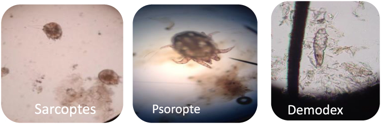 |
Figure 1 Mange mite genera. |
Site of Attachment
Based on the site of infestation, the highest infested body part was the shoulder whereas the least was the leg. There is strong association between body part and infestation of mange mite (P ≤ 0.05) (Figure 2 and Table 3).
 |
Table 3 Prevalence of Bovine Mange Mites Based on Site of Infestation |
 |
Figure 2 Mange mite lesion on the back part near to tail. |
Association with Potential Risk Factors
The prevalence of mange mite infestation was assessed in relation to age category. The prevalence in the adult and young age groups was 22.02% and 22.69%, respectively. However, there was no statistical significance (p ≥ 0.05) in the prevalence of the various species of mange mites and the two age categories (Table 4).
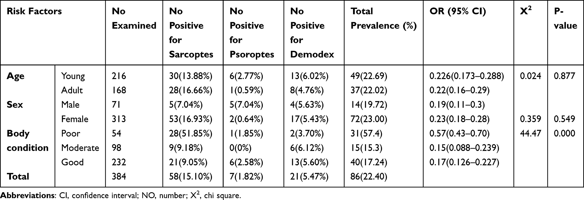 |
Table 4 Prevalence Mange Mite in Cattle According to Risk Factors |
The prevalence of bovine mange mites was also assessed in relation to sex. The prevalence mite infestation in females and males were found to be 23% and 19.72% respectively. There was no statistically significant association (p ≥ 0.05) between mite infestation and sex. Female animals were more infected with the mange mites than the male animals (Table 4).
Based on body condition score, the prevalence of mange mite infestation was assessed and a higher prevalence was recorded in poor body conditioned animals (57.4%), followed by good (17.24%) and (15.3%) moderate body conditioned animals by score. There was a statistically significant association (p ≤ 0.05) found for different body condition categories (Table 4).
Discussion
The study revealed that skin diseases due to mange mite were common in cattle in the study area. The overall prevalence of mange mite on cattle was 22.40%. It is slightly higher than a previous study27 which reported 13.79% in Gondor town and another28 which reported 11.78% in central zone of Tigrai and 3.54% in Womberta woreda. Another29 reported a 3.13% prevalence of mange mite in and around Hawassa, southern Ethiopia. The variations might be due to the difference in the weather conditions, inadequate veterinary service and types of management practiced in the study areas.
The observed mange mite genera were Sarcoptes, Demodex and Psoroptes. From those, Sarcoptes was more prevalent in the study area and accounted for 15.10%. This is higher than the reports of Tewodros et al27 who reported 4.367% in the Gondor town and Abebayehu et al30 who reported 0.57% in and around Kombolcha. This higher prevalence of sarcoptic mange mite might be attributed to the types of management practices and weather condition of the area.
The second commonly observed mange mite in the study site was Demodex with a prevalence of 21 (5.47%). This is higher than the previous reports of Yacob et al31 and Chalachew32 in the Adama and Wolaita soddo area with a prevalence of 1.88% and 1.63% in cattle, respectively. However, it was lower than the reports of Marsha et al33 and Tewodros et al27 which were 15.5% and 9.43% prevalence of bovine demodicosis from Gondor areas, respectively. This difference might be due to variations in climatic condition, management and feed accessibility and usage of acaricides.
The third most commonly observed mange mite in the study area was Psoroptes with a prevalence of 7 (1.82%). This is higher than the result of 0.9% reported by Onu and Shiferaw6 in the Bench Maji zone of south west Ethiopia and Marsha et al33 with 0.6% in and around Bishoftu town, central Ethiopia and 0.4% reported by Abebayehu et al30 in and around Kombolcha, northern Ethiopia. This higher prevalence of mange mite might be attributed to the cattle management system and weather condition differences which can exacerbate the disease conditions.
The prevalence of mange mite in different age categories was found to be statistically not significant (p>0.05), even though young cattle were more infected than the adults. This finding was in close agreement with the work of Tewodros et al27 who reported mange mites on young and adult with a prevalence of 7 (20%) and 14 (13.3%) respectively. When the level of infestation of mange mites were calculated for the two sex groups of cattle, a prevalence of 23.00% and 19.72% were observed in female and male cattle, respectively. However, there was no significance difference (p > 0.05) in sex groups. This finding was in agreement with previous observations made elsewhere in the country. Yacob et al31 and Tewodros et al27 reported that sex has no significant effect on the prevalence of mange mite. This indicated that the mange mite infestation was not age and sex selective. The difference might come about due to the poorly developed immune system of young cattle from lack of exposure, compared to the older age categories.
The prevalence of mange mite was higher in females than males. This might be due to factors like pregnancy and lactation which can decrease the immunity of females which might enhance the susceptibility of the female animal to mange mite infestation. In the present study, the prevalence was compared in animals which have poor, moderate and good body conditions. The highest level of prevalence (57.4%) was observed in animals which had poor body condition while the lowest prevalence (15.3%) was observed in animals with moderate body condition. This result is consistent with Meseret et al34 who reported 26.3% and 3.8% prevalence for poor and moderate body conditions respectively in and around Bishoftu town. This might be due to nutritional status; as well fed animals can better withstand parasitic infestation than animals on an inadequate diet which can influence the level of immunity. As Wall and Shearer17 indicated, there is an increase in mite infestation in animals with poor body condition.
The distribution of mange mites was found on six anatomical sites, indicating that mange mites were detected at the highest frequency from the shoulder, 24 (6.25%) followed by the neck and face region, 18 (4.69%) and the region of the ear, 14 (3.65%). The level of detection of mites was less in the regions of the tail, 8 (2.08%) and leg, 4 (1.02%). This distribution of mange mite agrees with Tewodros et al27 who reported the more preferable sites were shoulder, neck and back area of the body.
The variation on site of infestation might be due to the living condition of the parasite as commensals that lead suddenly to pathogenic states or due to the frequent exposure of neck and shoulder for various stress conditions like yoke sore, traumatic injury, and injury due to ploughing instruments which facilitates the mite to feed easily by puncturing the host skin and sucking out the tissues of the injured area.
Conclusion and Recommendations
The present study revealed a high prevalence of mange mite in cattle in the study area and the major genera of mange mite identified were Sarcoptes, Demodex and Psoroptes. From those genera the highest was sarcoptes. From risk factors, body condition is important factors that significantly associated with mange mites’ infestation in cattle. Mite infestation varies based on their preference site; shoulder is the most preferred site whereas the leg region is the less preferred one. Compared to the prevalence found in other sites, the current study indicated high prevalence that needs interventions to decrease the economic losses due to the parasite. In line with the above conclusion, the following recommendations are made:
- Appropriate control measures should be taken during early spring and end of winter or during summer because, during these seasons, the spread of the disease is high.
- The farmers should be advised about the possible risk factors like stress condition and poor nutrition which can aggravate the disease, so farmers should be aware regarding to the importance of appropriate feeding and to give more care for their cattle.
- Both preventive and control measures should be taken to minimize the economic losses associated with this problem.
- The regional and other concerned authorities should give attention and launch control measures against these parasites.
Abbreviations
CSA, Central Statistical Authority; GPD, gross domestic product; KOH, potassium hydroxide; NDAO, Nekemte District Agricultural Office; STATA, Statistical Analysis program.
Acknowledgments
Our deepest gratitude goes to Haramaya University College of Veterinary Medicine and Agriculture for supporting this research work through financial support. Also, we are grateful to Wollega University, School of Veterinary Medicine for supporting needed materials for the research.
Disclosure
The authors read and agreed on the manuscript and there is no conflict of idea on publishing this manuscript. The authors report no conflicts of interest for this work.
References
1. Central Statistics Authority(CSA). Federal democratic republic of Ethiopia. agricultural sample enumeration statistical, abstract. Addis Ababa, Ethiopia; 2008.
2. Sertse T. Investigation in ectoparasites of small ruminants in selected sites of Amhara Regional State and their impact on the tanning industry. MSc Thesis. Debrezeit, Ethiopia: Faculty of Veterinary Medicine, Addis Ababa University; 2004.
3. Bennett R, Ijpelar J. Updated estimate of the cost associated with thirty four endemic livestock diseases in Great Britain: a note. J Agric Econ. 2005;56
4. Bekele T. Studies on seasonal dynamics of ticks of Ogaden cattle and individual variation in resistance to ticks in eastern Ethiopia. J Vet Med. 2002;49:285–288. doi:10.1046/j.1439-0450.2002.00567.x
5. Ectoparasites: WR. Future challenges in a changing world. Vet Parasitol. 2007;148:62–74. doi:10.1016/j.vetpar.2007.05.011
6. Onu SH, Shiferaw TZ. Prevalence of ectoparasite infestations of cattle in bench. Maji zone, southwest Ethiopia. Vet World. 2013;6:291–294. doi:10.5455/vetworld.2013.291-294
7. Muller G, Kirk RW, Scott DW. Small Animal Dermatology.
8. Bornstein S, Morner T, Samuel WM. Sarcoptes scabiei and sarcoptic mange. In: Samuel WM, Pybus MJ, Kocan AA, editors. Parasitic Diseases of Wild Mammals.
9. Kahn CM, Line S, Allen DG, Anderson DP, Jeffcoh LB. Acariasis Mite Infestation. In the Merck Veterinary Manual.
10. Taylor MA, Coop RL, Wall RL. Veterinary Parasitology.
11. Tyler H, Ensminger H. Dairy cattle science; external parasites; 2006:279.
12. Wall R. Veterinary Ectoparasites: Biology, Pathology & Control. Oxford: Blackwell Science Ltd; 2001.
13. Krantz GW, Walter DE. A Manual of Acarology.
14. Bochkov AV, Mironov SV. Phylogeny and systematic of mammal- associated psorptidian mites (acariformes, astigmata, psorptidia) derived from external morphology. Invertebr Syst. 2011;25:22–59. doi:10.1071/IS10023
15. Urquhart GM, Armour J, Duncan JL, Dunn AM, Jennings FM. Veterinary Parasitology.
16. Krauss H, Weber A, Appel M, Enders B. Diseases Transmissible from Animal to Humans; Parasitic Zoonosis.
17. Wall R, Shearer D. Veterinary Ectoparasites; Biology, Pathology and Control.
18. Esmael S. Survey on cattle ectoparasites in and around Wolayta Sodo, Southern, Ethiopia. In: Ettinger S, Feldman E, editors. Text Book of Veterinary Internal Medicine; Other External Parasites.
19. Bowman D. Georgis Parasitology for Veterinarians.
20. Andrews A, Blowey W, Boyd H, Eddy R. Bovine Medicine; Diseases and Husbandry of Cattle.
21. Serrano E, Cross PC, Beneria M, Ficapal A, Curia J. Decreasing prevalence of brucellosis in red deer via efforts to control disease in livestock. Epidemiol Infect. 2011;139:1626–1630. doi:10.1017/S0950268811000951
22. Nekemte District Agricultural Office(NDAO). Annual report on population size and agriculture of the district. Nekemte, 2007.
23. Gatenby R. The Tropical Agriculture. London and Beging stock Mc Millan Education. Ltd; 1991:6–10.
24. Nicholsen M, Butterworth M. A Guide to Condition Scoring of Zebu Cattle International Livestock Center for Africa. Ethiopia: Addis Ababa; 1986.
25. OIE. Mange. In: Manual of Diagnostic Tests and Vaccines for Terrestrial Animals.
26. Thrusfield M. The control and eradication of diseases. In: Veterinary Epidemiology.
27. Tewodros F, Mekash A, Mersha C. Demodex and sarcoptes mites of cattle: an extravagance for leather industry. Am-Eurasian J Sci Res. 2012;7(3):131–135.
28. Ashenafi H, Tibbo M. Major skin diseases of cattle in the Central Zone of Tigray region, Northern Ethiopia. J Agric Environ Sci. 2003;7:1–8.
29. Addise A, Achenef M. Major skin diseases of cattle: prevalence and risk factors in and around Hawassa, Southern Ethiopia. J Adv Vet Res. 2013;3:147–153.
30. Abebayehu T, Endris F, Berhanu M, Rahmeto A, Solomon M, Endris Z. Study on the prevalence of ectoparasite infestation of ruminants in and around kombolcha and damage to fresh goat pelts and wet blue (pickled) skin at kombolcha Tannary, Northeastern Ethiopia. Hawassa University. Ethiop Vet j. 2011;15(2):87–101.
31. Yacob HT, Netsanet B, Dinka A. Prevalence of major skin diseases in cattle, sheep and goats at adama veterinary clinic, Oromia Regional State, Ethiopia. Ethiop Vet J. 2008;3:12–14.
32. Chalachew N. Study on skin diseases in cattle, sheep and goat in and around Wolayta Soddo, Southern Ethiopia. DVM Thesis. DebreZeit: Addis Ababa University, Faculty of Veterinary Medicine; 2001.
33. Marsha C, Solomon T, Basaznew B. Prevalence of bovine demodicosis in Gondar Zuria district Amhara Region, North West Ethiopia. Glob Vet. 2013;11(1):30–35.
34. Meseret G, Fikre Z, Gebremedhin R. Identification and prevalence of ectoparasites in cattle and sheep in and around Bishoftu Town, Central Ethiopia. Anim Vet Sci. 2014;2(4):124–129. doi:10.11648/j.avs.20140204.17
 © 2022 The Author(s). This work is published and licensed by Dove Medical Press Limited. The full terms of this license are available at https://www.dovepress.com/terms.php and incorporate the Creative Commons Attribution - Non Commercial (unported, v3.0) License.
By accessing the work you hereby accept the Terms. Non-commercial uses of the work are permitted without any further permission from Dove Medical Press Limited, provided the work is properly attributed. For permission for commercial use of this work, please see paragraphs 4.2 and 5 of our Terms.
© 2022 The Author(s). This work is published and licensed by Dove Medical Press Limited. The full terms of this license are available at https://www.dovepress.com/terms.php and incorporate the Creative Commons Attribution - Non Commercial (unported, v3.0) License.
By accessing the work you hereby accept the Terms. Non-commercial uses of the work are permitted without any further permission from Dove Medical Press Limited, provided the work is properly attributed. For permission for commercial use of this work, please see paragraphs 4.2 and 5 of our Terms.