")
Back to Journals » International Journal of General Medicine » Volume 16
Plasma Exosome-Derived microRNAs Profiles in Patients with Serofast Status: A Cross-Sectional Study
Authors Liu J , Zhang R, Lian T, Chen Z, Zhang RL, Wang Q
Received 1 February 2023
Accepted for publication 7 April 2023
Published 20 April 2023 Volume 2023:16 Pages 1455—1469
DOI https://doi.org/10.2147/IJGM.S404545
Checked for plagiarism Yes
Review by Single anonymous peer review
Peer reviewer comments 3
Editor who approved publication: Prof. Dr. Héctor Mora-Montes
Jinquan Liu,1 Ruihua Zhang,1 Tingting Lian,2 Zuoxi Chen,1 Rui-Li Zhang,3 Qianqiu Wang1
1Institute of Dermatology, Chinese Academy of Medical Sciences & Peking Union Medical College, National Center for STD Control, China Centers for Disease Control and Prevention, Nanjing, 210042, People’s Republic of China; 2Department of Dermatology, Zhongshan Hospital, Fudan University, Shanghai, 200032, People’s Republic of China; 3Department of Dermatology, the Second Affiliated Hospital of Nanjing Medical University, Nanjing, 210003, People’s Republic of China
Correspondence: Rui-Li Zhang; Qianqiu Wang, Email [email protected]; [email protected]
Purpose: Syphilis is a sexually transmitted bacterial infection caused by Treponema pallidum (T. pallidum), which can lead to chronic morbidity and adverse complications. In clinical practice, serofast status (SF) patients present with clinical symptoms that are very similar to those of healthy individuals or syphilis-cured patients, and often require prolonged follow-up for diagnosis. Currently, there is increasing interest in the potential of plasma exosome-derived miRNA as a biomarker for the detection of infectious diseases. In this study, we aimed to explore the diagnostic potential of miRNA in SF and its possible biological implications.
Patients and Methods: Exosome-derived miRNAs were isolated from peripheral plasma samples obtained from 20 patients with secondary syphilis (SS), SF, serologically cured syphilis (SC), and healthy controls (HC), and differentially expressed miRNAs (DEmiRNAs) were identified by microarray analysis. Prediction of potential target genes, functional annotation, gene ontology (GO) and Kyoto Encyclopedia of Genes and Genomes (KEGG) pathway analysis were then performed. The expression of selected miRNAs was confirmed in 37 patients by quantitative reverse transcription polymerase chain reaction (RT-qPCR). A receiver operating characteristic (ROC) analysis was performed to evaluate the diagnostic performance of these miRNAs in differentiating syphilis from HC or SC.
Results: The expression profile of plasma exosome-derived miRNA was discovered in individuals with SF through microarray analysis. The targeted genes of DEmiRNAs were found to be involved in diverse biological processes according to GO and KEGG analysis, such as regulation of transcription, mitochondria, Golgi, immune system, apoptosis, Ras signaling pathway, etc. Using RT-qPCR validation, miR-1273g-3p, miR-4485-5p, miR-197-3p, and miR-1908-3p showed significant upregulation in patients with SF. These miRNAs exhibited a superior diagnostic ability, either individually or combined, to distinguish SF from SC or HC.
Conclusion: The DEmiRNAs in plasma exosomes may play a role in the pathogenesis of SF and have the potential to become a noble and effective diagnostic method.
Keywords: serofast status, syphilis, microRNA, exosome, biomarker
Introduction
Syphilis is still a serious worldwide public health problem as a sexually transmitted infectious disease. The spirochete Treponema pallidum (T. pallidum) is difficult to culture in vitro, making syphilis a disease with unknown pathogenic mechanisms. Global estimates show that there were 7 million new syphilis infections in 2020.1 In China, the incidence of syphilis has continued to rise, with up to 36 cases per 100,000 population reported in 2018.2 Syphilis is a multistage, progressive systemic disease that presents with multiple characteristic symptoms, including chancres, gummas, disseminated skin lesions, and cardiovascular and brain involvement.3 The effects of syphilis are far-reaching, with untreated maternal syphilis causing an estimated 6.2% and 9.7% of global neonatal deaths and stillbirths, respectively.4 Although most patients are cured after recommended syphilis therapy, some individuals may experience a “serofast status (SF)”. The SF refers to a situation in which persons with a positive TPPA and a low pretreatment titers of TRUST results (also sometimes said<4-fold or no change following standard treatment).5,6 A study reported that 20.6% of 465 early syphilis patients who were randomized to receive penicillin or azithromycin had transferred into SF at six months, including 13% with primary syphilis, 14.2% with secondary syphilis, and 37.9% with early latent syphilis.7 In China, a study including 1327 syphilis patients confirmed that the prevalence of the SF is 34.4% at 12 months after recommended therapy, and the likelihood of a substantial decline in the SF proportion over time is low.8
The persistent positive serological reaction following syphilis treatment remains a puzzling clinical problem, with uncertain indicators of occult foci of T. pallidum infection or a common state of immune response in circulating blood. One study found that 48.6% of SF patients with early syphilis achieved serological cure 12 months after retreatment with 2.4 million units of benzathine penicillin weekly for three weeks.9 However, another study suggested that the serological cure rate dropped to only 27% with a single dose of benzathine penicillin for retreatment.10 Although some studies have shown changes in the immune status of SF patients,11–13 the pathogenic mechanism of SF remains unclear. The lack of highly sensitive and specific diagnostic tests to detect the activity of T. pallidum in the body is a major issue in syphilis diagnosis. Therefore, the diagnosis of SF often relies on clinical and experimental tests with long-term follow-up visits to the patients after treatment.
Exosomes have been widely accepted as possible novel biomarkers for the detection of infectious diseases.14,15 They are small (approximately 30–100 nm) vesicles formed inside secreting cells and then secreted into various body fluids, including blood, urine, milk, and cerebrospinal fluid.16 Exosomes are relatively stable due to their lipid bilayer, which encapsulates a variety of proteins, lipids, and nucleotides. As proficient bioinformatics transporters, exosomes protect their contents from degradation and deliver them to target cells, leading to changes in the biological behavior of the receiving cells.17 The main function of exosomes is mediated by miRNAs. As a subtype of small non-coding RNAs composed of approximately 21 nucleotides, miRNAs regulate mRNA translational regulation in cells and affect virtually all biological functions.18 Previous studies have shown changes in host miRNA profiles in response to infection with diverse pathogens, including viruses, bacteria, parasites, and fungi.19–22 Regarding T. pallidum infection, the miRNA profiles in peripheral blood mononuclear cells (PBMCs) of patients with syphilis were investigated.23–25 Exosomes were isolated from plasma and cerebrospinal fluid, and DEmiRNAs were associated with neurosyphilis.26 In our previous studies,27 we found that exosome-derived miR-146a-5p from T. pallidum-stimulated macrophages targets JAM-C, reducing endothelial cells permeability and monocyte transendothelial migration. Therefore, studying exosome-derived miRNAs in SF would be beneficial for exploring new detection indicators for SF and understanding the associated regulatory mechanisms of syphilis.
In this study, we collected peripheral blood samples from healthy volunteers and patients with different stages of syphilis. We then used miRNA microarray analysis to identify DEmiRNAs in exosomes derived from plasma. Specific miRNAs were subsequently screened and identified using quantitative reverse transcription polymerase chain reaction (RT-qPCR), and their functions were explored through bioinformatics techniques. Our study aims to provide new insights into the diagnosis and pathogenesis of SF.
Material and Methods
Study Group and Samples
Blood samples were collected from 40 syphilitic patients and 17 healthy volunteers, aged 18–86 visited to the Department of Dermatology and Venereal Diseases at the Institute of Dermatology, Chinese Academy of Medical Sciences & Peking Union Medical College between August 2018 and October 2021. After obtaining consent, the patients were divided into three groups according to the diagnostic criteria for syphilis issued by the National Health Commission of the People’s Republic of China (WS 273–2018).28 SS is defined as a disease manifested by skin rash often seen on the palms of the hands and soles, condylomata lata, mucocutaneous lesions, and generalized lymphadenopathy combined with positive laboratory testing results from rapid plasma reagin test(RPR) and T. pallidum Particle Agglutination(TPPA). SC was defined as a syphilis patient with positive TPPA and negative RPR test after treatment. SF is inconsistently defined but generally entails a lack of a four-fold decline and/or persistence of baseline low titer (≤ 1:4 or ≤ 1:8 in different studies), 6–12 months after adequate treatment.29 17 healthy volunteers were recruited. For all of the participants, pregnant or breastfeeding women and patients who had comorbidities like AIDS, tuberculosis, viral hepatitis, autoimmune disease, cancer, and other systemic or infective diseases were excluded. The clinical characteristics of this study are presented in Table 1. It is worth mentioning that blood samples from SS were collected before treatment. Blood collected from patients were centrifuged to obtain plasma, then stored at −80°C before miRNA microarray and RT-qPCR validation.
 |
Table 1 Information on the Clinical Samples Used for the Microarray Study and RT-qPCR Validation Study |
Extraction of Exosome-Derived miRNA
Exosome-derived miRNAs were collected from 3mL of plasma using exoRNeasy plasma/serum Midi Kit (Exiqon QIAGEN, #77044). Operation adhered to the manufacturer’s instructions. In brief, the plasma was pipetted to the exoEasy spin column and centrifuged in turn. Then the lysate was collected from the column after QIAzol washing. Then we mixed the lysate and chloroform, shook it vigorously, and centrifuged it for 15 min at 4°C. After this we mixed the upper aqueous phase with ethanol and then added the mixture into a new RNeasy MinElute Spin Column, centrifuged for 15 second at room temperature. Finally, miRNAs were collected from the column after RNase-free water washing.
Microarray Analysis
Exosome-derived miRNA samples were analyzed using the Agilent Human miRNA (8*60K) V21.0 microarray at Shanghai Biotechnology Co., Ltd. (Shanghai, China), and following the manufacturer’s protocols for labeling and hybridization. We removed spots with a flag < 0 and normalized spot intensities before transforming them into gene expression log2 ratios. Genes with |log2 ratio| ≥ 1.0 and a p-value < 0.05 were considered significantly differentially expressed.
RT-qPCR Assay
The extracted miRNA was reversed into cDNA using the Mir-X™ miRNA First-Strand Synthesis Kit (Takara Biomedicals, Shiga, Japan) according to the manufacturer’s instructions. The expression levels of target miRNAs were measured using the SYBR Premix Ex Taq kit (Takara Biomedicals, Shiga, Japan). U6 was selected as the internal reference. All specimens were evaluated twice. The primer sequences were as follows: 5′- gttcaccaccttctccac-3′ for miR-197-3p; 5′ -ggccgccggct-3′ for miR-1908-3p; 5′ -gaccgcctgccca −3′ for miR-4485-5p; 5′-cactgcactccagcct-3′ for miR-1273g-3p and 5′-TGGAACGCTTCACGAATTTGCG-3′ (forward) and 5′-GGAACGATACAGAGAAGATTAGC-3′ (reverse) for U6. Triplicate samples were assessed by the 2−ΔΔCt method. Differential expression of miRNAs was determined by the Independent variance t-test.
MicroRNA-Predicted Target Genes, Gene Ontology (GO), and Pathway Analyses
Potential target genes of miRNAs were predicted and analyzed by the bioinformatics algorithm TargetScan. Gene ontology and KEGG pathway enrichment analysis were performed to elucidate the biological annotation and potential pathways, respectively.
Statistics
R 3.5.0 software was used to analyze Microarray data, and GraphPad Prism (V.7.0, GraphPad Software) software was used to analyze RT-qPCR data. Subsequently, MedCalc 15.2.2 was used to draw receiver operating characteristic (ROC) curves and calculate the area under the ROC curve (AUC), optimal diagnostic value (largest Youden’s index), sensitivity, and specificity. A significance level of p < 0.05 was considered.
Results
Sample Quality Control and Principal Component Analysis
To assess the biological separation of the groups, principal component analysis (PCA) was performed based on probe intensities. Samples were divided into four groups according to the clinical diagnosis (Figure 1). The principal component of the SS group was significantly different from that of the HC group. The principal components one accounted for 85.7% of the contribution rate. Only one sample “SC006” showed a large degree of dispersion and difference from that of the group of SC. As a means of quality control, PCA analysis verified the rationality of the grouping of samples and experimental design, and the homogeneity of biological repeated samples.
 |
Figure 1 Principal component analysis (PCA) of miRNA expression data from human plasma exosomes. Red, SF; yellow, HC; green, SC; blue, SS. |
DEmiRNAs in Plasma Exosomes of Patients with SF
Based on the microarray results, we observed distinct patterns of differentially expressed microRNAs (DEmiRNAs) between different stages of syphilis. The volcano plots indicate that only miR-1908-3p was down-regulated in HC compared with SF (Figure 2a, Supplementary Table 1). We also observed 24 downregulated miRNAs and 20 upregulated miRNAs in SS compared with SF (Figure 2b, Supplementary Table 2). Conversely, seven miRNAs were downregulated and four miRNAs were upregulated in SC compared with SF (Figure 2c, Supplementary Table 3). The numbers of DEmiRNAs between different groups are further displayed in Figure 2d. The Venn diagram highlights miRNAs that exhibit either concurrent upregulation (Figure 2e) or downregulation (Figure 2f) in different group comparisons. Notably, miR-4419b is down-expressed in both SS and SC compared with SF (Figure 2f).
 |
Figure 2 DEmiRNAs across different stages of syphilis. Changes in miRNA expression (|log2 ratio| ≥ 1.0, P < 0.05) are illustrated by volcano plot. Up-expressed miRNAs were shown as red dots, and down-expressed ones were marked as blue dots. (a) DEmiRNAs between HC compared with SF. (b) DEmiRNAs between SS compared with SF. (c) DEmiRNAs between SC compared with SF. (d) Number of DEmiRNAs corresponding to all groups. (e) Venn diagram of up-expressed miRNAs. (f) Venn diagram of down-expressed miRNAs. “A” represents DEmiRNAs in SS compared with HC. “B” represents DEmiRNAs in SC compared with HC. “C” represents DEmiRNAs in HC compared with SF in (a). “D” represents DEmiRNAs in SS compared with SC. “E” represents DEmiRNAs in SS compared with SF in (b). “F” represents DEmiRNAs in SC compared with SF in (c). |
Additionally, heat maps are created by utilizing clustering algorithms that enable the analysis of gene expression patterns in large-scale gene expression datasets. Through classification and clustering of genes, genes that share similarities can be grouped together to form clusters that provide a more visual representation of the overall structure and pattern of the data. Upon comparing the SS and SF samples, a heat map was generated, which effectively segregated the samples into two clusters in each respective group. This distinction was visibly apparent and was represented in Figure 3a. Further comparisons were made between SC and SF samples, most SF samples clustered together in an independent group, with all SC samples forming their own individual cluster. However, a sample “SF017” was observed to cluster with the SC samples in Figure 3b. Despite this discrepancy, the sample was retained within Group SF due to the implementation of biological repetition. It was deemed justifiable to retain this sample given its resemblance to other SF samples across different inter-group comparisons and its negligible influence on the overall precision and credibility of the study.
 |
Figure 3 miRNA screening in SC, SF, and SS. Red boxes indicate up-regulated miRNAs, and blue boxes indicate down-regulated miRNAs. The brightness indicates the magnitude of the difference. Changes in miRNA expression (|log2 ratio| ≥ 1.0, P < 0.05) are illustrated by the heat map. MiRNAs and samples with similar expression are clustered together. (a) SS vs SF, (b) SC vs SF. |
Differential Gene Target Sequence Prediction and Pathway Enrichment Analysis
To investigate the possible functions of the DEmiRNAs between SC and SF, the target sequences of miRNAs were predicted by using the Targetscan database. The target genes were then subjected to GO and KEGG pathway enrichment analysis. GO classification analysis (Figure 4a) and the Top 30 GO enrichment analysis (Figure 4b, Supplementary Table 4) showed that the DEmiRNA target genes are associated with various biological processes such as DNA-binding transcription factor activity, regulation of transcription, RNA biosynthetic process, GTP binding, nucleoside binding, aromatic compound biosynthetic process, and regulation of the structure and function of mitochondria and Golgi. The KEGG pathway classification (Figure 5a) and enrichment analysis (Figure 5b, Supplementary Table 5) revealed that DEmiRNAs are involved in a range of pathways including Immune system, Endocrine system, Signal transduction, Apoptosis, Signaling pathways regulating pluripotency of stem cells, VEGF signaling pathway, p53 signaling pathway, Fc epsilon RI signaling pathway, ErbB signaling pathway, HIF-1 signaling pathway, Rap1 signaling pathway, and Ras signaling pathway. These pathways play critical roles in regulating various biological processes and are often interconnected, highlighting the complex regulatory network controlled by miRNAs in syphilis patients. Therefore, the dysregulation of miRNAs may affect the expression of genes involved in immune cell function, leading to impaired immune responses and persistent infection, which would be one of the contributing factors for SF. Thus, understanding the roles of miRNAs in regulating immune cell function in syphilis patients could provide insights into the mechanisms underlying the pathogenesis of syphilis.
 |
Figure 4 GO analysis of target genes of DEmiRNAs between SC and SF. (a) GO classification of target genes. (b) The top 30 most significantly enriched GO terms associated with target genes. Analysis Method: Fisher’s Exact Test (Precision Inspection), p-value < 0.05, q-value < 0.05, minimum difference count of 4. |
 |
Figure 5 Pathway analysis of target genes of DEmiRNAs between SC and SF. (a) KEGG classification of target genes. (b) Pathways associated with target genes. Analysis Method: Fisher’s Exact Test (precision inspection), p-value < 0.05, with a minimum difference count of 4. |
Validation of the Microarray Results by RT-qPCR
A total of 37 patients from different groups were recruited for the validation of the microarray results (Table 1). Exosome-derived miRNAs were extracted in the same way as described before. Among the diverse DEmiRNAs, four miRNAs were selected for RT-qPCR validation. As shown in Figure 6, the expression of miR-197-3p, miR-4485-5p, miR-1273g-3p was significantly higher expressed at higher levels in SF compared to SC. And the expression of miR-1908-3p was significantly higher expressed at higher levels in the SF compared to HC. These results were consistent with microarray analysis and suggested that miR-197-3p, miR-1908-3p, miR-4485-5p, miR-1273g-3p may be the potential biomarkers to predict SF.
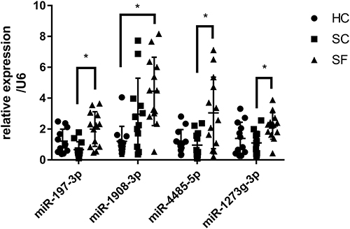 |
Figure 6 Detection of exosomes miRNAs of different groups by the RT-qPCR assay. The expression of four miRNAs was measured in 37 samples. Four miRNAs (miR-197-3p, miR-1273g-3p, miR-4485-5p, miR-1908-3p) were selected from the microarray data. Relative expression was used to normalize the gene expression data. U6 was set as the reference gene. Statistical analysis of the two groups was performed using Student’s t-test. *P < 0.05. |
The Diagnostic Value of the Individual miRNAs or Multiple miRNAs in SF
ROC curves were generated to evaluate the diagnostic value of the four miRNAs in syphilis (Table 2). MiR-1908-3p was useful in discriminating SF from HC (AUC = 0.901, sensitivity = 92.3%, specificity = 91.7%). However, the combination of four miRNAs had limited improved diagnostic value (AUC = 0.949, sensitivity = 92.3%, specificity = 91.7%) (Figure 7a and Table 2). To examine their capacity to discriminate between SF and SC, miR-1273g-3p, miR-4485-5p, and miR-197-3p showed better diagnostic capability than miR-1908-3p (AUC = 0.904, sensitivity = 100%, specificity = 83.3%) (Figure 7b and Table 2). Moreover, the combination of four miRNAs improved diagnostic value. In addition, miR-1908-3p and miR-197-3p could separate SC from HC (Figure 7c and Table 2). The combination of four miRNAs improved this capacity, with an AUC value of 0.868 (sensitivity = 83.3%, specificity = 75.0%).
 |
Table 2 Discrimination Power of Single or Multiple miRNAs in Syphilis |
 |
Figure 7 ROC curve analysis of single or multiple miRNAs evaluating the diagnostic value among different stages of syphilis. (a) SF vs HC. (b) SF vs SC. (c) SC vs HC. |
Discussion
At present, there is no consistent definition of the SF, with the primary difference being the follow-up time. In the USA, a follow-up at 6 and 12 months after treatment for primary and secondary syphilis, and 24 months for early latent syphilis, is recommended.30 In the UK, recommended clinical and serological (RPR or VDRL) follow-up is at 3, 6 and 12 months, and then, if indicated, every six months until VDRL/RPR negative or SF.31 However, according to Chinese diagnostic criteria for syphilis, patients are considered to be in a SF after 12 months of recommended therapy for primary syphilis, 2 years for secondary syphilis, and 3 years for late-stage of syphilis, if their nontreponemal test remains positive and the titers neither increased nor decrease by at least four-fold (two dilutions).8 To optimize practicality, the selection of samples for this survey was predicated on the Chinese diagnostic criteria for syphilis, and the follow-up period for SF patients was at minimum 12 months (Table 1). This particular sampling method affords us the opportunity to acquire disease specimens that satisfy varying diagnostic criteria.
Exosomes are nanovesicles involved in cell-to-cell communication, found in bodily fluids such as blood, milk, urine, saliva, and cerebrospinal fluid. They can deliver bioactive substances such as protein, lipids, and RNAs, making them attractive biomarkers detectable in liquid biopsies. Exosome-derived miRNAs detection is a noninvasive diagnostic method that benefits from the protective lipid bilayer of exosome, and specially promising biomarkers due to their stability and high sensitivity in peripheral blood compared to other body fluids.32–35
Prior to our study, also for the exosome-derived microRNA, a study has been carried out on neurosyphilis.26 Exosome-derived miRNAs are derived from serum and cerebrospinal fluid, and the results show that miR-590-5p, miR-570-3p, and miR-570-5p are significantly upregulated in patients with neurosyphilis in both cerebrospinal fluid and serum. The other kinds of research focus on miRNAs from PBMCs that serve as novel biomarkers for syphilis diagnosis, especially for the diagnosis of SF.25,36 MiR-223-3p and miR-195-5p from PBMCs varied from patients with primary syphilis or SF to HC, despite the difficulty of distinguishing latent syphilis from the SF.37 In addition, known and novel predicted (np)-miRNAs in PBMCs in syphilitic patients with SF were identified and verified, such as miR-338-5p, np-miR-163, np-miR-128, np-miR-244, and np-miR-5.23 It is evident that the exosome-derived miRNAs from serum and cerebrospinal fluid in neurosyphilis and the miRNAs from PBMCs in SF show distinct expression patterns from the exosome-derived miRNAs in plasma from SF. It is challenging to identify common DEmiRNAs across these groups, possibly due to differences in subject selection and individual variation.
Our study identified several potential exosome-derived miRNAs as biomarkers for SF diagnosis, including miR-197-3p, miR-1908-3p, miR-1273g-3p, and miR-4485-5p. Among these, miR-1908-3p had the highest diagnostic accuracy, sensitivity, and specificity with an AUC value of 0.901, 92.3% sensitivity, and 91.7% specificity. When combined, all four miRNAs showed a strong capacity to differentiate SF from SC or HC, with an AUC value of 0.904–0.949, 92.3–100% sensitivity, and 83.3–91.7% specificity. These findings are clinically significant due to the vague definition of SF and the lengthy follow-up times required for diagnosis.8,30 The potential implications or applications of exosome-derived miRNAs in SF are worth further research.
Nowadays, the exact mechanisms that lead to the SF in syphilis patients are not fully understood. However, it is believed that several factors may contribute to the development of SF, including incomplete or inadequate treatment of the infection, host immune responses, and genetic factors. One possible explanation is that the initial infection triggers the activation of host immune responses, which, in turn, result in the formation of long-lived plasma cells that continue to produce antibodies against T. pallidum, even after the bacteria have been cleared from the body. Another possible explanation is that the bacteria may be able to evade or suppress the immune system, allowing them to persist in the host and cause chronic infection.
Previous studies have suggested that the SF may be associated with the latent infection of T. pallidum,38 such as the special molecular subtypes of T. pallidum repeat gene found in SF,39 which was known to be linked with immune evasion by pathogens and chronic syphilis infection. Impaired immune function and dysfunction have been observed in SF patients.40,41 Thus, we aim to explore the potential connection between DEmiRNAs and the development of SF. By performing GO and KEGG enrichment analysis, we have discovered that the target genes of DEmiRNAs are linked with a range of biological processes, including regulation of transcription, regulation of the structure and function of mitochondria and Golgi, Immune system, Endocrine system, Apoptosis, VEGF signaling pathway, p53 signaling pathway, etc. These pathways are frequently interconnected, indicating the intricate regulatory network that miRNAs maintain in syphilis patients. Therefore, if miRNA expression is dysregulated, it may disrupt gene expression levels that encourage the function of immune cells. Consequently, this may result in compromised immune responses and ongoing infection, which may be one of the underlying factors for SF in syphilis patients.
Further analysis of the miRNA-protein coding gene relationships revealed that miR-197-3p, as one of the miRNAs exhibiting high differential expression in circulating exosomes between SS or SF and HC, may be involved in regulating the host immune system. Several studies have explored the possible roles of miR-197-3p in different types of cancer and ailments. These studies have found that miR-197-3p can inhibit prostate cancer cell proliferation by regulating the VDAC1/AKT/β-catenin signaling axis,42 reduce epithelial-mesenchymal transition in ovarian cancer cells by targeting ABCA7,43 regulate endothelial cell proliferation and migration in Kawasaki disease by targeting IGF1R and BCL2,44 and modulate inflammation in monocytes and synovial fibroblasts related to familial Mediterranean fever by targeting the IL1R1 gene.45 The facts suggest that miR-197-3p could be a significant candidate molecule for understanding the pathogenesis of SF. It would be worthwhile to conduct further investigations on the differential exosome-derived miRNAs to ascertain their contribution to the development of SF. Such investigations could provide valuable insights into the mechanisms that drive the development of syphilis by elucidating the roles of miRNAs in regulating immune cell activity.
Conclusion
In summary, our research effectively characterized DEmiRNAs within plasma exosomes isolated from individuals with SF, subsequently conducting preliminary investigations on their respective target genes. Our findings shed valuable insights into the potential significance of miRNA target genes in the pathogenesis of SF. Additionally, we successfully confirmed the differential expression levels of identified DEmiRNAs, further validating their potential as diagnostic markers for SF. Notably, miR-197-3p, miR-1908-3p, miR-1273g-3p, and miR-4485-5p demonstrated a favorable capacity to distinguish SF from HC or SC. The combination of all four miRNAs further improved the accuracy of distinguishing SF from SC. Nevertheless, our study is limited by a relatively small sample size, and further research and clinical trials are necessary to validate our findings and develop novel diagnostic methods. Considering the clinical difficulties in diagnosing and predicting the prognosis of SF, our findings suggest that using exosome-derived miRNAs from plasma may present a new and effective diagnostic approach.
Abbreviations
HC, healthy controls; SS, secondary syphilis; SF, serofast status; SC, serologically cured syphilis; DEmiRNAs, differentially expressed miRNAs; T. pallidum, Treponema pallidum; PBMCs, peripheral blood mononuclear cells; ROC, receiver operating characteristic; AUC, area under the curve; RT-qPCR, quantitative reverse transcription polymerase chain reaction; PCA, Principal component analysis; GO, Gene ontology; KEGG, Kyoto Encyclopedia of Genes and Genomes; np, novel predicted; RPR, rapid plasma reagin test; TPPA, Treponema pallidum Particle Agglutination.
Data Sharing Statement
All data and material can be available from one of the first authors and corresponding authors, who can be contacted on the email address.
Ethics and Data Quality Assurance
The study is in compliance with relevant institutional, national, and international guidelines and legislation. This study was approved by the ethics committee of the Institute of Dermatology and Skin Disease Hospital, Chinese Academy of Medical Sciences (No. 2017-KY-010). All methods were carried out in accordance with relevant guidelines and regulations. Informed consent was obtained from all subjects. The study complied with the Declaration of Helsinki.
Acknowledgments
We want to thank Wenlong Hu and Caixia Kou for their help in conducting the experiments related to this work. We thank the researchers of Shanghai Biotechnology, Co., Ltd. (Shanghai, China) for their technical support, as well as the patients and the clinics, nurses and counselors who identified the patients and referred them to our study.
Author Contributions
All authors made a significant contribution to the work reported, whether that is in the conception, study design, execution, acquisition of data, analysis and interpretation, or in all these areas; took part in drafting, revising or critically reviewing the article; gave final approval of the version to be published; have agreed on the journal to which the article has been submitted; and agree to be accountable for all aspects of the work.
Funding
National Natural Science Foundation of China (81772209 and 81601804). Union Innovation Team Project of CAMS (2016-I2M-3021). CAMS Innovation Fund for Medical Sciences (CIFMS-2021-I2M-1-001). Nanjing Incubation Program for National Clinical Research Center (2019060001).
Disclosure
The authors report no conflicts of interest in this work.
References
1. World Health Organization. Sexually transmitted infections (STIs). Available from: https://www.who.int/news-room/fact-sheets/detail/sexually-transmitted-infections-(stis).
2. Chinese Center for Disease Control and Prevention. An overview of the epidemic situation of notifiable infectious diseases in China in 2018; 2019.
3. Peeling RW, Mabey D, Kamb ML, Chen XS, Radolf JD, Benzaken AS. Syphilis. Nat Rev Dis Primers. 2017;3(Suppl):17073. doi:10.1038/nrdp.2017.73
4. World Health Organization. WHO Guidelines for the Treatment of Treponema Pallidum (Syphilis). Geneva Switzerland: World Health Organization; 2016.
5. Workowski KA, Bachmann LH, Chan PA, et al. Sexually transmitted infections treatment guidelines, 2021. MMWR Recomm Rep. 2021;70(4):1–187. doi:10.15585/mmwr.rr7004a1
6. Arlene C. A systematic review of syphilis serological treatment outcomes in HIV-infected and HIV-uninfected persons: rethinking the significance of serological non-responsiveness and the serofast state after therapy. BMC Infect Dis. 2015; 2015:1.
7. Seña AC, Wolff M, Martin DH, et al. Predictors of serological cure and Serofast State after treatment in HIV-negative persons with early syphilis. Clin Infect Dis. 2011;53(11):1092–1099. doi:10.1093/cid/cir671
8. Tong ML, Lin LR, Liu GL, et al. Factors associated with serological cure and the serofast state of HIV-negative patients with primary, secondary, latent, and tertiary syphilis. PLoS One. 2013;8(7):e70102. doi:10.1371/journal.pone.0070102
9. Wang Z-S, Liu X-K LJ, Li J. Serological response to therapy following retreatment of serofast early syphilis patients with benzathine penicillin. J Antimicrob Chemother. 2018;73(5):1348–1351. doi:10.1093/jac/dky006
10. Seña AC, Wolff M, Behets F, et al. Response to therapy following retreatment of serofast early syphilis patients with benzathine penicillin. Clin Infect Dis. 2013;56(3):420–422. doi:10.1093/cid/cis918
11. Seña AC, Zhang XH, Li T, et al. A systematic review of syphilis serological treatment outcomes in HIV-infected and HIV-uninfected persons: rethinking the significance of serological non-responsiveness and the serofast state after therapy. BMC Infect Dis. 2015;15:479. doi:10.1186/s12879-015-1209-0
12. Pastuszczak M, Gozdzialska A, Jakiela B, Obtulowicz A, Jaskiewicz J, Wojas-Pelc A. Robust pro-inflammatory immune response is associated with serological cure in patients with syphilis: an observational study. Sex Transm Infect. 2017;93(1):11–14. doi:10.1136/sextrans-2016-052681
13. Li J, Wang LN, Zuo YG, et al. 梅毒血清抵抗患者临床分析及免疫功能研究 [Clinical analysis and study of immunological function in syphilis patients with seroresistance]. Zhonghua Yi Xue Za Zhi. 2009;89(12):813–816. Chinese.
14. Chaudhari P, Ghate V, Nampoothiri M, Lewis S. Multifunctional role of exosomes in viral diseases: from transmission to diagnosis and therapy. Cell Signal. 2022;94:110325. doi:10.1016/j.cellsig.2022.110325
15. Yang J, Shin TS, Kim JS, Jee YK, Kim YK. A new horizon of precision medicine: combination of the microbiome and extracellular vesicles. Exp Mol Med. 2022;54(4):466–482. doi:10.1038/s12276-022-00748-6
16. Tkach M, Kowal J, Théry C. Why the need and how to approach the functional diversity of extracellular vesicles. Biol Sci. 2018;373(1737). doi:10.1098/rstb.2016.0479
17. Villarroya-Beltri C, Gutiérrez-Vázquez C, Sánchez-Cabo F, et al. Sumoylated hnRNPA2B1 controls the sorting of miRNAs into exosomes through binding to specific motifs. Nat Commun. 2013;4:2980. doi:10.1038/ncomms3980
18. Anfossi S, Babayan A, Pantel K, Calin GA. Clinical utility of circulating non-coding RNAs - an update. Nat Rev Clin Oncol. 2018;15(9):541–563. doi:10.1038/s41571-018-0035-x
19. Zhang H, Li QY, Guo ZZ, et al. Serum levels of microRNAs can specifically predict liver injury of chronic hepatitis B. World J Gastroenterol. 2012;18(37):5188–5196. doi:10.3748/wjg.v18.i37.5188
20. Mourenza Á, Lorente-Torres B, Durante E, et al. Understanding microRNAs in the context of infection to find new treatments against human bacterial pathogens. Antibiotics. 2022;11(3):356. doi:10.3390/antibiotics11030356
21. Yuan S, Wu Q, Wang Z, et al. miR-223: an immune regulator in infectious disorders. Front Immunol. 2021;12:781815. doi:10.3389/fimmu.2021.781815
22. He X, Pan W. Host-parasite interactions mediated by cross-species microRNAs. Trends Parasitol. 2022;38(6):478–488. doi:10.1016/j.pt.2022.02.011
23. Jia X, Wang Z, Liu X, Zheng H, Li J. Peripheral blood mononuclear cell microRNA profiles in syphilitic patients with serofast status. Mol Biol Rep. 2020;47(5):3407–3421. doi:10.1007/s11033-020-05421-7
24. Jya B, Tao HB, Pz B, et al. MicroRNA-101-3p, MicroRNA-195-5p, and MicroRNA-223-3p in peripheral blood mononuclear cells may serve as novel biomarkers for syphilis diagnosis. Microb Pathog. 2021;152:104769.
25. Huang T, Zhang J, Ke W, et al. MicroRNA expression profiling of peripheral blood mononuclear cells associated with syphilis. BMC Infect Dis. 2020;20(1):165. doi:10.1186/s12879-020-4846-x
26. Chen H, Zhou Y, Wang ZY, et al. Exosomal microRNA profiles from serum and cerebrospinal fluid in neurosyphilis. Sex Transm Infect. 2019;95(4):246–250. doi:10.1136/sextrans-2018-053813
27. Hu W, Xu B, Zhang J, et al. Exosomal miR-146a-5p from Treponema pallidum-stimulated macrophages reduces endothelial cells permeability and monocyte transendothelial migration by targeting JAM-C. Exp Cell Res. 2020;388(1):111823. doi:10.1016/j.yexcr.2020.111823
28. ICS. National Standard of the People’s Republic of China: Diagnosis for Syphilis(WS 273—2018). Beijing: Standards Press of China; 2018.
29. Mehta N, Bhari N, Gupta S. Asian guidelines for syphilis. J Infect Chemother. 2022;28(8):1084–1091. doi:10.1016/j.jiac.2022.04.023
30. Workowski KA, Berman S. Sexually transmitted diseases treatment guidelines, 2010. MMWR Recomm Rep. 2010;59(Rr–12):1–110.
31. Kingston M, French P, Higgins S, et al. UK national guidelines on the management of syphilis 2015. Int J STD AIDS. 2016;27(6):421–446. doi:10.1177/0956462415624059
32. Dieckmann K, Radtke A, Geczi L, et al. Serum levels of MicroRNA-371a-3p (M371 test) as a new biomarker of testicular germ cell tumors: results of a prospective multicentric study. J Clin Oncol. 2019;37(16):1412–1423. doi:10.1200/jco.18.01480
33. Thomou T, Mori M, Dreyfuss J, et al. Adipose-derived circulating miRNAs regulate gene expression in other tissues. Nature. 2017;542(7642):450–455. doi:10.1038/nature21365
34. Shan Z, Qin S, Li W, et al. An endocrine genetic signal between blood cells and vascular smooth muscle cells: role of MicroRNA-223 in smooth muscle function and atherogenesis. J Am Coll Cardiol. 2015;65(23):2526–2537. doi:10.1016/j.jacc.2015.03.570
35. Mitchell PS, Parkin RK, Kroh EM, et al. Circulating microRNAs as stable blood-based markers for cancer detection. Proc Natl Acad Sci. 2008;105(30):10513–10518. doi:10.1073/pnas.0804549105
36. Yang Z, Yang X, Gan Z, Yuan L, Liu W, Si C. Genome-wide MicroRNA analysis of peripheral blood mononuclear cells reveals elevated miR-142-3p expression as a potential biomarker for secondary syphilis. Biomed Res Int. 2021;2021:5520053. doi:10.1155/2021/5520053
37. Yang J, Huang T, Zhao P, et al. MicroRNA-101-3p, MicroRNA-195-5p, and MicroRNA-223-3p in peripheral blood mononuclear cells may serve as novel biomarkers for syphilis diagnosis. Microb Pathog. 2021;152:104769. doi:10.1016/j.micpath.2021.104769
38. Lin LR, Tong ML, Fu ZG, et al. Evaluation of a colloidal gold immunochromatography assay in the detection of Treponema pallidum specific IgM antibody in syphilis serofast reaction patients: a serologic marker for the relapse and infection of syphilis. Diagn Microbiol Infect Dis. 2011;70(1):10–16. doi:10.1016/j.diagmicrobio.2010.11.015
39. Yang WL, Lin WS, Yang J, Zheng HP. Relationship between seroresistance and molecular subtypes of treponema pallidum repeat gene. China J Mod Med. 2011 2011:1.
40. Leader BT, Hevner K, Molini BJ, Barrett LK, Van Voorhis WC, Lukehart SA. Antibody responses elicited against the Treponema pallidum repeat proteins differ during infection with different isolates of Treponema pallidum subsp. pallidum. Infect Immun. 2003;71(10):6054–6057. doi:10.1128/iai.71.10.6054-6057.2003
41. Morgan CA, Lukehart SA, Van Voorhis WC. Protection against syphilis correlates with specificity of antibodies to the variable regions of Treponema pallidum repeat protein K. Infect Immun. 2003;71(10):5605–5612. doi:10.1128/iai.71.10.5605-5612.2003
42. Huang Q, Ma B, Su Y, et al. miR-197-3p represses the proliferation of prostate cancer by regulating the VDAC1/AKT/β-catenin signaling axis. Int J Biol Sci. 2020;16(8):1417–1426. doi:10.7150/ijbs.42019
43. Xie W, Shui C, Fang X, Peng Y, Qin L. miR-197-3p reduces epithelial-mesenchymal transition by targeting ABCA7 in ovarian cancer cells. Biotech. 2020;10(8):375. doi:10.1007/s13205-020-02362-7
44. Li Y, Wu X, Gao F, Wang X. MiR-197-3p regulates endothelial cell proliferation and migration by targeting IGF1R and BCL2 in Kawasaki disease. Int J Clin Exp Pathol. 2019;12(11):4181–4192.
45. Akkaya-Ulum YZ, Akbaba TH, Tavukcuoglu Z, et al. Familial Mediterranean fever-related miR-197-3p targets IL1R1 gene and modulates inflammation in monocytes and synovial fibroblasts. Sci Rep. 2021;11(1):685. doi:10.1038/s41598-020-80097-4
 © 2023 The Author(s). This work is published and licensed by Dove Medical Press Limited. The full terms of this license are available at https://www.dovepress.com/terms.php and incorporate the Creative Commons Attribution - Non Commercial (unported, v3.0) License.
By accessing the work you hereby accept the Terms. Non-commercial uses of the work are permitted without any further permission from Dove Medical Press Limited, provided the work is properly attributed. For permission for commercial use of this work, please see paragraphs 4.2 and 5 of our Terms.
© 2023 The Author(s). This work is published and licensed by Dove Medical Press Limited. The full terms of this license are available at https://www.dovepress.com/terms.php and incorporate the Creative Commons Attribution - Non Commercial (unported, v3.0) License.
By accessing the work you hereby accept the Terms. Non-commercial uses of the work are permitted without any further permission from Dove Medical Press Limited, provided the work is properly attributed. For permission for commercial use of this work, please see paragraphs 4.2 and 5 of our Terms.