Back to Journals » Infection and Drug Resistance » Volume 13
Phenotypic and Genotypic Characteristics of Clostridium difficile Isolates in Patients with Type 2 Diabetes in Iran
Authors Shoaei P ![]() , Shojaei H, Shirani K
, Shojaei H, Shirani K ![]()
Received 2 August 2019
Accepted for publication 21 February 2020
Published 27 February 2020 Volume 2020:13 Pages 683—690
DOI https://doi.org/10.2147/IDR.S225829
Checked for plagiarism Yes
Review by Single anonymous peer review
Peer reviewer comments 2
Editor who approved publication: Dr Eric Nulens
Parisa Shoaei,1 Hasan Shojaei,2 Kiana Shirani1
1Nosocomial Infection Research Center, Isfahan University of Medical Sciences, Isfahan, Iran; 2Department of Microbiology, School of Medicine, Isfahan University of Medical Sciences, Isfahan, Iran
Correspondence: Hasan Shojaei
Department of Microbiology, School of Medicine, Isfahan University of Medical Sciences, Hezar Jerib Ave, Isfahan, Iran
Tel +98 31 37929119
Email [email protected]
Purpose: This study aimed to investigate the phenotypic and genotypic characterization of Clostridium difficile isolates in type 2 diabetes patients with hospital-acquired diarrhea in four teaching hospitals in Isfahan, Iran.
Patients and Methods: A total of 104 hospitalized patients with type 2 diabetes and nosocomial diarrhea were included in the current study over a 2-year period (2015– 2017). C. difficile isolates were characterized by conventional microbiological methods including the presence of toxin genes, antibiotic resistance testing and molecular methods including multilocus sequence typing (MLST) and random amplification of polymorphic DNA (RAPD).
Results: All 21 C. difficile isolates (20.2%) were detected from 104 studied patients. All isolates were susceptible to metronidazole and vancomycin. The antimicrobial resistance rates were distinctly higher for clindamycin and for moxifloxacin. Based on PCR amplification of tcdA and tcdB, 13 isolates (12.5%) carried both of these genes and were considered toxigenic. Thirteen toxigenic C. difficile strains were classified into two sequence types (STs), that is, ST54 and ST2 types. The RAPD-PCR amplification patterns of the detected toxigenic C. difficile revealed three distinct but related RAPD clusters. RAPD cluster 1 had the highest similarity with RAPD types 2 and 3.
Conclusion: A relatively high rate of CDI was observed in patients with type 2 diabetes and was associated with poorer health outcomes. These patients were exposed to multiple antibiotics and other therapeutic agents. We recommend close screening for the coexistence of CDI and type 2 diabetes in patients with diarrhea using a combination of conventional and molecular methods.
Keywords: Clostridium difficile, hospital-acquired diarrhea, RAPD, MLST, molecular characterization, risk factor, type 2 diabetes
Introduction
Clostridium difficile is a strictly anaerobic, gram-positive, sporulating bacillus, identified as an important opportunistic and nosocomial pathogen.1,2 Clostridium difficile infection (CDI) is the most commonly recognized cause of infectious diarrhea in healthcare settings leading to diseases from the asymptomatic carriage diarrhea to life-threatening colitis.1,3,4
Type 2 diabetes is a common chronic metabolic disease and its prevalence is growing at an alarming rate worldwide.5 The American Society of diabetes has estimated that a minimum of 285 million people is affected by type 2 diabetes worldwide, and the prevalence is expected to reach 693 million by the year 2045.6,7
The numbers for diabetes indicate that over 5 million adults in 2017 (8.9% of the adult Iranian population have diabetes) and it is estimated that by the year 2030, 9.2 million Iranian individuals will have diabetes.6,8 increases the risk of recurrent CDI Diabetes-related hospitalization is a serious challenge to the health care system, a situation which may be further aggravated by nosocomial CDI and recurrent CDI.2,9
Epidemiologic characteristics and molecular typing of CDI cases are important tools for the investigation of hospital outbreaks and understanding the modes of transmission.10 Considering the increased duration of inpatient hospitalization due to type 2 diabetes-related sequelae. Existing data lack information on CDI status in type 2 diabetes patients in our region. The current study aimed to provide a relatively more precise data that might cast light on the issue from a microbiological standpoint using conventional and molecular approaches.
Materials and Methods
Study Design
The current descriptive cross-sectional study involved 104 hospitalized patients ≥18 years of age with type 2 diabetes and nosocomial diarrhea admitted to four teaching hospitals in Isfahan, Iran, between April 2015 to May 2017. All cases had acquired diarrhea during hospitalization.
The patients who had been diagnosed with CDI within the last three months before admission to hospitals were excluded from our study. To prevent over-representing, only the first stool sample from each patient was examined. Stool samples were also screened for E. coli, Salmonella spp, Shigella spp, and Campylobacter jejuni.
The diagnosis of type 2 diabetes was based on typical symptoms and a fasting plasma glucose level >126 mg/dl (7.0 mmol/l) or a two-hour post-load glucose level of >200 mg/dl (11.1 mmol/l).11,12
A case C. difficile infection (CDI) was defined based on clinical symptoms of diarrhea and a positive toxigenic culture.10 Data on demographic characteristics, recent (within 4 weeks) hospital admission, recent (within 8 weeks) antibiotic treatment and prior medications use including metformin and insulin were collected for all patients.
C. difficile Culture
Stool samples were examined by inoculation into C. difficile moxalactam norfloxacin (CDMN) broth supplemented with cysteine hydrochloride, norfloxacin, moxalactam, and 0.1% sodium taurocholate (Oxoid, UK) in an anaerobic jar for 5–7 days. Preliminary treatment with alcohol shock was performed in order to recover C. difficile from stool specimens. The treated stool samples were inoculated onto the CDMN- agar surface supplemented with 7% sheep blood and incubated anaerobically for 48 h at 37C°. Plates were examined for suspect colonies with 2–3 mm in diameter, the characteristics p-cresol odor unique to C.difficile, ultraviolet light (365 nm) for yellow fluorescence within 1 hr of removal from the anaerobic atmosphere, Gram stain morphology, and positive reaction to L-proline aminopeptidase test (Prodisk, Remeb, Lenexa, KS,USA)16,27
DNA Extraction
Intact chromosomal DNA was extracted using the modified Pitcher et al, (1989) with slight modification for extraction of large-scale DNA13 Briefly, cultures of toxigenic C. difficile isolates grown in Brain Heart Infusion (BHI) broth for 18 h were centrifuged and washed cells were treated with lysozyme and suspended in TE (Tris, 10 mM; EDTA, 50 mM; pH 8.0). Guanidium thiocyanate and sarkosyl were added to the mixture for protein denaturation. The DNA was purified by phenol-chloroform-isoamyl alcohol. The precipitate was washed in 70% ethanol, dehydrated and dissolved in deionized water and stored in a −20°C freezer until use.13–15
Molecular Identification
All isolates were confirmed by testing the existence of triose phosphate isomerase (tpi) gene and screened for the presence of the genes encoding toxin A, B (tcdA, tcdB) and binary toxins (cdtA, cdtB). C. difficile ribotype 027 was used as a positive control for molecular and microbiological analysis and C. perfringens 450 MTCC (Microbial Type Culture Collection) served as the negative control16,17
The patients were divided into 3 groups: i) C. difficile culture-negative, ii) non-toxigenic C. difficile carriers, and iii) CDI patients.
Multilocus Sequence Typing (MLST)
MLST was performed on all toxigenic C. difficile isolates using the primers and methods as described previously by Griffiths et al.18 Seven housekeeping genes (adk, atpA, dxr, glyA, recA, sodA, and tpi) were amplified and the amplified products were sent to Bioneer Corporation in South Korea for sequencing. DNA sequences were analysed using the PubMLST database (http://pubmlst.org/cdifficile/) to obtain the allele numbers and sequence types (STs).
RAPD Analysis
The PCR-RAPD assay was optimized to ensure the reproducibility and discriminatory power of the method [22,23]. The PCR amplification for RAPD fingerprinting was carried out with the primers AP3 (5ʹ-TCACGATGCA-3ʹ) and AP4 (5ʹ-TCACGCTGCA- 3ʹ) independently in a low-stringency PCR amplification. Each isolate was tested under the same conditions at least twice with these primers.14,19
The PCR products were separated by electrophoresis and scanned on an Uvi-doc gel documentation system. The software DNA FRAG version 3.03 (Nash, 1991) was used to estimate DNA fragment sizes in the RAPD profiles.13,20 GelCompar 6.6 software from Applied Maths was used for the construction of dendrogram based on the unweight pair-group method with averages (UPGMA) to estimate the relationships between the isolates. Only major bands were considered and band intensity was not used as a criterion.13
Antibiotic Susceptibility Testing
Minimum inhibitory concentrations (MICs) of clindamycin, metronidazole, moxifloxacin, rifampin, vancomycin and fusidic acid was determined using the Etest (bioMérieux, France) with log-phase inocula of 106 cfu/mL in tubes containing 5 mL of phosphate-buffered saline. All tests performed on pre-reduced Brucella Blood Agar plates containing vitamin K1 (1 mg/mL), haemin (5 mg/L) and 5% defibrinated sheep blood. The plates were incubated anaerobically with a chemical indicator (Microbiology Anaerotest® Strips; MERCK, Germany) at 37°C for 48 h.21 Interpretation of the results and determination of the MICs were carried out according to the European Committee on Antimicrobial Susceptibility Testing (EUCAST) and the Clinical Laboratory Standards Institute CLSI 2011 guidelines. In EUCAST there are only breakpoints for Vancomycin and metronidazole and for other antibiotics, only epidemiologic cut-offs are available.
Streptococcus sp. MTCC 689 and Clostridium perfringens MTCC 13124 strains were included in each run as controls.21,22 The antimicrobial agents tested were chosen because of the emergence of reduced susceptibility.
Statistical Analysis
Data were expressed as the means ± standard deviations and a P <0.05 was considered statistically significant. The logistic regression model was used to determine the clinical factors associated with C.difficle infection. First, a univariate logistic regression model was fitted on each clinical factor, and then a multivariate regression model with adjustment for the effects of other covariates was used. Variables that were significant in univariate models were entered into the multivariate model.
We estimated odds ratios (ORs) and 95% confidence intervals (CIs) for each of the variables using logistic regression models. Data were analysed using Statistical Package for Social Sciences (SPSS, Chicago, IL, USA) version 16.0.
Results
A total of 21 C. difficile isolates (20.2%) were detected from 104 hospitalized patients with type 2 diabetes based on positive culture and the presence of tpi gene. Based on PCR amplification of tcdA and tcdB, 13 isolates (12.5%) carried both of these genes and were considered toxigenic, while the remaining eight isolates (7.7%) were nontoxigenic (Table 1). Salmonella spp, Shigella spp, E. coli, and Campylobacter jejuni were not detected in any of the patient samples tested.
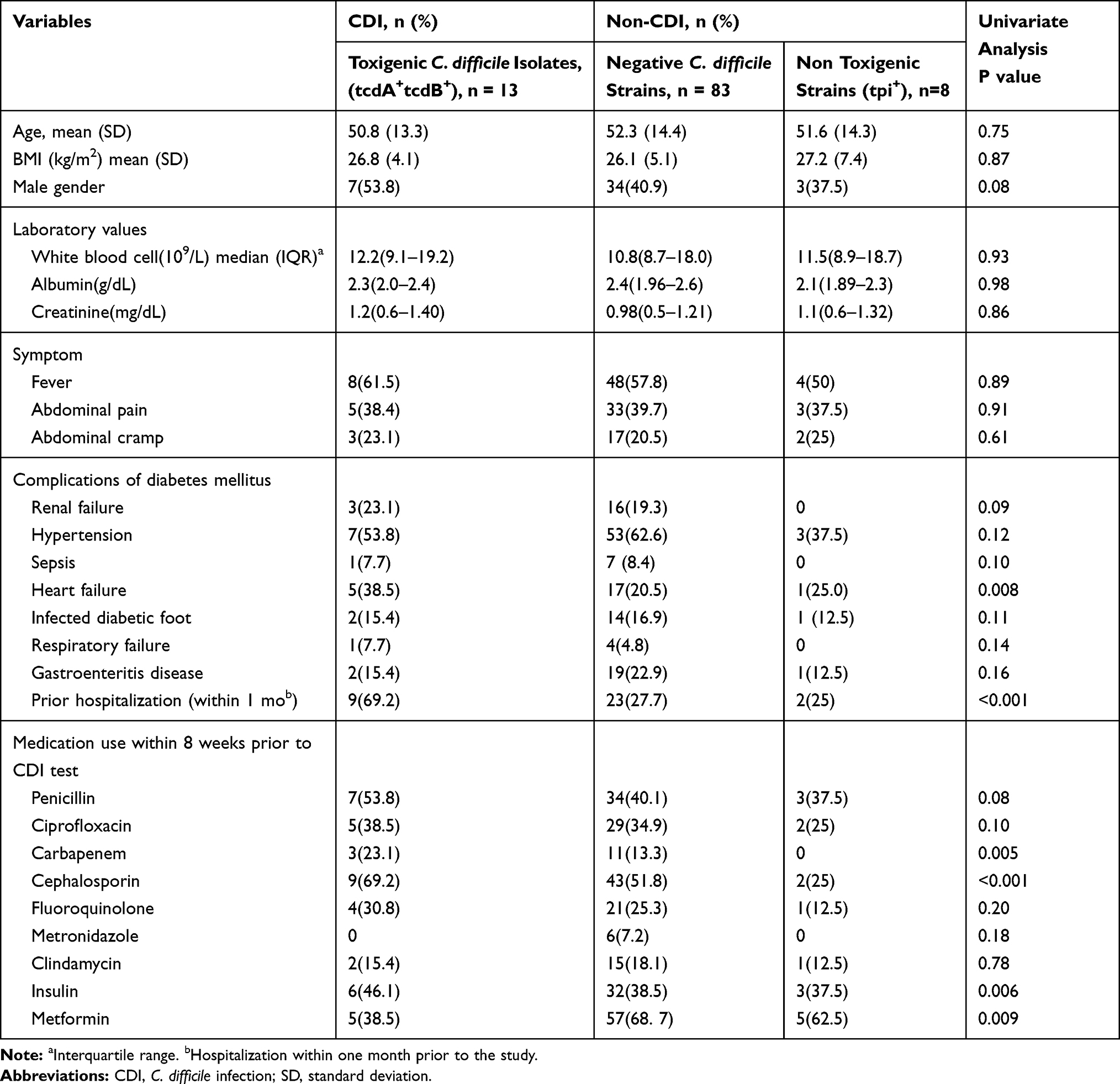 |
Table 1 Clinical Features of 104 Hospitalized Patients with Type 2 Diabetes in Toxigenic C. difficile Strains, Negative C. difficile Strains and Non-Toxigenic C. difficile Strains Groups Admitted to the 4 University Hospitals, Isfahan, Iran (2015–2017) |
The antibiotic susceptibility patterns of 21 C. difficile isolates are displayed in Table 2.
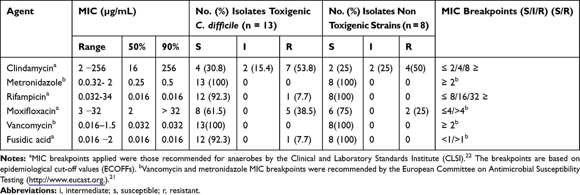 |
Table 2 Antimicrobial Susceptibility Testing of 21 Isolates to 6 Antimicrobial Agents |
All isolates were susceptible to metronidazole and vancomycin and were inhibited by a low concentration of the antibiotics. The MIC50/MIC90 of metronidazole and vancomycin were 0.25/0.5 μg/mL, 0.032/0.032 μg/mL, respectively. Approximately 50% (11/21) of C. difficile isolates were resistant to clindamycin and 33% (7/21) of the isolates to moxifloxacin (Table 2).
We found no obvious relationship between the resistance and toxin genotypes or CDI (P= 0.21).
The use of antibiotic was identified in all patients in the 8 weeks prior to CDI diagnosis (Table 1). The stepwise multivariate logistic regression model revealed in patients with diabetes, a history of heart failure (OR 1.35; 95% CI, 1.0–2.6; P= 0.04) and prior hospitalization (OR 3.2; 95% CI, 1.3–8.2; P = 0.001) were risk factors for CDI. Our findings suggest that in patients with diabetes metformin treatment seems to have a protective effect against the development of CDI (OR 0.52; 95% CI, 0.32–0.89; P = 0.02).
Genotypes of C. difficile Isolates
Of the 21 C. difficile isolates, 13 (61.9%) tested positive for both tcdA and tcdB (A+B). There were no deletions in tcdC genes in all toxigenic isolates tested. No isolate examined positive for the binary toxin genes cdtA and cdtB.
The 21 toxigenic C. difficile isolates were classified into two STs groups including, ST54 type (n= 8, 61.5%, tcdA+, tcdB+, cdtA−, cdtB−) and ST2 type (n= 5, 38.4%, tcdA+, tcdB+, cdtA−, cdtB (Figure 1). There was no significant difference between males and females in the prevalence of ST types (P=0.07).
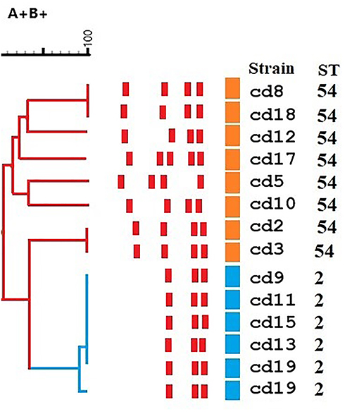 |
Figure 1 Dendrogram of the detected isolates of C. difficile, RAPD-PCR DNA fingerprints with the primer AP4. Dendrogram is color coded according to sequence types (STs) and toxin types. |
RAPD analysis of C. difficile isolates with the primer AP4 produced more informative banding patterns and generated four different RAPD types. RAPD profiles consisted of three to five amplicons ranging from 365 to 1196 base pairs in length. All studied isolates generated three major common bands, with the fragment sizes of 365, 400 and 510 base pairs. The most prevalent detected RAPD type was RT1 type with 7 isolates (53.8%) followed by RT-2 (4/13, 30.8%), RT-3 (1/13, 7.7%) and RT- 4 (1/13, 7.7%).
The RAPD-PCR amplification patterns of the detected toxigenic C. difficile revealed three distinct but related RAPD clusters. RAPD cluster 1 had 94% similarity with RAPD type 2 and 73% similarity with RAPD type 3. The remaining four isolates occurred in two small groups that showed a lower similarity with the other two clusters (Figure 1, Supplementary file).
The reference ribotype 027 formed a unique RAPD type pattern, which was distinctly separate from the other isolates.
Discussion
Type 2 diabetes mellitus often leads to an immunocompromised state, subsequent hospitalization, and an increased risk of CDI.5 CDI is the most prevalent cause of hospital-acquired diarrhea and is associated with a high mortality rate among elderly and immunocompromised patients who are hospitalized3,5 Epidemics of CDI over recent decades and the epidemiology of CDI in patients with type 2 diabetes is well studied in North America and Europe.9 However, there is relatively little evidence on those associations in developing countries. The need for surveillance of the international mobility of C. difficile strains has been emphasized. Circulating strains have capability to spread regionally and internationally, providing adequate ground for close monitoring of the prevalence and molecular epidemiology of CDI in the region. Due to the inaccessibility of health care facilities for C. difficile culture and toxin testing in many hospitals of Iran, awareness of circulating strains and their prevalence has been limited. The current study, to our knowledge, is the first cross-sectional study of patients with type 2 diabetes and hospital-acquired CDI in a developing country, Iran.2,12
The incidence of CDI in our patients was estimated at 12.5%.In two similar studies performed in Israel and Malaysia CDI prevalence were reported 30.8% and 8.8% respectively.12,23 None of our isolates carried cdtA and cdtB binary toxin genes, the presence of which is associated with increased mortality of the patients.17,24
The toxigenic C. difficile isolates from the studied hospitals exhibited similar epidemic genotype profiles (ST54, ST2). Most of CDI in previous studies were found to be due to A+B+ strains (ST2, ST54) and A−B+ strains (ST37, ST15).25–27 The main epidemic genotypes described recently in different regions of China were ST54, ST37, ST3, ST2 and ST35, however remarkable variations in molecular epidemiology of C. difficile across different countries worldwide have been observed.1 For example, in Korea and Japan, ST17 and ST2 are the predominant types; however, ribotype 027/ST1 is the most common C. difficile strain in the United States and Europe, for most Asian countries, ST1 cases have only been reported sporadically.1,24,28
We used RAPD typing method as a simple, fast and low-cost epidemiologic procedure to find the molecular variation of C. difficile isolates. C. difficile population structure is clonal and virulence-associated genes exhibited a moderate polymorphism.14,19
RAPD fingerprinting can rapidly classify isolates into clusters and the relationship of strains can be determined.13,19 The RAPD typing showed high discriminatory power and stability for analysis of these strains29 This method provide reliable results within one or 2 days and allow for early and more precise implementation of infection control measures.30 Our results strongly suggest that two major RAPD types of C. difficile, isolated from 53.8% (7/13) and 30.8% (4/13) of the patients, were prevalent circulating strains in this studied diabetic population. These cases occurred as unique endogenous CDI and might be spread by patient to patient transmission. We combined MLST method as a confirmation step to enhance specificity. Our C. difficile isolates were not resistant to metronidazole and vancomycin and in settings where access to vancomycin or fidaxomicin is limited, using metronidazole for an initial episode of nonsevere CDI only is recommended.10
The rates of resistance to other antimicrobials ranged from 4.8% to 53.8%, which was identical to another study described in Iran, which 5.3% and 89.3% of C. difficile strains were resistant to metronidazole and clindamycin respectively.31 High-level resistance to these antimicrobial agents can be attributed to different antibiotic regimens used. Antimicrobial use, including many chemotherapeutic agents, was prevalent in both CDI and non-CDI groups, however, the role of antimicrobials in CDI can definitely not be dismissed by these data. The patient age, long length of hospital stay, exposure to antibiotics and immunosuppressant are known main risk factors of CDI.2,5,32
In this study, recent hospitalization and heart failure were risk factors associated with CDI in patients with type 2 diabetes. In agreement with these findings, other studies also identified that severe diseases increase the risk of mortality and morbidity, which is explained by the potential for worsening immune state of these patients.5,12
Metformin treatment has strong effects on the gut microbiome and our findings provide support for the notion that altered gut microbiota could be considered to have a protective effect against the development of CDI.23,33
Conclusion
CDI is not thoroughly studied in Asia and in particular in the Middle East and therefore the extent of diagnosing needs to be more precisely known. Lack of close monitoring or antibiotic use and unreasonable prescribing in our country indicates that CDI could be widespread in the whole country where proper surveillance is currently absent. The widespread prevalence of strains in Asia indicates that the surveillance for toxin A (tcdA) or toxin B (tcdB) are more desirable to toxin A assays for diagnosis of CDI. The most virulent strains 027 and 078 have not established in Asia, while ST2 and ST54 types appear to be more prevalent.
In conclusion, our study demonstrated a relatively high CDI rate in patients with type 2 diabetes. The study population was exposed to multiple antibiotics and other therapeutic agents (i.e. metformin, insulin). The use of antibiotics was identified in all patients in the 8 weeks prior to CDI diagnosis and a history of heart failure and prior hospitalization were risk factors for CDI. Metformin treatment seems to have a protective effect against the development of CDI. We recommend close screening for CDI in hospitalized patients with type 2 diabetes using a combination of a rapid genotyping method such as RAPD and a robust high throughput MLST scheme.
Ethics Approval and Consent to Participate
The study was approved by the human research ethics committee at Isfahan University of Medical Sciences and the study was carried out in accordance with the approved guidelines. All the patients provided written informed consent before study entry to their stool being collected and for use in this study. This study was conducted in accordance with the Declaration of Helsinki.
Data Sharing Statement
Data generated or analyzed during this study are available and some are included in this article. (Gel pictures of different PCRs are available).
Acknowledgments
We would like to thank Dr. Behrooz Ataei and Mr. Abbas Daei naser for their special co-operation in this study.
Author Contributions
Shoaei P: contributed to acquisition of data and drafting the work and performed microbial and molecular experiments. Shojaei H: Designed and supervised the research, interpreted data, and co-wrote the paper. Shirani K: contributed to the conception of design and revising the draft critically and collected the patient data. All authors read and approved the final manuscript and agreed to be accountable for all aspects of the work in ensuring that questions related to the accuracy or integrity of any part of the work are appropriately investigated and resolved.
Funding
The current study was supported by a grant (No, 2933090) from Isfahan University of Medical Sciences, Isfahan, Iran.
Disclosure
The authors have no conflicts of interest to declare regarding this paper.
References
1. Luo Y, Zhang W, Cheng JW, et al. Molecular epidemiology of Clostridium difficile in two tertiary care hospitals in Shandong Province, China. Infect Drug Resist. 2018;11:489–500. doi:10.2147/IDR.S152724
2. Shakov R, Salazar RS, Kagunye SK, Baddoura WJ, DeBari VA. Diabetes mellitus as a risk factor for recurrence of Clostridium difficile infection in the acute care hospital setting. Am J Infect Control. 2011;39(3):194–198. doi:10.1016/j.ajic.2010.08.017
3. Azimirad M, Krutova M, Nyc O, et al. Molecular typing of Clostridium difficile isolates cultured from patient stool samples and gastroenterological medical devices in a single Iranian hospital. Anaerobe. 2017;47:125–128. doi:10.1016/j.anaerobe.2017.05.004
4. Gillespie W, Marya N, Fahed J, Leslie G, Patel K, Cave DR. Clostridium difficile in inflammatory bowel disease: a retrospective study. Gastroenterol Res Pract. 2017;2017:4803262. doi:10.1155/2017/4803262
5. Olanipekun TO, Salemi JL, Mejia de Grubb MC, Gonzalez SJ, Zoorob RJ. Clostridium difficile infection in patients hospitalized with type 2 diabetes mellitus and its impact on morbidity, mortality, and the costs of inpatient care. Diabetes Res Clin Pract. 2016;116:68–79. doi:10.1016/j.diabres.2016.04.021
6. Cho NH, Shaw JE, Karuranga S, et al. IDF Diabetes Atlas: global estimates of diabetes prevalence for 2017 and projections for 2045. Diabetes Res Clin Pract. 2018;138:271–281. doi:10.1016/j.diabres.2018.02.023
7. Kaiser AB, Zhang N, Van der Pluijm W. Global prevalence of type 2 diabetes over the next ten years (2018-2028). Am Diabetes Assoc. 2018;67:202–LB. doi:10.2337/db18-202-LB
8. Esteghamati A, Larijani B, Aghajani MH, et al. Diabetes in Iran: prospective analysis from first nationwide diabetes report of national program for prevention and control of diabetes (NPPCD-2016). Sci Rep. 2017;7(1):13461. doi:10.1038/s41598-017-13379-z
9. Qu HQ, Jiang ZD. Clostridium difficile infection in diabetes. Diabetes Res Clin Pract. 2014;105(3):285–294. doi:10.1016/j.diabres.2014.06.002
10. McDonald LC, Gerding DN, Johnson S, et al. Clinical practice guidelines for Clostridium difficile infection in adults and children: 2017 update by the Infectious Diseases Society of America (IDSA) and Society for Healthcare Epidemiology of America (SHEA). Clin Infect Dis. 2018;66(7):e1–e48. doi:10.1093/cid/cix1085
11. care ADAJD. Diagnosis and classification of diabetes mellitus. Diabetes Care. 2014;37(Supplement1):S81–S90. doi:10.2337/dc14-S081
12. Hassan S, Rahman R, Huda N, Wan Bebakar W, Lee Y. Hospital-acquired Clostridium difficile infection among patients with type 2 diabetes mellitus in acute medical wards. J JR Coll Physicians Edinb. 2013;43(2):103–107. doi:10.4997/JRCPE.2013.203
13. Hashemi A, Shojaei H, Heidarieh P, Aslani MM, Naser AD. Genetic diversity of Iranian clinical isolates of Mycobacterium tuberculosis. New Microbiol. 2012;35(1):61–65.
14. Barbut F, Mario N, Delmée M, Gozian J. Genomic fingerprinting of Clostridium difficile isolates by using a random amplified polymorphic DNA (RAPD) assay. FEMS Microbiol Lett. 1993;114(2):161–166. doi:10.1111/j.1574-6968.1993.tb06567.x
15. Pitcher D, Saunders N, Owen R. Rapid extraction of bacterial genomic DNA with guanidium thiocyanate. Lett Appl Microbiol. 1989;8(4):151–156. doi:10.1111/j.1472-765X.1989.tb00262.x
16. Lemee L, Dhalluin A, Testelin S, et al. Multiplex PCR targeting tpi (triose phosphate isomerase), tcdA (Toxin A), and tcdB (Toxin B) genes for toxigenic culture of Clostridium difficile. J Clin Microbiol. 2004;42(12):5710–5714. doi:10.1128/JCM.42.12.5710-5714.2004
17. Stubbs S, Rupnik M, Gibert M, Brazier J, Duerden B, Popoff M. Production of actin-specific ADP-ribosyltransferase (binary toxin) by strains of Clostridium difficile. FEMS Microbiol Lett. 2000;186(2):307–312. doi:10.1111/fml.2000.186.issue-2
18. Griffiths D, Fawley W, Kachrimanidou M, et al. Multilocus sequence typing of Clostridium difficile. J Clin Microbiol. 2010;48(3):770–778. doi:10.1128/JCM.01796-09
19. Mobasherizadeh S, Shojaei H, Havaei SA, et al. Application of the Random Amplified Polymorphic DNA (RAPD) fingerprinting to analyze genetic variation in community associated-methicillin resistant staphylococcus aureus (CA-MRSA) isolates in Iran. Glob J Health Sci. 2016;8(8):53822.
20. Nash JJIfBS. DNAfrag, Version 3.03. Ottawa (ON): National Research Council of Canada; 1991.
21. Testing ECoAS. Breakpoint tables for interpretation of MICs and zone diameters. Version 9.0, valid from 2019-01-01.
22. Wayne P. Performance Standards for Antimicrobial Susceptibility Testing. Clinical and laboratory standards institute; 2011.
23. Eliakim-Raz N, Fishman G, Yahav D, et al. Predicting Clostridium difficile infection in diabetic patients and the effect of metformin therapy: a retrospective, case–control study. Eur J Clin Microbiol Infect Dis. 2015;34(6):1201–1205. doi:10.1007/s10096-015-2348-3
24. Cheng VC, Yam WC, Chan JF, To KK, Ho PL, Yuen KY. Clostridium difficile ribotype 027 arrives in Hong Kong. Int J Antimicrob Agents. 2009;34(5):492–493. doi:10.1016/j.ijantimicag.2009.04.004
25. Shoaei P, Shojaei H, Jalali M, et al. Clostridium difficile isolated from faecal samples in patients with ulcerative colitis. BMC Infect Dis. 2019;19(1):361. doi:10.1186/s12879-019-3965-8
26. Shoaei P, Shojaei H, Khorvash F, et al. Clostridium difficile infection in cancer patients with hospital acquired diarrhea at the teaching hospitals in Iran: multilocus sequence typing analysis (MLST) and Antimicrobial resistance pattern. Anni Ig. 2019;31(4):365–373. doi:10.7416/ai.2019.2298
27. Shoaei P, Shojaei H, Khorvash F, et al. Molecular epidemiology of Clostridium difficile infection in Iranian hospitals. Antimicrob Resist Infect Control. 2019;8(1):12. doi:10.1186/s13756-018-0454-6
28. Jin D, Luo Y, Huang C, et al. Molecular epidemiology of clostridium difficile infection in hospitalized patients in Eastern China. J Clin Microbiol. 2017;55(3):801–810. doi:10.1128/JCM.01898-16
29. Azimirad M, Alebouyeh M, Rashidan M, Aslani MM, Zali MR. Comparison of common molecular typing methods for differentiation of clostridium difficile strains in the study of hospital acquired infections. Kowsarmedical. 2018;13(5):e61030–e61030.
30. Mutters NT, Heeg K, Späth I, Henny N, Günther F. Improvement of infection control management by routine molecular evaluation of pathogen clusters. Diagn Microbiol Infect Dis. 2017;88(1):82–87. doi:10.1016/j.diagmicrobio.2017.01.013
31. Goudarzi M, Goudarzi H, Alebouyeh M, et al. Antimicrobial susceptibility of clostridium difficile clinical isolates in iran. Iran Red Crescent Med J. 2013;15(8):704–711. doi:10.5812/ircmj
32. Olsen MA, Yan Y, Reske KA, Zilberberg MD, Dubberke ER. Recurrent Clostridium difficile infection is associated with increased mortality. Clin Microbiol Infect. 2015;21(2):164–170. doi:10.1016/j.cmi.2014.08.017
33. Wu H, Esteve E, Tremaroli V, et al. Metformin alters the gut microbiome of individuals with treatment-naive type 2 diabetes, contributing to the therapeutic effects of the drug. Nat Med. 2017;23(7):850–858. doi:10.1038/nm.4345
 © 2020 The Author(s). This work is published and licensed by Dove Medical Press Limited. The
full terms of this license are available at https://www.dovepress.com/terms
and incorporate the Creative Commons Attribution
- Non Commercial (unported, 3.0) License.
By accessing the work you hereby accept the Terms. Non-commercial uses of the work are permitted
without any further permission from Dove Medical Press Limited, provided the work is properly
attributed. For permission for commercial use of this work, please see paragraphs 4.2 and 5 of our Terms.
© 2020 The Author(s). This work is published and licensed by Dove Medical Press Limited. The
full terms of this license are available at https://www.dovepress.com/terms
and incorporate the Creative Commons Attribution
- Non Commercial (unported, 3.0) License.
By accessing the work you hereby accept the Terms. Non-commercial uses of the work are permitted
without any further permission from Dove Medical Press Limited, provided the work is properly
attributed. For permission for commercial use of this work, please see paragraphs 4.2 and 5 of our Terms.