Back to Journals » Journal of Inflammation Research » Volume 16
NUP62CL as an Immunological and Prognostic Biomarker of Oral Squamous Cell Carcinoma
Authors Yu X ![]() , Xu L, Zhou Y, Zhou X
, Xu L, Zhou Y, Zhou X ![]() , Yang L, Zhou Y
, Yang L, Zhou Y
Received 16 June 2023
Accepted for publication 23 August 2023
Published 29 August 2023 Volume 2023:16 Pages 3799—3809
DOI https://doi.org/10.2147/JIR.S426277
Checked for plagiarism Yes
Review by Single anonymous peer review
Peer reviewer comments 3
Editor who approved publication: Professor Ning Quan
Xin Yu,1,* Lijun Xu,2,* Yongqiang Zhou,3 Xiaorong Zhou,4 Lei Yang,2 Yan Zhou1
1Department of Orthodontics and Periodontology, Affiliated Nantong Stomatological Hospital of Nantong University, Nantong, People’s Republic of China; 2Department of Clinical Biobank & Institute of Oncology, Affiliated Hospital of Nantong University & Medical School of Nantong University, Nantong, People’s Republic of China; 3Department of Oral and Maxillofacial Surgery, Affiliated Nantong Stomatological Hospital of Nantong University, Nantong, People’s Republic of China; 4Department of Immunology, School of Medicine, Nantong University, Nantong, People’s Republic of China
*These authors contributed equally to this work
Correspondence: Yan Zhou, Affiliated Nantong Stomatological Hospital of Nantong University, Nantong, 226001, People’s Republic of China, Tel +86 513-89898899, Fax +86 513-89898890, Email [email protected] Lei Yang, Affiliated Hospital of Nantong University & Medical School of Nantong University, Nantong, 226001, People’s Republic of China, Tel +86 513-85052806, Email [email protected]
Background: Oral squamous cell carcinoma (OSCC) is the most common head and neck malignancy with a high mortality rate and poor prognosis. The exploration and understanding of biomarkers will help to further improve the diagnosis and treatment of OSCC.
Methods: Tumor tissue samples from 319 OSCC patients were retrospectively collected, along with their clinical information. In combination with bioinformatics tools and multiplex immunohistochemistry (mIHC) analyses, we evaluated NUP62CL protein expression and its relationship to tumor-infiltrating immune cells (TIICs) and immune checkpoints in the tumor microenvironment (TME), as well as its association with clinical features and prognosis of OSCC.
Results: We identified high-NUP62CL expression in OSCC tissues, and high-NUP62CL protein expression was associated with large tumor size, advanced clinical stage and poor prognosis. In addition, NUP62CL protein expression was positively associated with the abundance of CD3+CD4+ T cells (P< 0.01), CD3+CD8+ T cells (P< 0.01), CD56+ NK cells (P< 0.05), CD68+CD86+ macrophages (P< 0.01) and CD68+CD163+ macrophages (P< 0.01), as well as the immune checkpoints, including PD-1 (P< 0.001), PD-L1 (P< 0.001), and CTLA-4 (P< 0.001) protein expression.
Conclusion: In conclusion, NUP62CL could be an effective prognostic and immunological biomarker for OSCC patients.
Keywords: oral squamous cell carcinoma, NUP62CL, mIHC, prognosis, tumor-infiltrating immune cells, biomarker
Introduction
Oral squamous cell carcinoma (OSCC) is the most common oral malignancy, which originates from oral epithelial cells and develops into carcinoma in situ.1 The most common sites are the lips, tongue, palate, buccal mucosa, gums, floor of the mouth, vestibule, and posterior molars.2 Oral tissue is the main organ for the human body to eat and obtain nutrients. Chewing betel nut, chewing tobacco and irritation of tooth stump are the reasons for the high incidence of OSCC, which will hinder human nutrition intake through the oral pathway and lead to high mortality.3 Globally, there will be 377,713 new cases and 177,757 deaths in 2020.4,5 Although great progress has been made in the diagnosis and treatment of OSCC in recent years, the prognosis for OSCC has not improved significantly. According to clinical data, the 5-year survival rate of patients is about 50%.6 Therefore, the exploration and understanding of biomarkers for OSCC will help to further improve the diagnosis and treatment of OSCC.
Recently, Tumor microenvironment (TME), an environment for tumor occurrence, as the focus of current research, plays a key role in tumor progression.7,8 Tumor-infiltrating immune cells (TIICs) are important regulatory factors in TME, which can either anti-tumor or promote tumor occurrence.9 Studies have shown that TIICs are potential prognostic indicators of head and neck squamous cell carcinoma (HNSC).10 Immune checkpoint molecules, commonly known as programmed cell death receptor 1 (PD-1) and its ligand programmed cell death ligand 1 (PD-L1), induce immunosuppression and protect tumors from immune attack.11 Immunotherapy can activate the host’s natural defense system and transform TME from tumor friendly to tumor suppressor, which is a very promising new strategy for cancer treatment.12
The NUP family has been observed to play an important role in mitochondrial transport, DNA repair, gene transcription, and microtubule regulation.13,14 High Nup98 expression is associated with a poor prognosis in triple-negative breast cancer and predicts response to anthracycline-based chemotherapy as a novel marker.15 Nup153 shows an association with colorectal cancer cell proliferation and tumor growth.16 In addition, NUP62 is highly expressed in squamous cell carcinomas (SCCs). NUP62 absence inhibits SCCs cell proliferation and enhances differentiation.17 Further studies are needed to determine whether NUP62CL is involved in cancer development.
In this study, we aimed to investigate the immunological and prognostic value of NUP62CL in the immune microenvironment of OSCC. The association of NUP62CL expression with clinical features of OSCC and its influence on prognosis were explored. In addition, NUP62CL protein expression and its relationship to TIICs and immune checkpoints were evaluated by multiplex immunohistochemistry (mIHC) staining in the tissue microarray. Our study provided evidence for NUP62CL as an immune and prognostic biomarker, and provided a reference for immunotherapy for OSCC.
Materials and Methods
Human Tissue Samples and Patient Clinical Information
Transcriptome profiles of tumor tissues from 329 OSCC patients and oral mucosal tissues from 32 healthy individuals were downloaded from The Cancer Genome Atlas (TCGA) (https://portal.gdc.cancer.gov/), along with their clinical information. Tumor tissue samples from 319 OSCC patients in the clinical Biobank of Nantong Stomatological Hospital Affiliated to Nantong University during 2012–2018 were retrospectively collected, as well as their clinical data, including age, sex, anatomic classification, tumor size, lymph node metastasis (N), distant metastasis (M) and clinical stage. The patient had no radiation, chemotherapy, immunotherapy or other treatments before surgery. Informed consent to participate in this study was obtained from all enrolled subjects. This study has been approved by the Ethics Committee of Affiliated Nantong Stomatological Hospital of Nantong University (PJ 2021-014-01), and all methods and procedures followed the Declaration of Helsinki.
Immune Cells Infiltration Analysis
Xiantao tool (http://www.xiantao.love/) is a useful bioinformatics analysis web tool for visualization, which provided information on the relationship between NUP62CL mRNA expression and TIICs abundances, based on TCGA data. The correlation module in the Tumor Immune Estimation Resource (TIMER) 2.0 database (http://timer.cistrome.org/) was employed to assess the association between NUP62CL and immune checkpoint mRNA expression. In addition, the distribution of TIICs, and the association of TIICs and immune checkpoints with NUP62CL protein expression were examined by the R software (version 3.6.0).
Tissue Microarray and Multiplex Immunohistochemical Staining
Although protein levels generally correlate with the levels of the corresponding mRNA, a wide range of inconsistent expression patterns between proteins and mRNA can also be observed.18 As a result, we performed mIHC to explore the relationship between TIICs and immune checkpoints with NUP62CL protein expression. The tissue microarray was constructed by the manual Tissue Microarrayer System Quick Ray (UNITMA, Seoul, Korea). Continuous specimens of OSCC removed by operation were prepared with TMA slides, fixed with formalin and embedded in paraffin. The sample is represented on TMA as a core with a diameter of 2 mm.
TMA slides dewaxed and dehydrated using xylene and alcohol. The slices were heated in microwave with sodium citrate buffer (10 mM, pH 6.0) for antigen extraction. Section staining includes incubation of primary and secondary antibodies. Subsequently, opal fluorophore conjugated tyramide signal amplification (Akoya Biosciences, USA) was performed to amplify the signal. Repeat staining until the tissue sample is labeled with a set of markers. Details of the antibodies used in this study are as follows: anti-NUP62CL (1:500, orb312372, Biorbyt, UK); anti-cytokeratin (1:4000, orb69073, Biorbyt, UK); anti-CD56 antibody (1: 200, 3576s, CST, USA); anti-CD66b (1:2000, arg66287, Arigo, China); anti-CD68 (1: 2000, 76437s, CST, USA); anti-CD86 (1: 200, orb388891, Biorbyt, UK); anti-CD163 (1: 400, 93498s, CST, USA); anti-CD3 antibody (1: 400, 85061s, CST, USA); anti-CD4 (1:50, ab133616, Abcam, USA); anti-CD8 (1: 200, 85336s, CST, USA); anti-PD-1 (1:200, 13684T, CST, USA); anti-PD-L1 (1:400, 18616s, CST, USA); anti-CTLA-4 (1:400, orb527271, Biorbyt, UK).
We used the Vectra3-automated imaging software to capture each sample at 20X magnification. Using inForm4.1.0 (Perkin Elmer) to analyze and score the images, the pathologist analyzed the staining intensity distribution across the entire tissue section and determined the threshold for positive staining for each marker. Positive or negative cells are quantified according to a specified threshold. The percentage of cells in each area was assessed and scored (0–100). We divided patients into high and low expression groups by the median NUP62CL staining score, 14.36 points.
Statistical Analysis
The correlation between NUP62CL protein expression and clinical features of OSCC were evaluated by Pearson’s χ2. Survival prognostic analysis was performed by Cox regression models and the Kaplan-Meier survival curves. TIICs abundance and immune checkpoint expression were compared between high and low NUP62CL expression groups using student t-test. The correlation between TIICs or immune checkpoint expression and NUP62CL protein expression was analyzed using Pearson’s test. Data was analyzed using SPSS statistical software (version 24.0) and R software (version 3.6.0), and the significance level was set as P<0.05.
Results
NUP62CL mRNA Was Significantly Upregulated in OSCC Compared with Normal Oral Mucosal Tissue and Associated with Poor Survival
The expression of NUP62CL mRNA in tumor tissues was significantly higher than that in normal oral mucosal tissues (P<0.001, Figure 1A) from the TCGA database, while NUP62CL expression was significantly increased in 32 cases of OSCC tumor tissues compared with their paired normal tissue (P<0.001, Figure 1B). According to NUP62CL expression level, tumor cases were classified as high-NUP62CL expression cases (>median expression level) and cases with low-NUP62CL expression (≤median expression level). Log-rank survival analysis indicated that high-NUP62CL expression in OSCC was associated with poor survival (HR=1.48 (1.07–2.04), Log-rank P=0.017, Figure 1C).
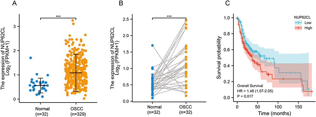 |
Figure 1 (A) NUP62CL mRNA expression in normal and OSCC tumor tissues. (B) NUP62CL mRNA expression in OSCC tumor tissues and their paired normal tissue. (C) Patients with high NUP62CL expression had a poor prognosis. ***P < 0.001. |
NUP62CL Protein Expression in OSCC and Para-Carcinoma Tissues
Due to post-transcriptional regulatory mechanisms, mRNA expression does not always correlate with corresponding protein expression patterns.19 We next evaluated NUP62CL protein expression in OSCC and para-carcinoma tissues by mIHC technology. As a result, the expression of NUP62CL protein in OSCC tissue was significantly higher than that in para-carcinoma tissue (Figure 2). Our results indicated that the expression of NUP62CL protein in OSCC tissues was higher and consistent with mRNA expression level.
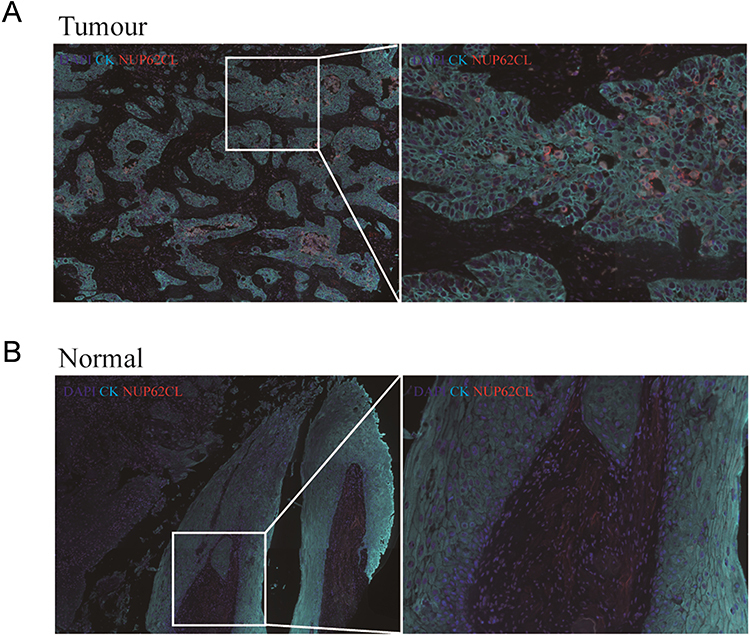 |
Figure 2 The multiplex immunohistochemistry revealed the expression of the NUP62CL protein in tumor tissues (A) and normal tissues (B). |
Correlation Between NUP62CL Expression and Clinical Features of OSCC
We divided these cases into high-expression and low-expression groups with the median expression level of NUP62CL as a boundary. The relationship between NUP62CL expression and clinicopathological features was further analyzed (Table 1). It was noticed that the NUP62CL protein expression was correlated with tumor size (Pearson χ2=6.416, P=0.011) and clinical stage (Pearson χ2=10.268, P=0.006). However, no statistically significant correlation was found between NUP62CL protein expression and other clinical features, including age, sex, anatomic classification, lymph node metastasis and distant metastasis (P>0.05).
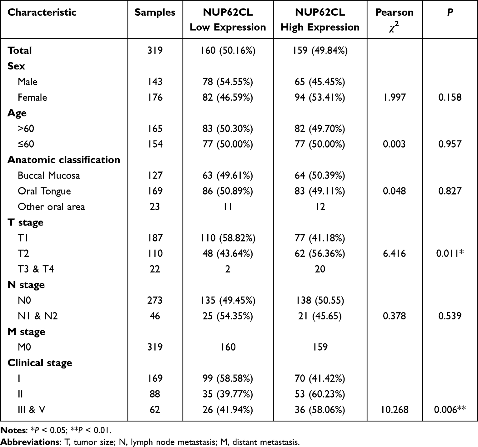 |
Table 1 Relationship Between NUP62CL Expression and Clinicopathological Features |
Prognostic Potential of NUP62CL Protein Expression in OSCC
A cohort of 240 patients with specific prognostic information was included to further assess prognostic factors for OSCC by univariate and multivariate Cox regression analysis. According to the univariate Cox regression analysis, NUP62CL protein expression (P=0.009), age (P<0.001), anatomic classification (Pother vs buccal=0.003), T stage (PT2 vs T1=0.001, PT3&T4 vs T1<0.001), N stage (P<0.001) and clinical stage (PII vs I<0.001, PIII&IV vs I<0.001) were significantly associated with the 5-year survival rate of OSCC patients. By the multivariate Cox regression analysis, NUP62CL protein expression (P=0.043), age (P<0.001), anatomic classification (Pother vs buccal<0.001), clinical stage (PII vs I=0.003, PIII&IV vs I<0.001) were found to be independent prognostic factors correlated with an inferior overall survival of these patients (Figure 3).
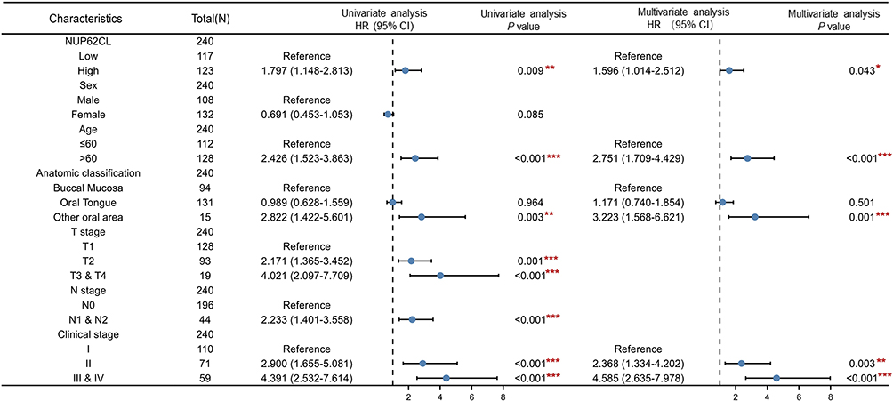 |
Figure 3 Univariate and multivariable analyses of prognostic factors for 5-year survival in patients with OSCC. *P<0.05, **P<0.01, ***P<0.001. |
Kaplan-Meier curve analysis showed that OSCC patients with high expression of NUP62CL protein had a poor prognosis (HR =1.78 (1.17–2.71), Log-rank P=0.009). Meanwhile, old age, high TMN grade and high clinical grade were also factors contributing to the poor prognosis (Figure 4). The above results suggested that NUP62CL protein expression was a reliable factor for predicting the prognosis of OSCC patients.
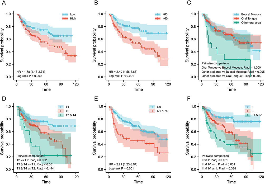 |
Figure 4 The impacts of NUP62CL expression levels (A), age (B), anatomic classification (C), T stage (D), N stage (E) and clinical stage (F) on the overall survival of OSCC patients. |
Correlation Between NUP62CL Expression and TIICs Abundance in OSCC
The relative abundance of 24 TIICs was assessed by single-sample gene set enrichment analysis (ssGSEA) algorithm in each sample from TCGA. The group comparison diagram showed the overall immune cell landscape of OSCC (Figure 5A). In addition, NUP62CL expression was significantly correlated with the abundance of macrophages (Spearman, r=−0.318, P<0.001), neutrophils (Spearman, r=−0325, P<0.001), NK cells (Spearman, r =−0.199, P<0.001), T cells (Spearman, r=−0.210, P<0.001) (Figure 5B).
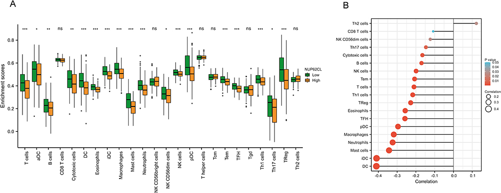 |
Figure 5 (A) The group comparison diagram represents the proportions of 24 distinct immune cell types. (B) NUP62CL expression was correlated with tumor-infiltrating immune cells (TIICs) abundance. *P<0.05, **P<0.01, ***P<0.001. |
To further verify the correlation between NUP62CL protein expression and TIICs abundance in OSCC immune microenvironment, we performed mIHC on OSCC tissues to determine the proportion of TIICs (Figure 6A). There was an extensive infiltration of CD3+CD4+ T cells, CD3+CD8+ T cells, CD56+ NK cells, CD68+CD163+ macrophages, and CD68+CD86+ macrophages the OSCC microenvironment while the infiltration of CD66b+ neutrophils was low (Figure 6B). Besides, the expression of NUP62CL was positively associated with the abundance of T cells (P<0.01), NK cells (P<0.05), and macrophages (P<0.01). However, no significant correlation was observed between the infiltration of NUP62CL expression and neutrophils (Figure 6C).
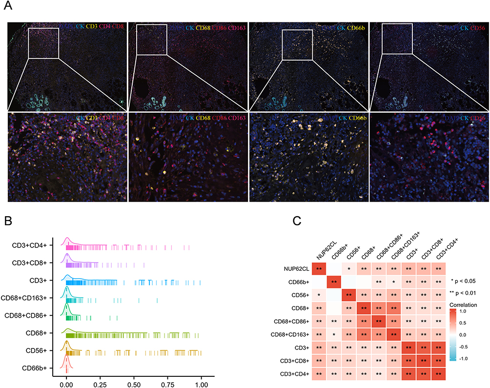 |
Figure 6 (A) Representative multiplex immunofluorescence staining images detecting different immune cell types in OSCC tissues. (B) The infiltration levels of TIICs in OSCC. (C) NUP62CL expression was related to TIICs abundance. *P<0.05, **P<0.01. |
Association of NUP62CL Protein Expression with the Immune Checkpoints
It was found mRNA that NUP62CL expression was positively correlated with PD-L1 (CD274) mRNA expression (r=0.280, P=0.0165), and inversely correlated with PD-1 (PDCD1) mRNA (r=−0.305, P=0.009) and CTLA-4 (CTLA4) mRNA expression (r=−0.306, P=0.008) after adjusting by tumor purity (Figure S1).
We further verified the correlation of NUP62CL protein expression with the immune checkpoints in OSCC tissues, including PD-1, PD-L1 and CTLA-4 (Figure 7A). These tissues were divided into high-expression and low-expression groups with the median expression level of NUP62CL as a boundary. The PD-L1 and CTLA-4 protein expression were increased in the high-expression group (P<0.001), as well as PD-1 protein (P=0.036) (Figure 7B). In addition, NUP62CL protein expression demonstrated a positive correlation with PD-1 (r=0.203, P<0.001, Figure 7C), PD-L1 (r=0.308, P<0.001, Figure 7D), and CTLA-4 (r=0.717, P<0.001, Figure 7E) protein expression.
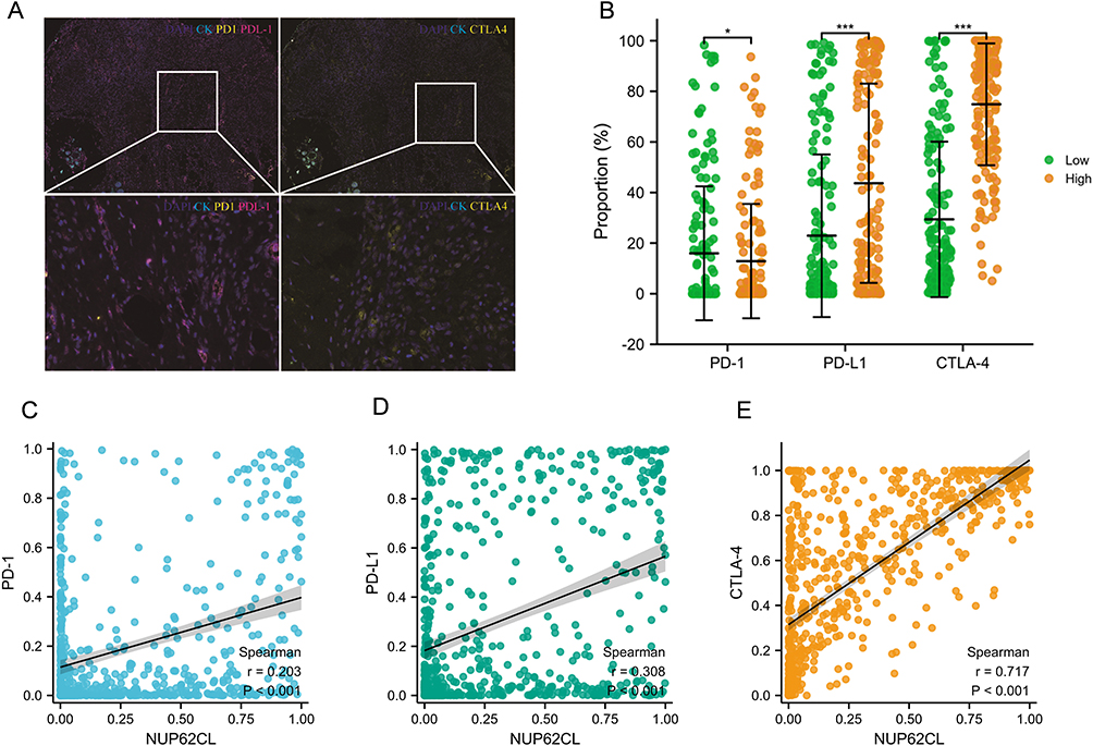 |
Figure 7 (A) The representative mIHC images of PD-1, PD-L1 and CTLA-4 protein expression. (B) The PD-1, PD-L1 and CTLA-4 protein expression in NUP62CL high-expression and low-expression groups. The correlation of NUP62CL protein expression with the immune checkpoints in OSCC tissues, including PD-1 (C), PD-L1 (D) and CTLA-4 (E). *P<0.05, ***P<0.001. |
Discussion
OSCC is the most common head and neck malignancy with a high mortality rate and poor prognosis.20 Recently, researchers have been exploring new targets to improve clinical outcomes for OSCC patients. In the present study, NUP62CL was found significantly upregulated in OSCC tissue compared with normal oral mucosal tissue and associated with poor survival in the TCGA database. In addition, Higher NUP62CL protein expression was related to large tumor size and advanced clinical stage. Kaplan-Meier curve analysis showed that OSCC patients with high expression of NUP62CL protein had a poor prognosis. These results indicate the clinical value of NUP62CL as a marker for OSCC diagnosis.
The relationship between immune cells and tumor cells is extremely complex,21 and the growth, progression and metastasis of tumors are affected by the interaction between immune cells and tumor cells in the TME.22 In this study, the expression of NUP62CL was positively associated with the abundance of CD3+CD4+ T cells, CD3+CD8+ T cells, CD56+ NK cells, CD68+CD163+ macrophages, and CD68+CD86+ macrophages. Patients with high NUP62CL expression and high degree of immune cell infiltration have a poor prognosis. Similarly, a retrospective study of head and neck squamous cell carcinoma (HNSCC) showed that a high abundance of CD3+CD4+ and CD3+CD8+ T cell infiltration indicated a higher risk of death.23 However, the prognostic role of tumor associated macrophages in different types of cancer is unclear. It was found that the generic macrophage marker CD68+ had no prognostic value in patients with HNSCC, but the M2-like marker CD163+ predicted poor prognosis.24
In the TME, infiltrating immune cells cause functional damage through the expression of PD-1/PD-L1, CTLA-4, and other inhibitory signals on the cell surface, leading to tumor immunosuppression. These inhibitory signals are also called immune checkpoints.25 PD-1 is expressed on activated T cells and B cells with PD-L1 as its ligand.26 The interaction between PD-1 and PD-L1 negatively regulates the immune response by reducing the production of cytokines and inducing T lymphocyte energy and apoptosis.27 CTLA-4 is expressed on activated T cells and negatively regulates T cell proliferation and IL-2 production. CTLA-4 blocking results in reduced regulatory T cell frequency and increased T cell function,28 improving the efficiency of long-term remission in patients with head and neck cancer and increasing response to treatment.29 The immune checkpoint inhibitors as a type of immunotherapy that targets tumor cells by reactivating T cells, have been approved for the treatment of head and neck cancer with significant results.26,30,31 Herein, NUP62CL protein expression demonstrated a positive correlation with PD-1, PD-L1 and CTLA-4 in OSCC tissues. NUP62CL as a new immunological biomarker will provide new insights into the immunological treatment of OSCC.
However, there are some limitations to this study that warrant consideration. First, the reliability of our conclusions was limited by the size of the sample studied. Second, the population of labeled immune cells in our study was also limited. For example, T cells can have an immunosuppressive phenotype, but also exhibit an immune-effect phenotype, depending on their subtype, which greatly affects the progress and diagnosis of OSCC. In addition, we did not identify the regulatory pathway of NUP62CL expression in immune cells. In order to reveal the mechanism of NUP62CL action in the OSCC tumor immune microenvironment, sufficient experiments are needed.
Conclusion
In conclusion, this is the first study on the role of NUP62CL in OSCC patients. We identified high-NUP62CL expression in OSCC tissues, and high-NUP62CL expression was associated with poor survival. In addition, our results elucidated the association between NUP62CL expression and immune cell abundance and immune checkpoint expression in OSCC. NUP62CL could be an effective prognostic and immunological biomarker for OSCC patients.
Acknowledgments
We thank all the study participants, research staff, and students who contributed to this study. This work was supported by the National Natural Science Foundation of China (82172931 and 32170915), Jiangsu Province Capability Improvement Project through Science, Technology and Education (ZDXK202234), the fifth orthodontic Committee of Jiangsu Stomatological Association (Joa-2022-6), Nantong Municipal Health Committee Scientific Research Project (QA2021058), Nantong Science and Technology Project (MS22022072) and Nantong University Clinical Medicine special project (2022JQ007).
Disclosure
The authors report no conflicts of interest in this work.
References
1. Mao L, Zhuang R, Qin L, et al. CCL18 overexpression predicts a worse prognosis in oral squamous cell carcinoma (OSCC). Neoplasma. 2020;67(3):700–706. doi:10.4149/neo_2020_190821N802
2. Peres MA, Macpherson LMD, Weyant RJ, et al. Oral diseases: a global public health challenge. Lancet. 2019;394(10194):249–260. doi:10.1016/S0140-6736(19)31146-8
3. McCord C, Kiss A, Magalhaes MA, Leong IT, Jorden T, Bradley G. Oral squamous cell carcinoma associated with precursor lesions. Cancer Prev Res. 2021;14(9):873–884. doi:10.1158/1940-6207.CAPR-21-0047
4. Jiang M, Li B. STAT3 and its targeting inhibitors in oral squamous cell carcinoma. Cells. 2022;11(19):3131. doi:10.3390/cells11193131
5. Chen Y, Feng Y, Yan F, Zhao Y, Zhao H, Guo Y. A novel immune-related gene signature to identify the tumor microenvironment and prognose disease among patients with oral squamous cell carcinoma patients using ssGSEA: a bioinformatics and biological validation study. Front Immunol. 2022;13:922195. doi:10.3389/fimmu.2022.922195
6. Zhao X, Sun S, Zeng X, Cui L. Expression profiles analysis identifies a novel three-mRNA signature to predict overall survival in oral squamous cell carcinoma. Am J Cancer Res. 2018;8(3):450–461.
7. Wang PC, Hu ZQ, Zhou SL, et al. The spatial distribution of immune cell subpopulations in hepatocellular carcinoma. Cancer Sci. 2022;113(2):423–431. doi:10.1111/cas.15202
8. Tang N, Ning Q, Wang Z, Tao Y, Zhao X, Tang S. Tumor microenvironment based stimuli-responsive CRISPR/Cas delivery systems: a viable platform for interventional approaches. Colloids Surf B Biointerfaces. 2022;210:112257. doi:10.1016/j.colsurfb.2021.112257
9. Liu T, Tan J, Wu M, et al. High-affinity neoantigens correlate with better prognosis and trigger potent antihepatocellular carcinoma (HCC) activity by activating CD39(+)CD8(+) T cells. Gut. 2021;70(10):1965–1977. doi:10.1136/gutjnl-2020-322196
10. Schneider K, Marbaix E, Bouzin C, et al. Immune cell infiltration in head and neck squamous cell carcinoma and patient outcome: a retrospective study. Acta Oncol. 2018;57(9):1165–1172. doi:10.1080/0284186X.2018.1445287
11. Yi C, Chen L, Lin Z, et al. Lenvatinib targets FGF receptor 4 to enhance antitumor immune response of anti-programmed cell death-1 in HCC. Hepatology. 2021;74(5):2544–2560. doi:10.1002/hep.31921
12. Seiwert TY, Burtness B, Mehra R, et al. Safety and clinical activity of pembrolizumab for treatment of recurrent or metastatic squamous cell carcinoma of the head and neck (KEYNOTE-012): an open-label, multicentre, phase 1b trial. Lancet Oncol. 2016;17(7):956–965. doi:10.1016/S1470-2045(16)30066-3
13. Ren S, Wang W, Zhang C, et al. The low expression of NUP62CL indicates good prognosis and high level of immune infiltration in lung adenocarcinoma. Cancer Med. 2021;10(10):3403–3412. doi:10.1002/cam4.3877
14. Singh U, Bindra D, Samaiya A, Mishra RK. Overexpressed Nup88 stabilized through interaction with Nup62 promotes NF-kappaB dependent pathways in cancer. Front Oncol. 2023;13:1095046. doi:10.3389/fonc.2023.1095046
15. Mullan PB, Bingham V, Haddock P, et al. NUP98 - a novel predictor of response to anthracycline-based chemotherapy in triple negative breast cancer. BMC Cancer. 2019;19(1):236. doi:10.1186/s12885-019-5407-9
16. Wu Y, Fang G, Wang X, et al. NUP153 overexpression suppresses the proliferation of colorectal cancer by negatively regulating Wnt/beta-catenin signaling pathway and predicts good prognosis. Cancer Biomark. 2019;24(1):61–70. doi:10.3233/CBM-181703
17. Hazawa M, Lin DC, Kobayashi A, et al. ROCK-dependent phosphorylation of NUP62 regulates p63 nuclear transport and squamous cell carcinoma proliferation. EMBO Rep. 2018;19(1):73–88. doi:10.15252/embr.201744523
18. Chen F, Chandrashekar DS, Varambally S, Creighton CJ. Pan-cancer molecular subtypes revealed by mass-spectrometry-based proteomic characterization of more than 500 human cancers. Nat Commun. 2019;10(1):5679. doi:10.1038/s41467-019-13528-0
19. Agle K, Vincent BG, Piper C, et al. Bim regulates the survival and suppressive capability of CD8(+) FOXP3(+) regulatory T cells during murine GVHD. Blood. 2018;132(4):435–447. doi:10.1182/blood-2017-09-807156
20. Togni L, Caponio VCA, Zerman N, et al. The emerging impact of tumor budding in oral squamous cell carcinoma: main issues and clinical relevance of a new prognostic marker. Cancers. 2022;14(15):3571. doi:10.3390/cancers14153571
21. Sangro B, Sarobe P, Hervas-Stubbs S, Melero I. Advances in immunotherapy for hepatocellular carcinoma. Nat Rev Gastroenterol Hepatol. 2021;18(8):525–543. doi:10.1038/s41575-021-00438-0
22. Arneth B. Tumor Microenvironment. Medicina. 2019;56(1):15. doi:10.3390/medicina56010015
23. Borsetto D, Tomasoni M, Payne K, et al. Prognostic significance of CD4+ and CD8+ tumor-infiltrating lymphocytes in head and neck squamous cell carcinoma: a meta-analysis. Cancers. 2021;13(4):781. doi:10.3390/cancers13040781
24. Troiano G, Caponio VCA, Adipietro I, et al. Prognostic significance of CD68(+) and CD163(+) tumor associated macrophages in head and neck squamous cell carcinoma: a systematic review and meta-analysis. Oral Oncol. 2019;93:66–75. doi:10.1016/j.oraloncology.2019.04.019
25. Wang Y, Wang Y, Ren Y, Zhang Q, Yi P, Cheng C. Metabolic modulation of immune checkpoints and novel therapeutic strategies in cancer. Semin Cancer Biol. 2022;86(Pt 3):542–565. doi:10.1016/j.semcancer.2022.02.010
26. Damasio MPS, Nascimento CS, Andrade LM, de Oliveira VL, Calzavara-Silva CE. The role of T-cells in head and neck squamous cell carcinoma: from immunity to immunotherapy. Front Oncol. 2022;12:1021609. doi:10.3389/fonc.2022.1021609
27. Kao HF, Lou PJ. Immune checkpoint inhibitors for head and neck squamous cell carcinoma: current landscape and future directions. Head Neck. 2019;41(Suppl 1):4–18. doi:10.1002/hed.25930
28. Yu GT, Bu LL, Zhao YY, et al. CTLA4 blockade reduces immature myeloid cells in head and neck squamous cell carcinoma. Oncoimmunology. 2016;5(6):e1151594. doi:10.1080/2162402X.2016.1151594
29. Qi X, Jia B, Zhao X, Yu D. Advances in T-cell checkpoint immunotherapy for head and neck squamous cell carcinoma. Onco Targets Ther. 2017;10:5745–5754. doi:10.2147/OTT.S148182
30. Sarkizova S, Hacohen N. How T cells spot tumour cells. Nature. 2017;551(7681):444–446. doi:10.1038/d41586-017-07267-9
31. Luksza M, Riaz N, Makarov V, et al. A neoantigen fitness model predicts tumour response to checkpoint blockade immunotherapy. Nature. 2017;551(7681):517–520.
 © 2023 The Author(s). This work is published and licensed by Dove Medical Press Limited. The
full terms of this license are available at https://www.dovepress.com/terms
and incorporate the Creative Commons Attribution
- Non Commercial (unported, 3.0) License.
By accessing the work you hereby accept the Terms. Non-commercial uses of the work are permitted
without any further permission from Dove Medical Press Limited, provided the work is properly
attributed. For permission for commercial use of this work, please see paragraphs 4.2 and 5 of our Terms.
© 2023 The Author(s). This work is published and licensed by Dove Medical Press Limited. The
full terms of this license are available at https://www.dovepress.com/terms
and incorporate the Creative Commons Attribution
- Non Commercial (unported, 3.0) License.
By accessing the work you hereby accept the Terms. Non-commercial uses of the work are permitted
without any further permission from Dove Medical Press Limited, provided the work is properly
attributed. For permission for commercial use of this work, please see paragraphs 4.2 and 5 of our Terms.
Recommended articles
A Novel Risk Score (P-score) Based on a Three-Gene Signature, for Estimating the Risk of Prostate Cancer-Specific Mortality
Söderdahl F, Xu LD, Bring J, Häggman M
Research and Reports in Urology 2022, 14:203-217
Published Date: 11 May 2022
Serum NOX4 as a Promising Prognostic Biomarker in Association with 90-Day Outcome of Severe Traumatic Brain Injury
Jiang F, Chen Z, Hu J, Liu Q
International Journal of General Medicine 2022, 15:5307-5317
Published Date: 30 May 2022
Identification of KRBA1 as a Potential Prognostic Biomarker Associated with Immune Infiltration and m6A Modification in Hepatocellular Carcinoma
Liu Y, Fu B, Yu Z, Song G, Zeng H, Gong Y, Ding Y, Huang D
Journal of Hepatocellular Carcinoma 2022, 9:497-516
Published Date: 31 May 2022
Expression, Clinical Significance, Immune Infiltration, and Regulation Network of miR-3940-5p in Lung Adenocarcinoma Based on Bioinformatic Analysis and Experimental Validation
Lin Z, Huang W, Xie Z, Yi Y, Li Z
International Journal of General Medicine 2022, 15:6451-6464
Published Date: 6 August 2022
CSTF2 Acts as a Prognostic Marker Correlated with Immune Infiltration in Hepatocellular Carcinoma
Zhang W, Wan Y, Zhang Y, Liu Q, Zhu X
Cancer Management and Research 2022, 14:2691-2709
Published Date: 12 September 2022