
Back to Journals » International Journal of Nanomedicine » Volume 20
Nanozymes Targeting Redox Imbalance: A Novel Weapon for Immunomodulation and Organ Protection in Sepsis
Received 28 September 2025
Accepted for publication 8 December 2025
Published 18 December 2025 Volume 2025:20 Pages 15167—15183
DOI https://doi.org/10.2147/IJN.S570887
Checked for plagiarism Yes
Review by Single anonymous peer review
Peer reviewer comments 2
Editor who approved publication: Professor Dong Wang
Junlong Gao,1,2 Xiaobo Pang,3 Yulan Li1,4
1The First School of Clinical Medicine, Lanzhou University, Lanzhou, Gansu, People’s Republic of China; 2Intensive Care Unit, Gansu Provincial Hospital, Lanzhou, Gansu, People’s Republic of China; 3State Key Laboratory of Applied Organic Chemistry (SKLAOC), College of Chemistry and Chemical Engineering, Lanzhou University, Lanzhou, Gansu, People’s Republic of China; 4Department of Anesthesiology, The First Hospital of Lanzhou University, Lanzhou, Gansu, People’s Republic of China
Correspondence: Yulan Li, Email [email protected]
Abstract: Sepsis remains a major challenge in critical care, with high mortality despite ongoing improvements in treatment. The early uncontrolled burst of reactive oxygen and nitrogen species (RONS) and cytokine storms form a vicious cycle, ultimately leading to multiple organ dysfunction syndrome (MODS). The absence of effective therapies to interrupt this process is likely a key reason for poor outcomes. In recent years, the emergence of nanozymes has represented a transformative breakthrough in addressing this challenge. With strong antioxidant capacity, high stability, and low cost, nanozymes surpass conventional antioxidants and offer a promising therapeutic strategy for sepsis, especially through effective redox regulation. Nanozymes not only efficiently scavenge diverse RONS but also inhibit hyperactivated inflammatory pathways, thereby breaking the fatal vicious cycle between oxidative stress and cytokine storms. This provides a novel approach for immunomodulation and organ protection in sepsis. This review summarizes the key role of redox imbalance in sepsis progression and the therapeutic potential of nanozymes targeting redox imbalance, discusses their in vivo metabolic distribution and biosafety, and outlines prospects for future clinical translation and development. The objective is to provide insights that facilitate the development of innovative therapies targeting the RONS-inflammation axis in sepsis.
Keywords: nanozymes, sepsis, redox imbalance, cytokine storm, immunosuppression, organ protection
Introduction
Epidemiology of Sepsis
Sepsis is a life-threatening organ dysfunction caused by a dysregulated host response to infection.1 It is prevalent in intensive care units (ICUs) and exhibits extremely high mortality. From a global epidemiological perspective, approximately 48.9 million new sepsis cases occur annually, resulting in around 11 million deaths, which account for 19.7% of all global deaths.2 Sepsis imposes a huge economic burden on healthcare systems worldwide and remains one of the most intractable challenges in acute and critical care medicine.3
Lack of Effective Therapies for Redox Imbalance and Immune Dysregulation
Despite considerable advances in sepsis management (including fluid resuscitation, antimicrobial therapy, and organ support),1 mortality remains unacceptably high, with approximately one-third of patients dying before hospital discharge.4–6 The lack of effective interventions targeting the central pathophysiological drivers of sepsis—redox imbalance and the closely associated cytokine storm and immune suppression—may be a key contributor to the persistently high mortality.5,7
Anti-Inflammatory and Antioxidant Strategies in Sepsis
Recent international guidelines recommend the use of corticosteroids in refractory septic shock to exert anti-inflammatory effects. However, their therapeutic benefit remains highly controversial. Numerous studies have shown that glucocorticoids do not reduce sepsis-related mortality and are associated with increased adverse events, such as a heightened risk of secondary infections. Therefore, guidelines do not support their routine early use.1,7 Antioxidant therapies have been explored to alleviate oxidative stress and inflammation.8 For example, high-dose vitamin C can mitigate inflammatory response in COVID-19 and prevent disease progression.9 Nevertheless, numerous studies indicate that natural antioxidants, such as vitamin C and E, are ineffective in disrupting the “sepsis redox cycle” and reducing mortality, primarily due to their low catalytic activity and poor stability. Thus, most antioxidant strategies remain at the preclinical stage.10,11
Nanozymes Offer New Hope for Overcoming Sepsis
Nanozymes, a new class of artificial enzymes, possess many advantages including low cost, facile synthesis, high stability, and tunable activity.12,13 They hold significant promise as replacements for natural antioxidants in treating redox imbalance related diseases.14,15 In animal models of sepsis, nanozymes efficiently reduce reactive oxygen and nitrogen species (RONS) by mimicking multiple natural antioxidant enzymes. Furthermore, nanozymes disrupt pathological crosstalk between RONS and inflammation, modulate immune imbalance, achieve multi-organ protection, and reduce mortality (Table 1). Hence, nanozymes are highly promising candidates to address the critical therapeutic gap related to redox imbalance and immune dysregulation in sepsis.16,17 The representative nanozymes for sepsis treatment described in this article are summarized in Table 1.
 |
Table 1 Representative Nanozymes for Sepsis Treatment |
This review systematically elaborates sepsis pathophysiology of immune dysregulation and organ injury induced by oxidative stress and cytokine storms. It then discusses the mechanisms and therapeutic effects of intravenously administered nanozymes in immunomodulation and organ protection, analyzes their in vivo metabolic distribution and biosafety, and outlines the potential applications of nanozymes in sepsis treatment.
Literature Search and Selection Strategy
This review searched the PubMed and Web of Science databases for studies published before August 2025, with the language restricted to English. The search strategy employed the following keywords: “nanozymes”, “nanoparticles”, “antioxidant”, “sepsis”, “septic shock”, “inflammation”, “oxidative stress” and “organ injury”. We reviewed and selected each publication according to the following eligibility criteria: (1) disease models of sepsis or lipopolysaccharide (LPS)-induced organ injury; (2) investigations focusing on the pathogenesis of sepsis or LPS-induced organ injury; and (3) treatment of sepsis or LPS-induced organ injury with nanozymes.
Mechanisms of Oxidative Stress-Mediated Injury in Sepsis
Excessive RONS Production During Sepsis Progression
Endogenous RONS mainly include superoxide anion (∙ O2−), hydrogen peroxide (H2O2), hydroxyl radical (∙ OH), nitric oxide (∙ NO), and peroxynitrite anion (ONOO−), which can interconvert under certain conditions5 (Figure 1). Under physiological conditions, approximately 2% of O2 in mammals is reduced to ∙ O2− and released due to “leakage” in the mitochondrial electron transport chain. Furthermore, enzymatic systems such as intracellular nicotinamide adenine dinucleotide phosphate oxidase (NOX), xanthine oxidase (XO), cyclooxygenase (COX), and inducible nitric oxide synthase (iNOS) regulate the production of ∙ O2− and ∙ NO, contributing to physiological functions including pathogen elimination, liver detoxification, and microvascular dilation regulation.30 Endogenous antioxidant systems including SOD, catalase (CAT), and glutathione peroxidase (GPx) counteract RONS to maintain redox homeostasis.31 However, under pathological conditions such as sepsis, respiratory bursts in neutrophils, macrophages, and endothelial cells, overactivation of enzymatic systems like NOX, XO, and iNOS, and impaired mitochondrial function collectively lead to massive production of ∙ O2− and ∙ NO, and their secondary derivatives, such as H2O2, ∙ OH, and ONOO−. The radical ∙ OH and ONOO− further induce lipid peroxidation, generating toxic lipid peroxidation products (LPPs), such as lipid radicals (L ∙), lipid peroxyl radicals (LOO ∙), and malondialdehyde (MDA)5,6,32,33 (Figure 1). Excessive RONS trigger and amplify inflammatory cascades by oxidatively damaging biomolecules (proteins, lipids, DNA) and impairing organelle functions (eg, mitochondrial), resulting in cell death, tissue damage, and multiple organ dysfunction5,6,31 (Figure 2). Studies demonstrate a strong correlation between elevated RONS levels and sepsis severity and prognosis.34,35
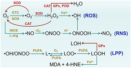 |
Figure 1 Biosynthetic and metabolic pathways of Oxidative stress related reactive molecules. Abbreviations: ETC, electron transport chain; L-arg, L-arginine; PUFA, polyunsaturated fatty acid; LPP, lipid peroxidation product; L, lipid radical; LOO, lipid peroxyl radical; LOOH, lipid hydroperoxide; LOH, hydroxylated lipid; MDA, malondialdehyde; 4-HNE, 4-hydroxynonenal. |
 |
Figure 2 Mechanism of Oxidative stress-mediated injury in sepsis. Pathogen-associated molecular patterns (PAMPs) and damage-associated molecular patterns (DAMPs) activate Toll-like receptors (TLRs) and NOD-like receptors (NLRs), inducing the production of reactive oxygen and nitrogen species (RONS). Excessive RONS deplete endogenous antioxidant defenses, such as superoxide dismutase (SOD) and glutathione (GSH). On the other hand, RONS cause diverse cellular damage, including exacerbated inflammatory responses, vascular leakage and microcirculatory disorders, mitochondrial injury, endoplasmic reticulum stress, ferroptosis, and damage to DNA, proteins, and lipids. These injuries further amplify RONS release, and resulting in systemic cytokine storms, immunosuppression, multiple organ dysfunction syndrome (MODS), and even death in severe cases. |
Vicious Cycle Between Cytokine Storm and RONS
Oxidative stress and inflammatory responses exhibit a mutually amplifying relationship during sepsis, driving immunosuppression, energy metabolism disorders, and multiple organ failure—key determinants of sepsis progression and mortality.6,36,37 During sepsis, Pathogen- and damage-associated molecular pattern (PAMP and DAMP) activate innate immunity via pattern recognition receptor (PRR), provoking excessive release of proinflammatory mediators and RONS. These mediators damage DNA, proteins, and lipids, disrupt mitochondrial function, and promote further DAMP release, thus exacerbating inflammation. Concurrently, heightened NOX and iNOS activities amplify RONS production that drives irreversible sepsis30,33,36 (Figure 2). All known NLR family pyrin domain-containing 3 (NLRP3) activators can trigger intracellular ROS release, while ROS scavenging inhibits NLRP3 activation and attenuates inflammation-induced pyroptosis.37,38 Mimicking cytokine storms with tumor necrosis factor-alpha (TNF-α) and interferon-gamma (IFN-γ) induces high expression of iNOS and ∙ NO, whereas reducing ∙ NO release markedly decreases mortality in models of cytokine storm-related diseases, including COVID-19, sepsis, and hemophagocytic syndrome.6 Thus, sepsis can be viewed as an uncontrolled oxidative stress and inflammation triggered by pathogens, whereby the RONS system rapidly amplifies and induces the body to fall into a self-destructive vicious cycle.5,39,40
RONS Promote Inflammation and Immunosuppression
Excessive RONS during sepsis enhance innate immune cell responses to proinflammatory stimuli and induce inflammatory apoptosis, which rapidly triggers compensatory anti-inflammatory and immunosuppressive brake mechanisms, thereby contributing to sepsis-induced immunosuppression37,40 (Figure 2). H2O2 activates macrophages and endothelial cells, enhances responses to lipopolysaccharides (LPS), promotes M1 macrophage polarization, and induces high mobility group box 1 (HMGB-1) release alongside increased Toll-like receptor 4 (TLR4) expression. HMGB1 activates downstream NF-κB and p38 MAPK pathways via the TLR4 signaling, triggering proinflammatory cascades.30,41,42 RONS activate the NLRP3 inflammasome through multiple pathways, promoting the upregulation of IL-1β.43,44 For example, LPS-stimulated macrophages generate mitochondrial ROS via reverse electron transport (RET) at complex I, which is crucial for NLRP3 inflammasome activation. Inhibition of mtROS can suppress NLRP3 inflammasome activation, attenuate pyroptosis, and reduce neutrophil infiltration in peritonitis models.37 The cytokine storm and oxidative stress in sepsis rapidly induce immunosuppressive responses, including elevated levels of anti-inflammatory cytokines (IL-10 and TGF-β), expansion of immunosuppressive cells populations (immature neutrophils, M2 macrophages, Treg cells, and myeloid-derived suppressor cells), and increased apoptosis of CD4 cells. Moreover, RONS directly damage immune cells, collectively exacerbating sepsis-induced immunosuppression.40,45,46
RONS Disrupt Mitochondrial Function
Mitochondria act as both sources and targets of ROS. During sepsis, excessive ROS damage mitochondrial ultrastructure and disrupt the electron transport chain (ETC), causing electron leakage, opening of permeability transition pores, ROS-induced ROS release, and membrane potential collapse. These events are closely associated with energy metabolism disorders and cell apoptosis.47–49 Mitochondrial damage also releases mitochondrial DAMPs (eg, mitochondrial DNA, ATP, cytochrome c), exacerbating systemic inflammatory injury. Effective ROS scavenging can mitigate mitochondrial structural and functional damage.5,50
RONS Induce Ferroptosis and Aggravate Endoplasmic Reticulum Stress
Ferroptosis is one of the key mechanisms underlying multiple organ damage in sepsis. ROS interact with iron to deplete GPx4, leading to ferroptosis characterized by intense lipid peroxidation and necrotic features.51,52 Inhibiting ferroptosis by activating the nuclear factor erythroid 2-related factor 2 (Nrf2)/GPx4 antioxidant pathway exerts therapeutic effects in sepsis.53 Endoplasmic reticulum (ER) stress is also associated with sepsis progression.54 Excessive RONS significantly promote ER stress, leading to the accumulation of unfolded/misfolded proteins, activation of downstream inflammation pathways, and further ROS generation via ER-dependent pathways, thereby exacerbating oxidative damage.55,56
Physicochemical and Biological Characteristics of Nanozymes
Classification of Nanozymes by Catalytic Activity and Material Type
Nanozymes exhibit catalytic functions analogous to one or more natural enzymes, including catalase (CAT), superoxide dismutase (SOD), glutathione peroxidase (GPx), peroxidase (POD), and oxidase (OXD).13 Based on material composition, nanozymes are primarily classified as metallic nanozymes (eg, Au, Pt, Cu, Mn),16 metal oxide nanozymes (eg, CeO2, MnO2, Fe3O4),57,58 metal sulfide nanozymes (eg, WS2, FeS2),19,59 carbon-based nanozymes (eg, carbon dots),21 and composite nanozymes (including inorganic-inorganic composite nanozymes such as Au/CeO2 nanozymes, and inorganic-organic composite nanozymes such as CeCH nanozymes synthesized by coordinating CeO2 nanozymes with curcumin).24,28,60 Recent advances in nanozyme fabrication and material optimization, the catalytic activity of some nanozymes has surpassed those of natural enzymes. Notably, single-atom nanozymes (SAzymes) exhibit advantages including nearly 100% atom utilization, uniformly distributed catalytic sites, and tunable electronic structures and coordination environments, showing great potential for further development and application.61
Mechanisms of High-Efficiency Catalysis in Nanozymes
The catalytic reactions of nanozymes generally follow classical Michaelis-Menten kinetics, characterized by parameters Vmax (maximum reaction rate) and Km (Michaelis constant).13,60 The quantum size effect inherent to nanozymes increases the number of exposed catalytic active sites and enhances surface energy for substrate adsorption. Meanwhile, the energy band structure changes (upward shift of the conduction band and downward shift of the valence band), which optimizes electron transfer efficiency and enhances redox catalytic activity.61,62
The catalytic activity and stability of nanozymes can be effectively enhanced by modifying size, morphology, element doping, functional groups, and coating with polymers or biomolecules.63,64 For example, compared with cubic CeO2 nanozymes (6.4 nm), ultrathin CeO2 nanozymes (approximately 1.2 nm in thickness) demonstrate significantly increased antioxidant activities, with SOD-like and CAT-like activities increased approximately 2.6-fold (comparable to natural SOD) and 7.5-fold, respectively.65 Au-doped CeO2 (Au/CeO2) nanozymes exhibit POD-like activity double that of undoped CeO2 nanozymes (95.36 U/g vs 48.34 U/g) and nearly 3-fold higher oxidase-like activity (28.9 U/g vs 9.74 U/g).24 Hydrothermally synthesized carbon dots (Hy-CDs) retain abundant amino, hydroxyl, and other functional groups inherited from the honeysuckle precursor. These surface functionalities reduce catalytic activation energy and optimize electron transfer, thereby yielding superior SOD-like activity compared to those synthesized by the carbonization method (3519.14 U mg−1 VS 426.52 U mg−1).21
Mimicking the metal coordination structure of natural enzymes and the enzymatic cascade reactions in organisms represents an effective approach to designing highly active nanozymes. For example, by mimicking the electronic and structural features of natural copper-only superoxide dismutase (SOD5), a copper single-atom nanozyme (Cu-SAzyme) featuring Cu-N4 active sites was synthesized. Increasing its copper loading from 0.5% to 2% enhanced SOD-like activity from 58.89 U/mg to 448.72 U/mg. Analogous nanozymes substituting Cu with other metals (Co > Mn > Zn > Ni) displayed lower catalytic activity than Cu-SAzyme.16 Pt@CNDs nanozymes, synthesized by combining Pt nanozymes with carbon dots, enhance the SOD-like activity of carbon dots and integrate the CAT-like activity of Pt, solving the toxicity problem of H2O2 (a product of SOD nanozyme catalysis) and achieving safe and effective cascade amplification of catalysis.60 Integrating natural enzymes or antioxidants with nanozymes to construct cascade systems allows interaction with multiple substrates, significantly improving therapeutic catalytic effects.66 Additionally, surface modification with biological membranes and polyethylene glycol (PEG) can further enhance biocompatibility and stability, and improve cell uptake efficiency.16,67
In vivo Distribution and Pharmacokinetics of Nanozymes
The distribution, metabolic pathways, and half-life of nanozymes in vivo are highly related to their engineering design and the disease microenvironment.18,21,25 At the cellular level, a high ROS microenvironment increases membrane permeability, promoting the enrichment of nanozymes in oxidatively damaged cells.17,21 For example, Hy-CDs predominantly localize in macrophages and bronchial epithelial cells that generate high ROS levels, and efficiently target mitochondria, enabling ROS clearance at the source21 (Figure 3). In animal models, after intravenous injection, nanozymes are distributed in major organs such as the heart, liver, spleen, lungs, and kidneys.17 However, nanozymes preferentially accumulate in inflammatory foci, metabolic organ (liver), or the excretory organ (kidney).16,18,19 For instance, the blood half-life of PEG-modified Cu-SAzyme (PEG-Cu-SAzyme) is approximately 1 hour after intravenous injection and selectively distributes in the abdominal lesions near the cecum in cecal ligation and puncture (CLP) sepsis animal models during the early phase, whereas control animals show minimal abdominal distribution16 (Figure 3). After intravenous injection, Pt@CNDs nanozymes, with a plasma half-life of 48 minutes, mainly distributed in the liver at 3 hours, and redistribute to the kidneys by 6 hours, where they undergo renal excretion.60 Surface modification with PEG and targeting peptides can prolong nanozyme plasma half-life.16 Moreover, nanozymes with size smaller than the renal filtration threshold (5.5 nm), such as Ce12V6 clusters, can be directly filtered by the kidneys and excreted in urine, thereby minimizing in vivo accumulation and associated toxicity risks.25
 |
Figure 3 Distribution and Pharmacokinetics of Nanozymes In Vivo. (A and B) illustrate the Pearson’s correlation coefficient (PCC), Manders’ colocalization coefficients, and plot profiles of fluorescence-labeled Hy - CDs and mitochondria. Reprinted with permission from ref.21 Copyright 2025, Wiley. (C) Blood circulation curve of intravenously administered PEG-Cu-SAzyme. (D) displays in vivo near-infrared fluorescence images of cecal ligation and puncture (CLP) and sham-operated mice at 1, 4 and 8 hours post-injection of fluorescent PEG-Cu-SAzyme. Insets: cecal fluorescence. Reprinted with permission from ref.16 Copyright 2022, Wiley. |
Mechanisms of Nanozyme Therapy for Sepsis
Scavenging RONS and Restoring Endogenous Antioxidant Systems
With the innovative enhancement of nanozymes’ catalytic activity and stability, coupled with the cascade of multi-enzyme functions, nanozymes are not only significantly more effective than natural antioxidants in scavenging intracellular and extracellular RONS generated during sepsis but also reverse the depletion of endogenous antioxidant systems18,26,68 (Figure 4). The SOD-like activity of Hy-CDs reaches 3519.14 U mg−1, comparable to that of natural SOD, and the efficiency of scavenging ∙ O2− reaches 83.8%.21 Au10 clusters can scavenge 85% of ABTS+• (a cationic free radical) at 150 ng/μL concentration, and their antioxidant capacity is 5.7 times higher than that of Trolox (a vitamin E analog). Moreover, their CAT-like activity has a Vmax of 5.24 mM/h, and Km of 1.32 mM, much lower than natural CAT’s Km of 28.8 mM, showing stronger substrate and higher catalytic efficiency.17 In vital organs of sepsis animal models, H2O2 and MDA accumulate significantly, while activities of endogenous SOD and glutathione/oxidized glutathione (GSH/GSSG) ratio are markedly diminished. Treatment with Ce12V6 clusters nanozymes can restore H2O2, MDA, SOD, and GSH/GSSG ratio in vital organs to nearly normal levels.25 Furthermore, WS2 nanozymes can be reused to continuously scavenge ∙ O2−, H2O2, ∙ OH−, and ∙ NO, demonstrating the advantage of multi-enzyme activity for synergistical RONS elimination.19
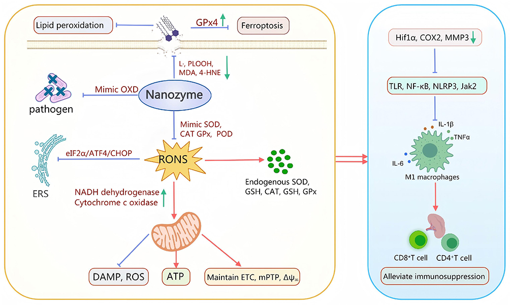 |
Figure 4 Mechanism of nanozymes therapy for sepsis. Nanozymes mimic oxidase (OXD) to directly kill pathogens; mimic superoxide dismutase (SOD), catalase (CAT), glutathione peroxidase (GPx), peroxidase (POD), etc. to scavenge reactive oxygen/nitrogen species (RONS) and restore the activity of endogenous SOD, CAT, GSH, etc. in the body; regulate the eIF2α/ATF4/CHOP pathway to inhibit endoplasmic reticulum (ER) stress; upregulate mitochondrial respiratory chain oxidoreductases (such as NADH dehydrogenase), maintain mitochondrial structures and functions such as electron transport chain (ETC), mitochondrial permeability transition pore (mPTP), and mitochondrial membrane potential (ΔΨm), promote ATP production, and reduce the release of mitochondria-derived DAMPs and ROS; inhibit lipid peroxidation-related damage, and restore GPx4 activity to prevent ferroptosis. These effects collectively inhibit oxidative stress pathways, including Hif1α, COX2, and MMP3, and proinflammatory pathways such as TLR, NF-κB, and NLRP3; reduce M1 macrophage polarization and the release of proinflammatory cytokines, including IL-1β, TNFα, and IL-6; increase the counts of CD8+ and CD4+ T cells and protect the spleen. |
Controlling Cytokine Storm
By scavenging excessive RONS in sepsis, nanozymes modulate macrophage metabolism and polarization, inhibit neutrophil tissue infiltration, reduce the release of proinflammatory mediators, thereby intercepting the cascade amplification between oxidative stress and inflammatory pathways, and restoring immune homeostasis.16,24 For example, application of Ce12V6 cluster nanozymes in sepsis animal models rapidly reduces IFN-γ in the blood from 1000 pg/mg to 500 pg/mg, and decreases TNF-α, interleukin-6 (IL-6), and interleukin-1 beta (IL-1β), thereby comprehensively alleviating cytokine storms and preventing multiple organ failure.25 Hy-CDs nanozymes reduce proinflammatory cytokines and alleviate lung pathology by downregulating the GBP protein family and inhibiting Caspase11/GSDMD-dependent atypical pyroptosis. Concurrently, they upregulate antibacterial Defa protein expression to promote lung infection recovery.21 The anti-inflammatory efficacy of carbon dots derived from Armeniacae Semen Amarum Carbonisata (ASAC-CDs) is comparable to that of dexamethasone, a commonly used clinical glucocorticoid.69 Moreover, nanozymes combined with natural active substances through cascade design can achieve multitarget regulation of RONS and cytokines. For instance, multifunctional polyphenol-copper (Cu-CA) nanozymes demonstrate broad-spectrum antibacterial activity and strong SOD and CAT like activities. They downregulate proinflammatory signaling pathways, including TLRs, COX-2, and NF-κB, and inhibit M1 macrophage polarization and neutrophil infiltration.29 Tannic acid-Zn-coated selenium (TZn@CSe) nanozymes significantly inhibit oxidative stress-related gene expression such as hypoxia-inducible factor 1α (Hif1α), janus kinase 2 (Jak2), and matrix metalloproteinase 3 (Mmp3) in sepsis animal models.70 Therefore, nanozymes can synergistically block oxidative stress and cytokine storms, effectively curbing sepsis progression (Figure 4).
Protecting Mitochondrial Function
Nanozymes can selectively accumulate in mitochondria, thereby effectively preserving both the structural and functional integrity of mitochondria in sepsis animal models. This preservation includes maintaining mitochondrial membrane potential and ETC function, preventing abnormal opening of permeability transition pores, mitigating mitochondrial cristae damage, restoring mitochondrial quality control, and preventing oxidative damage to mitochondrial DNA. These effects collectively alleviate energy metabolism crises, reduces the release of mitochondria-derived DAMPs, and inhibit ROS-induced ROS release21,29,49,66 (Figure 4). Hy-CDs accumulate in mitochondria and efficiently scavenge mitochondria-derived∙ O2−, thereby alleviating ROS mediated inhibition of respiratory chain complexes, and upregulating the expression of respiratory chain-related oxidoreductase genes. This process restores respiratory chain function and energy metabolism, reduces electron leakage to form ROS, and establishes a positive feedback loop.21 Additionally, CeCH nanozymes effectively prevent ferroptosis-related mitochondrial damage, upregulate GPx4 expression, and decrease levels of 4-hydroxynonenal (4-HNE) and MDA.28 Other studies have also confirmed the strong mitochondrial protective effect of nanozymes.29,71
Alleviating Endoplasmic Reticulum Stress
Nanozymes effectively inhibit ER stress signaling pathways, including the eIF2α/ATF4/CHOP axis, disrupt deleterious oxidative stress feedback loop between ER and mitochondria, and suppress ER-dependent ROS generation. These combined effects reduce apoptotic proteins expression, ameliorate ER structural abnormalities, and mitigate systemic inflammatory responses29,66 (Figure 4). Cu-CA nanozymes downregulate the gene expression of ER stress markers such as Atf4, Ddit3, and Hspa5, as well as proapoptotic protein Bax. They also reduce the levels of p-eIF2α, ATF4, and CHOP in liver tissues, alleviate ER dilation, vesiculation, structural swelling, and reduce hepatocyte apoptosis. Notably, the ER stress agonist tunicamycin abolishes the protective effect of nanozymes.29 Additionally, RosA-Mn nanozymes reverse oxidative stress-induced upregulation of ER stress markers (BIP/GRP78, p-eIF2α, and ATF4), and simultaneously inhibit the p38 MAPK pathway to alleviate inflammatory signal cascades.66
Improving Immunosuppression and Reducing Secondary Infections
Repeated stimulation of the immune system by cytokine storms and oxidative stress can rapidly induce immunosuppression,72 which is strongly correlated with hospital-acquired secondary infections and increased mortality risk.73 Nanozymes demonstrate significant potential in preventing and treating sepsis-induced immunosuppression and secondary infections (Figure 4). Both CeO2 nanozymes and Au/CeO2 nanozymes can effectively increase blood CD4+ and CD8+ T cell counts and prevent the reduction of the white pulp structure of the spleen. Notably, Au/CeO2 nanozymes exhibit significantly superior therapeutic efficacy compared to CeO2 nanozymes.24 Similarly, treatment of sepsis with AuO cluster nanozymes alleviates T cell exhaustion in septic blood and reverse CD4+ T cell counts in the spleen.23 Treatment with single-atom cobalt nanozymes (Co/PMCS) in animal models of bacteremia, induced by intraperitoneal injection of a lethal dose of Escherichia coli, efficiently reduces RONS levels, alleviates oxidative stress and immune dysregulation, thereby reducing bacterial load in the blood and mitigates secondary infections in vital organs such as the liver, lungs, kidneys, and intestines.26
In addition, some nanozymes directly exert antibacterial effects by generating ROS via OXD activity, but excessive ROS production in vivo may exacerbate sepsis (Figure 4). To address this issue, researchers have developed BiO2-X nanozymes with OXD, CAT, and SOD cascade activities.27 In S. aureus-induced septic models, treatment with BiO2-X nanozymes increased survival rate from 0% to 77%. Further analyses revealed significant reduced bacterial loads in multiple organs (liver, kidneys, spleen, and lungs), alongside effective scavenging of excessive ROS without exacerbating oxidative damage.
Protective Effects of Nanozymes on Vital Organ Functions
By mimicking the activity of natural antioxidant enzymes, nanozymes effectively scavenge excessive RONS during sepsis, restore endogenous antioxidant enzyme activity, control cytokine storms, and alleviate immunosuppression. Through these mechanisms, nanozymes provide robust protection to vital organs15,23,70 (Figure 5).
 |
Figure 5 Protective effects of nanozymes on vital organs in sepsis. Nanozymes confer protective effects on vital organs under sepsis conditions. Brain: Increase NeuN expression, reduce neuronal apoptosis, and enhance tolerance to cerebral hypoxia. Lungs: Decrease lung injury scores, alleviate pathological changes, and improve respiratory function. Heart: Reduce creatine kinase (CK) and lactate dehydrogenase (LDH) levels, alleviate pathological changes, restore cardiac function (LVEF, CO), and enhance myocardial hypoxia tolerance. Kidneys: Reduce proteinuria and blood levels of urea nitrogen (BUN) and creatinine (CRE). Liver: Decrease levels of alanine aminotransferase (ALT), aspartate aminotransferase (AST), and total bilirubin (TBIL) and alleviate pathological changes. |
Cardioprotection
Heart failure is a severe complication of sepsis, which increases mortality by approximately 80%. Direct myocardial injury and functional inhibition caused by cytokine storms and oxidative stress are important contributors. Clinical manifestations include ventricular dilation, diffuse weakening of ventricular wall motion, and decreased left ventricular ejection fraction (LVEF).64 Nanozymes mitigate oxidative damage to septic cardiomyocytes, reduce myocardial inflammatory infiltration and necrosis, lower levels of myocardial injury markers such as troponin and creatine kinase (CK), and restore LVEF.22,74 Treatment with CeCH nanozymes restores LVEF from an acute heart failure state (~50% reduction) to animal levels in sepsis.28 Zero-valent iron nanozymes reduce myocardial ROS and NOX2 levels and reverse the downregulation of antioxidant stress system (Nrf2, NQO1, HO-1) in sepsis animal models. Furthermore, they alleviate pathological myocardial changes including inflammation, fibrosis, interstitial edema, and cell necrosis, reverse increased myocardial injury markers (lactate dehydrogenase, CK), and successfully improve ultrasonic parameters such as cardiac output. Further studies demonstrate that zero-valent iron nanozymes alleviate septic myocardial injury via modulation of ER stress and apoptotic pathways (upregulating GRP78, CHOP, ATF6, and Bcl2 expression and downregulating p-PERK, PERK/p-PERK, and Bax levels).74 Recently, sulfide-modified zero-valent iron nanozymes also exert significant protective effects on septic myocardial injury by activating the AMPK/PPARγ pathway to inhibit myocardial inflammation and oxidative stress. Importantly, these effects were reversed by AMPK inhibitors.22 Additionally, in animal models of myocardial infarction and myocardial ischemia-reperfusion injury, nanozymes also display positive cardioprotective effects. For example, bimetallic nanozymes (Cu-TCPP-Mn) reduce the infarct size from 30.7% to 6.74% in myocardial infarction animal models and promote angiogenesis, ventricular remodeling, and recovery of cardiac ejection.64
Neuroprotection
Systemic oxidative stress and inflammatory responses induced by sepsis damage the blood-brain barrier, activate brain microglia and astrocytes, exacerbate neuroinflammation. These effects, combined with hypoxia and metabolic disorders, lead to sepsis-associated encephalopathy, clinically manifested by delirium, coma, and convulsions. Nanozymes significantly mitigate sepsis-associated encephalopathy by inhibiting oxidative damage and inflammatory responses.17,23,24 Au10 cluster and AuO clusters nanozymes effectively reduce H2O2 and MDA levels in brain tissues of sepsis animal models, restore intrinsic antioxidant defenses including SOD and GSH/GSSG ratio, decrease astrocyte activation, and exert effective neuroprotection.17,23 Ce12V6 cluster nanozymes also exert efficient anti-neuroinflammatory effects, reverse the reduction of neuron-specific nuclear protein (NeuN), and attenuate neuronal damage in septic models.25 Furthermore, nanozymes also demonstrate neuroprotective effects in other brain injury models. For example, treatment with ultrathin CeO2 nanozymes reduces cerebral infarct size from 30.1% to 8.5%, outperforming edaravone, a commonly used clinical drug.65
Pulmonary Protection
Sepsis complicated with acute lung injury (ALI) is common, with 25–50% of sepsis patients developing ALI or acute respiratory distress syndrome (ARDS), which further increases mortality.75 With superior antioxidative capacity and the ability to inhibit cytokine storm, nanozymes are promising therapeutic drugs for septic pulmonary protection.17,24,71 Prussian blue nanozyme (PBzyme) significantly reduces lung injury scores in sepsis animal models, ameliorates pathological changes (alveolar wall thickening, alveolar interstitial hemorrhage, inflammatory cell infiltration, etc), and decreases apoptosis rates of lung tissue cells. Mechanistic studies indicate that PBzyme increases the expression of HO-1 (heme oxygenase-1, an antioxidant enzyme) and reverses the elevation of proinflammatory cytokines in lung tissues and bronchoalveolar lavage fluid. Blocking HO-1 activity with zinc protoporphyrin (ZnPP) eliminates the septic pulmonary protective effect of PBzyme.20 In LPS-induced ALI animal models, Hy-CDs nanozymes significantly alleviate lung tissue pathology and restore levels of lung barrier markers (ZO-1 and Occludin).21 The septic pulmonary protective efficacy of ASAC-CDs nanozymes is comparable to that of dexamethasone.69 In addition, Hy-CDs also demonstrate protective effects in animal models of lung ischemia-reperfusion injury.21 Our team was the first to confirm that CeO2 nanozymes have a positive therapeutic effect on hyperoxic acute lung injury animal models.58
Renal Protection
Almost half of sepsis patients develop acute kidney injury (AKI), with sepsis being the leading cause of AKI in ICU patients. Clinical manifestations include water and sodium retention, accumulation of nitrogenous toxins, electrolyte imbalances, and fluid overload, which further impair other organ functions and increase hospitalization costs and mortality risk.76 Sepsis-associated AKI is closely linked to oxidative stress, cytokine storms, and insufficient organ perfusion. Therefore, scavenging RONS is crucial for AKI treatment.77,78 In sepsis animal models, AuO cluster nanozymes effectively alleviate oxidative stress and inflammatory responses in renal tissues, reduce renal tubular damage, and decrease urinary protein leakage. In addition, AuO cluster nanozymes demonstrate urease-like activity, facilitating scavenging nitrogenous toxins. These mechanisms reverse elevated blood urea nitrogen (BUN) and creatinine (Cr) levels.23 Ziziphi Spinosae Semen, a kidney-protective traditional Chinese medicine, was used to synthesize carbon dots (Z-CDs) via hydrothermal methods. In LPS-induced sepsis models, Z-CDs effectively reduce serum Cr and BUN levels, alleviate inflammatory infiltration and tubular epithelial cell damage, with a therapeutic effect comparable to dexamethasone.78 Other nanozymes such as Ce12V6 cluster, Au10 clusters, Au/CeO2, and RosA-Mn have also demonstrated remarkable efficacy in rescuing septic AKI.17,24,25,66,71 The renal protective effects of nanozymes have been further validated in ischemia-reperfusion models and AKI models induced by glycerol and cisplatin.71,79,80 For example, all animals in the glycerol-induced rhabdomyolysis AKI model died within 5 days, but all AKI mice treated with CeLutNCs nanozymes survived.71
Hepatic Protection
Acute liver injury occurs in 34–46% of sepsis patients and is closely associated with oxidative stress and inflammatory storms. These patients typically exhibit increased serum alanine transaminase (ALT), aspartate transaminase (AST), and total bilirubin (TBIL) levels.81 As a metabolic organ, the liver tends to accumulate nanozymes, even reaching the highest concentration, which facilitates resistance against sepsis-induced hepatic injury.24,60,65 Au10 cluster nanozymes reduce H2O2, MDA, and proinflammatory cytokine levels in liver tissues, restore liver antioxidant capacity (SOD and GSH/GSSG ratio), and alleviate liver pathological changes and cell apoptosis. Consequently, serum levels of liver injury markers (ALT, AST, TBIL) are markedly reduced.17 In sepsis animal models, Cu-CA nanozymes demonstrate significant immunomodulatory effects by reducing hepatic neutrophil infiltration, inhibiting M1 macrophage activation, and promoting protective M2 phenotype polarization, ultimately attenuating liver tissue injury.29 Pt@CNDs nanozymes predominantly accumulate in the liver at 3 hours post intravenous administration. Treatment with Pt@CNDs nanozymes in acute liver injury models induced by intraperitoneal injection of CCl4 significantly downregulates hepatic pro-oxidative stress genes Nox2 and Cyp2e1, restores the activity of antioxidant enzymes SOD, CAT, and GPx to normal levels, and reduces the extent of liver necrosis.60
Improving Survival Prognosis
Nanozymes significantly enhance the survival rate of sepsis model animals through their excellent and stable antioxidant activities combined with anti-inflammation, immunomodulation, and organ protection.24,25,71 (Table 1) Liu et al demonstrated that mortality of untreated LPS-induced sepsis model within 36 hours was 100%, while treatment with TZn@CSe nanozymes reduced the mortality to 50%.70 Li et al reported that PBzyme decreased mortality of LPS-induced acute lung injury mice from 100% to 40%.20 In untreated CLP sepsis models, animals exhibited rapid hypothermia, circulatory collapse, and 100% mortality within 24 hours. Treatment with PEG-Cu-SAzyme maintained the body temperature, mitigated symptoms, achieved an 80% survival rate within 24 hours, and sustained a 40% survival rate 3 days after treatment.16
Safety and Limitations of Nanozymes in Sepsis Treatment
Most nanozymes have demonstrated favorable biosafety profiles in animal models across various experimental doses. Safety assessments included cytotoxicity assays, blood compatibility evaluations, hematological and biochemical analyses, as well as monitoring of animal body weight and histopathological examination of vital organs.20,25,27,64 CeO2 and Au/CeO2 nanozymes showed no hemolytic effects on red blood cells across multiple concentrations. Following intravenous administration, these nanozymes distributed throughout major organs, with no observed organ function damage, pathological changes, or any abnormalities in biochemistry and routine blood tests.24 Similarly, no significant hematological or pathological abnormalities were observed on days 10 and 30 following Pt@CNDs administration.60
However, nanozymes may cause adverse reactions related to dosage, particle size, or element composition. For example, high concentrations of ASAC-CDs nanozymes (31.25–1000 μg/mL) inhibit cell proliferation.69 Nanoparticles exceeding the renal filtration threshold (>5.5 nm) may not be excreted rapidly, potentially leading to in vivo accumulation.25 Some metal-based nanozymes may release free metal ions in vivo, which could accumulate and cause bodily damage. Silver nanoparticles (AgNPs) exhibit 70–98% of their toxicity due to released Ag+ ions; however, controlling particle size, surface modification, or exposure conditions can regulate Ag+ release, thereby reducing toxicity.82
Nevertheless, concerns about nanozyme toxicity may be exaggerated. Catalytically active nanominerals have existed on Earth since its formation. For example, a large number of iron sulfide nanominerals (FS2) have long existed in hydrothermal areas at the bottom of the ocean.83 Notably, some scholars propose that nanozymes may have played a key role in the origin of life, while substances exhibiting nanozyme-like activity have been identified within living organisms.84,85 Furthermore, by mimicking the electronic and structural features of natural antioxidant enzymes, nanozymes with higher catalytic activity can be obtained. Interestingly, iron oxide nanoparticles (Feraheme®) clinically used for treating iron deficiency anemia have recently been found to exhibit nanozyme activity, which provides an excellent opportunity for nanozymes to enter in vivo clinical research in humans.86
Future Perspectives of Nanozymes Therapy for Sepsis
Despite the remarkable potential of nanozymes in sepsis treatment, numerous challenges remain. In addition to safety concerns, critical issues such as insufficient targeting, low catalytic activity and specificity, and a mismatch with clinical demands must be addressed.60,63,87 Moreover, nanozymes currently lack standardized quality control systems as well as clinical evaluation criteria tailored for sepsis therapy. Looking forward, future research should focus on several key directions to overcome these bottlenecks. First, the design of nanozymes with high catalytic activity and multifunctional enzyme-mimicking properties should be prioritized to synergistically eliminate diverse RONS and thereby enhance therapeutic efficacy. Second, it is essential to further elucidate the core mechanisms by which nanozymes intervene in the pathological progression of sepsis, particularly their roles in modulating the cytokine storm and reversing immune suppression, in order to provide a solid theoretical basis for precision therapy. Third, the development of smart targeting strategies is needed to enable nanozymes to accumulate specifically in diseased organs or to be activated within particular pathological microenvironments (eg, low pH, high ROS), thereby improving efficacy while minimizing off-target side effects. Fourth, given that human biosafety is the most prominent concern in nanozyme clinical translation, we need to systematically develop adverse reaction early warning systems and long-term safety monitoring strategies in human clinical trials, thereby fortifying the safety barrier for their clinical application via multi-dimensional comprehensive assessment. Fifth, further exploration of the combination of nanozymes, which have been strictly verified for human safety, and existing standard therapies for sepsis is required, focusing on evaluating the clinical benefits of such combinations. Most importantly, breaking disciplinary barriers and fostering deep collaboration between materials scientists and clinicians are imperative. Establishing a research and development paradigm driven by clinical needs will be crucial to accelerate the translation of promising nanozyme candidates from fundamental research into clinical application.
Conclusion
The vicious cycle triggered by uncontrolled RONS outburst and cytokine storms in sepsis leads to immunosuppression and multiple organ failure. Current treatment strategies are difficult to effectively interrupt this pathological process, resulting in persistently high mortality. The emergence of nanozymes offers a revolutionary approach to overcoming the limitations of sepsis therapy. Nanozymes, characterized by high antioxidant activity, good stability, and capacity to target and accumulate at inflammatory sites, efficiently scavenge excessive RONS in sepsis, disrupt RONS–inflammation cascade, thereby alleviating immunosuppression, reducing secondary infection risks, and reversing organ dysfunction. These effects collectively improve the prognosis in sepsis animal models. In the future, clinical studies on nanozymes with clinical safety for sepsis treatment should be gradually conducted. Meanwhile, further optimization of their targeted delivery, catalytic activity, and long-term safety is needed to promote the translation from preclinical research to clinical application, bringing new therapeutic hope to sepsis patients.
Data Sharing Statement
No datasets were generated or analysed during the current study.
Author Contributions
All authors made a significant contribution to the work reported, whether that is in the conception, study design, execution, acquisition of data, analysis and interpretation, or in all these areas; took part in drafting, revising or critically reviewing the article; gave final approval of the version to be published; have agreed on the journal to which the article has been submitted; and agree to be accountable for all aspects of the work.
Funding
This study was supported by grants from National Natural Science Foundation of China (82460017).
Disclosure
All authors declare that they have no conflicts of interest in this work.
References
1. Singer M, Deutschman CS, Seymour CW, et al. The third international consensus definitions for sepsis and septic shock (Sepsis-3). JAMA. 2016;315:801–810. doi:10.1001/jama.2016.0287
2. Rudd KE, Johnson SC, Agesa KM, et al. Global, regional, and national sepsis incidence and mortality, 1990–2017: analysis for the Global Burden of Disease Study. Lancet. 2020;395:200–211. doi:10.1016/S0140-6736(19)32989-7
3. La Via L, Sangiorgio G, Stefani S, et al. The global burden of sepsis and septic shock. Epidemiologia. 2024;5:456–478. doi:10.3390/epidemiologia5030032
4. Das A, Ariyakumar G, Gupta N, et al. Identifying immune signatures of sepsis to increase diagnostic accuracy in very preterm babies. Nat Commun. 2024;15:388. doi:10.1038/s41467-023-44387-5
5. Andrades MÉ, Morina A, Spasić S, et al. Bench-to-bedside review: sepsis - from the redox point of view. Crit Care. 2011;15:230. doi:10.1186/cc10334
6. Karki R, Sharma BR, Tuladhar S, et al. Synergism of TNF-α and IFN-γ triggers inflammatory cell death, tissue damage, and mortality in SARS-CoV-2 infection and cytokine shock syndromes. Cell. 2021;184:149–168.e17. doi:10.1016/j.cell.2020.11.025
7. Cao M, Wang G, Xie J. Immune dysregulation in sepsis: experiences, lessons and perspectives. Cell Death Discov. 2023;9:465. doi:10.1038/s41420-023-01766-7
8. Dhalla NS, Ostadal P, Tappia PS. Involvement of oxidative stress and antioxidants in modification of cardiac dysfunction due to ischemia-reperfusion injury. Antioxidants. 2025;14:340. doi:10.3390/antiox14030340
9. Sun L, Zhao JH, Fan WY, et al. Therapeutic effects of high-dose vitamin C supplementation in patients with COVID-19: a meta-analysis. Nutr Rev. 2024;82:1056–1068. doi:10.1093/nutrit/nuad105
10. Rosengrave P, Spencer E, Williman J, et al. Intravenous vitamin C administration to patients with septic shock: a pilot randomised controlled trial. Crit Care. 2022;26:26. doi:10.1186/s13054-022-03900-w
11. Sahoo DK, Wong D, Patani A, et al. Exploring the role of antioxidants in sepsis-associated oxidative stress: a comprehensive review. Front Cell Infect Microbiol. 2024;14:1348713. doi:10.3389/fcimb.2024.1348713
12. Zeng M, Wang Y, Liu M, et al. Potential efficacy of herbal medicine-derived carbon dots in the treatment of diseases: from mechanism to clinic. Int J Nanomed. 2023;18:6503–6525. doi:10.2147/IJN.S431061
13. Zandieh M, Liu J. Nanozymes: definition, Activity, and Mechanisms. Adv Mater. 2024;36:2211041. doi:10.1002/adma.202211041
14. Yang Y, Li Z, Fan X, et al. Nanozymes: potential therapies for reactive oxygen species overproduction and inflammation in ischemic stroke and traumatic brain injury. ACS Nano. 2024;18:16450–16467. doi:10.1021/acsnano.4c03425
15. Chen L, Huang Q, Zhao T, et al. Nanotherapies for sepsis by regulating inflammatory signals and reactive oxygen and nitrogen species: new insight for treating COVID-19. Redox Biol. 2021;45:102046. doi:10.1016/j.redox.2021.102046
16. Yang J, Zhang R, Zhao H, et al. Bioinspired copper single-atom nanozyme as a superoxide dismutase-like antioxidant for sepsis treatment. Exploration. 2022;2:20210267. doi:10.1002/EXP.20210267
17. Xin Q, Zhou S, Zhang L, et al. Biocatalytic Au10 clusters for sepsis management via modulation of systemic inflammation. Chem Eng J. 2024;489:150988. doi:10.1016/j.cej.2024.150988
18. Soh M, Kang DW, Jeong HG, et al. Ceria-zirconia nanoparticles as an enhanced multi-antioxidant for sepsis treatment. Angew Chem Int Ed. 2017;56:11399–11403. doi:10.1002/anie.201704904
19. Yim D, Lee DE, So Y, et al. Sustainable nanosheet antioxidants for sepsis therapy via scavenging intracellular reactive oxygen and nitrogen species. ACS Nano. 2020;14:10324–10336. doi:10.1021/acsnano.0c03807
20. Li XY, Qiu CM, Yang FY, et al. Protective effects of Prussian blue nanozyme against sepsis-induced acute lung injury by activating HO-1. Eur J Pharmacol. 2024;968:176354. doi:10.1016/j.ejphar.2024.176354
21. Deng Z, Zhang Y, Li R, et al. Honeysuckle‐derived carbon dots with robust catalytic and pharmacological activities for mitigating lung inflammation by inhibition of Caspase11/GSDMD‐dependent pyroptosis. Adv Funct Mater. 2025;35:2418683. doi:10.1002/adfm.202418683
22. Wang D, Zhao H, Deng C, et al. Sulfide-modified nanoscale zero-valent iron as a novel therapeutic remedy for septic myocardial injury. J Adv Res. 2024;55:145–158. doi:10.1016/j.jare.2023.02.008
23. Sun S, Liu D, Song N, et al. Single active Au1O5 clusters for metabolism-inspired sepsis management through immune regulation. Nano Today. 2024;58:102416. doi:10.1016/j.nantod.2024.102416
24. Xin Q, Zhang S, Sun S, et al. Multienzyme active nanozyme for efficient sepsis therapy through modulating immune and inflammation inhibition. ACS Appl Mater Interfaces. 2024;16:36047–36062. doi:10.1021/acsami.4c04994
25. Liu D, Sun S, Qiao H, et al. Ce12V6 clusters with multi-enzymatic activities for sepsis treatment. Adv Healthcare Mater. 2025;14:2401581. doi:10.1002/adhm.202401581
26. Cao F, Zhang L, You Y, et al. An enzyme-mimicking single-atom catalyst as an efficient multiple reactive oxygen and nitrogen species scavenger for sepsis management. Angew Chem Int Ed. 2020;59:5108–5115. doi:10.1002/anie.201912182
27. Liu F, Zhang K, Lu B, et al. Oxygen‐vacancy‐rich monolayer BiO2−X nanosheets for bacterial sepsis management via dual physically antibacterial and chemically anti‐inflammatory functions. Adv Healthcare Mater. 2024;13:2304002. doi:10.1002/adhm.202304002
28. Jiang C, Shi Q, Yang J, et al. Ceria nanozyme coordination with curcumin for treatment of sepsis-induced cardiac injury by inhibiting ferroptosis and inflammation. J Adv Res. 2024;63:159–170. doi:10.1016/j.jare.2023.10.011
29. Che D, Xiao Y, Zhang X, et al. Multifunctional integrated polyphenol-copper nanozymes for sepsis-induced acute liver injury via ameliorating endoplasmic reticulum stress and reprogramming inflammatory microenvironment. Chem Eng J. 2025;507:160293. doi:10.1016/j.cej.2025.160293
30. Lugrin J, Rosenblatt-Velin N, Parapanov R, et al. The role of oxidative stress during inflammatory processes. Biol Chem. 2014;395:203–230. doi:10.1515/hsz-2013-0241
31. Jomova K, Raptova R, Alomar SY, et al. Reactive oxygen species, toxicity, oxidative stress, and antioxidants: chronic diseases and aging. Arch Toxicol. 2023;97:2499–2574. doi:10.1007/s00204-023-03562-9
32. Breda CNS, Davanzo GG, Basso PJ, et al. Mitochondria as central hub of the immune system. Redox Biol. 2019;26:101255. doi:10.1016/j.redox.2019.101255
33. Visnes T, Cázares-Körner A, Hao W, et al. Small-molecule inhibitor of OGG1 suppresses proinflammatory gene expression and inflammation. Science. 2018;362:834–839. doi:10.1126/science.aar8048
34. Bime C, Zhou T, Wang T, et al. Reactive oxygen species-associated molecular signature predicts survival in patients with sepsis. Pulm Circ. 2016;6:196–201. doi:10.1086/685547
35. Mathru M, Rooney MW, Dries DJ, et al. Urine hydrogen peroxide during adult respiratory distress syndrome in patients with and without sepsis. Chest. 1994;105:232–236. doi:10.1378/chest.105.1.232
36. Joffre J, Hellman J. Oxidative stress and endothelial dysfunction in sepsis and acute inflammation. Antioxid Redox Signal. 2021;35:1291–1307. doi:10.1089/ars.2021.0027
37. Casey AM, Ryan DG, Prag HA, et al. Pro-inflammatory macrophages produce mitochondria-derived superoxide by reverse electron transport at complex I that regulates IL-1β release during NLRP3 inflammasome activation. Nat Metab. 2025;7:493–507. doi:10.1038/s42255-025-01224-x
38. Tschopp J, Schroder K. NLRP3 inflammasome activation: the convergence of multiple signalling pathways on ROS production? Nat Rev Immunol. 2010;10:210–215. doi:10.1038/nri2725
39. Wong LR, Perlman S. Immune dysregulation and immunopathology induced by SARS-CoV-2 and related coronaviruses - are we our own worst enemy? Nat Rev Immunol. 2022;22:47–56. doi:10.1038/s41577-021-00656-2
40. Gao X, Cai S, Li X, et al. Sepsis-induced immunosuppression: mechanisms, biomarkers and immunotherapy. Front Immunol. 2025;16:1577105. doi:10.3389/fimmu.2025.1577105
41. Thoppil J, Mehta P, Bartels B, et al. Impact of norepinephrine on immunity and oxidative metabolism in sepsis. Front Immunol. 2023;14:1271098. doi:10.3389/fimmu.2023.1271098
42. Rendra E, Riabov V, Mossel DM, et al. Reactive oxygen species (ROS) in macrophage activation and function in diabetes. Immunobiology. 2019;224:242–253. doi:10.1016/j.imbio.2018.11.010
43. Zhao S, Chen F, Yin Q, et al. Reactive oxygen species interact with NLRP3 inflammasomes and are involved in the inflammation of sepsis: from mechanism to treatment of progression. Front Physiol. 2020;11:571810. doi:10.3389/fphys.2020.571810
44. Surabhi S, Jachmann LH, Shumba P, et al. Hydrogen peroxide is crucial for NLRP3 inflammasome-mediated IL-1β production and cell death in pneumococcal infections of bronchial epithelial cells. J Innate Immun. 2022;14:192–206. doi:10.1159/000517855
45. Belikov AV, Schraven B, Simeoni L. T cells and reactive oxygen species. J Biomed Sci. 2015;22:85. doi:10.1186/s12929-015-0194-3
46. Hoetzenecker W, Echtenacher B, Guenova E, et al. ROS-induced ATF3 causes susceptibility to secondary infections during sepsis-associated immunosuppression. Nat Med. 2011;18:128–134. doi:10.1038/nm.2557
47. Zorov DB, Juhaszova M, Sollott SJ. Mitochondrial reactive oxygen species (ROS) and ROS-induced ROS release. Physiol Rev. 2014;94:909–950. doi:10.1152/physrev.00026.2013
48. Zhang H, Feng YW, Yao YM. Potential therapy strategy: targeting mitochondrial dysfunction in sepsis. Mil Med Res. 2018;5:41. doi:10.1186/s40779-018-0187-0
49. Preau S, Vodovar D, Jung B, et al. Energetic dysfunction in sepsis: a narrative review. Ann Intensive Care. 2021;11:104. doi:10.1186/s13613-021-00893-7
50. Grazioli S, Pugin J. Mitochondrial damage-associated molecular patterns: from inflammatory signaling to human diseases. Front Immunol. 2018;9:832. doi:10.3389/fimmu.2018.00832
51. He R, Liu B, Xiong R, et al. Itaconate inhibits ferroptosis of macrophage via Nrf2 pathways against sepsis-induced acute lung injury. Cell Death Discov. 2022;8:43. doi:10.1038/s41420-021-00807-3
52. Liu J, Kang R, Tang D. Signaling pathways and defense mechanisms of ferroptosis. FEBS J. 2022;289:7038–7050. doi:10.1111/febs.16059
53. Wang Y, Wang W, Zhang Y, et al. Targeting ferroptosis offers therapy choice in sepsis-associated acute lung injury. Eur J Med Chem. 2025;283:117152. doi:10.1016/j.ejmech.2024.117152
54. Gong T, Liu Y, Tian Z, et al. Identification of immune-related endoplasmic reticulum stress genes in sepsis using bioinformatics and machine learning. Front Immunol. 2022;13:995974. doi:10.3389/fimmu.2022.995974
55. Chen X, Shi C, He M, et al. Endoplasmic reticulum stress: molecular mechanism and therapeutic targets. Signal Transduct Target Ther. 2023;8:352. doi:10.1038/s41392-023-01570-w
56. Laurindo FR, Pescatore LA, Fernandes DDC. Protein disulfide isomerase in redox cell signaling and homeostasis. Free Radic Biol Med. 2012;52:1954–1969. doi:10.1016/j.freeradbiomed.2012.02.037
57. Gao L, Zhuang J, Nie L, et al. Intrinsic peroxidase-like activity of ferromagnetic nanoparticles. Nat Nanotechnol. 2007;2:577–583. doi:10.1038/nnano.2007.260
58. Guo H, Huang RR, Qu SS, et al. FAM134B deletion exacerbates apoptosis and epithelial-to-mesenchymal transition in rat lungs exposed to hyperoxia. iScience. 2024;27:110385. doi:10.1016/j.isci.2024.110385
59. Shen X, Ma R, Huang Y, et al. Nano-decocted ferrous polysulfide coordinates ferroptosis-like death in bacteria for anti-infection therapy. Nano Today. 2020;35:100981. doi:10.1016/j.nantod.2020.100981
60. Zhang Y, Gao W, Ma Y, et al. Integrating Pt nanoparticles with carbon nanodots to achieve robust cascade superoxide dismutase-catalase nanozyme for antioxidant therapy. Nano Today. 2023;49:101768. doi:10.1016/j.nantod.2023.101768
61. Shi X, Wen Z, Gu Q, et al. Metal-support frontier orbital interactions in single-atom catalysis. Nature. 2025;640:668–675. doi:10.1038/s41586-025-08747-z
62. Shen X, Wang Z, Gao XJ, et al. Reaction mechanisms and kinetics of nanozymes: insights from theory and computation. Adv Mater. 2024;36:2211151. doi:10.1002/adma.202211151
63. Zhang R, Jiang B, Fan K, et al. Designing nanozymes for in vivo applications. Nat Rev Bioeng. 2024;2:849–868. doi:10.1038/s44222-024-00205-1
64. Xiang K, Wu H, Liu Y, et al. MOF-derived bimetallic nanozyme to catalyze ROS scavenging for protection of myocardial injury. Theranostics. 2023;13:2721–2733. doi:10.7150/thno.83543
65. Liu C, Gui L, Zheng JJ, et al. Intrinsic strain-mediated ultrathin ceria nanoantioxidant. J Am Chem Soc. 2023;145:19086–19097. doi:10.1021/jacs.3c07048
66. Yuan H, Wang F, Wang Z, et al. Natural metal polyphenol nanozyme: free radical scavenging and antioxidation for the treatment of acute kidney injury. ACS Mater Lett. 2023;5:2807–2819. doi:10.1021/acsmaterialslett.3c00906
67. Zhang B, Li Q, Xu Q, Li B, Dong H, Mou Y. Polydopamine modified ceria nanorods alleviate inflammation in colitis by scavenging ROS and regulating macrophage M2 polarization. Int J Nanomed. 2023;18:4601–4616. doi:10.2147/IJN.S416049
68. Zhang D, Shen N, Zhang J, et al. A novel nanozyme based on selenopeptide-modified gold nanoparticles with a tunable glutathione peroxidase activity. RSC Adv. 2020;10:8685–8691. doi:10.1039/c9ra10262k
69. Zhao Y, Zhang Y, Kong H, et al. Protective effects of carbon dots derived from armeniacae semen amarum carbonisata against acute lung injury induced by lipopolysaccharides in rats. Int J Nanomed. 2022;17:1–14. doi:10.2147/IJN.S338886
70. Liu K, Chen S, Geng X, et al. Multifunctional selenium-based metal polyphenol nanoparticles impede the pathological cross-talk between reactive oxygen species and inflammation in sepsis. ACS Mater Lett. 2024;6:2434–2445. doi:10.1021/acsmaterialslett.4c00084
71. Gu J, Zhang P, Li H, et al. Cerium-luteolin nanocomplexes in managing inflammation-related diseases by antioxidant and immunoregulation. ACS Nano. 2024;18:6229–6242. doi:10.1021/acsnano.3c09528
72. Meyer NJ, Prescott HC. Sepsis and Septic Shock. N Engl J Med. 2024;391:2133–2146. doi:10.1056/NEJMra2403213
73. Pei F, Yao RQ, Ren C, et al. Expert consensus on the monitoring and treatment of sepsis-induced immunosuppression. Mil Med Res. 2022;9:74. doi:10.1186/s40779-022-00430-y
74. Wang D, Wang C, Liang Z, et al. Protection of zero-valent iron nanoparticles against sepsis and septic heart failure. J Nanobiotechnol. 2022;20:405. doi:10.1186/s12951-022-01589-1
75. Huang X, Zhang R, Fan G, et al. Incidence and outcomes of acute respiratory distress syndrome in intensive care units of mainland China: a multicentre prospective longitudinal study. Crit Care. 2020;24:515. doi:10.1186/s13054-020-03112-0
76. Molinari L, Rio-Pertuz GD, Priyanka P, et al. Distribution of acute and chronic kidney disease across clinical phenotypes for sepsis. Chest. 2024;166:480–490. doi:10.1016/j.chest.2024.03.006
77. Yu H, Jin F, Liu D, et al. ROS-responsive nano-drug delivery system combining mitochondria-targeting ceria nanoparticles with atorvastatin for acute kidney injury. Theranostics. 2020;10:2342–2357. doi:10.7150/thno.40395
78. Wang L, Li ZY, Zhong CL, et al. Therapeutic potential of naturally derived carbon dots in sepsis-associated acute kidney injury. Chin Med. 2025;20:49. doi:10.1186/s13020-025-01103-3
79. Guo W, Wang T, Li H, et al. Defect-engineered nanoscale metal-organic frameworks as self-cascade antioxidation nanozymes for targeted therapy of cisplatin-induced acute kidney injury. Chem Eng J. 2025;516:163893. doi:10.1016/j.cej.2025.163893
80. Xia F, Xiang S, Qiu Y, et al. Macrophage membrane‐camouflaged nanozymes for combating the oxidative stress‐microvascular perfusion negative feedback in ischemia‐reperfusion induced acute kidney injury. Adv Healthcare Mater. 2025;14:2405090. doi:10.1002/adhm.202405090
81. Guo Y, Guo W, Chen H, et al. Mechanisms of sepsis-induced acute liver injury: a comprehensive review. Front Cell Infect Microbiol. 2025;15:1504223. doi:10.3389/fcimb.2025.1504223
82. Yan N, Wang Y, Wong TY, et al. Differential mapping of intracellular metallic nanoparticles and ions and dynamic modeling prediction. ACS Nano. 2025;19:35013–35028. doi:10.1021/acsnano.5c04379
83. Yücel M, Gartman A, Chan CS, et al. Hydrothermal vents as a kinetically stable source of iron-sulphide-bearing nanoparticles to the ocean. Nat Geosci. 2011;4:367–371. doi:10.1038/ngeo1148
84. Ma L, Liang Z, Hou Y, et al. Nanozymes and their potential roles in the origin of life. Adv Mater. 2025;37:2412211. doi:10.1002/adma.202412211
85. Ma L, Zheng JJ, Zhou N, et al. A natural biogenic nanozyme for scavenging superoxide radicals. Nat Commun. 2024;15:233. doi:10.1038/s41467-023-44463-w
86. Wang Q, Qian W, Han Y, et al. Ferumoxytol promotes haematopoietic stem cell post-injury regeneration as a reactive oxygen species scavenger. Nat Nanotechnol. 2025;20:959–969. doi:10.1038/s41565-025-01907-2
87. Wang SH, Xu XL, Chen W. How do organelle-targeting nanotherapeutics treat inflammatory diseases? A comprehensive review of the literature. Int J Nanomed. 2025;20:7133–7152. doi:10.2147/IJN.S516260
 © 2025 The Author(s). This work is published and licensed by Dove Medical Press Limited. The
full terms of this license are available at https://www.dovepress.com/terms
and incorporate the Creative Commons Attribution
- Non Commercial (unported, 4.0) License.
By accessing the work you hereby accept the Terms. Non-commercial uses of the work are permitted
without any further permission from Dove Medical Press Limited, provided the work is properly
attributed. For permission for commercial use of this work, please see paragraphs 4.2 and 5 of our Terms.
© 2025 The Author(s). This work is published and licensed by Dove Medical Press Limited. The
full terms of this license are available at https://www.dovepress.com/terms
and incorporate the Creative Commons Attribution
- Non Commercial (unported, 4.0) License.
By accessing the work you hereby accept the Terms. Non-commercial uses of the work are permitted
without any further permission from Dove Medical Press Limited, provided the work is properly
attributed. For permission for commercial use of this work, please see paragraphs 4.2 and 5 of our Terms.
Recommended articles

Electroacupuncture at ST36 (Zusanli) Prevents T-Cell Lymphopenia and Improves Survival in Septic Mice
Lv ZY, Shi YL, Bassi GS, Chen YJ, Yin LM, Wang Y, Ulloa L, Yang YQ, Xu YD
Journal of Inflammation Research 2022, 15:2819-2833
Published Date: 3 May 2022
Advances in the Study of Immunosuppressive Mechanisms in Sepsis
Fu X, Liu Z, Wang Y
Journal of Inflammation Research 2023, 16:3967-3981
Published Date: 8 September 2023
The Role of Dectin-1-Akt-RNF146 Pathway in β-Glucan Induced Immune Trained State of Monocyte in Sepsis
Guo C, Xu P, Luo W, Zhang J, Sun X, Hoang H, Ma D, Wu D, Zhong J, Miao C
Journal of Inflammation Research 2025, 18:1147-1165
Published Date: 24 January 2025