
Back to Journals » International Journal of Nanomedicine » Volume 21

Nanomaterials for Photodynamic Therapy in Cancer: Classifications, Recent Advances, and Combination Applications
Authors Zhang L, Song L, Pei Y, Zhao M, Shi J, Li J, Qin Y, Liu Y
Received 24 December 2025
Accepted for publication 24 June 2026
Published 21 July 2026 Volume 2026:21 591447
DOI https://doi.org/10.2147/IJN.S591447
Checked for plagiarism Yes
Review by Single anonymous peer review
Peer reviewer comments 3
Editor who approved publication: Dr Kamakhya Prakash Misra
Lan Zhang,1,* Lei Song,1,* Yuhe Pei,2 Minghui Zhao,3 Jianhong Shi,4,5 Jinghua Li,4,6 Yan Qin,4,5,7 Yawei Liu1
1Department of Integrated Traditional Chinese Medicine and Western Medicine, Affiliated Hospital of Hebei University, Baoding, Hebei, People’s Republic of China; 2Clinical Medical College, Heibei University, Baoding, Hebei, People’s Republic of China; 3Department of Radiology, Affiliated Hospital of Hebei University, Baoding, Hebei, People’s Republic of China; 4Central Laboratory, Hebei International Joint Research Center of Digital Twins to Precision Diagnosis and Treatment of Gastrointestinal Cancers, Baoding Key Laboratory for Interdisciplinary Tumor Microecology Metabolism Research, Affiliated Hospital of Hebei University, Baoding, Hebei, People’s Republic of China; 5Hebei Collaborative Innovation Center of Tumor Microecological Metabolism Regulation, Clinical Medical College, Hebei University, Baoding, Hebei, People’s Republic of China; 6Department of Hepatobiliary Surgery, Affiliated Hospital of Hebei University, Baoding, Hebei, People’s Republic of China; 7Hebei Key Laboratory of Precise Imaging of Inflammation Related Tumors, Affiliated Hospital of Hebei University, Baoding, Hebei, People’s Republic of China
*These authors contributed equally to this work
Correspondence: Yawei Liu, Department of Integrated Traditional Chinese Medicine and Western Medicine, Affiliated Hospital of Hebei University, No. 21 Yuhua East Road, Baoding, Hebei, People’s Republic of China, Tel +86 03125981182; +86 18903363756, Email [email protected] Yan Qin, Central Laboratory, Baoding Key Laboratory for Interdisciplinary Tumor Microecology Metabolism Research, Affiliated Hospital of Hebei University, No. 21 Yuhua East Road, Baoding, Hebei, People’s Republic of China, Tel +86 03125981736, Email [email protected]
Abstract: Photodynamic therapy (PDT) has emerged as a promising non-invasive therapeutic modality for cancer treatment. Compared with conventional antineoplastic therapies, PDT offers targeted cytotoxic effects and relatively low systemic toxicity, making it an attractive therapeutic option for patients with cancer. The therapeutic efficacy of PDT is largely determined by the intrinsic properties of photosensitizers (PSs), which are the central components of this therapeutic approach. However, currently available PSs still present several inherent limitations, including inadequate penetration into deep tumor tissues, suboptimal accumulation and delivery at tumor sites, and substantial dependence on oxygen availability within the tumor microenvironment. Consequently, the development of high-performance PSs remains a key research priority in the field of PDT. Recent advances in photosensitizer technology have enabled the rational integration of PDT with other therapeutic modalities, including radiotherapy, chemotherapy, and immunotherapy, to achieve synergistic therapeutic effects, thereby significantly improving overall treatment outcomes. This review systematically summarizes the major classifications, key characteristics, and recent advances in the application of nanomaterials for PDT. It evaluates the current progress in the development of nanomaterial-based PSs and examines their potential applications in combination therapeutic strategies for cancer treatment. Collectively, these findings provide valuable insights into the future development of PDT-based anticancer therapies.
Keywords: combination therapy, drug delivery, nanomaterials, photodynamic therapy, photosensitizers
Introduction
Cancer remains one of the most significant global health challenges, imposing a substantial burden on healthcare systems worldwide and representing a leading cause of morbidity and mortality. Globally, approximately 2,114,850 new cases of malignant neoplasms were diagnosed in 2026, with an estimated 626,140 cancer-related deaths.1 Current cancer treatment modalities include surgical resection, chemotherapy, radiotherapy, immunotherapy, and targeted therapy. Despite their widespread clinical application and substantial therapeutic benefits, these conventional approaches are associated with notable limitations, including systemic adverse effects, treatment-related toxicity to normal tissues, and procedural complexity, all of which may restrict therapeutic efficacy and patient compliance.
Against this background, photodynamic therapy (PDT) has emerged as a promising alternative therapeutic strategy owing to its selective mechanism of action, relatively low systemic toxicity, and potential for synergistic integration with other treatment modalities. Since its initial application in cancer diagnosis in the 1950s, PDT has undergone continuous development and has evolved into a minimally invasive therapeutic modality that selectively eradicates malignant lesions through photochemical reactions initiated by light-activated photosensitizers (PSs). However, significant challenges remain in both the research and clinical application of PDT, particularly with respect to the treatment of deep-seated tumors, limited tumor-specific targeting, and the lack of effective oxygen-independent photosensitizer systems.2,3 Although thousands of PSs have been synthesized and assessed to date, only a small proportion have progressed to clinical trials, highlighting the need for the development of next-generation PSs with improved efficacy and safety.
In recent years, nanomaterials have attracted considerable attention in the field of cancer therapy due to their inherent advantages, including structural stability, ease of surface modification, and tunable physicochemical properties, which facilitate enhanced tumor targeting and improved drug delivery. Accumulating evidence indicates that the rational integration of PDT with other therapeutic modalities, such as chemotherapy, radiotherapy, and immunotherapy, can significantly improve overall therapeutic efficacy through synergistic effects, thereby overcoming the limitations associated with single-modality treatment.4–6 In this context, this review summarizes recent advances in the development of nanomaterial-based PSs and systematically examines their applications in combination with other therapeutic strategies for cancer treatment. The objective is to provide a comprehensive overview of current progress in the field and offer valuable insights into the future development and clinical translation of PDT-based anticancer therapies.
This review also provides a distinct perspective by examining the transition from conventional Type II-dominant photodynamic mechanisms to emerging Type I strategies designed to overcome tumor hypoxia. It critically evaluates recent advances in combination approaches involving chemotherapy, radiotherapy, and immunotherapy, while highlighting the important challenges of biosafety, toxicity, and standardization that continue to influence the clinical development of nanomaterial-based PDT.
Application of PDT in Tumor Management
PDT can selectively eradicate localized primary and recurrent tumors while minimizing damage to surrounding healthy tissues, demonstrating considerable potential as an adjunctive therapeutic modality. The principal mechanism of PDT involves the administration of PSs that accumulate within tissues and are subsequently activated by irradiation with light of an appropriate wavelength. Following photoexcitation, PSs generate reactive oxygen species (ROS) through two primary pathways:
Type I reaction: Excited-state PSs interact with surrounding biological substrates through electron or hydrogen atom transfer processes, generating radical intermediates that subsequently react with molecular oxygen to produce ROS, including hydrogen peroxide, superoxide anion radicals, and hydroxyl radicals.
Type II reaction: Excited-state PSs transfer energy directly to molecular oxygen, resulting primarily in the generation of singlet oxygen (1O2), with additional production of other reactive oxygen species under certain conditions.
The intracellular accumulation of ROS induces irreversible damage to subcellular structures, including the plasma membrane, mitochondria, lysosomes, and organelles, as well as to essential biomolecules such as DNA, proteins, and lipids.4,7–11
The therapeutic efficacy of PDT is determined primarily by three key components: the light source, the photosensitizer, and oxygen availability. Additional variables that influence treatment outcomes include light dose, tumor location, histological subtype, lesion depth, photosensitizer properties, and local tissue oxygenation.
Despite its considerable therapeutic potential, PDT continues to face several limitations in oncologic applications. The limited penetration depth of light in biological tissues reduces its effectiveness in the treatment of deep-seated tumors. The delivery efficiency and tumor accumulation of many PSs remain suboptimal, limiting their therapeutic effectiveness. Furthermore, the hypoxic tumor microenvironment compromises photosensitizer activity and ROS generation, thereby reducing treatment efficacy. Collectively, these challenges constitute major barriers to the successful translation of PDT from preclinical research into clinical practice.
To address these limitations, combination strategies integrating PDT with chemotherapy, radiotherapy, or other therapeutic modalities have demonstrated promising synergistic effects. By leveraging complementary mechanisms of action, these multimodal approaches can enhance therapeutic efficacy and may provide more effective strategies for cancer treatment (Figure 1).
 |
Figure 1 Mechanisms of reactive oxygen species generation during PDT. The schematic diagram illustrates the two principal photochemical pathways involved in PDT. In the Type I pathway, an excited-state photosensitizer interacts with surrounding biological substrates through electron or hydrogen atom transfer processes, generating radical intermediates that subsequently react with molecular oxygen to produce reactive oxygen species, including hydrogen peroxide, superoxide anion radicals, and hydroxyl radicals. In the Type II pathway, the excited-state photosensitizer transfers energy directly to molecular oxygen, resulting primarily in the generation of singlet oxygen. |
Mechanisms of PDT in Tumor Management
PDT exerts antineoplastic effects through three principal mechanisms:
Direct Cytotoxicity Toward Malignant Cells
PDT initiates a series of photochemical and photobiological reactions following irradiation with light of an appropriate wavelength, resulting in selective damage to malignant cells while minimizing injury to surrounding healthy tissues. Currently, PDT is primarily used in the treatment of early-stage, superficial, and hollow-organ malignancies, including esophageal carcinoma, breast carcinoma, and melanoma.12–17
Disruption of Tumor-Associated Vasculature
PDT induces damage to the tumor vasculature, leading to impaired oxygen and nutrient delivery and subsequently promoting tumor cell apoptosis and necrosis. In pancreatic carcinoma, for example, PDT has been shown to induce tumor cell apoptosis while disrupting vascular networks, thereby enhancing antitumor immune responses.18
Activation of Antitumor Immune Responses
PDT can stimulate antitumor immunity through the induction of immunogenic cell death and the enhancement of immune surveillance. Evidence suggests that combining PDT with selective cyclooxygenase-2 (COX-2) inhibitors may improve therapeutic outcomes in patients with skin and oral malignancies.19
Despite these therapeutic mechanisms, PDT continues to face two major limitations in the treatment of solid tumors: ① the limited penetration depth of light within tumor tissues and ② the hypoxic conditions characteristic of many tumor microenvironments, both of which significantly compromise therapeutic efficacy. Consequently, current research efforts are focused on the development of more efficient photosensitizer formulations, particularly nanomaterial-based formulations, as well as combination therapeutic strategies designed to overcome these challenges (Figure 2).
 |
Figure 2 Mechanisms underlying the antitumor effects of PDT. The schematic diagram depicts three principal mechanisms by which PDT exerts antitumor activity: (1) direct cytotoxicity toward malignant cells through reactive oxygen species-mediated cellular damage; (2) disruption of tumor-associated vasculature, resulting in impaired oxygen and nutrient supply and subsequent cell apoptosis and necrosis; and (3) activation of antitumor immune responses through the induction of immunogenic cell death and enhancement of immune surveillance. |
Applications of Nanomaterial-Based Photosensitizers in Tumor Management
PSs are the key determinants of PDT efficacy. An ideal PS should demonstrate high tumor-selective accumulation, rapid and safe metabolic clearance, a well-defined molecular structure, minimal systemic toxicity, ease of administration, a high singlet oxygen quantum yield, and stable activation under irradiation at appropriate wavelengths.20–24
However, most conventional PSs are limited by poor aqueous solubility, low chemical and photostability, inadequate in vivo delivery efficiency and safety, limited cellular and tissue specificity, and dependence on oxygen availability, all of which restrict their clinical applicability.
In recent years, nanomaterials have emerged as a promising platform for the development of next-generation PSs, enabling the design of a wide range of photodynamic nanomaterials. Nanomaterial-based PSs can be broadly classified into organic nanomaterials, such as liposomes, polymeric micelles, and polymeric nanoparticles (NPs), and inorganic nanomaterials, including gold nanoparticles (AuNPs) and silica NPs. Owing to their high specific surface area, nanoscale dimensions, mechanical flexibility, and excellent photostability, these nanomaterials exhibit considerable potential in tumor diagnosis and therapy.
Table 1 summarizes the major classifications and key characteristics of nanomaterials used in PDT.
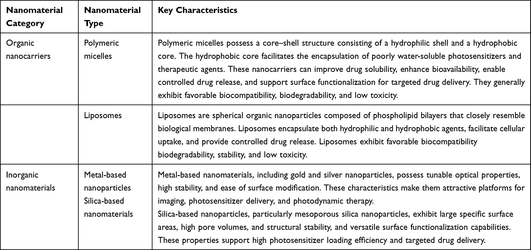 |
Table 1 Classification and Key Characteristics of Nanomaterial Platforms Used in PDT |
NPs are colloidal structures composed of natural, synthetic, or semisynthetic polymers. Encapsulation of PSs within NPs significantly enhance their uptake by tumor cells.20 As delivery vehicles, NPs can address several limitations associated with conventional PSs, including poor tumor-targeting efficiency and the hypoxic tumor microenvironment. This approach improves the accumulation and retention of PSs in tumor tissues, thereby enhancing the therapeutic efficacy of PDT.25–27
To improve tumor-targeting efficiency, NP surfaces can be functionalized with specific antibodies, aptamers, or other targeting ligands that facilitate selective delivery to malignant cells. Ligand-modified NPs can bind specifically to receptors overexpressed on cancer cell surfaces and are subsequently internalized through receptor-mediated endocytosis, thereby resulting in enhanced intracellular accumulation of PSs.4,7
For example, by exploiting the Warburg effect, Kataoka et al developed a glucose-conjugated chlorin-based photosensitizer that selectively targets glucose transporters GLUT1, GLUT3, and GLUT4, which are frequently overexpressed in cancer cells. This modification enhances selective photosensitizer accumulation and improves PDT efficacy. Similarly, many cancer cells exhibit increased expression of low-density lipoprotein (LDL) receptors, resulting in higher intracellular LDL levels than those observed in normal cells. Conjugation of PSs with LDL can therefore enhance selective tumor cell uptake.8
Another widely used targeting strategy exploits the overexpression of folate receptors, which are particularly abundant in ovarian, breast, and lung carcinomas. Co-encapsulation of the anticancer agent camptothecin and the photosensitizer chlorin e6 (Ce6) within metal-organic framework NPs enables selective binding to folate receptors, thereby enhancing tumor-cell uptake and targeting efficiency.12 This approach not only reduces the non-specific distribution of conventional PSs, such as porphyrin derivatives, naphthalocyanines, phthalocyanines, and chlorin compounds, but also facilitates the co-delivery of PSs and chemotherapeutic agents within a single nanoplatform, supporting precision imaging and therapy.28
Encapsulation of PSs within NPs can help address hypoxia-related limitations during PDT. Sun et al developed a co-delivery system incorporating the photosensitizer Ce6 and the hypoxia-activated agent tirapazamine (TPZ).27 Ce6 generates large amounts of singlet oxygen (1O2) upon irradiation with 660 nm light, whereas TPZ is activated under hypoxic conditions, thereby further enhancing cytotoxicity toward hypoxic tumor cells. This combination of PDT with hypoxia-activated therapy resulted in significantly improved antitumor efficacy.
Inorganic NPs derived from metal-based nanomaterials have also demonstrated considerable potential in PDT. Commonly used metal-based nanomaterials include gold-, silver-, and silica-based systems.29,30 Gold- and silver-based nanomaterials are widely employed in PDT owing to their high extinction coefficients, chemical stability, resistance to oxidation, and ease of surface modification. For example, AuNPs have been reported to preferentially accumulate in the mitochondria of breast cancer cells through surface charge modulation, thereby enhancing intracellular ROS generation and improving photodynamic efficacy.31 Similarly, silver NPs conjugated with conventional PSs can generate novel nano-PSs capable of inducing tumor-cell apoptosis through oxidative stress-mediated mechanisms.32
Nanomaterial engineering has facilitated a paradigm shift from conventional Type II-dominated PDT toward Type I photodynamic mechanisms under hypoxic conditions. Traditional PSs primarily rely on oxygen-dependent singlet oxygen generation through Type II reactions; consequently, the hypoxic tumor microenvironment often limits their therapeutic efficacy. Advanced nanoplatforms, particularly semiconductor heterojunctions, metal-organic frameworks, and organic NPs with enhanced electron-transfer capabilities, can promote the formation of radical intermediates through Type I photochemical pathways, leading to the generation of reactive species such as superoxide anions and hydroxyl radicals with reduced dependence on molecular oxygen. This mechanistic transition helps overcome hypoxia-associated therapeutic resistance, enabling effective cytotoxic activity even in oxygen-deficient tumor regions and expanding the applicability of PDT to hypoxic solid tumors that are less responsive to conventional approaches.
Among inorganic nanomaterials, silica NPs, particularly mesoporous silica nanoparticles (MSNs), have attracted considerable attention in PDT applications. Silica-based matrices can protect photosensitizer molecules from degradation in vivo while providing high drug-loading capacity, thereby enhancing ROS generation efficiency. Owing to their structural stability, large specific surface area, high pore volume, and versatile functionalization potential, MSNs are considered promising platforms for PDT and related therapeutic applications.33 For example, Ce6-loaded MSNs have exhibited enhanced cellular uptake, high drug-loading efficiency, and significant antitumor activity in cisplatin-resistant lung cancer cells.4
Beyond PDT, nanomaterials have been primarily used in biomedical imaging and cancer therapy, including magnetic resonance imaging, computed tomography, positron emission tomography (PET), single-photon emission computed tomography, optical imaging, ultrasound imaging, and photoacoustic imaging.34,35 Numerous innovative nanoparticle-based platforms have been developed for both diagnostic and therapeutic purposes.36 For example, NPs co-loaded with temozolomide and oligonucleotide esters have been investigated for the treatment of glioblastoma, enabling penetration across the blood–brain barrier while simultaneously enhancing imaging performance and therapeutic efficacy.37 Calcium phosphate NPs have been utilized for PET imaging of brain tumors, facilitating in vivo monitoring of nanoparticle biodistribution.38
Although nanomaterial-based PSs that combine favorable biocompatibility, selective tumor-targeting, modulation of hypoxic tumor microenvironments, and controlled in vivo biodegradation remain under active development, advances in composite nanomaterials have created new opportunities for the design of next-generation nano-PSs. By integrating multiple functional components into a single platform, composite nanomaterials can combine complementary properties to achieve multifunctional synergistic effects, thereby providing novel approaches to improve the efficacy and specificity of PDT. A detailed comparison of the advantages, limitations, and clinical translational potential of different nanomaterial platforms is presented in Table S1.
Application of PDT as an Adjunctive Therapy in Cancer Treatment
Although PDT has demonstrated therapeutic benefits in cancer treatment, its ability to achieve complete eradication of solid tumors remains limited. Advances in nanomaterial-based PSs have facilitated the development of integrated delivery platforms capable of the co-delivery of PSs and therapeutic agents. These platforms enable the integration of multiple treatment modalities, promoting synergistic interactions between PDT and other therapeutic approaches, thereby enhancing overall therapeutic efficacy.
Combination strategies integrating PDT with chemotherapy, radiotherapy, and immunotherapy have demonstrated promising therapeutic outcomes in cancer treatment. Nanomaterial-based systems enable targeted delivery of PSs, chemotherapeutic agents, and immunomodulatory molecules, thereby improving treatment specificity and efficacy. PDT enhances tumor-cell sensitivity to chemotherapeutic agents, potentiate the effects of radiotherapy, and induces immunostimulatory responses that improves the efficacy of immunotherapy. Consequently, multimodal therapeutic approaches have the potential to overcome several limitations associated with monotherapies and support the development of more comprehensive and targeted strategies for cancer treatment. The synergistic mechanisms, key therapeutic outcomes, and current clinical translation status of PDT combined with different therapeutic modalities are summarized in Table S2.
Light penetration depth is a critical determinant of PDT efficacy. Visible light (400–600 nm) exhibits limited tissue penetration (< 5–10 mm), whereas near-infrared (NIR) light offers substantially greater penetration into biological tissues. Specifically, NIR-I (650–950 nm) and NIR-II (1000–1700 nm) wavelengths can penetrate approximately 5–10 mm and > 10 mm, respectively, thereby facilitating the treatment of deep-seated lesions. However, the clinical translation of nanoplatforms requires a balance between optical performance and regulatory feasibility. Liposomal formulations and AuNPs exhibit relatively high translational potential owing to their favorable safety profiles and scalable manufacturing processes. In contrast, more complex multifunctional systems, such as up-conversion NPs and metal–organic frameworks, continue to face substantial challenges related to large-scale production, standardization, and long-term safety evaluation, which presently limit their clinical applicability.
PDT Combined with Chemotherapy
Chemotherapy remains one of the principal treatment modalities for cancer in clinical practice. However, prolonged administration of chemotherapeutic agents is often associated with substantial systemic toxicity, limited tumor selectivity, and the development of drug resistance. Consequently, improving targeted drug delivery, enhancing chemosensitivity, and overcoming therapeutic resistance remain important clinical objectives.
The combination of PDT and chemotherapy has demonstrated considerable potential to enhance the antitumor efficacy while reducing the required dosage of chemotherapeutic agents, minimizing systemic toxicity, and improving patient quality of life. Several nanomaterial-based delivery systems have been developed to facilitate the synergistic integration of these treatment modalities. For example, hollow porous lead-based NPs coated with red blood cell membranes were engineered to evade immune surveillance, and improve biodistribution. These biomimetic NPs enabled the co-delivery of doxorubicin and PDT, promoting synergistic therapeutic effects through enhanced tumor accumulation and uptake.39 Wang et al developed molybdenum diselenide/bismuth selenide nanosheets for combined PDT and chemotherapy. These nanosheets exhibited high photothermal conversion efficiency and facilitated the transfer of photoexcited electrons, leading to enhanced ROS generation. Following doxorubicin loading, the nanosystem achieved substantial photodynamic activity and significantly improved chemotherapeutic efficacy.40
Overall, nanocarrier-based platforms represent a promising approach for integrating PDT and chemotherapy, offering improved therapeutic precision, enhanced antitumor efficacy, and increased potential for clinical translation in oncology.
PDT Combined with Radiotherapy
The combination of PDT and radiotherapy has demonstrated considerable potential in cancer treatment. Studies conducted by Zhang and Li, as well as Saravana et al reported that PDT mediated by second-generation PSs, when combined with radiotherapy, significantly improved clinical outcomes in patients with locally advanced esophageal cancer who were unresponsive to chemoradiotherapy.41,42 In patients with advanced malignancies, this combinatorial approach not only enhanced tumor growth inhibition but helped address limitations of conventional PDT, including restricted tissue penetration and insufficient ROS generation. These findings suggest that integrating PDT with radiotherapy may improve therapeutic efficacy while overcoming some of the limitations associated with either modality alone.
Recent advances have focused on the development of multifunctional nanocarriers. Wang et al and Zhang et al proposed nanoplatforms incorporating scintillators and semiconductors within a single carrier, enabling the activation of PDT by ionizing radiation and thereby addressing key limitations of conventional PDT, such as poor tissue penetration and oxygen dependence.43,44 Mou et al developed a multifunctional nanocarrier composed of titanium dioxide and conventional nanocomposite materials, enabling the integration of PDT with image-guided radiotherapy for tumor treatment.45 By compensating for the limitations of single-modality therapies, the use of PDT and radiotherapy represents a promising strategy for enhancing antitumor efficacy and holds considerable potential for clinical translation.
PDT Combined with Immunotherapy
Immunotherapy exerts its therapeutic effects by activating the immune system to recognize and eliminate malignant cells while promoting the establishment of durable immune memory capable of preventing tumor recurrence and metastasis. The combination of PDT and immunotherapy has attracted considerable attention, with numerous studies demonstrating enhanced antitumor efficacy.
For example, Kim et al used a B16F10 melanoma model in which Ce6-loaded nanoparticle-based PDT was combined with anti-programmed death-ligand 1 (PD-L1) antibodies and immune checkpoint blockade.46 This combination strategy significantly inhibited tumor metastasis and prolonged survival through the promotion of dendritic cell maturation, cytokine production, and CD8+ T-cell proliferation, thereby suppressing distant tumor dissemination. Gao et al investigated integrin αvβ6-targeted PDT using phthalocyanine dye-labeled probes in combination with anti-PD-1 immune checkpoint inhibitors and demonstrated enhanced antitumor activity.47 Similarly, Hwang et al reported that PDT combined with the toll-like receptor 5 agonist flagellin and tumor-specific peptide vaccines elicited robust systemic antitumor immune responses, which were further enhanced by the addition of PD-1 checkpoint blockade.48,49
Nanocarrier-based systems have also been engineered to simultaneously modulate tumor immunity and deliver photodynamic agents. For example, core–shell NPs encapsulating oxaliplatin within the core and photosensitizer–lipid conjugates within the shell have been shown to reduce tumor-mediated immunosuppression and enhance therapeutic efficacy.50
The synergistic potential of PDT-based combination therapies extends beyond simple additive cytotoxic effects to encompass broader immunomodulatory mechanisms. PDT-induced immunogenic cell death promotes the release of damage-associated molecular patterns (DAMPs) and tumor-associated antigens, thereby enhancing antigen presentation, dendritic cell activation, and T-cell priming. When combined with immune checkpoint inhibitors, such as anti-PD-1 or anti-CTLA-4 antibodies, or with dendritic cell-based vaccines these immune-stimulatory effects can generate systemic antitumor immunity, including abscopal responses against metastatic lesions. Preclinical studies have demonstrated significantly prolonged survival and enhanced tumor suppression, with improvements of approximately 40–60% compared with monotherapy approaches.
In addition to immunotherapy, emerging PDT–radiotherapy nanoplatforms exploit scintillation-mediated activation or catalytic oxygen-generation mechanisms to amplify ROS production under hypoxic conditions. These approaches help overcome the oxygen-dependent limitations of conventional PDT while simultaneously inducing synergistic DNA damage through complementary oxidative stress pathways.
Overall, the combination of PDT and immunotherapy has demonstrated significant efficacy against primary tumors while effectively suppressing metastatic spread and tumor recurrence. The integration of nanocarrier-based delivery systems enables precise control over drug delivery and enhances tumor-specific targeting, thereby improving the therapeutic efficacy of PDT and providing a promising multimodal strategy for cancer treatment.
Despite encouraging preclinical outcomes, the clinical translation of nanomaterial-based PDT remains limited. Table S3 summarizes the current clinical status of available PSs, including approved indications, the incorporation of nano-delivery systems, and the developmental stages of nano-PDT formulations currently undergoing clinical evaluation.
Biosafety, Toxicity, and Regulatory Considerations
The widespread preclinical success of nanomaterial-based PDT has increased attention to biosafety concerns and unresolved regulatory challenges that continue to hinder clinical translation.51 Toxicological risks observed in vivo primarily arise from the intrinsic physicochemical properties of engineered nanomaterials, including particle size, surface charge, morphology, composition, and surface functionalization. Following systemic administration, many inorganic and synthetic polymeric NPs exhibit prolonged accumulation within the liver, spleen, kidneys, and reticuloendothelial system due to limited metabolic degradation and clearance. The long-term retention of these nanomaterials may induce chronic inflammation, organ injury, and oxidative stress from sustained ROS generation beyond therapeutic requirements.52,53 Uncontrolled off-target diffusion of ROS into adjacent healthy tissues during light irradiation may damage cellular membranes, mitochondria, and intracellular biomacromolecules, thereby exacerbating treatment-related adverse effects.54 Silver-based NPs, which are widely used in PDT formulations, may pose unique systemic toxicity concerns because of their prolonged in vivo retention and potential to induce non-specific oxidative damage in healthy tissues and organs.55 Immunogenicity represents another major biosafety concern in nanomaterial-based PDT.56 Exogenous nano-systems may be recognized and internalized by tissue-resident macrophages and dendritic cells, thereby triggering innate immune responses accompanied by the release of pro-inflammatory cytokines. In addition, surface-conjugated targeting ligands, residual synthesis-related impurities, and degradation products may act as exogenous antigens that activate adaptive immune responses, leading to accelerated in vivo clearance of PSs and reduced PDT efficacy, while increasing the risk of hypersensitivity reactions.57,58 Variability in nanocarrier-mediated photosensitizer delivery may contribute to unpredictable systemic adverse effects during PDT-based combination therapies.59 From a regulatory perspective, existing pharmaceutical regulatory frameworks lack harmonized guidelines specifically tailored to nano-PDT formulations.51 Regulatory requirements vary considerably across regions with respect to raw material qualification, in vitro toxicity assessment, in vivo biodistribution evaluation, and clinical trial approval criteria.56 Most current evaluation frameworks were developed for conventional small-molecule PSs and therefore do not fully address the unique challenges associated with nano-formulations, including nanotoxicity, long-term biocompatibility, and delayed cumulative adverse effects. As a result, the regulatory evaluation and clinical development of novel nano-photosensitizer platforms are often prolonged and resource-intensive.
The absence of standardized requirements for impurity profiling, manufacturing quality control, and product characterization further impedes the industrial-scale development and clinical implementation of nano-PDT formulations.60
Standardization Challenges and Evaluation Metrics in Nano-PDT
Large-scale manufacturing and the establishment of standardized performance evaluation systems remain major challenges limiting the clinical translation and widespread adoption of nanomaterial-mediated PDT. During manufacturing, achieving batch-to-batch consistency in nanoparticle size distribution, surface functionalization, photosensitizer encapsulation efficiency, and morphological uniformity remains technically challenging. Even subtle variations in synthesis parameters can directly influence tumor accumulation, ROS generation efficiency, and systemic toxicological profiles in vivo, resulting in variability in therapeutic outcomes across different production batches.61 Furthermore, the diversity of formulation strategies used for polymeric and inorganic nanocarriers contributes additional variability during industrial-scale production.62
The lack of universally accepted quantitative evaluation metrics further complicates the standardized assessment of nano-PDT systems. Currently, no comprehensive assessment framework encompasses the full range of parameters required for nano-PDT characterization, including physicochemical properties, in vitro biological performance, pharmacokinetics, biodistribution, and in vivo antitumor efficacy.63 Conventional evaluation indicators primarily include singlet oxygen quantum yield, drug-loading efficiency, and in vitro cancer cell inhibition rates. However, standardized quantitative criteria for assessing Type I photochemical activity under hypoxic conditions, active tumor-targeting capability, in vivo biodegradation kinetics, and long-term toxicity remain insufficiently developed.
In addition, substantial heterogeneity exists among preclinical studies with respect to experimental design, irradiation parameters, tumor models, and efficacy endpoints. This variability limits the comparability of published findings and contributes to challenges in reproducibility and cross-study validation.64
The establishment of standardized clinical evaluation frameworks has not kept pace with advances in preclinical nano-PDT research. Currently, no universally accepted clinical evaluation guidelines have been established for nano-PDT with respect to optimal light irradiation parameters, dosing regimens, post-treatment imaging assessment criteria, and long-term safety monitoring.65 The absence of standardized evaluation metrics not only hinders objective comparisons among emerging nano-photosensitizer platforms but also complicates the development of unified clinical protocols for multimodal nano-PDT regimens combined with chemotherapy, radiotherapy, or immunotherapy.66
Potential Challenges and Current Limitations of Nanomaterials in PDT Applications
Despite the substantial advances enabled by nanomaterials in PDT, important concerns continue to hinder their clinical translation and widespread application. In particular, unresolved issues related to long-term biosafety, biodegradability, and immunogenicity remain significant barriers to clinical application.
Long-term in vivo safety remains insufficiently characterized, and potential toxicological risks have not been fully defined. Due to their small size (typically <100 nm), nanomaterials exhibit biological behaviors that differ significantly from those of conventional materials. Following systemic administration, many nanomaterials may evade efficient clearance and accumulate within organs, including the liver, spleen, and kidneys, potentially resulting in organ dysfunction. The physicochemical properties of nanomaterials, such as particle size, surface charge, morphology, and surface chemistry can substantially influence biodistribution, cellular uptake, and toxicity profiles. Currently, systematic long-term toxicological data, particularly regarding chronic toxicity, genotoxicity, and reproductive toxicity, remain limited. Excessive ROS generated during PDT may diffuse into surrounding normal tissues, inducing oxidative stress and damaging cellular components, thereby further exacerbating potential toxic effects.
Limited biodegradability makes long-term in vivo accumulation difficult to avoid. Ideally, nanomaterials should undergo controlled degradation into non-toxic small molecules that can be efficiently eliminated following completion of their therapeutic function. However, many commonly used nanomaterials, including metallic NPs, carbon-based nanomaterials, and certain synthetic polymers, exhibit poor biodegradability. For example, gold and silver NPs are highly resistant to enzymatic degradation, whereas carbon nanotubes and graphene-based materials degrade very slowly because of their chemical stability, potentially leading to chronic inflammation and fibrosis. Even biodegradable nanomaterials present formulation challenges, as rapid degradation may result in premature PS release and reduced therapeutic efficacy, whereas delayed degradation may increase material accumulation within tissues. Moreover, the biological effects and toxicological profiles of degradation products remain incompletely understood.
Immunogenicity of nanomaterials also remains a significant concern, as these materials can readily trigger immune responses. As exogenous substances, nanomaterials may be recognized by the immune system and internalized by macrophages, dendritic cells, and other phagocytic cells, thereby activating innate immune responses and promoting the release of inflammatory cytokines. Excessive immune activation may result in tissue injury or, in some cases, contribute to immune-mediated disorders. In addition, surface-modifying agents and degradation products may act as antigens capable of stimulating adaptive immune responses and promoting the production of specific antibodies. This process can accelerate the clearance of nanomaterials, thereby reducing therapeutic efficacy, and may increase the risk of hypersensitivity reactions. Currently, knowledge regarding the long-term immunological consequences of repeated nanomaterial exposure remains limited, and effective strategies for immune modulation require further investigation.
Beyond these core challenges, several related issues remain to be addressed. The large-scale manufacturing of high-performance nanomaterials remains technically challenging, and maintaining consistent control of particle size, morphology, and surface characteristics is often difficult to achieve, thereby increasing uncertainty regarding clinical performance. Studies evaluating the pharmacokinetics and pharmacodynamics of nanomaterial-based PDT systems remain limited. Comprehensive data describing in vivo absorption, biodistribution, metabolism, clearance, and long-term retention are still lacking.
Clinical translation continues to face both technical and regulatory challenges. Dedicated regulatory pathways, standardized characterization methods, and harmonized quality-control requirements for PDT-related nanomaterials have yet to be fully established. These challenges contribute to the slow progression of many promising nano-PDT technologies from preclinical research to clinical application.
Conclusion and Outlook
PDT, whether used as a standalone treatment modality or in combination with chemotherapy, radiotherapy, or immunotherapy, has emerged as a promising strategy for cancer treatment because of its selective antitumor activity, favorable safety profile, and potential for synergistic therapeutic effects. However, its broader clinical application continues to be constrained by several persistent challenges, including the limited penetration depth of activating light, hypoxia-mediated resistance within the tumor microenvironment, and insufficient tumor-specific accumulation of conventional PSs. The development of nanomaterial-based delivery systems has provided promising solutions to many of these limitations. By improving photosensitizer solubility, enhancing tumor-targeting efficiency, and enabling modulation of the tumor microenvironment, nanomaterials have substantially expanded the therapeutic potential of PDT.
Future optimization of PDT should encompass both technological and biological innovations. Refinement of irradiation parameters, including wavelength selection, power density, and irradiation duration, should proceed in parallel with the development of next-generation nano-photosensitizers capable of deep-tissue activation, improved tumor selectivity, and modulation of the tumor microenvironment. Such advances may further facilitate the transition of PDT toward more precise and personalized cancer treatment strategies. A systematic annual assessment of developments in PDT technology, clinical translation, combination therapeutic approaches, and mechanistic understanding is summarized in Table S4.
Several priorities should be considered for future development: ① the design of PSs with enhanced tissue penetration to improve the treatment of deep-seated tumors; ② the development of advanced targeting strategies that increase photosensitizer accumulation and retention within tumor tissues; ③ the incorporation of oxygen-generating photosensitizer systems to overcome hypoxia-associated therapeutic resistance; and ④ the optimization of PS selectivity and safety profiles to maximize therapeutic efficacy while minimizing off-target toxicity.
The integration of nanotechnology and oncology represents a promising direction for future cancer therapy. Engineered nanomaterials possess favorable biocompatibility, tunable physicochemical properties, and enhanced permeability and retention properties that support both passive accumulation and active targeting within tumor tissues. These characteristics help address several key limitations of conventional PDT, including nonspecific biodistribution, poor aqueous solubility of hydrophobic PSs, and hypoxia-related reductions in therapeutic efficacy. Consequently, nanomaterial-mediated PDT has emerged as a promising therapeutic approach for the treatment of malignant tumors.
Despite these advances, the translation of preclinical findings into routine clinical practice remains challenging. Major barriers include unresolved biosafety concerns, limited long-term toxicity data, manufacturing and quality-control requirements, regulatory complexity, and the biological heterogeneity of human tumors. Realizing the full clinical potential of nano-PDT will require sustained interdisciplinary collaboration among researchers in materials science, photochemistry, pharmacology, and clinical oncology. Future research should focus on the rational design of stimuli-responsive and clinically translatable photosensitizer systems, improved strategies for tumor microenvironment modulation, and rigorous evaluation through well-designed clinical studies. Addressing these challenges will facilitate the development of safer, more effective, and clinically applicable nano-PDT platforms, ultimately advancing the role of PDT in precision cancer therapy.
Abbreviations
PDT, Photodynamic therapy; ROS, Reactive oxygen species; PS, Photosensitizer; NPs, Nanoparticles; G-chlorin, Glucose-conjugated chlorin; LDL, Low-density lipoprotein; AuNPs, Gold nanoparticles; MRI, Magnetic resonance imaging; CT, Computed tomography; PET, Positron emission tomography; SPECT, Single-photon emission computed tomography.
Data Sharing Statement
The datasets used or analysed during the current study are available from the corresponding author Yawei Liu on reasonable request.
Author Contributions
All authors made a significant contribution to the work reported, whether that is in the conception, study design, execution, acquisition of data, analysis and interpretation, or in all these areas; took part in drafting, revising or critically reviewing the article; gave final approval of the version to be published; have agreed on the journal to which the article has been submitted; and agree to be accountable for all aspects of the work.
Funding
This work was supported by research grants from National Natural Science Foundation of China (No.82103181); Natural Science Foundation of Hebei Province grant (No. H2026201038); S&T Program of Baoding (No. 2463P025); Government Foundation of Clinical Medicine Talents Training Program of Hebei Province (No. ZF2025257); Innovation and Training Program for University Students of Hebei Province (No. CXZZSS2025010; No.HBU2026SS013; No. XJLXZ52179). Innovation and Training Program for University Students of Hebei University (No. HBU2024SS004, S202410075068); Hospital fund of the Affiliated Hospital of Hebei University (No. 2023ZA01); the Outstanding Young Talents Project of Life Science and Green Development Discipline (No. 050001-521100532034); Tumor Microecological Metabolism Regulation Research Innovation Team of Hebei University; Scientific research Plan Project of the Administration of Traditional Chinese Medicine of Hebei Province (No.2023330, 2024071, 2025060).
Disclosure
The authors declare that they have no conflict of interest regarding this work.
References
1. Siegel RL, Kratzer TB, Wagle NS, Sung H, Jemal A. Cancer statistics, 2026. CA Cancer J Clin. 2026;76(1):e70043. PMID: 41528114; PMCID: PMC12798275. doi:10.3322/caac.70043
2. Yin Y, Han J, Jin F, Du YZ. Recent strategies to address hypoxic tumor environments in photodynamic therapy. Pharmaceutics. 2022;14(10):2027. PMID: 36296996; PMCID: PMC9505114. doi:10.3390/pharmaceutics14102027
3. Zhang L, Wang H, Gu Z. Innovative strategies for photodynamic therapy against hypoxic tumor. Eur J Med Chem. 2023;246:115028. PMID: 36896447. doi:10.1016/j.ejmech.2023.115028
4. van der Veen AH, Gurusamy KS, van der Meer PF, et al. Photodynamic therapy for pancreatic adenocarcinoma: preclinical progress and clinical translational challenges. World J Gastroenterol. 2022;28(32):4512–14. PMID: 36064928, PMCID: PMC9493512. doi:10.3748/wjg.v28.i32.4512
5. Karimnia V, Slack FJ, Celli JP. Photodynamic therapy for pancreatic ductal adenocarcinoma. Cancers. 2021;13(17):4354. doi:10.3390/cancers13174354
6. Karimnia V, Slack FJ, Celli JP. Photodynamic therapy for pancreatic ductal adenocarcinoma: endoscopic ultrasound-guided clinical progress and translational challenges. Cancers. 2021;13(17):4354. PMID: 34503259; PMCID: PMC8431269. doi:10.3390/cancers1317435
7. Correia JH, Rodrigues JA, Pimenta S, et al. Photodynamic therapy review: principles, photosensitizers, applications, and future directions. Pharmaceutics. 2021;13(9):1332. PMID: 34577595; PMCID: PMC8465193. doi:10.3390/pharmaceutics13091332
8. Xiao Q, Wu J, Pang X, et al. Discovery and development of natural products and their derivatives as photosensitizers for photodynamic therapy. Curr Med Chem. 2017;24(32):3626–3649. PMID: 28831916. doi:10.2174/0929867324666170823143137
9. Gunaydin G, Gedik ME, Ayan S. Photodynamic therapy for the treatment and diagnosis of cancer–a review of the current clinical status. Front Chem. 2021;9:686303. doi:10.3389/fchem.2021.686303
10. Gunaydin G, Gedik ME, Ayan S. Photodynamic therapy-current limitations and novel approaches. Front Chem. 2021;9:691697. doi:10.3389/fchem.2021.691697
11. van Straten D, Mashayekhi V, de Bruijn HS, Oliveira S, Robinson DJ. Oncologic photodynamic therapy: basic principles, current clinical status and future directions. Cancers. 2017;9(2). doi:10.3390/cancers9020019
12. Tamaoki M, Yokoyama A, Horimatsu T, et al. Repeated talaporfin sodium photodynamic therapy for esophageal cancer: safety and efficacy. Esophagus. 2021;18(4):817–824. doi:10.1007/s10388-021-00853-x
13. Nkune NW, Abrahamse H. Nanoparticle-based drug delivery systems for photodynamic therapy of metastatic melanoma: a review. Int J Mol Sci. 2021;22(22):12549. doi:10.3390/ijms222212549
14. Tian W, Wang S, Tian Y, et al. Periodic mesoporous organosilica coupled with chlorine6 and catalase for enhanced photodynamic therapy to treat triple-negative breast cancer. J Colloid Interface Sci. 2022;610:634–642. doi:10.1016/j.jcis.2021.11.107
15. Sato H, Hikichi T, Kato T, et al. Combination of photodynamic therapy and endoscopic mucosal resection for recurrent esophageal squamous cell carcinoma after chemoradiotherapy. Clin J Gastroenterol. 2022;15(6):1035–1040. doi:10.1007/s12328-022-01695-5
16. Zhang P, Han T, Xia H, et al. Advances in photodynamic therapy based on nanotechnology and its application in skin cancer. Front Oncol. 2022;12:836397. doi:10.3389/fonc.2022.836397
17. Li J, Wang Y, Tao J, et al. Mitochondria-targeting and oxygen self-supplying eccentric hollow nanoplatform for enhanced breast cancer photodynamic therapy. Bioinorg Chem Appl. 2024;2024:6618388. doi:10.1155/2024/6618388
18. Zhang D, Xie Q, Liu Y, et al. Photosensitizer IR700DX-6T- and IR700DX-mbc94-mediated photodynamic therapy markedly elicits anticancer immune responses during treatment of pancreatic cancer. Pharmacol Res. 2021;172:105811. doi:10.1016/j.phrs.2021.105811
19. de Oliveira MG, de Souza AC, Silva MR, et al. Targeting tumour microenvironment components to improve photodynamic therapy efficacy: anti-angiogenic and COX-2 combinatorial strategies. Int J Mol Sci. 2023;24(10):8762. PMID: 37238298; PMCID: PMC10181753. doi:10.3390/ijms24108762
20. Kaur A, Sharma S, Garg S, et al. Nanocarrier-based delivery of photosensitizers for enhanced cancer photodynamic therapy: an updated review. Pharmaceutics. 2022;14(11):2285. doi:10.3390/pharmaceutics14112285
21. Kruger CA, Mishra A, Abrahamse H, Zhang T. Advancements in nanoparticle-facilitated cancer photodynamic therapy: recent preclinical progress and translational hurdles. Int J Mol Sci. 2023;24(17):13329. PMID: 37687197; PMCID: PMC10488592. doi:10.3390/ijms241713329
22. Liew HS, Mai C-W, Zulkefeli M, et al. Recent emergence of rhenium(I) tricarbonyl complexes as photosensitisers for cancer therapy. Molecules. 2020;25(18):4176. doi:10.3390/molecules25184176
23. Aebisher D, Serafin I, Batóg-Szczęch K, Dynarowicz K, Chodurek E. Photodynamic therapy in the treatment of cancer—the selection of synthetic photosensitizers. Pharmaceuticals. 2024;17(7):932. PMID: 39065781; PMCID: PMC11279632. doi:10.3390/ph17070932
24. Dragicevic N, Predic-Atkinson J, Nikolic B, et al. Nanocarriers in topical photodynamic therapy. Expert Opin Drug Deliv. 2024;21(2):279–307. doi:10.1080/17425247.2024.2318460
25. Hamblin MR. Novel synthetic and natural photosensitizer development for PDT. Photochem Photobiol. 2021;97(5):1192–1208. PMID: 33821462.
26. Zhang H. Carbon and inorganic nanomaterial-based PDT agents: preclinical status. J Control Release. 2023;359:422–438. PMID: 37233263.
27. Li X, Ding J, Li X. Albumin nanomedicine for hypoxia-responsive PDT-chemo combination therapy. Eur J Pharm Sci. 2022;175(106221). PMID: 35649125. doi:10.1016/j.ejps.2022.106213
28. Hong SH, Kaoud TS, Stoy H. Recent advances in multifunctional nanoparticulate photosensitizer carriers. Int J Mol Sci. 2022;23(15):8679. PMID: 35956778. doi:10.3390/ijms23158679
29. Hasan M, Gulzar H, Zafar A, et al. Multiplexing surface anchored functionalized iron carbide nanoparticle: a low molecular weight proteome responsive nano-tracer, Colloids and Surfaces B. Biointerfaces. 2021;203:111746. doi:10.1016/j.colsurfb.2021.111746
30. Liu Y, Meng Y, Zhu Y, et al. Comparative time-dependent proteomics reveal the tolerance of cancer cells to magnetic iron oxide nanoparticles. Regen Biomater. 2024;11:rbae065. PMID: 38921657. doi:10.1093/rb/rbae065
31. Wang L, Zhang H, Li X, et al. Low-dose hypericin phototreatment triggers protective autophagy-mediated photoresistance in U937 human histiocytic lymphoma cells. J Photochem Photobiol B. 2023;241:112689. PMID: 37986211.
32. Saha S, Ray S, Das S, et al. Selective aPKC-ι/ζ small-molecule inhibitor triggers intrinsic apoptosis in human U87/T98 malignant glioma cell lines. Int J Mol Sci. 2024;25(12):6892. PMID: 38998765. doi:10.3390/ijms25136892
33. Ma S, Shi Y, Hu L, et al. From basic research to clinical translation: advances and perspectives of photodynamic nanodrugs. Pharmaceutics. 2025;17(6):1528. PMID: 40463906. doi:10.3390/pharmaceutics17121528
34. Sathuvan M, Narayanan K, Cheong KL, Thangam R. Nanoplatforms for multimodal imaging and targeted cancer therapy: recent advances and future perspectives. Bioengineering. 2026;13(2):174. PMID: 41749714; PMCID: PMC12938062. doi:10.3390/bioengineering13020174
35. Siddique S, Chow JCL. Recent advances in functionalized nanoparticles in cancer theranostics. Nanomaterials. 2022;12(16):2826. doi:10.3390/nano12162826
36. Chow JCL. Application of nanomaterials in biomedical imaging and cancer therapy II. Nanomaterials. 2024;14(20):1627. doi:10.3390/nano14201627
37. Thomas RG, Kim S, Tran T-A-T, et al. Magnet-guided temozolomide and ferucarbotran loaded nanoparticles to enhance therapeutic efficacy in glioma model. Nanomaterials. 2024;14(11):939. doi:10.3390/nano14110939
38. Petronek MS, Teferi N, Lee CY, Magnotta VA, Allen BG. MRI detection and therapeutic enhancement of ferumoxytol internalization in glioblastoma cells. Nanomaterials. 2024;14(2):189. doi:10.3390/nano14020189
39. Trotta F, Barbero N, Alsayari A, Wahab S, Kesharwani P. β-Cyclodextrin supramolecular inclusion with modified chlorophyll a as biogenic photosensitizer for tumor PDT: solubility improvement and in vitro antitumor evaluation. Int J Pharm. 2025;678:125721. PMID: 39821647. doi:10.1016/j.ijpharm.2025.125721
40. Li H, Liu X, Wang H. Bi2Se3/MoSe2 Z-scheme nanoheterostructure for dual-modal CT/photothermal imaging-guided synergistic PTT-PDT-chemo trimodal cancer therapy. Biomaterials. 2024;302:122389. PMID: 38912451.
41. Zhang L, Chen Q, Bruno E. Advances in combinatorial photodynamic-based multimodal cancer therapy: mechanisms and preclinical translation. Int J Mol Sci. 2023;24(18):13962. PMID: 37763989. doi:10.3390/ijms241813962
42. Wajda BG, Van der Meer PF, Abbott AG, Elmi Assadzadeh G, Monument MJ, Kendal JK. Non-radiotherapy palliative management strategies for painful metastatic bone disease: targeted drug, immunotherapy and minimally invasive options. Cancers. 2025;17(3):388. PMID: 39941757. doi:10.3390/cancers17030388
43. Marques NPM, Guidelli EJ. Europium-doped fluoride nanoscintillator mediated low-dose X-ray activated photodynamic therapy combining radiotherapy and deep-tissue PDT. Theranostics. 2025;15(8):2891–2905. PMID: 38630101.
44. Wang B, Xie X. Organic AIE nanoscintillator-semiconductor hybrid for hypoxia-insensitive synchronous radiotherapy and deep X-PDT. Angew Chem Int Ed. 2024;63(32):e202405178. PMID: 38927613.
45. Zhao X, Dong Y. Oxygen-deficient black titania theranostic nanoplatform for single NIR-II laser triggered dual-modal imaging guided synergistic PTT/PDT. Biomaterials. 2025;312:122765. PMID: 39768922.
46. Kim S, Kim SA, Nam G-H, et al. In situ immunogenic clearance induced by a combination of photodynamic therapy and rho-kinase inhibition sensitizes immune checkpoint blockade response to elicit systemic antitumor immunity against intraocular melanoma and its metastasis. J Immunother Cancer. 2021;9(1):e001481. doi:10.1136/jitc-2020-001481
47. Liu Y, Zhao W. αvβ6-targeted peptide-photosensitizer conjugate mediated photodynamic therapy synergizes anti-PD-L1 checkpoint blockade against epithelial-origin carcinoma. Theranostics. 2025;15(11):4122–4138. PMID: 39972156.
48. Park J, Lee S. TLR5 agonist flagellin-based cancer vaccine combined with ICD-inducing PDT potentiates anti-PD-1 immunotherapy against B16 melanoma. Cells. 2024;13(19):2381. PMID: 39367228.
49. Dudzik A, Makuch S, Treviño-Almaguer D. Modulation of systemic anti-tumor immunity triggered by photodynamic therapy: preclinical progress and translational perspective. Int J Mol Sci. 2024;25(7):5692. PMID: 38724194. doi:10.3390/ijms25115692
50. Horváth T, Kovács M, Maneesai P. Hypericin-PDT combined with hyperforin suppresses pro-angiogenic factors in colorectal microtumors grown on chick chorioallantoic membrane. Int J Mol Sci. 2025;26(3):1897. PMID: 39886541. doi:10.3390/ijms26051897
51. Dobrovolskaia MA, McNeil SE. Biosafety profiling of photodynamic nanomedicines: immunotoxicity, organ accumulation and global regulatory gaps. Adv Drug Deliv Rev. 2022;188:114498. PMID: 35772647. doi:10.1016/j.addr.2022.114498
52. Overchuk M, Weersink RA, Wilson BC, et al. Photodynamic and photothermal therapies: synergy opportunities for nanomedicine. ACS Nano. 2023;17(9):7979–8003. PMID: 37129253. doi:10.1021/acsnano.3c00891
53. El-Sayed AM. Inorganic polymer-based PDT nanoformulation: degradation behavior and chronic organ toxicity. J Inorg Organomet Polym Mater. 2022;32(4):1422–1435. PMID: 35427899. doi:10.1007/s10904-022-02219-7
54. Li H, Abrahamse H, Liu P. Off-target ROS-mediated normal tissue damage during nano-PDT: oxidative stress biosafety evaluation. Biomed Pharmacother. 2021;141:111896. PMID: 34284132. doi:10.1016/j.biopha.2021.111896
55. Saddik MS, Al-Hakkani MF, Abu-Dief AM, et al. Formulation and evaluation of azithromycin-loaded silver nanoparticles for the treatment of infected wounds. Int J Pharm X. 2024;7:100245. PMID: 38633410; PMCID: PMC11021372. doi:10.1016/j.ijpx.2024.100245
56. Hu X, Li J, Long Q, et al. A self-assembly ICG nanoparticle potentiating targeted photothermal and photodynamic therapy in NSCLC. ACS Biomater Sci Eng. 2022;8(10):4535–4546. PMID: 36070516. doi:10.1021/acsbiomaterials.2c00620
57. Wang H, He Z, Gao Y, et al. Dual-pronged attack: pH-driven membrane-anchored NIR dual-type nano-photosensitizer excites immunogenic pyroptosis and sequester immune checkpoint for enhanced prostate cancer photo-immunotherapy. Adv Sci (Weinh). 2023;10(28):e2302422. PMID: 37544896. doi:10.1002/advs.202302422
58. Rashwan MS, Khalil KD, Abu-Dief AM, El-Atawy MA. Biopolymer-based photosensitizer nanocarriers: biodegradation, byproduct toxicity and immunogenicity for nano-PDT. Int J Biol Macromol. 2023;253:126856. PMID: 37689425. doi:10.1016/j.ijbiomac.2023.126856
59. Nkune NW, Abrahamse H, Nakamura Y. Hypersensitivity and adaptive immune responses triggered by surface-modified PDT nanocarriers: preclinical immunogenicity data. Int J Mol Sci. 2023;24(15):12289. PMID: 37569633. doi:10.3390/ijms241512289
60. Siddique S, Chow JCL, Zhang Q. Unresolved chronic toxicity endpoints hindering clinical translation of theranostic nano-PDT formulations. Nanomaterials. 2025;15(2):298. PMID: 39836217. doi:10.3390/nano15020298
61. Khan HA, Lee YK, Shaik MR, et al. Hybrid nanoparticles of manganese oxide and highly reduced graphene oxide for photodynamic therapy. Front Biosci (Landmark Ed). 2023;28(1):19. PMID: 36722275. doi:10.31083/j.fbl2801019
62. Obaid G, Hasan T. Harmonized preclinical evaluation metrics for nano-photosensitizer: unified testing criteria for singlet oxygen yield, hypoxia-adaptive Type-I PDT efficiency. Theranostics. 2022;12(19):8921–8939. PMID: 36381795. doi:10.7150/thno.75912
63. Alzahrani SM, Alsehli M, Al-Enizi A, Nafady A. Nanocarrier mediated photosensitizer delivery for PDT: in vivo biodistribution & systemic toxicity assessment. Curr Drug Deliv. 2022;19(4):436–450. PMID: 35103692. doi:10.2174/1567201819666220117112522
64. Ren H, Liu J, Su F, et al. Relighting photosensitizers by synergistic integration of albumin and perfluorocarbon for enhanced photodynamic therapy. ACS Appl Mater Interfaces. 2017;9(4):3463–3473. PMID: 28067039. doi:10.1021/acsami.6b14885
65. Lu XX, Xue C, Dong JH, et al. Nanoplatform-based strategies for enhancing the lethality of current antitumor PDT. J Mater Chem B. 2024;12(13):3209–3225. PMID: 38497405. doi:10.1039/d4tb00008k
66. Wang J, Chen Q, Shan Q, et al. Clinical development of immuno-oncology therapeutics. Cancer Lett. 2025;617:217616. PMID: 40054657. doi:10.1016/j.canlet.2025.217616
 © 2026 The Author(s). This work is published and licensed by Dove Medical Press Limited. The
full terms of this license are available at https://www.dovepress.com/terms
and incorporate the Creative Commons Attribution
- Non Commercial (unported, 4.0) License.
By accessing the work you hereby accept the Terms. Non-commercial uses of the work are permitted
without any further permission from Dove Medical Press Limited, provided the work is properly
attributed. For permission for commercial use of this work, please see paragraphs 4.2 and 5 of our Terms.
© 2026 The Author(s). This work is published and licensed by Dove Medical Press Limited. The
full terms of this license are available at https://www.dovepress.com/terms
and incorporate the Creative Commons Attribution
- Non Commercial (unported, 4.0) License.
By accessing the work you hereby accept the Terms. Non-commercial uses of the work are permitted
without any further permission from Dove Medical Press Limited, provided the work is properly
attributed. For permission for commercial use of this work, please see paragraphs 4.2 and 5 of our Terms.
Recommended articles

Global Trends and Research Progress of Photodynamic Therapy in Skin Cancer: A Bibliometric Analysis and Literature Review
Sun J, Zhao H, Fu L, Cui J, Yang Y
Clinical, Cosmetic and Investigational Dermatology 2023, 16:479-498
Published Date: 21 February 2023
Photodynamic Therapy for Oral Squamous Cell Carcinoma: Current Status, Challenges, and Prospects

Zhang W, Chen S, Bai Z, Gan M, Chen M, Zhang Y, Liu S, Liu D
International Journal of Nanomedicine 2024, 19:10699-10710
Published Date: 22 October 2024
Classification of Nanomaterial Drug Delivery Systems for Inflammatory Bowel Disease
Wang H, Zhou F, Shen M, Ma R, Yu Q
International Journal of Nanomedicine 2025, 20:1383-1399
Published Date: 3 February 2025
Nanotechnology in Orthopedic Care: Advances in Drug Delivery, Implants, and Biocompatibility Considerations
Xia L, Zhou C, Li Q, Liu L, Jiang C, Dai H, Zhang H, Zhao J, Liang W
International Journal of Nanomedicine 2025, 20:9251-9274
Published Date: 21 July 2025
An Experimental Study into the Photodynamic/Sonodynamic Therapy of Triple-Negative Breast Cancer Mediated by the Novel Nano-Photonic Sensitizer NBDPBr Nanoparticles
Huang Q, Wang L, Zhang Y, Zhao L, Zhang T
International Journal of Nanomedicine 2026, 21:576927
Published Date: 28 March 2026