
Back to Journals » Journal of Inflammation Research » Volume 19
Monocyte/Macrophage Subsets in the Peripheral Blood, Their Related Cytokines, and Circulating Markers of Fibrosis in Active Takayasu Arteritis and Longitudinal Changes Following Immunosuppressive Therapy
Authors Qamar T  , Singh D, Chauhan RS, Abid S, Rathore U , Lawrence A, Behera MR , Jain N, Ora M , Misra DP
Received 23 February 2026
Accepted for publication 13 May 2026
Published 27 May 2026 Volume 2026:19 602674
DOI https://doi.org/10.2147/JIR.S602674
Checked for plagiarism Yes
Review by Single anonymous peer review
Peer reviewer comments 3
Editor who approved publication: Dr Qing Lin
Tooba Qamar,1 Deeksha Singh,1 Ranjeet Singh Chauhan,1 Sara Abid,1 Upendra Rathore,1 Able Lawrence,1 Manas Ranjan Behera,2 Neeraj Jain,3 Manish Ora,4 Durga Prasanna Misra1
1Department of Clinical Immunology and Rheumatology, Sanjay Gandhi Postgraduate Institute of Medical Sciences (SGPGIMS), Lucknow, Uttar Pradesh, India; 2Department of Nephrology, Sanjay Gandhi Postgraduate Institute of Medical Sciences (SGPGIMS), Lucknow, Uttar Pradesh, India; 3Department of Radiodiagnosis, Sanjay Gandhi Postgraduate Institute of Medical Sciences (SGPGIMS), Lucknow, Uttar Pradesh, India; 4Department of Nuclear Medicine, Sanjay Gandhi Postgraduate Institute of Medical Sciences (SGPGIMS), Lucknow, Uttar Pradesh, India
Correspondence: Durga Prasanna Misra, Department of Clinical Immunology and Rheumatology, Sanjay Gandhi Postgraduate Institute of Medical Sciences (SGPGIMS), Lucknow, Uttar Pradesh, 226014, India, Email [email protected]; [email protected]
Purpose: Takayasu arteritis (TAK) is a rare, stenosing large vessel vasculitis characterised by arterial wall fibrosis. Longitudinal changes in circulating monocyte/macrophage populations, which drive both inflammation and fibrosis in TAK, have not been studied.
Patients and Methods: Circulating monocyte/macrophage populations, related cytokines, and markers of fibrosis were evaluated longitudinally in active TAK and compared with healthy controls. Flow cytometry was performed on peripheral blood mononuclear cells to identify overall CD14+CD66b-, M1-like, and M2-like monocytes/macrophages. Serum M1-like (TNF-α, IL-1β, IL-6, IP-10, IL-12p40, IL-23) and M2-like cytokines (IL-1RA, IL-10, CCL17) and circulating proteins indicating arterial wall fibrosis [plasma hyaluronic acid (HA), procollagen-III aminoterminal propeptide (PIIINP), tissue inhibitor of metalloproteinase 1 (TIMP-1), serum transforming growth factor-beta (TGF-β)] were estimated using cytokine bead array or ELISA. Mann–Whitney U-test was used for unpaired analysis, and Wilcoxon matched-pairs signed-rank test for paired analysis; p< 0.05 was considered statistically significant (after correction for multiple testing).
Results: Eighteen patients with active TAK and 8 age- and sex-similar healthy controls were recruited. Patients with active TAK had higher circulating overall, M1-like, and M2-like monocytes/macrophages than controls (p< 0.001). M1-like cytokines TNF-⍺, IL-6, and IL-23, and M2-like cytokine IL-1RA were higher in active TAK than controls (p< 0.001). Circulating markers of fibrosis HA and PIIINP were significantly elevated in active TAK than controls (p< 0.05). Following immunosuppressive therapy, a significant decrease in overall and M1-like monocytes/macrophages and related cytokines (TNF-⍺, IL-1β, IL-6, IL-12p40, IP-10, IL-23) concomitant with an increase in M2-like monocytes/macrophages, related M2-like cytokines (IL-10, CCL17) and circulating markers of fibrosis (HA, TIMP-1, PIIINP, TGF-β) was observed (p< 0.05).
Conclusion: A transition from M1-like to M2-like monocyte/macrophage populations with an increase in circulating markers of fibrosis is seen in the peripheral blood following immunosuppressive therapy in active TAK. Future treatment strategies should evaluate a combination of anti-fibrotic agents with immunosuppressive therapy in TAK. The diagram illustrates the process of blood collection and analysis for patients with active TAK, inactive TAK after treatment and healthy controls. Blood is drawn using plain and EDTA tubes. Peripheral blood mononuclear cells, serum and plasma are collected. Flow cytometry and cytokine bead array are used for analysis, along with ELISA. M1-like monocytes/macrophages produce cytokines such as TNF-alpha, IL-1beta, IL-6, IL12p40, IL-23 and IP-10. M2-like monocytes/macrophages produce IL-1RA, IL-10 and CCL-17. Circulating proteins associated with fibrosis include HA, TIMP-1, PIIINP and TGF-beta. Comparisons are made between active TAK and healthy controls, showing increased monocytes/macrophages, M1-like and M2-like cytokines and fibrosis markers in active TAK. After immunosuppressive therapy, inactive TAK show decreased overall monocytes/macrophages and M1-like monocytes/macrophages and their related cytokines, increased M2-like monocytes/macrophages and their related cytokines and circulating fibrosis markers.Diagram of blood analysis and cytokine profiles in active/inactive TAK patients and healthy controls.
Keywords: aortoarteritis, inflammation, M1-like, M2-like, macrophage polarization
Introduction
Takayasu arteritis (TAK) is an uncommon large vessel vasculitis (LVV) that is relatively more frequent in Asia and South America.1–3 Distinct genetic pathways and pathophysiologic mechanisms operate in TAK and Giant Cell Arteritis (GCA), the other subtype of LVV.4 TAK more often affects young females.1–3 TAK is associated with significant morbidity and a greater risk of mortality due to ischemic events such as stroke, myocardial infarction and intestinal ischemia.5 Granulomatous arteritis underlies the pathology of TAK.6 The arterial inflammation in TAK heals with a stenosed arterial lumen resulting from exuberant arterial wall fibrosis.7 Owing to the inflammatory arteritis, corticosteroids and immunosuppressive agents are used for the management of TAK.2,3 However, their use is supported by observational studies and a low level of evidence. High-quality randomized controlled trials in patients with TAK are limited.8–11
The assessment of disease activity in patients with TAK is challenging. The traditional markers of an acute phase response, erythrocyte sedimentation rate (ESR) and C-reactive protein (CRP), poorly reflect disease activity in patients with TAK.12–14 Constitutional features or arterial tenderness on palpation indicate active disease on clinical examination. Disease activity scores such as the National Institutes of Health (NIH) score, Indian TAK Clinical Disease Activity Score 2010 (ITAS 2010), and the Disease Extent Index in TAK (DEI.TAK) better predict disease activity on follow-up visits when compared with the initial presentation. The uptake of 18-F fluorodeoxyglucose (18-F FDG) on positron emission tomography (PET) provides complementary information to the elevation of ESR or CRP to reflect the disease activity of patients with TAK. Involvement of new arterial territories on serial angiography is also indicative of active disease in patients with a previous angiography available for comparison. Therefore, active disease is inferred in a patient with TAK by the clinician after considering information together from clinical features, acute phase markers, and angiography or PET.2,3,15,16
Macrophages have long been recognized in the inflammatory infiltrates within the arterial wall of patients with TAK.17,18 Over the past two decades, two sub-populations of macrophages have been delineated: the inflammatory M1-like and reparative M2-like macrophages. M1-like macrophages express markers such as CD80, CD86, Toll-like receptor (TLR-2), and TLR-4. M2-like macrophages express markers such as CD163, CD204, and CD206.19 Increasingly, M1-like and M2-like macrophages are recognized as part of a continuum with overlapping functions and plasticity at the tissue level. Circulating monocyte/macrophage populations have also been identified which share the characteristics of M1-like and M2-like sub-populations.20
Considerable evidence from genetic studies also supports the role of monocytes and macrophages in the pathogenesis of TAK. The gene locus IL12B has been implicated in susceptibility to TAK based on genome-wide association studies (GWAS). A functional polymorphism in Interleukin (IL) 12B (rs6871626) has been associated with higher circulating levels of IL-12p40 and IL-12p40 secretion from cultured monocytes from patients with TAK, particularly when the risk allele is present in the homozygous or heterozygous state.21,22 IL-12p40 is common to both IL-12 and IL-23, therefore, it drives both Th1 and Th17 activation.23–26 Another gene locus encoding IL-6 drives susceptibility towards TAK. This is now understood to be driven by the repression of glycoprotein non-metastatic melanoma protein B (GPNMB), whose expression inhibits monocyte/macrophage activation.27 A single nucleotide polymorphism (SNP) rs665268 in MLX2, which results in heightened macrophage activation, has been associated with susceptibility towards and severity of arteritis in patients with TAK.28 The gene locus ETS2, which drives monocyte and macrophage activation, has been recently identified as a common driver of susceptibility towards TAK and other immune-mediated inflammatory diseases such as spondyloarthritis, inflammatory bowel diseases and primary sclerosing cholangitis.29
Epithelioid cell granulomas with multinucleate giant cells derived from macrophages are a characteristic feature of the arterial pathology of TAK.3,6 Arterial wall fibrosis resulting in stenotic lesions is typical of TAK.7 Although more widely studied in GCA, the literature on circulating monocyte/macrophage populations in patients with TAK is sparse.30 Recent literature has also identified perturbations in the populations of circulating monocytes in patients with TAK when compared with healthy controls. While the overall numbers or proportions of circulating monocytes/macrophages were similar between patients with TAK and healthy controls, intermediate monocytes were more frequent in TAK than controls. Patients with active TAK had higher circulating total monocytes/macrophages and the sub-populations of classical and intermediate monocytes than those with inactive TAK.31 Histopathology of the arterial tissue from patients with TAK revealed a higher proportion of overall and M1-like macrophages when compared with control arterial tissue from individuals with atherosclerosis.17 In patients with TAK who are not on immunosuppressive therapy, there is a dominance of M1-like macrophages and the macrophage cytokine CCL2 in the media-adventitia interface. Conversely, in those patients with TAK who have received immunosuppressive therapy, a dominance of M2-like macrophages and CCL2 expression at the intima-medial interface has been observed on histopathology, suggesting a transition from M1-like to M2-like macrophage populations upon treatment with immunosuppressive medications.18 This corresponds to the observation that arterial wall stenosis often occurs despite immunosuppressive therapy in patients with TAK.7
Single-cell transcriptomics of TAK arterial tissue has revealed a dominance of CD14+ monocytes/macrophages in patients with TAK than in healthy controls and a higher expression of the ferroptosis-related gene PTGS2 in M1-like macrophages.32,33 The macrophage populations in patients with TAK revealed an activation of pathways related to nuclear factor kappa B, NOD-like receptor, and complement activation upon analysis of single-cell transcriptomics data.34 The use of positron emission tomography (PET) ligands targeted towards macrophages, such as 11C PK11195 and somatostatin receptor 2 (SST2), has shown promise as markers of disease activity in TAK.35,36
Arterial tissue in patients with TAK is difficult to access other than during open surgical procedures, which are infrequent. Circulating populations of monocytes/macrophages in patients with TAK and the relative abundance of circulating M1-like or M2-like monocytes/macrophages before and after immunosuppressive therapy in TAK have not been reported. This study aimed to assess circulating monocyte/macrophage populations, their related cytokines and markers of arterial wall fibrosis in patients with active TAK and healthy controls, and changes in the frequencies of these cell populations and circulating proteins after the attainment of inactive disease in patients with active TAK. We hypothesized that treatment with immunosuppressive therapy in patients with active TAK would result in a pro-fibrotic milieu reflected by an increase in the the frequency of circulating M2-like monocytes/macrophages, cytokines reflecting M2-like monocyte/macrophage polarization, and circulating markers of fibrosis.
Material and Methods
Inclusion and Exclusion Criteria
Adult patients (≥18 years of age) with active TAK attending the outpatient services of the Department of Clinical Immunology and Rheumatology at Sanjay Gandhi Postgraduate Institute of Medical Sciences (SGPGIMS), Lucknow, a tertiary care training and referral center in North India, were included after obtaining written informed consent for participation. All the included patients fulfilled the 2022 American College of Rheumatology (ACR) - European Alliance of Associations for Rheumatology (EULAR) classification criteria for TAK.37 Patients belonging to vulnerable groups, such as pregnant or lactating women, or critically sick patients requiring intensive care, were excluded. All the recruited patients with TAK had active disease at enrolment and were not on glucocorticoids. Disease activity was determined using a composite assessment of clinical features suggestive of active disease, such as carotidynia or constitutional symptoms, elevated acute-phase reactants, new or worsening arterial involvement on computed tomography angiography, or active arteritis on 18F-fluorodeoxyglucose positron emission tomography. Disease activity scores using the National Institutes of Health (NIH) criteria, Indian TAK Clinical Disease Activity Score 2010 (ITAS2010), and Disease Extent Index in TAK (DEI.TAK) were also recorded.38–40 At recruitment, peripheral blood was collected for flow cytometry and estimation of circulating proteins. A repeat blood sample was obtained after a period of follow-up once inactive disease (defined as the cessation of signs and symptoms suggestive of disease activity) was attained after immunosuppressive therapy and glucocorticoids were initiated as per standard recommendations.41 Age- and sex-similar healthy controls were also included for comparison after seeking written informed consent. The study was approved by the Institute Ethics Committee, SGPGIMS, Lucknow (document submission number 2023–115-PhD-131, date of approval 16 July 2023). The study complies with the Declaration of Helsinki regarding ethical considerations for research on human subjects.
Flow Cytometry for Macrophage Populations
Five milliliters of peripheral blood in ethylene diamine tetraacetic acid (EDTA) were collected from patients with TAK and healthy controls on the morning of the outpatient visit. Peripheral blood mononuclear cells (PBMCs) were isolated using density gradient centrifugation with the help of a Histopaque gradient and then counted by the Trypan Blue exclusion method using a hemocytometer. Trypan Blue selectively stains dead cells blue, while viable ones remain unstained or colourless. To achieve this, the cell suspension was mixed in a ratio of 1:1 with 0.4% Trypan Blue solution and incubated for 3 minutes at room temperature. The mixture was then loaded onto a hemocytometer chamber for further microscopic visualization. Under the microscope, viable (unstained) and dead (blue-stained) cells were counted separately. A concentration of 1 million PBMCs per mL was used for staining of monocyte/macrophage subsets after the cell count was determined using a hemocytometer. Per million PBMCs, 10 µL of human serum was added followed by gentle mixing and incubation for 10–15 minutes at room temperature, thereafter, staining for the different surface markers without washing, to maintain Fc block. These PBMCs were then resuspended in FACS buffer (PBS with 0.2% BSA and 0.01% NaN3) and treated with fluorochrome-conjugated mouse anti-human monoclonal antibodies and left for incubation with isotype-matched IgG controls for 30 minutes at 4°C. Flow cytometry was performed using the BD FACS Canto-II flow cytometer (BD Biosciences, San Jose, CA, USA) and the data analyzed using BD-FACS Diva software. To exclude debris, cells were gated based on light-scattering properties to identify overall monocytes/macrophages on forward and side scatter plots. Monocytes/macrophages were further identified by gating on CD14+ cells negative for the granulocyte marker CD66b. M1-like monocytes/macrophages were characterized by CD80+, CD86+, and TLR4+ markers, while M2-like monocytes/macrophages were identified as CD163+, CD204+ and CD206+ (Supplementary Table S1).19 All the antibodies used in flow cytometry were directed towards cell surface markers. The frequencies of monocyte/macrophage populations were compared between patients with active TAK and healthy controls, and between TAK patients before and after immunosuppressive therapy. The mean fluorescence intensity (MFI) of M1-like and M2-like markers on the overall population of PBMCs was calculated from the flow cytometry data after subtracting the MFI of the corresponding isotype control for each marker to quantify the level of marker expression. MFI values were derived from flow cytometry data and served as a direct measure of fluorescence signal intensity for each marker in the PBMC population. The reported values indicate the delta MFI (ie., MFI of the particular marker minus the MFI of the corresponding isotype control). The following antibodies and reagents were used for flow cytometry experiments: CD14 APC-H7 MphiP9 (Cat No. 641394), Human CD66b PerCP-Cy5.5 G10F5 (Cat No. 562254), Human CD80 PE-Cy7 L307.4 (Cat No. 561135), Human CD86 APC 2331(FUN-1) (Cat No. 555660), Human MSR1 (CD204) BV480 (Cat No. 746318), Human TLR4 (CD284) BV421 TF901 (Cat No. 564401), Human CD163 FITC GHI/61 (Cat No. 563697), Human CD206 (MMR) PE 15–2 (Cat No. 566884), BD FACS Lysing Solution (Cat No. 349202), all purchased from Pharmingen BD Biosciences (San Jose, CA, USA). Histopaque-1077 (Cat No. 10771), Phosphate Buffer Saline (PBS) (Cat No. R027) were purchased from Sigma (Germany) and G Biosciences (USA), respectively. For the calculation of absolute counts of monocytes/macrophages and their sub-populations in patients with TAK serially during active disease and after treatment with immunosuppressive therapy, a dual-platform method was used. Total leukocyte counts in the peripheral blood at the same visit which was done as part of routine monitoring of treatment of these patients was estimated using an automated hematology analyzer, and this was used as the numerator to enumerate overall monocyte/macrophage and M1-like/ M2-like sub-populations.42,43 Absolute counts could not be estimated for healthy controls as the corresponding total leukocyte count at study recruitment was unavailable.
Estimation of M1-Like and M2-Like Cytokines
Cytometric Bead Array (CBA) Flex Sets (BD Biosciences, USA) were used to measure serum levels of M1-like cytokines TNF-α (Cat No. 558273), IL-1β (Cat No. 558279), IL-6(Cat No. 558276), IP-10(Cat No. 558280) and M2-like cytokines IL-1RA (CD121a) (Cat No. 560276), and IL-10(Cat No. 558274). Commercially available enzyme-linked immunosorbent assays (ELISA, R&D Systems, USA and Canada) were used to estimate M1-like cytokines IL-12p40(Cat No. DY1240) and IL-23 (Cat No. DY1290) and M2-like cytokine CCL17 (Cat No. DY364) in sera of patients with TAK and healthy controls.
A multiplexed bead cocktail was created by combining capture beads coated with particular antibodies against each cytokine. Samples, standards, and beads were incubated together such that cytokines in the sample bound to their corresponding capture beads. TNF-α, IL-1β, IL-6, IP-10, IL-1RA(CD121a) and IL-10 specific PE-conjugated detection antibodies were then added to create sandwich immunocomplexes. In order to ensure optimal incubation periods and reagent volumes, the assay was carried out in compliance with the manufacturer’s instructions. Beads were acquired using a flow cytometer that was set up to distinguish between different bead populations based on distinct fluorescence intensities and to identify PE signals that indicate cytokine levels after unbound reagents were removed. CBA data were then analyzed using the Flow Cytometric Analysis Program (FCAP) Array Software v3.0 (BD Biosciences).
Estimation of Circulating Proteins Indicative of Fibrosis
Plasma levels of the constituents of the enhanced liver fibrosis (ELF) score [hyaluronic acid (HA) (Cat. No). DY3614, tissue inhibitor of metalloproteinase 1 (TIMP-1) (Cat. no. DY970), and procollagen-III aminoterminal propeptide (PIIINP) (Cat. no. ITLK02621) from ImmunoTag(USA), recently described as a marker of vascular damage in TAK,44 and serum levels of the profibrotic cytokine Transforming growth factor-beta (TGF-β) (Cat. no. DY240) were estimated using ELISA in patients with TAK and healthy controls by using commercially available enzyme-linked immunosorbent assays (ELISA, from R&D Systems, USA and Canada), according to the manufacturer’s instructions.45 Biotek ELISA reader ELX800 (Biotek, VT, USA) was used to obtain the optical density of each sample at a wavelength of 450 nm.
Sample Size Calculation and Statistical Analysis
The sample size was calculated based on a previous study where using flow cytometry, the median (interquartile range) of CD14+ leukocytes (macrophage/monocyte population) in peripheral blood in active TAK was 976.1 (495.0–2438.4) X 10 6 /L (n=8) and in healthy controls was 461.3 (314.0–544.1) X 10 6 /L (n=30).31 Mean and standard deviation (SD) were imputed [mean = (q1+median+q3)/3; median = (q3-q1)/1.35].46 The mean (SD) of total CD14+ leukocytes for active TAK was 1303.2 (1439.6) X 10 6 /L and for healthy controls was 439.8 (170.4) X 10 6 /L. Using an online sample size calculator,47 assuming an ⍺ error of 0.01, a β error of 0.01 (99% power), and the ratio of patients: controls as 3:1, the minimum sample size was calculated as 7 patients with active TAK and 3 healthy controls. In this ratio, we enrolled 18 patients with active TAK and 8 age- and sex-similar healthy controls.
The presented data were summarized using medians with interquartile range (25th quartile – 75th quartile). Monocyte/macrophage populations [medians with interquartile range – IQR (Q1-Q3)] were compared between active TAK and healthy controls using the Mann–Whitney U-test, and between TAK with active disease before and after immunosuppressive therapy using the Wilcoxon matched-pairs signed rank test. Post-hoc subgroup analyses were conducted including the sixteen patients treated with glucocorticoids and mycophenolate (ie., excluding the two patients on tacrolimus) to account for variability in monocyte/macrophage polarization arising from differences in immunosuppressive treatments. Statistical significance was inferred at p≤0.05. All the p-values were corrected for multiple testing using the Bonferroni-Sidak method. The absolute values of uncorrected and corrected p values (up to 10 decimal places) are presented as Supplementary Tables. Corrected p values in the figures and tables in the main document are presented up to 3 decimal places. Statistical analyses were conducted using GraphPad Prism 10 for macOS (version 10.6.0).
Results
Characteristics of the Cohort
Eighteen patients with active TAK [median (IQR) age 29.5 (23–38) years, 14 females, duration of disease at recruitment 3.11 (0.96–7.01) years] and eight healthy controls of a similar age [median (IQR) 28.0 (23.5–31.3) years, p-value 0.556] and sex (7 females, Fisher’s exact test p-value >0.999) were recruited. Vascular bruits, hypertension, and constitutional features were the most frequent clinical features. Two-thirds of the patients belonged to Cluster 1 as per Misra’s phenotypic clusters.48 Hata’s angiographic subtype was most often observed (11/18).49 Computed tomographic (CT) angiography (16/18) and 18-F fluorodeoxyglucose (FDG) positron emission tomography (PET)-CT (15/18) were the imaging modalities most often used at diagnosis (Table 1).
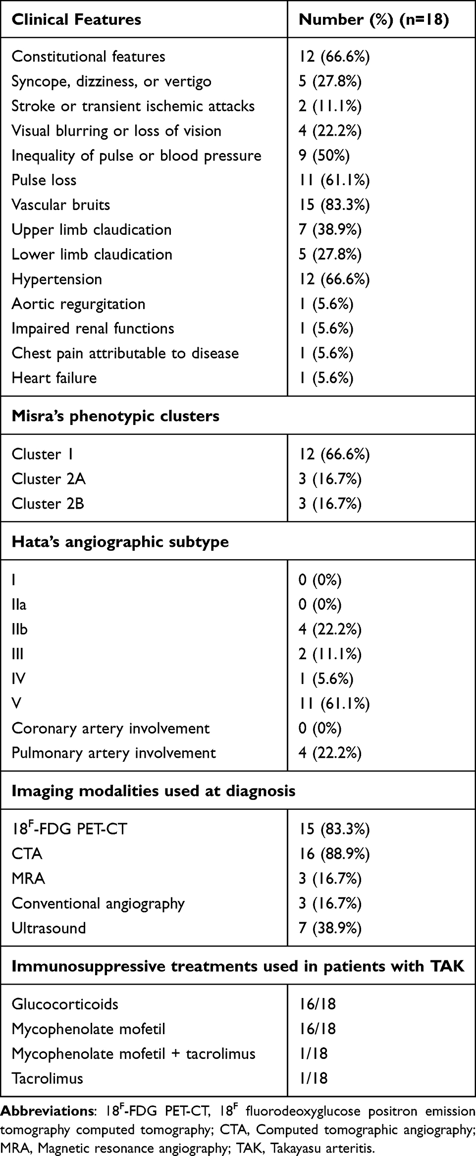 |
Table 1 Characteristics of the Cohort |
All the recruited patients with TAK had active disease as per the physician global assessment and were not on glucocorticoids; fifteen had never been treated with glucocorticoids. None of the patients were on disease-modifying antirheumatic drugs except for two patients who had relapsed while on tacrolimus. After recruitment, sixteen patients were initiated on glucocorticoids [median (IQR) daily prednisolone dose 25.0 (11.9–26.3) mg] and mycophenolate mofetil (2 gram daily in 14 patients, 1.5 gram daily in two patients) to treat active disease. For one patient, MMF 2 gram daily was added to tacrolimus 1.5 mg/day, and for the other, the dose of tacrolimus was increased from 1 mg to 3 mg daily. Follow-up blood samples were collected at a median of 5.1 (3.8–5.8) months after treatment with immunosuppressive therapy for active disease. At the time of collection of the follow-up sample, all the patients had attained inactive disease by the physician global assessment and had registered significant reductions in NIH disease activity score, ITAS2010, ITAS-A-ESR, ITAS-A-CRP, and DEI.TAK (Table 2). At the collection of follow-up samples, the median (IQR) daily prednisolone dose was 5.0 (4.7–7.5) mg.
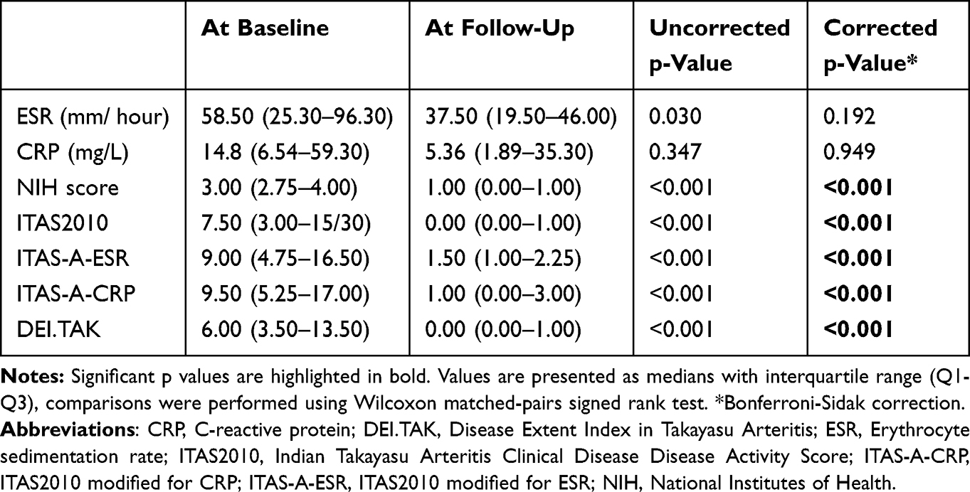 |
Table 2 Disease Activity Parameters for Patients with Takayasu Arteritis at Baseline and Follow-up Visits |
Flow Cytometry for Overall Monocyte/Macrophage Populations and Macrophage Sub-Populations
Representative flow-cytometry plots depicting the different populations in patients with active TAK (Figure 1), inactive TAK (following immunosuppressive therapy, Figure 2), and healthy controls (Figure 3) are depicted. Figure 4 depicts representative plots for the MFI of various antibodies related to monocyte/macrophage populations (CD14, TLR4, CD80, CD86, CD163, CD204, CD206) on the overall PBMCs for patients with active TAK, inactive TAK, and healthy controls. Supplementary Figure S1 provides the corresponding isotype control data for the MFI of the various antibodies.
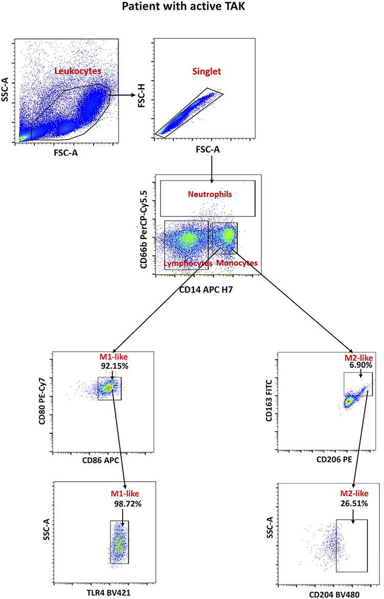 |
Figure 1 Representative Plots of flow cytometry for patients with active TAK (before immunosuppressive therapy. The leukocyte population was gated on forward scatter area (FSC-A) versus side scatter area (SSC-A). Singlet cells were selected by plotting FSC-A against FSC-H to eliminate doublets and aggregates, ensuring only individual cells were analyzed. Monocytes/macrophages were identified using CD66b versus CD14 plot. Neutrophils were gated as CD66b⁺, lymphocytes as CD14⁻/CD66b⁻, and monocytes/macrophages as CD14⁺/CD66b⁻. Within the monocyte/macrophage population, M1-like monocytes/macrophages were identified based on co-expression of CD86 and CD80. CD14+ CD80+ CD86+ M1-like monocyte/macrophage population were further gated for TLR4 positivity. Within the same monocyte/macrophage population, M2-like monocytes/macrophages were identified based on positivity for CD163 and CD206. CD14+ CD163+ CD206+ M2-like monocytes/macrophages were further gated for CD204 positivity. |
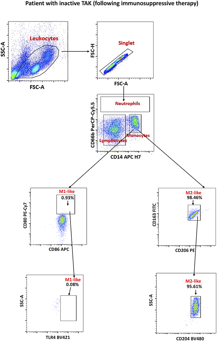 |
Figure 2 Representative plots of flow cytometry for patients with inactive TAK (after immunosuppressive therapy). |
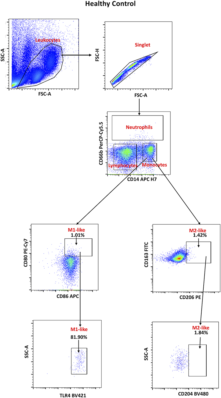 |
Figure 3 Representative plots of flow cytometry for healthy controls. |
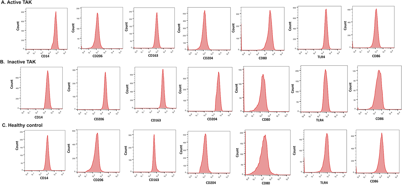 |
Figure 4 Representative histograms showing the mean fluorescence intensity (MFI) for surface expression of CD14 monocyte/macrophage marker, M1-like (CD80, CD86 & TLR4) and M2-like (CD163, CD204 & CD206) monocyte/macrophage markers for patients with active TAK (A), inactive TAK (B) and healthy controls (C). The x-axis indicates fluorescence intensity for each marker, while the y-axis shows cell counts. |
Comparison Between Patients with Active TAK and Healthy Controls
The overall population of circulating monocytes/ macrophages was significantly higher in patients with TAK than in healthy controls. Using two gating strategies (CD80+ CD86+ or CD14+ CD80+ CD86+), the proportions of M1-like monocytes/macrophages or M2-like monocytes/macrophages (CD163+ CD206+ or CD163+ CD206+ CD204+) were significantly greater in patients with active TAK than in healthy controls (Table 3 and Supplementary Table S2). Subgroup analyses including only the sixteen patients treated with glucocorticoids and mycophenolate revealed similar results (Supplementary Tables S3 and S4).
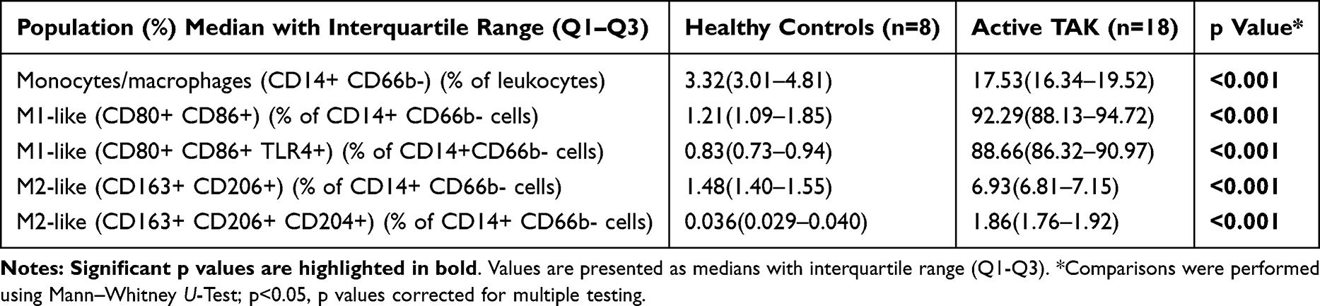 |
Table 3 Comparison of Overall Monocytes/Macrophage and Monocyte/Macrophage Sub-Populations Between Patients with TAK with Active Disease and Healthy Controls |
On the population of overall PBMCs, the MFI for CD14 was significantly higher for patients with TAK than for healthy controls. Analysis of the MFI of surface markers related to M1-like monocytes/macrophages revealed a higher intensity of CD80, TLR4, and CD86 expression in patients with active TAK than in healthy controls. For surface markers related to M2-like monocytes/macrophages, the intensity of CD163, CD206, and CD204 were also higher in patients with active TAK than healthy controls (Figure 5 and Supplementary Table S5). Subgroup analyses with the patients treated with glucocorticoids and mycophenolate (n=16) revealed similar findings (Supplementary Figure S2, and Supplementary Table S6).
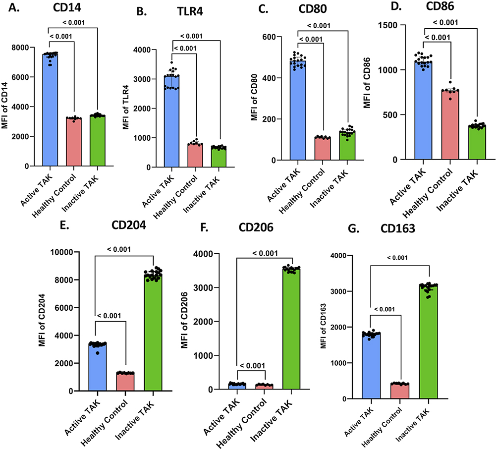 |
Figure 5 Mean Fluorescence Intensity (MFI) of monocyte/macrophage marker CD14, M1-like and M2-like monocyte/macrophage markers on overall peripheral blood mononuclear cells. (A) CD14 (B) TLR4 (C) CD80 (D) CD86 (E) CD204 (F) CD206 (G) CD163. Delta MFI (MFI of the marker minus that of the corresponding isotype control) are plotted. Comparison were made between patients with TAK & healthy controls using Mann–Whitney U-Test and between patients with TAK before and after immunosuppressive therapy using Wilcoxon matched pairs signed rank test, p values were corrected for multiple testing using the Bonferroni-Sidak method. |
Comparison Between Patients with Active TAK Before and After Immunosuppressive Treatment
Flow cytometry on peripheral blood was repeated after a median 5.1 months of receiving immunosuppressive therapy when these patients had attained inactive disease. In the follow-up samples, paired comparisons revealed a significant decrease in the overall population of circulating monocytes/macrophages and M1-like monocytes/macrophages (CD80+ CD86+ or CD14+ CD80+ CD86+) with a corresponding increase in M2-like monocytes/macrophages (CD163+ CD206+ or CD163+ CD206+ CD204+) (Table 4 and Supplementary Table S2). The same results were replicated with the absolute counts of overall and M1-like or M2-like monocytes/macrophages (Table 5 and Supplementary Tables S7). Analysis of the subgroup including only the sixteen patients treated with glucocorticoids and mycophenolate revealed similar results with proportions (Supplementary Tables S4 and S8) or absolute counts of monocytes/macrophages (Supplementary Tables S9 and S10.
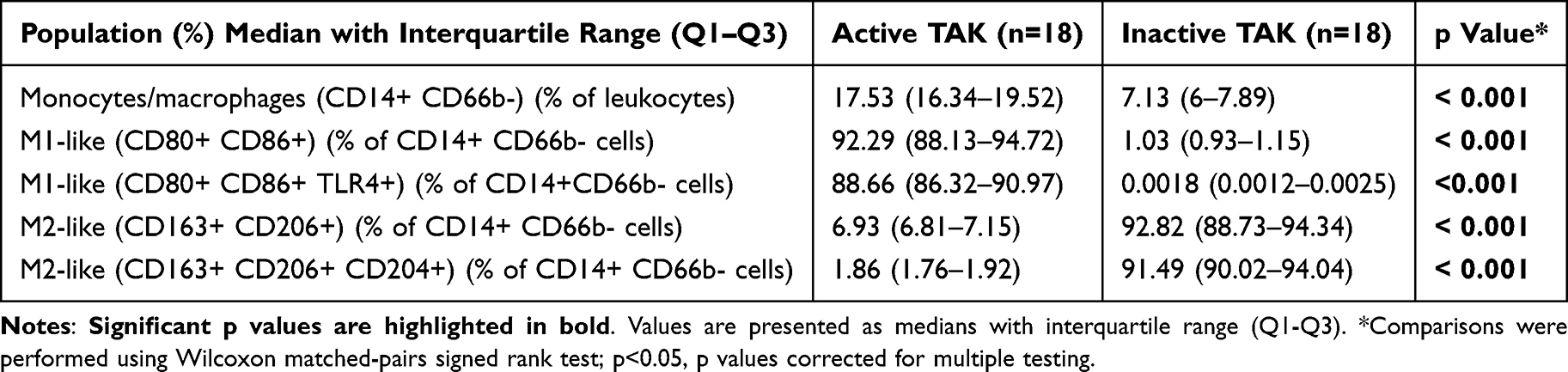 |
Table 4 Comparison of Overall Monocytes/Macrophage and Monocyte/Macrophage Sub-Populations in Patients with TAK Before (Active TAK) and After Immunosuppressive Therapy (Inactive TAK) |
 |
Table 5 Comparison of Absolute Counts of Overall Monocytes/Macrophage and Monocyte/Macrophage Sub-Populations in Patients with TAK Before (Active TAK) and After Immunosuppressive Therapy (Inactive TAK) |
On the population of overall PBMCs, the MFI for CD14 expression was significantly higher for patients with TAK at the time of active disease than at the follow-up sample when the disease was inactive. Analysis of the MFI of antibodies related to M1-like monocytes/macrophages revealed a reduction in the intensity of TLR4, CD80, and CD86 expression following immunosuppressive therapy. Conversely, the intensity of expression of antibodies related to M2-like monocytes/macrophages, viz., CD163, CD204, and CD206, was significantly increased following immunosuppressive therapy (Figure 5 and Supplementary Table S5). Similar results were obtained for the subgroup of 16 patients treated with glucocorticoids and mycophenolate (Supplementary Figure S2 and Supplementary Table S6).
Circulating Cytokines Related to Monocyte/Macrophage Sub-Populations
For cytokines related to M1-like monocytes/macrophages, higher serum TNF-⍺, IL-6, and IL-23 in patients with active TAK but similar IL-1β, IL-12p40, and IP-10 were observed when compared with healthy controls. For cytokines related to M2-like monocytes/macrophages, patients with active TAK had higher serum IL1RA but similar IL-10 and lower CCL17 than healthy controls (Figure 6 and Supplementary Table S11). Identical results were obtained with the subgroup of sixteen patients treated with glucocorticoids and mycophenolate both for M1-like cytokines and M2-like cytokines (Supplementary Figure S3 and Supplementary Table S12). Upon paired analyses following immunosuppressive therapy in patients with TAK, a significant decrease in M1-like cytokines TNF-⍺, IL-1β, IL-6, IP-10, IL-12p40, and IL-23 were observed. Regarding M2-like cytokines, a significant increase in IL-10 and CCL17 but similar levels of IL-1RA were observed (Figure 6 and Supplementary Table S11). For the subgroup of sixteen patients treated with glucocorticoids and mycophenolate, a significant decrease in M1-like cytokines TNF-⍺, IL-1β, IL-6, IL-12p40, and IL-23 but similar levels of IP-10 were observed. Identical results were observed with M2-like cytokines to the previous analysis (Supplementary Figure S3 and Supplementary Table S12).
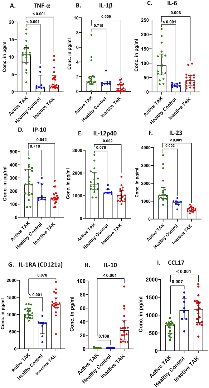 |
Figure 6 Serum levels of M1-like cytokines, TNF-α (A), IL-1β (B), IL-6 (C), IP-10 (D), IL-12p40 (E), IL-23 (F) and M2-like cytokines, IL-1RA (G) and IL-10 (H), and CCL17 (I). Comparison were made between patients with active TAK & healthy controls using Mann–Whitney U-Test and between patients with TAK before (active disease) and after immunosuppressive therapy (inactive disease) using Wilcoxon matched pairs signed rank test, p values were corrected for multiple testing using the Bonferroni-Sidak method. |
Circulating Markers of Fibrosis
Higher circulating levels of HA and PIIINP but similar levels of TIMP-1 and TGF-β were observed in patients with active TAK than in healthy controls. Following immunosuppressive therapy, a significant increase in all the circulating markers of fibrosis (HA, TIMP-1, PIIINP, TGF-β) was observed (Figure 7 and Supplementary Table S13). The same results were replicated in the subgroup of 16 patients treated with glucocorticoids and mycophenolate (Supplementary Figure S4 and Supplementary Table S14).
 |
Figure 7 Plasma levels of circulating markers of fibrosis, individual components of the Enhanced Liver Fibrosis score (Hyaluronic Acid (HA) (A), tissue inhibitor of metalloproteinases-1 (TIMP-1) (B), procollagen type III N-terminal propeptide (PIIINP) (C), and serum levels of TGF-β (D). Comparison were made between patients with active TAK & healthy controls using Mann–Whitney U-Test and between patients with TAK before (active disease) and after immunosuppressive therapy (inactive disease) using Wilcoxon matched pairs signed rank test, p values were corrected for multiple testing using the Bonferroni-Sidak method. |
Discussion
Patients with active TAK had a higher frequency of overall circulating as well as M1-like and M2-like monocyte/macrophage populations than healthy controls. The intensity of expression of CD14 (reflecting the overall circulating monocyte/macrophage population), M1-like markers TLR4, CD80, and CD86, and M2-like markers CD163 and CD204 on PBMCs was higher in patients with active TAK than in control subjects. Cytokines reflecting M1-like activation (TNF-⍺, IL-6, and IL-23), M2-like activation (IL1RA), and circulating markers of fibrosis HA, PIIINP, and TGF-β were higher in patients with active TAK than healthy controls. Paired comparisons before and after immunosuppressive therapy in patients with active TAK revealed a significant reduction in the frequency of overall circulating monocytes/macrophages and M1-like monocytes/macrophages with a concomitant increase in the frequency of M2-like monocytes/macrophages. Similar findings were observed with the intensity of expression of CD14 and markers of M1-like monocytes/macrophages showing a reduction along with an increase in the intensity of markers of M2-like monocytes/macrophages on the PBMCs following immunosuppressive therapy and attainment of inactive disease. Consistent changes in circulating cytokines were observed. M1-like cytokines TNF-⍺, IL-1β, IL-6, IL-12p40, and IL-23 showed a reduction, whereas M2-like cytokines IL-10 and CCL7 were elevated following immunosuppressive therapy. Circulating markers of fibrosis, viz., HA, TIMP-1, PIIINP, and TGF-β, showed a significant increase on the follow-up samples consistent with a pro-fibrotic phenotype also reflected by the elevation of M2- monocytes/macrophages and related cytokines despite the control of disease activity in patients with TAK. These observations favour our hypothesis of the evolution of a pro-fibrotic milieu in patients with TAK following immunosuppressive therapy once they attain inactive disease. To the best of our knowledge, an increased overall circulating monocyte/macrophage population in patients with active TAK when compared with healthy controls has not been reported before. Also, our observations about the evolution of a profibrotic phenotype in the peripheral blood following immunosuppressive treatment in patients with active TAK, reflected by proportions and absolute numbers of circulating M2-like monocyte/macrophage populations, cytokines reflecting M2-like polarization, and circulating markers of fibrosis have not been reported earlier.
Only one previous paper had evaluated overall circulating monocyte/macrophage populations in patients with TAK. In a cross-sectional study, de Aguiar et al reported a similar number of circulating monocytes/macrophages in 32 patients with TAK and 30 healthy controls. However, only 8/32 of their patients with TAK had active disease.31 Based on this study, the present study was powered to detect a difference in circulating monocyte/macrophage populations between patients with active TAK and healthy controls. This likely explains why the present study could identify a greater frequency of overall circulating monocytes/macrophages in patients with active TAK than in control subjects. We have assessed longitudinal changes following treatment in circulating monocyte/macrophage populations, their related cytokines, and circulating markers of fibrosis which have not been reported before. Our findings on the peripheral blood tie in with previous observations of macrophage infiltration in the arterial lesions of patients with TAK based on histopathology and single-cell RNA sequencing.17,18,32
Kong et al reported a dominance of M1-like macrophages and immunostaining for the macrophage chemokine CCL2 at the adventitia-media interface in the arterial tissue of patients with TAK who were not on immunosuppressive therapy. Conversely, the arteries of those patients with TAK on immunosuppressive therapy showed a dominance of M2-like macrophages and immunostaining for CCL2 at the interface of the endothelium and medial layers.18 Based on these findings, we hypothesized that treatment with immunosuppressive agents and the attainment of inactive disease might result in a pro-fibrotic milieu reflected by circulating M2-like macrophages, their cytokines, and circulating markers of fibrosis. Indeed, we observed an increase in the frequency of M2-like macrophages, the intensity of cell surface markers reflecting M2-like polarization, an increase in cytokines IL-10 and CCL7 associated with M2-like polarization, and an increase in fibrotic markers following immunosuppressive therapy and the attainment of inactive disease in patients with TAK. Notably, such changes were observed a short while after the initiation of immunosuppressive therapy (median 5.1 months). The short duration of follow-up between these serial observations suggests that this could indicate an active reparative phase of the healing of arterial wall injury. Such a pro-fibrotic milieu, if unchecked, might result in permanent arterial scarring. Stojanovic et al have reported an association between the plasma ELF score and its individual components HA and PIIINP with clinically assessed damage using the Vasculitis Damage Index and angiographic damage using the Combined Arteritis Damage Score in a cross-sectional study.44 Serial evolution of the components of this score in patients with TAK following treatment with immunosuppressive therapy have not been reported earlier. The individual components of the ELF score (HA, TIMP1, and PIIINP) are validated as markers of vascular damage in different settings.50–53 Since the algorithm to calculate the ELF score is proprietary, we assessed the individual components of the ELF score, viz., HA, TIMP1, and PIIINP, and the pro-fibrotic cytokine TGF-β in healthy controls and serially in patients with TAK. Even during active disease, circulating HA, PIIINP, and TGF-β were elevated when compared with control subjects. Further, a consistent increase in all four circulating markers of fibrosis was observed following immunosuppressive therapy and the attainment of inactive disease. Coupled with the dominance of M2-like polarization in patients with TAK at this time point, these findings lend credence to the hypothesis that the resolution of the inflammatory milieu in patients with TAK leads on to the evolution of a pro-fibrotic milieu.7,25 Observations in patients with TAK suggest a progression of existing arterial stenosis in some patients despite the initiation of immunosuppressive therapy and the control of disease activity.54,55 Similarly, a significant proportion of patients with TAK show progression of clinically assessed damage scores on follow-up. Such progression has been associated with active disease at presentation.56 This reinforces the need to explore strategies combining immunosuppressive therapy with antifibrotic agents up front in patients with TAK with active disease instead of the prevalent strategy of using immunosuppressive therapy alone in such patients.7,25 Our findings on peripheral blood correspond to previously reported changes in infiltrating macrophage populations in the arterial tissue of patients with TAK reported by Kong et al18 Arterial tissue from patients with TAK is inaccessible for histopathologic analysis except during open vascular surgeries, which are infrequently performed to deal with vascular complications.2,16 Our study suggests that the relative frequencies of circulating macrophage sub-populations might be a surrogate reflection of inflammatory or fibrotic processes occurring in the arterial wall. This will require confirmation in the peripheral blood of patients with TAK collected at the time of arterial surgery in future studies.
Prior data on M2-like monocytes/macrophages and fibrosis in the context of inflammatory rheumatic diseases are largely derived from systemic sclerosis (a disease characterized by widespread systemic fibrosis, resulting in end-organ damage) and systemic lupus erythematosus (where renal fibrosis arising from glomerular injury drives worsening renal function and progress to chronic kidney disease, requiring renal replacement therapy).57,58 A lowered expression of interferon regulatory factor 8 (IRF8) in circulating monocytes/macrophages in the peripheral blood of patients with systemic sclerosis is associated with polarization towards CD68+ CD163+ M2-like monocytes/macrophages and increased secretion of cytokines such as TGF-β and interleukin-6, which have been implicated as drivers of fibrosis.20,59 Monocyte-derived macrophages from patients with systemic sclerosis cultured in vitro secrete periostin, which might increase collagen synthesis from fibroblasts to drive fibrosis in tissues.20,60 Bleomycin-induced lung fibrosis is an animal model of systemic sclerosis, where the secretion of Wnt7a from CD206+ macrophages is recognized to increase collagen synthesis from fibroblasts.20,61 An increased circulating population of CD163+ CD204+ monocytes/macrophages and elevated soluble CD163 have been observed in systemic sclerosis when compared with healthy controls.20,62,63 Levels of soluble CD163 have been associated with pulmonary hypertension in systemic sclerosis, a process which is driven in part by lung fibrosis.20,64 Macrophages expressing CD163 and CD204 are increased in skin biopsies from patients with systemic sclerosis, and they express genes associated with fibrosis, such as CX3CR1 and interleukin-10 receptor.20,62,65 Monocytes/macrophages expressing CD163 in the peripheral blood are associated with interstitial lung disease in patients with systemic sclerosis. In vitro, CD163-expressing monocytes/macrophages secrete proteins mediating fibrosis, such as CCL18 and IL-10.20,66 Therapies used in patients with systemic sclerosis also impact M2-like populations of monocytes/macrophages. The endothelin-1 receptor antagonist bosentan (which has vasodilatory actions and therefore is useful in the vasculopathy associated with systemic sclerosis), when treated with monocyte-derived macrophages in vitro, reduces the expression of M2-like markers CD163, CD204, and CD206.20,57,67 Nintedanib is another tyrosine kinase inhibitor that has been explored in systemic sclerosis.57 When circulating monocytes from patients with systemic sclerosis were differentiated in vitro to macrophages and treated with nintedanib, a reduction in the expression of M2-like markers CD163, CD204, and CD206 was observed.20,68 Tocilizumab is an inhibitor of interleukin-6 used in patients with systemic sclerosis as well as TAK.3,57 Patients with systemic sclerosis treated with tocilizumab show a reduction in circulating monocytes/macrophages expressing CD38.20,69 Janus kinase inhibitors such as tofacitinib are also being explored in systemic sclerosis and TAK. Janus kinase inhibitors reduce the differentiation of macrophages from circulating monocyte-derived macrophages derived from healthy controls.20,70 In the context of systemic lupus erythematosus, renal injury results in fibrosis and a greater risk of end-stage renal disease.58 Renal biopsies from patients with lupus show increased CD68+ macrophages with a M2-like phenotype depicted by CD163 positivity. Such macrophage populations activate complement 3a receptor, which in turn drives renal fibrosis, leading to a loss of renal function in systemic lupus erythematosus.20,71 M2-like macrophages also express arginase-1, which drives the conversion of L-arginine to L-ornithine. L-ornithine is involved in the synthesis of proline and hydroxyproline, both of which are required for collagen synthesis.72 M2-like macrophages show greater glutamine metabolism and fatty acid oxidation (as opposed to glycolysis in M1-like macrophages), which might drive their pro-fibrotic phenotype.73
IL-12p40 and IL-23, cytokines associated with M1-like polarization which also play a role in Th1 differentiation and Th17 lineage maintenance, showed a decrease following immunosuppressive therapy in patients with TAK. IL-12p40 and IL-23 also play key roles in the differentiation of Th1 lymphocytes and the maintenance ofTh1 and Th17 lymphocytes, whose role has been implicated as drivers of arterial inflammation in TAK from multiple studies.24–26,74–76 The dampening of these cytokines in peripheral blood collected during inactive disease likely reflects their role in driving Th1 and Th17 lymphocytes during active disease.
It is now well recognized that M1-like and M2-like monocytes/macrophages exhibit a phenotypic and functional continuum.19,20 Prior studies have assessed mixed M1/M2-like populations of circulating monocytes/macrophages expressing surface markers of both M1-like and M2-like populations.19 However, we have not reported these populations as they were too sparse in our patients with TAK to permit meaningful analysis.
It begs the question whether the increase in M2-like monocytes/macrophages observed in patients with TAK following the initiation of immunosuppressive therapy is amenable to therapeutic targeting? In a series of elegant experiments, Cui et al demonstrated that the treatment of in vitro cultured monocytes from patients with TAK with leflunomide reduced their polarization towards M2-like macrophages and induced apoptosis of existing M2-like macrophages.77 Similarly, Xiaojuan et al showed that GPNMB was linked with macrophage-fibroblast interactions in the arterial wall, leading to arterial fibrosis in patients with TAK. In vitro treatment with leflunomide reduced the secretion of GPNMB secretion (linked with macrophage-fibroblast interactions in the arterial wall) from cultured monocyte-derived macrophages from patients with TAK or macrophages derived in vitro from the THP-1 monocyte cell line.78 Mammalian target of rapamycin (mTOR) activation drives polarization to M2-like macrophages and is amenable to modulation by rapamycin(sirolimus).79 mTOR complex 1 (mTOR C1) is also implicated in endothelial cell activation and Th1/Th17 polarization in patients with TAK.80 Both T lymphocytes and macrophages are implicated in the arterial wall fibrosis of TAK.7 A serpin protein derived from myxomavirus, Serp-1, has demonstrated effective inhibition of T lymphocytes and macrophages in an arterial implant model derived from patients with Giant Cell Arteritis (GCA), another variant of LVV.81 Strategies such as CAR-T cells targeted towards platelet-derived growth factor receptor beta (PDGFR-β) have recently shown promise to reverse existing fibrosis in models of kidney injury.82
Recent studies have shown promise with PET using [11C]-PK11195 or SST2 to denote in vivo macrophage activation in LVV, including TAK.35,36 How changes in circulating M1-like and M2-like monocyte/macrophage populations or cytokines are associated with findings on [11C]-PK11195 or SST2 is a research agenda for future exploration. It is also reasonable to evaluate whether circulating populations of M2-like monocytes/macrophages or their related cytokines and circulating markers of fibrosis correlate with in vivo fibroblast activity in the arterial wall of patients with TAK, demonstrable using fibroblast activating protein inhibitor (FAPI) PET.83
There were limitations to our study. The use of a single platform method such as a bead-based assay to evaluate serial changes in circulating overall or M1-like/M2-like monocytes/macrophages was an alternative strategy to the dual-platform method used in our study. The morphological distinction between circulating monocytes and macrophages would have been possible through techniques such as imaging flow cytometry. However, this was beyond the scope of the present study. For this reason, we have labelled the observed cell populations as monocytes/macrophages. The concomitant assessment of monocyte/macrophage populations in the arterial tissue would have added value to the study. However, arterial tissue can only be obtained during major vascular surgery which are rarely performed in patients with TAK, and usually not during periods of active disease.84 This aspect merits exploration in future studies. Conducting in vitro experiments on cultured aortic adventitial fibroblasts and macrophages would help to further dissect the association between M2-like monocytes/macrophages and arterial wall fibrosis, and its therapeutic modulation in patients with TAK. While we have assessed serum cytokines related to monocyte/macrophage polarization, the inherent variability in serum cytokine measurement is a limitation. While the absolute number of patients with TAK was small, the study was powered to detect differences between patients with TAK and healthy controls. Differences in circulating monocyte/macrophage populations based on specific disease features of TAK would require a much larger sample size and multicentric sample collection, which were beyond the remit of this study. A longitudinal in-depth immunophenotyping through cell populations and multiple circulating proteins indicating monocyte/macrophage populations and fibrotic proteins revealed consistent results, indicating the emergence of a pro-fibrotic milieu after immunosuppressive therapy was initiated in patients with active TAK. To the best of our knowledge, this is the first report of M1-like and M2-like monocyte/macrophage populations and cytokines in the peripheral blood of patients with TAK.
Conclusion
To conclude, this paper reports for the first time an elevated overall monocyte/macrophage population and M1-like or M2-like sub-populations, and their related cytokines and circulating fibrotic proteins in patients with TAK than healthy controls. Following the initiation of immunosuppressive therapy, the circulating monocyte/macrophage populations and cytokines showed a transition to a dominant M2-like phenotype and a profibrotic milieu indicated by elevated levels of fibrotic proteins which could indicate a reparative phase that if goes unchecked, might result in permanent arterial scarring. Targeting M2-like monocytes/macrophages and fibrosis in patients with TAK after they attain inactive disease merits exploration in pre-clinical models and eventually in clinical trials.
Data Sharing Statement
All the data analyzed for this study has been reported in the main text or Supplementary Files. Data pertaining to the article will be made available on reasonable request to the corresponding author (Durga Prasanna Misra, [email protected]).
Acknowledgments
The abstract of this paper was presented as a poster with interim findings at the Indian Rheumatology Association Conference November 2024 and October 2025. The poster’s abstract was published in Indian J Rheumatol 2024; 19(4S):S211-212.
Author Contributions
Tooba Qamar: Conceptualization, Data curation, Formal Analysis, Funding acquisition, Investigation, Methodology, Resources, Software, Validation, Visualization, Writing – original draft. Deeksha Singh: Data curation, Formal Analysis, Investigation, Writing – review & editing. Ranjeet Singh Chauhan: Data curation, Formal Analysis, Investigation, Writing – review & editing. Sara Abid: Data curation, Formal Analysis, Investigation, Writing – review & editing. Upendra Rathore: Conceptualization, Data curation, Formal Analysis, Investigation, Supervision, Writing – review & editing. Able Lawrence: Data curation, Formal Analysis, Investigation, Writing – review & editing. Manas Ranjan Behera: Data curation, Formal Analysis, Investigation, Writing – review & editing. Neeraj Jain: Data curation, Formal Analysis, Investigation, Writing – review & editing. Manish Ora: Data curation, Formal Analysis, Investigation, Writing – review & editing. Durga Prasanna Misra: Conceptualization, Data curation, Formal Analysis, Funding acquisition, Investigation, Methodology, Project administration, Resources, Software, Supervision, Validation, Visualization, Writing – original draft. All authors took part in drafting, revising or critically reviewing the article; gave final approval of the version to be published; have agreed on the journal to which the article has been submitted; and agree to be accountable for all aspects of the work.
Funding
This study was funded by the Department of Science & Technology (DST)-Innovation in Science Pursuit for Inspired Research (INSPIRE) New Delhi, India (Grant ID IF210497). Tooba Qamar acknowledges the support of Department of Science & Technology (DST)-Innovation in Science Pursuit for Inspired Research (INSPIRE) New Delhi, India (Grant ID IF210497) for research fellowship support. Ranjeet Singh Chauhan acknowledges the support of Anusandhan National Research Foundation (ANRF) (Grant ID: CRG/2023/000769) for research fellowship support. Deeksha Singh acknowledges the support of Uttar Pradesh Council of Science & Technology (Grant ID: CST/D-2823) for research fellowship support. The sponsors of the research had no role in preparing this article for submission.
Disclosure
Ms Deeksha Singh reports grants, personal fees, non-financial support from Uttar Pradesh Council of Science & Technology (Grant ID: CST/D-2823), during the conduct of the study; Mr Ranjeet Chauhan reports grants, personal fees, non-financial support from Anusandhan National Research Foundation (ANRF) (Grant ID: CRG/2023/000769), during the conduct of the study; Ms Sara Abid reports grants, personal fees, non-financial support from Council of Scientific and Industrial Research (CSIR)(File No. 09/0590(19217)/2024-EMR-I, during the conduct of the study; The authors have no other conflicts of interest to declare.
References
1. Pugh D, Karabayas M, Basu N. et al. Large-vessel vasculitis. Nat Rev Dis Primers. 2022;7(1):93. doi:10.1038/s41572-021-00327-5
2. Misra DP, Singh K, Rathore U, et al. Management of Takayasu Arteritis. Best Pract Res Clin Rheumatol. 2023;37(1):101826. doi:10.1016/j.berh.2023.101826
3. Sawalha AH, Misra DP, Goel R, et al. Advances in the Pathophysiology, Diagnosis and Treatment of Takayasu Arteritis. Nat Rev Rheumatol. 2025;21:737–23. doi:10.1038/s41584-025-01309-7. Epub 2025/11/07
4. Watanabe R, Berry GJ, Liang DH, Goronzy JJ, Weyand CM. Pathogenesis of Giant Cell Arteritis and Takayasu Arteritis-Similarities and Differences. Curr Rheumatol Rep. 2020;22(10):68. doi:10.1007/s11926-020-00948-x. Epub 2020/08/28.
5. Jagtap S, Mishra P, Rathore U, et al. Increased Mortality Rate in Takayasu Arteritis Is Largely Driven by Cardiovascular Disease - a Cohort Study. Rheumatology. 2024;63(12):3337–3345. doi:10.1093/rheumatology/kead584
6. Arnaud L, Haroche J, Mathian A, Gorochov G, Amoura Z. Pathogenesis of Takayasu’s Arteritis: a 2011 Update. Autoimmunity Rev. 2011;11(1):61–67. doi:10.1016/j.autrev.2011.08.001. Epub 2011/08/23.
7. Misra DP, Singh K, Sharma A, Agarwal V. Arterial Wall Fibrosis in Takayasu Arteritis and Its Potential for Therapeutic Modulation. Front Immunol. 2023;14:1174249. doi:10.3389/fimmu.2023.1174249
8. Misra DP, Rathore U, Patro P, Agarwal V, Sharma A. Corticosteroid Monotherapy for the Management of Takayasu Arteritis-a Systematic Review and Meta-Analysis. Rheumatol Int. 2021;41(10):1729–1742. doi:10.1007/s00296-021-04958-5. Epub 2021/07/25.
9. Misra DP, Rathore U, Patro P, Agarwal V, Sharma A. Disease-Modifying Anti-Rheumatic Drugs for the Management of Takayasu Arteritis—a Systematic Review and Meta-Analysis. Clin Rheumatol. 2021;40(11):4391–4416. doi:10.1007/s10067-021-05743-2
10. Misra DP, Singh K, Rathore U, et al. The Effectiveness of Tocilizumab and Its Comparison with Tumor Necrosis Factor Alpha Inhibitors for Takayasu Arteritis: a Systematic Review and Meta-Analysis. Autoimmun Rev. 2023;22(3):103275. doi:10.1016/j.autrev.2023.103275. Epub 2023/01/19.
11. Misra DP, Mukhtyar CB, Chandwar K, Putman M, Walsh M. The Fragility of Randomized Controlled Trials in Large Vessel Vasculitis. Autoimmun Rev. 2025;24(12):103917. doi:10.1016/j.autrev.2025.103917. Epub 2025/08/28.
12. Lagneau P, Michel JB, Vuong PN. Surgical Treatment of Takayasu’s Disease. Ann Surg. 1987;205(2):157–166. doi:10.1097/00000658-198702000-00010. Epub 1987/02/01.
13. Hoffman GS. Takayasu Arteritis: lessons from the American National Institutes of Health Experience. Int J Cardiol. 1996;54:S99–102. Epub 1997/08/01.
14. Incerti E, Tombetti E, Fallanca F, et al. 18f-Fdg Pet Reveals Unique Features of Large Vessel Inflammation in Patients with Takayasu’s Arteritis. Eur J Nucl Med Mol Imaging. 2017;44(7):1109–1118. doi:10.1007/s00259-017-3639-y. Epub 2017/02/10.
15. Misra DP, Misra R. Assessment of Disease Activity in Takayasu’s Arteritis. Indian J Rheumatol. 2015;10(5):S43–S47. doi:10.1016/j.injr.2015.08.006. Epub 19/09/2015.
16. Misra DP, Jain N, Ora M, Singh K, Agarwal V, Sharma A. Outcome Measures and Biomarkers for Disease Assessment in Takayasu Arteritis. Diagnostics. 2022;12(10):2565. doi:10.3390/diagnostics12102565. Epub 2022/10/28.
17. Dos Santos JP, Artigiani Neto R, Mangueira CLP, et al. Associations between Clinical Features and Therapy with Macrophage Subpopulations and T Cells in Inflammatory Lesions in the Aorta from Patients with Takayasu Arteritis. Clin Exp Immunol. 2020;202(3):384–393. doi:10.1111/cei.13489. Epub 2020/07/09.
18. Kong X, Xu M, Cui X, et al. Potential Role of Macrophage Phenotypes and Ccl2 in the Pathogenesis of Takayasu Arteritis. Front Immunol. 2021;12:646516. doi:10.3389/fimmu.2021.646516. Epub 2021/06/04
19. Trombetta AC, Soldano S, Contini P, et al. A Circulating Cell Population Showing Both M1 and M2 Monocyte/Macrophage Surface Markers Characterizes Systemic Sclerosis Patients with Lung Involvement. Respir Res. 2018;19(1):186. doi:10.1186/s12931-018-0891-z. Epub 2018/09/27.
20. Cutolo M, Soldano S, Smith V, Gotelli E, Hysa E. Dynamic Macrophage Phenotypes in Autoimmune and Inflammatory Rheumatic Diseases. Nat Rev Rheumatol. 2025;21(9):546–565. doi:10.1038/s41584-025-01279-w
21. Nakajima T, Yoshifuji H, Shimizu M, et al. A Novel Susceptibility Locus in the Il12b Region Is Associated with the Pathophysiology of Takayasu Arteritis through Il-12p40 and Il-12p70 Production. Arthritis Res Ther. 2017;19(1):197. doi:10.1186/s13075-017-1408-8. Epub 2017/09/07.
22. Ortiz-Fernández L, Saruhan-Direskeneli G, Alibaz-Oner F, et al. Identification of Susceptibility Loci for Takayasu Arteritis through a Large Multi-Ancestral Genome-Wide Association Study. Am J Hum Genet. 2021;108(1):84–99. doi:10.1016/j.ajhg.2020.11.014. Epub 2020/12/15.
23. Misra DP. Ustekinumab Merits Further Exploration in Takayasu Arteritis Despite a Failed Randomized Controlled Trial. Rheumatol Adv Pract. 2025;9(2):rkaf038. doi:10.1093/rap/rkaf038. Epub 2025/04/17.
24. Misra DP, Chaurasia S, Misra R. Increased Circulating Th17 Cells, Serum Il-17a, and Il-23 in Takayasu Arteritis. Autoimmune Dis. 2016;2016:7841718. doi:10.1155/2016/7841718
25. Singh K, Rathore U, Rai MK, et al. Novel Th17 Lymphocyte Populations, Th17.1 and Pd1+Th17, Are Increased in Takayasu Arteritis, and Both Th17 and Th17.1 Sub-Populations Associate with Active Disease. J Inflamm Res. 2022;15:1521–1541. doi:10.2147/JIR.S355881
26. Saadoun D, Garrido M, Comarmond C, et al. Th1 and Th17 Cytokines Drive Inflammation in Takayasu Arteritis. Arthritis Rheumatol. 2015;67(5):1353–1360. doi:10.1002/art.39037. Epub 2015/01/22.
27. Kong X, Sawalha AH. Takayasu Arteritis Risk Locus in Il6 Represses the Anti-Inflammatory Gene Gpnmb through Chromatin Looping and Recruiting Mef2-Hdac Complex. Ann Rheumatic Dis. 2019;78(10):1388–1397. doi:10.1136/annrheumdis-2019-215567. Epub 2019/07/19.
28. Tamura N, Maejima Y, Matsumura T, et al. Single-Nucleotide Polymorphism of the Mlx Gene Is Associated with Takayasu Arteritis. Circ Genomic Precis Med. 2018;11(10):e002296. doi:10.1161/circgen.118.002296. Epub 2018/10/26.
29. Stankey CT, Bourges C, Haag LM, et al. A Disease-Associated Gene Desert Directs Macrophage Inflammation through Ets2. Nature. 2024;630(8016):447–456. doi:10.1038/s41586-024-07501-1. Epub 2024/06/06.
30. Watanabe R, Hashimoto M. Pathogenic Role of Monocytes/Macrophages in Large Vessel Vasculitis. Front Immunol. 2022;13:859502. doi:10.3389/fimmu.2022.859502. Epub 2022/08/16.
31. de Aguiar MF, Torquato H, Salu BR, et al. Monocyte Subsets and Monocyte-Related Chemokines in Takayasu Arteritis. Sci Rep. 2023;13(1):2092. doi:10.1038/s41598-023-29369-3. Epub 2023/02/07.
32. Qing G, Zhiyuan W, Jinge Y, et al. Single-Cell Rna Sequencing Revealed Cd14(+) Monocytes Increased in Patients with Takayasu’s Arteritis Requiring Surgical Management. Front Cell Dev Biol. 2021;9:761300. doi:10.3389/fcell.2021.761300. Epub 2021/10/22
33. Gao Q, Gao S, Li H, et al. Multi-Omics Exploration of the Role of Ptgs2 as a Hub Gene in Ferroptosis within the Artery of Takayasu Arteritis. J Inflamm Res. 2024;17:9135–9146. doi:10.2147/jir.S478413. Epub 2024/11/26
34. Gao N, Tang H, Li T, et al. Single-Cell Transcriptome Analysis Reveals Cellular Heterogeneity in the Aortas of Takayasu Arteritis. Arthritis Res Ther. 2025;27(1):55. doi:10.1186/s13075-025-03523-w. Epub 2025/03/11.
35. Pugliese F, Gaemperli O, Kinderlerer AR, et al. Imaging of Vascular Inflammation with [11c]-Pk11195 and Positron Emission Tomography/Computed Tomography Angiography. J Am Coll Cardiol. 2010;56(8):653–661. doi:10.1016/j.jacc.2010.02.063. Epub 2010/08/14.
36. Ćorović A, Wall C, Nus M, et al. Somatostatin Receptor Pet/Mr Imaging of Inflammation in Patients with Large Vessel Vasculitis and Atherosclerosis. J Am College Cardiol. 2023;81(4):336–354. doi:10.1016/j.jacc.2022.10.034. Epub 2023/01/26.
37. Grayson PC, Ponte C, Suppiah R, et al. 2022 American College of Rheumatology/Eular Classification Criteria for Takayasu Arteritis. Ann Rheum Dis. 2022;81(12):1654–1660. doi:10.1136/ard-2022-223482. Epub 2022/11/10.
38. Kerr GS, Hallahan CW, Giordano J, et al. Takayasu Arteritis. Ann Intern Med. 1994;120(11):919–929. Epub 1994/06/01.
39. Misra R, Danda D, Rajappa SM, et al. Development and Initial Validation of the Indian Takayasu Clinical Activity Score (Itas2010). Rheumatology. 2013;52(10):1795–1801. doi:10.1093/rheumatology/ket128. Epub 2013/04/19.
40. Aydin SZ, Yilmaz N, Akar S, et al. Assessment of Disease Activity and Progression in Takayasu’s Arteritis with Disease Extent Index-Takayasu. Rheumatology. 2010;49(10):1889–1893. doi:10.1093/rheumatology/keq171. Epub 2010/06/15.
41. Hellmich B, Agueda A, Monti S, et al. 2018 Update of the Eular Recommendations for the Management of Large Vessel Vasculitis. Ann Rheum Dis. 2020;79(1):19–30. doi:10.1136/annrheumdis-2019-215672. Epub 2019/07/05.
42. Hultin LE, Chow M, Jamieson BD, et al. Comparison of Interlaboratory Variation in Absolute T-Cell Counts by Single-Platform and Optimized Dual-Platform Methods. Cytometry B Clin Cytom. 2010;78(3):194–200. doi:10.1002/cyto.b.20500. Epub 2009/10/09.
43. Glencross D, Scott L, Aggett H, Sonday S, Scott CS. Microvolume Fluorimetry for the Determination of Absolute Cd4 and Cd8 Lymphocyte Counts in Patients with Hiv: a Comparative Evaluation. Clin Lab Haematol. 1999;21(6):391–395. doi:10.1046/j.1365-2257.1999.00276.x. Epub 2000/02/15.
44. Stojanovic M, Raskovic S, Milivojevic V, et al. Enhanced Liver Fibrosis Score as a Biomarker for Vascular Damage Assessment in Patients with Takayasu Arteritis-a Pilot Study. J Cardiovasc Dev Dis. 2021;8(12). doi:10.3390/jcdd8120187. Epub 2021/12/24.
45. Parkes J, Roderick P, Harris S, et al. Enhanced Liver Fibrosis Test Can Predict Clinical Outcomes in Patients with Chronic Liver Disease. Gut. 2010;59(9):1245–1251. doi:10.1136/gut.2009.203166. Epub 2010/08/03.
46. Wan X, Wang W, Liu J, Tong T. Estimating the Sample Mean and Standard Deviation from the Sample Size, Median, Range and/or Interquartile Range. BMC Med Res Method. 2014;14(1):135. doi:10.1186/1471-2288-14-135
47. Available from: https://Sample-Size.Net/Ssmeans9.Php.
48. Avcu A, Jose A, Ganapathy S, et al. Phenotypic Clusters of Patients with Takayasu Arteritis Predict Disease Activity at Presentation, Damage and Future Vascular Complications. Rheumatology. 2026;65(1):keaf505. doi:10.1093/rheumatology/keaf505. Epub 2025/09/20.
49. Hata A, Noda M, Moriwaki R, Numano F. Angiographic Findings of Takayasu Arteritis: new Classification. Int J Cardiol. 1996;54:S155–63. Epub 1997/08/01.
50. Abignano G, Blagojevic J, Bissell LA, et al. European Multicentre Study Validates Enhanced Liver Fibrosis Test as Biomarker of Fibrosis in Systemic Sclerosis. Rheumatology. 2019;58(2):254–259. doi:10.1093/rheumatology/key271. Epub 2018/09/22.
51. Chen C, Wang L, Wu J, et al. Circulating Collagen Metabolites and the Enhanced Liver Fibrosis (Elf) Score as Fibrosis Markers in Systemic Sclerosis. Front Pharmacol. 2022;13:805708. doi:10.3389/fphar.2022.805708. Epub 2022/02/19
52. Lieb W, Song RJ, Xanthakis V, Vasan RS. Association of Circulating Tissue Inhibitor of Metalloproteinases-1 and Procollagen Type Iii Aminoterminal Peptide Levels with Incident Heart Failure and Chronic Kidney Disease. J Am Heart Assoc. 2019;8(7):e011426. doi:10.1161/jaha.118.011426. Epub 2019/03/21.
53. Wang Y, Yuan JM, Pan A, Koh WP. Tissue Inhibitor Matrix Metalloproteinase 1 and Risk of Type 2 Diabetes in a Chinese Population. BMJ Open Diabetes Res Care. 2020;8(1). doi:10.1136/bmjdrc-2019-001051. Epub 2020/04/19
54. Misra DP, Aggarwal A, Lawrence A, Agarwal V, Misra R. Pediatric-Onset Takayasu’s Arteritis: clinical Features and Short-Term Outcome. Rheumatol Int. 2015;35(10):1701–1706. doi:10.1007/s00296-015-3272-7
55. Direskeneli H, Aydin SZ, Merkel PA. Assessment of Disease Activity and Progression in Takayasu’s Arteritis. Clin Exp Rheumatol. 2011;29(1):S86–91. Epub 2011/08/04.
56. Misra DP, Rathore U, Qamar T, et al. High Prevalent Early Damage Independently Predicts Future Mortality in an Inception Cohort of Takayasu Arteritis. Rheumatology. 2026;65(1):keaf508. doi:10.1093/rheumatology/keaf508. Epub 2025/09/25.
57. Volkmann ER, Andréasson K, Smith V. Systemic Sclerosis. Lancet. 2023;401(10373):304–318. doi:10.1016/s0140-6736(22)01692-0. Epub 2022/11/29.
58. Kaul A, Gordon C, Crow MK, et al. Systemic Lupus Erythematosus. Nat Rev Dis Primers. 2016;2:16039. doi:10.1038/nrdp.2016.39. Epub 2016/06/17
59. Ototake Y, Yamaguchi Y, Asami M, et al. Downregulated Irf8 in Monocytes and Macrophages of Patients with Systemic Sclerosis May Aggravate the Fibrotic Phenotype. J Invest Dermatol. 2021;141(8):1954–1963. doi:10.1016/j.jid.2021.02.015. Epub 2021/03/12.
60. Suzuki M, Ototake Y, Akita A, et al. Periostin-an Inducer of Pro-Fibrotic Phenotype in Monocytes and Monocyte-Derived Macrophages in Systemic Sclerosis. PLoS One. 2023;18(8):e0281881. doi:10.1371/journal.pone.0281881. Epub 2023/08/02.
61. Hou J, Shi J, Chen L, et al. M2 Macrophages Promote Myofibroblast Differentiation of Lr-Mscs and Are Associated with Pulmonary Fibrogenesis. CCS. 2018;16(1):89. doi:10.1186/s12964-018-0300-8. Epub 2018/11/25.
62. Higashi-Kuwata N, Jinnin M, Makino T, et al. Characterization of Monocyte/Macrophage Subsets in the Skin and Peripheral Blood Derived from Patients with Systemic Sclerosis. Arthritis Res Ther. 2010;12(4):R128. doi:10.1186/ar3066. Epub 2010/07/07.
63. Frantz C, Pezet S, Avouac J, Allanore Y. Soluble Cd163 as a Potential Biomarker in Systemic Sclerosis. Dis. Markers. 2018;2018:8509583. doi:10.1155/2018/8509583. Epub 2018/05/29.
64. Nakayama W, Jinnin M, Makino K, et al. Serum Levels of Soluble Cd163 in Patients with Systemic Sclerosis. Rheumatol Int. 2012;32(2):403–407. doi:10.1007/s00296-010-1691-z. Epub 2010/12/02.
65. Mahoney JM, Taroni J, Martyanov V, et al. Systems Level Analysis of Systemic Sclerosis Shows a Network of Immune and Profibrotic Pathways Connected with Genetic Polymorphisms. PLoS Comput Biol. 2015;11(1):e1004005. doi:10.1371/journal.pcbi.1004005. Epub 2015/01/09.
66. Mathai SK, Gulati M, Peng X, et al. Circulating Monocytes from Systemic Sclerosis Patients with Interstitial Lung Disease Show an Enhanced Profibrotic Phenotype. Lab Invest. 2010;90(6):812–823. doi:10.1038/labinvest.2010.73. Epub 2010/04/21.
67. Pechkovsky DV, Prasse A, Kollert F, et al. Alternatively Activated Alveolar Macrophages in Pulmonary Fibrosis-Mediator Production and Intracellular Signal Transduction. Clin Immunol. 2010;137(1):89–101. doi:10.1016/j.clim.2010.06.017. Epub 2010/08/03.
68. Cutolo M, Gotelli E, Montagna P, et al. Nintedanib Downregulates the Transition of Cultured Systemic Sclerosis Fibrocytes into Myofibroblasts and Their Pro-Fibrotic Activity. Arthritis Res Ther. 2021;23(1):205. doi:10.1186/s13075-021-02555-2. Epub 2021/08/05.
69. Chen H, Yang D, Shi Y, et al. The Effect of Tocilizumab Treatment for Skin Fibrosis by Inhibiting Cd38(+) Macrophages in Systemic Sclerosis. Cellular Immunol. 2025;408:104914. doi:10.1016/j.cellimm.2024.104914. Epub 2025/01/09
70. Lescoat A, Lelong M, Jeljeli M, et al. Combined Anti-Fibrotic and Anti-Inflammatory Properties of Jak-Inhibitors on Macrophages in Vitro and in Vivo: perspectives for Scleroderma-Associated Interstitial Lung Disease. Biochem Pharmacol. 2020;178:114103. doi:10.1016/j.bcp.2020.114103. Epub 2020/06/21
71. Tao J, Zhao J, Qi XM, Wu YG. Complement-Mediated M2/M1 Macrophage Polarization May Be Involved in Crescent Formation in Lupus Nephritis. Int Immunopharmacol. 2021;101(Pt A):108278. doi:10.1016/j.intimp.2021.108278. Epub 2021/10/27.
72. Long H, Lichtnekert J, Andrassy J, Schraml BU, Romagnani P, Anders HJ. Macrophages and Fibrosis: how Resident and Infiltrating Mononuclear Phagocytes Account for Organ Injury, Regeneration or Atrophy. Front Immunol. 2023;14:1194988. doi:10.3389/fimmu.2023.1194988. Epub 2023/10/23.
73. Braga TT, Agudelo JS, Camara NO. Macrophages During the Fibrotic Process: M2 as Friend and Foe. Front Immunol. 2015;6:602. doi:10.3389/fimmu.2015.00602. Epub 2015/12/05.
74. Misra DP, Agarwal V. Th17.1 Lymphocytes: emerging Players in the Orchestra of Immune-Mediated Inflammatory Diseases. Clin Rheumatol. 2022;41(8):2297–2308. doi:10.1007/s10067-022-06202-2. Epub 2022/05/14.
75. Jiang W, Sun M, Wang Y, et al. Critical Role of Notch-1 in Mechanistic Target of Rapamycin Hyperactivity and Vascular Inflammation in Patients with Takayasu Arteritis. Arthritis Rheumatol. 2022;74(7):1235–1244. doi:10.1002/art.42103. Epub 2022/02/26.
76. Zhang J, Zhao L, Wang J, et al. Targeting Mechanistic Target of Rapamycin Complex 1 Restricts Proinflammatory T Cell Differentiation and Ameliorates Takayasu Arteritis. Arthritis Rheumatol. 2020;72(2):303–315. doi:10.1002/art.41084. Epub 2019/08/21.
77. Cui X, Kong X, Chen R, Ma L, Jiang L. The Potential Role of Leflunomide in Inhibiting Vascular Fibrosis by Down-Regulating Type-Ii Macrophages in Takayasu’s Arteritis. Clin Experiment Rheumatol. 2020;38(2):69–78. Epub 2020/01/24.
78. Dai X, Sun Y, Ma L, et al. A Novel Molecular Mechanism of Vascular Fibrosis in Takayasu Arteritis: macrophage-Derived Gpnmb Promoting Adventitial Fibroblast Extracellular Matrix Production in the Aorta. Transl Res. 2023;255:128–139. doi:10.1016/j.trsl.2022.12.004. Epub 2022/12/25
79. Mercalli A, Calavita I, Dugnani E, et al. Rapamycin Unbalances the Polarization of Human Macrophages to M1. Immunology. 2013;140(2):179–190. doi:10.1111/imm.12126. Epub 2013/05/29.
80. Michailidou D, Gartshteyn Y, Askanase AD, Perl A. The Role of Mtor Signaling Pathway in Systemic Lupus Erythematosus and Systemic Vasculitis. Autoimmunity Rev. 2025;24(11):103910. doi:10.1016/j.autrev.2025.103910. Epub 2025/08/14 22:49.
81. Chen H, Zheng D, Ambadapadi S, et al. Serpin Treatment Suppresses Inflammatory Vascular Lesions in Temporal Artery Implants (Tai) from Patients with Giant Cell Arteritis. PLoS One. 2015;10(2):e0115482. doi:10.1371/journal.pone.0115482. Epub 2015/02/07.
82. Zhao S, Li R, Xia Y, et al. Targeting Ecm-Producing Cells with Car-T Therapy Alleviates Fibrosis in Chronic Kidney Disease. Cell Stem Cell. 2025;32(9):1390–402.e9. doi:10.1016/j.stem.2025.07.014. Epub 2025/08/24.
83. Röhrich M, Rosales JJ, Hoppner J, et al. Fibroblast Activation Protein Inhibitor-Positron Emission Tomography in Aortitis: fibroblast Pathology in Active Inflammation and Remission. Rheumatology. 2024;63(9):2473–2483. doi:10.1093/rheumatology/keae225. Epub 2024/04/23.
84. Rosa Neto NS, Shinjo SK, Levy-Neto M, Pereira RMR. Vascular Surgery: the Main Risk Factor for Mortality in 146 Takayasu Arteritis Patients. Rheumatol Int. 2017;37(7):1065–1073. doi:10.1007/s00296-017-3656-y. Epub 2017/02/23.
 © 2026 The Author(s). This work is published and licensed by Dove Medical Press Limited. The
full terms of this license are available at https://www.dovepress.com/terms
and incorporate the Creative Commons Attribution
- Non Commercial (unported, 4.0) License.
By accessing the work you hereby accept the Terms. Non-commercial uses of the work are permitted
without any further permission from Dove Medical Press Limited, provided the work is properly
attributed. For permission for commercial use of this work, please see paragraphs 4.2 and 5 of our Terms.
© 2026 The Author(s). This work is published and licensed by Dove Medical Press Limited. The
full terms of this license are available at https://www.dovepress.com/terms
and incorporate the Creative Commons Attribution
- Non Commercial (unported, 4.0) License.
By accessing the work you hereby accept the Terms. Non-commercial uses of the work are permitted
without any further permission from Dove Medical Press Limited, provided the work is properly
attributed. For permission for commercial use of this work, please see paragraphs 4.2 and 5 of our Terms.
Recommended articles

Theaflavin-3,3’-Digallate Ameliorates Collagen-Induced Arthritis Through Regulation of Autophagy and Macrophage Polarization

Zhang L, Li W, Hou Z, Wang Z, Zhang W, Liang X, Wu Z, Wang T, Liu X, Peng X, Yang X, Yang H, Geng D
Journal of Inflammation Research 2023, 16:109-126
Published Date: 10 January 2023
The Regulatory Network of Transcription Factors in Macrophage Polarization
Liu J, Wang M, Zhao Y
ImmunoTargets and Therapy 2025, 14:555-575
Published Date: 6 June 2025
Geniposide Stabilized Atherosclerosis Plaque by Induced M2 Polarization via PPARγ Signaling Pathway
Jin Z, Chu Q, Du Z, Li J, Wu W, Zhou X, Zhao H
Drug Design, Development and Therapy 2025, 19:8805-8821
Published Date: 30 September 2025
Repeated Mesenchymal Stem Cell Delivery Attenuates UHMWPE Wear Particle-Induced Osteolysis by Paracrine-Mediated Macrophage Reprogramming
Ding K, Fan D, Dong A, Ma C, Zeng T, Huang Z, Yang Y, Song K
Journal of Inflammation Research 2025, 18:15717-15735
Published Date: 11 November 2025
Mechanisms of Macrophage Polarization Regulated by Oridonin: A Review
Han Y, Zheng Z, Chen M, Xie K
Journal of Inflammation Research 2025, 18:16545-16560
Published Date: 26 November 2025