Back to Journals » International Journal of Chronic Obstructive Pulmonary Disease » Volume 21
Modulation of Inflammatory and Oxidative Stress Pathways in COPD: Unraveling the Mechanisms of Fenugreek Through Network Pharmacology and Single-Cell RNA Sequencing
Authors Aikeranmu B, Maimaitiyiming D, Aierken H
Received 15 September 2025
Accepted for publication 19 February 2026
Published 4 March 2026 Volume 2026:21 567735
DOI https://doi.org/10.2147/COPD.S567735
Checked for plagiarism Yes
Review by Single anonymous peer review
Peer reviewer comments 2
Editor who approved publication: Dr Jill Ohar
Bahaerguli Aikeranmu,1 Dilinuer Maimaitiyiming,2 Haidiya Aierken3
1Department of Respiratory Medicine, Bayingolin Mongolian Autonomous Prefecture People’s Hospital, Korla, Xinjiang Uygur Autonomous Region, People’s Republic of China; 2Department of Cardiology, The First Affiliated Hospital of Xinjiang Medical University, Urumqi, Xinjiang Uygur Autonomous Region, People’s Republic of China; 3Respiratory Center, The First Affiliated Hospital of Xinjiang Medical University, Urumqi, Xinjiang Uygur Autonomous Region, People’s Republic of China
Correspondence: Haidiya Aierken, Respiratory Center of the First Affiliated Hospital of Xinjiang Medical University, No. 137 Liyushan South Road, Xinshi District, Urumqi, Xinjiang Uygur Autonomous Region, 830011, People’s Republic of China, Tel +86-15022958851, Email [email protected]
Purpose: This study aims to investigate the potential targets and signaling pathways of fenugreek, a key component of Hanchuan Zupa Granule, in treating COPD using network pharmacology and single-cell RNA sequencing.
Patients and Methods: Utilizing single-cell RNA sequencing data on lung tissues from COPD patients (GSE173896 dataset) and network pharmacology, we analyzed alterations in cell populations, pseudotime trajectory of alveolar macrophages, intercellular communication networks, and performed functional enrichment and molecular docking of active fenugreek compounds with disease-related targets. Key bioinformatics tools and databases included Seurat, Monocle2, CellChat, Cytoscape, AutoDock Vina, PyMOL, TCMSP, SwissTargetPrediction, and Comparative Toxicogenomics Database.
Results: A total of 39,240 high-quality single cells were obtained from COPD patients, leading to the identification of 23 distinct cell clusters, including 13 major immune and epithelial cell types. Notably, the proportion of senescent alveolar macrophages was significantly higher in COPD samples compared to controls, indicating enhanced involvement in inflammation and oxidative stress (P < 0.05). Pseudotime trajectory analysis categorized nine alveolar macrophage subtypes, demonstrating distinct differentiation pathways concerning inflammation, tissue repair, and cellular senescence. Network pharmacology identified 148 overlapping genes between the targets of fenugreek and COPD, with functional enrichment analyses revealing significant associations with “cytokine-mediated signaling” and “oxidative stress response.” Molecular docking indicated that bioactive compounds of fenugreek exhibited strong binding affinities to CCL2 and IL1R1, confirming their roles in disrupting inflammatory signalling and limiting oxidative damage.
Conclusion: Our integrated analysis suggests that fenugreek in Hanchuan Zupa Granule holds promise as a therapeutic strategy for modulating inflammatory and oxidative pathways in COPD and provides a strong mechanistic hypothesis centered on the IL1R1/CCL2 axis for experimental validation.
Keywords: chronic obstructive pulmonary disease, Hanchuan Zupa Granules, molecular docking, network pharmacology
Introduction
Chronic obstructive pulmonary disease (COPD) is a progressive respiratory disorder characterized by persistent airway inflammation, structural changes in lung tissue, and an increase in mucus production, which collectively lead to airflow limitation. It is estimated to impact approximately 10% of the global population aged over 40, marking it as a significant public health concern that consumes considerable healthcare resources and imposes a heavy economic burden on individuals and societies.1 Standard treatment modalities, including bronchodilators and glucocorticoids, offer symptomatic relief; however, they frequently fall short in addressing the underlying pathophysiology of the disease.2 Moreover, the issues of medication-induced toxicity and the emerging problem of drug resistance complicate the management of COPD.3 Therefore, it is necessary to develop new therapeutic strategies for COPD.
In this context, traditional Chinese medicine (TCM) has attracted attention for its holistic approach to treating chronic complex diseases such as COPD.4 TCM treatments often focus on restoring balance within the immune system and enhancing the body’s intrinsic healing mechanisms. Specifically, the Uyghur medicinal formula Hanchuan Zupa Granule (with fenugreek as the core component) has shown promise in alleviating COPD symptoms and potentially modifying disease progression.5 However, its underlying mechanisms remain unclear. Recent studies suggest that the IL1R1/CCL2 axis-mediated cytokine storm may constitute a central link in COPD inflammatory cascades and play a pivotal role in the pathogenesis of the disease, leading to excessive tissue damage and exacerbated symptoms.6,7 Furthermore, the senescence-associated secretory phenotype has been implicated in accelerating COPD progression through the mechanism of oxidative stress, further complicating the inflammatory milieu.8
The elucidation of TCM formula mechanisms presents a significant challenge due to their multi-component, multi-target nature. Recent advancements in bioinformatics and genomics offer powerful solutions. Network pharmacology, a systems biology-based approach, is adept at predicting the potential targets and signaling pathways of herbal compounds by constructing interactive networks, and has been widely applied to study TCM formulae.9,10 Single-cell RNA sequencing technology, on the other hand, allows for the unbiased dissection of cellular heterogeneity and dynamic cell-state transitions within complex tissues, providing unprecedented resolution to study disease microenvironments, including in COPD.11,12 The integration of these two approaches—connecting compound-target predictions with high-resolution cellular phenotyping—provides a robust and innovative framework to deconvolute the systemic actions of TCM formulas at a molecular and cellular level.
Building upon this integrative framework, we aim to elucidate how fenugreek, the core component of Hanchuan Zupa Granule, inhibits alveolar macrophage (AM)-mediated inflammation-oxidative stress cascades by regulating the IL1R1/CCL2 axis and cytokine-cytokine receptor interaction pathway in COPD. The study theoretically demonstrates the feasibility of Hanchuan Zupa Granule for COPD treatment via immune microenvironment modulation. We anticipate that our findings will provide molecular pharmacological evidence for the modernization of ethnomedicine and contribute to novel therapeutic concepts targeting the COPD microenvironment.
Materials and Methods
Single-Cell Transcriptome Data Analysis
The single-cell transcriptome dataset GSE173896 was obtained from the Gene Expression Omnibus database.13 This dataset includes single-cell transcriptome profiles from lung tissues of COPD patients (n=5) and healthy controls (n=5). Preprocessing of the data was performed using Seurat (version 4.0). Briefly, the low-quality cells, defined as those exhibiting gene expressions of less than 200 or mitochondrial gene expression exceeding 20%, were excluded. All cells underwent normalization and centering with “LogNormalize” in R software (version 4.3.3). Then, the “Integration with Anchor” method was used to mitigate batch effects across samples, followed by dimensionality reduction using principal component analysis (PCA). Visualization of cell clustering results was achieved through Uniform Manifold Approximation and Projection (UMAP). Classic marker genes were employed to annotate the various cell subpopulations.
Single-Cell Pseudotime Trajectory Analysis
Utilizing the Monocle2 algorithm,14 the trajectories of AM subpopulations were classified into three branches based on pseudotime. Upon removing smoothing points, these branches represented descendant cells, thereby delineating distinct differentiation pathways. All differentially expressed genes (DEGs) along the pseudotime trajectory were classified into functional modules, with heatmap visualization and q-value clustering (q < 1×10⁻¹⁰). Functional annotation was conducted based on gene enrichment. Gene sets associated with oxidative stress and inflammatory responses were selected as candidate genes.
Intercellular Communication Network
Cell-cell communication networks were inferred and compared between COPD and healthy lung tissues using the CellChat package.15 The differences in ligand-receptor interactions among cells between COPD and healthy tissues were analyzed, with a focus on signaling pathways that significantly changed under disease conditions, such as ICAM1-ITGA4/ITGB1 and TNFSF12-TNFRSF13C.The analysis quantified overall communication probability, interaction strength, and pathway activity. Significantly altered ligand-receptor pairs and pathways (p < 0.05) were identified through differential analysis. The signaling pathways exhibiting the most prominent changes in interaction number and strength were prioritized for subsequent in-depth analysis.
Screening of Active Drug Components and Disease Targets of Fenugreek
The active components of fenugreek (Trigonella foenum-graecum) were screened using the Traditional Chinese Medicine Systems Pharmacology Database (accessed Jan 17, 2025), applying the criteria of an oral bioavailability ≥ 30% and a druglikeness ≥ 0.18.16 The targets of fenugreek were obtained through the SwissTarget Prediction (accessed Jan 17, 2025) and the Comparative Toxicogenomics Database (accessed Jan 17, 2025).
Screening of COPD-Related Targets
COPD-related targets were identified by searching the GeneCards (version 2.53, accessed Jan 17, 2025), OMIM (accessed Jan 17, 2025), and DisGeNET (version 25.4, accessed Jan 17, 2025) databases using the combined keywords “Chronic Obstructive Pulmonary Disease” AND “oxidative stress”. The DEGs between COPD and control samples were identified through the Wilcoxon rank-sum test, applying threshold criteria of |log2FC| > 1 and adj. p < 0.05. Volcano plots and Venn diagrams were generated utilizing the ggplot2 package in R software (version 4.3.1). The Venny tool (version 2.1.0) was subsequently utilized to determine the intersection of the DEGs of COPD and the targets of fenugreek.
Network Construction
The “drug-active component-target genes”, “drug-active component-target genes-disease”, and “drug-pathway-genes-disease” networks were constructed and visualized with Cytoscape software (version 3.9.0).17
Functional Enrichment Analysis
Gene Ontology (GO) and Kyoto Encyclopedia of Genes and Genomes (KEGG) pathway enrichment analyses were performed using the DAVID bioinformatics resource (version 6.8),18 with the selection criteria of false discovery rate < 0.05. The significantly enriched GO terms in biological processes, molecular functions, and cellular components were analyzed to elucidate the mechanisms of action of fenugreek. Pathway visualization was performed using the “pathview” R package (version 1.40.0) with KEGG REST API.
Molecular Docking
The crystal structures of CCL2 (PDB ID: 1DOK) and IL1R1 (PDB ID: 4GAF) were downloaded from the RCSB PDB database. AutoDock Tools (version 1.5.6)19 was used to remove water molecules, add hydrogen atoms, and assign charges. The 3D structures of the active components of fenugreek were obtained from PubChem and converted into PDBQT format. Molecular docking was conducted using AutoDock Vina software (version 4.2.6), with the grid box centered on the ligand binding pocket and set to a size of 20×20×20 Å to ensure docking accuracy. The exhaustiveness parameter was set to 8 to better meet the requirements of this docking study. A binding energy threshold of less than −5.0 kcal/mol was considered indicative of binding affinity,20 while values below −8.0 kcal/mol indicated strong binding capability.21 After docking, PyMOL (version 2.5)22 software was used to visualize the binding conformations post-docking, selecting the conformation with the lowest energy for subsequent analysis.
Results
Single-Cell Transcriptome Atlas of COPD
In this study, we analyzed 10 samples from the GSE173896 dataset. After excluding low-quality cells, 39240 cells were retained for further analysis. Before performing “anchor point” integration, all cells underwent normalization and centering. After dimensionality reduction using PCA, 23 principal components were obtained for subsequent clustering analysis. The distribution of each cluster was observed through UMAP visualization. A total of 23 distinct cell clusters were generated from 39240 single cells (Figure 1A). Combining previously published literature,23 we identified 13 major cell clusters (Figure 1B). Figure 1C illustrates the transcriptomic profiles of tissue single-cell RNA sequencing from both healthy control and COPD groups. The classification of cell populations was primarily based on their classic markers: myofibroblasts expressing LMOD1; alveolar type II cells expressing SFTPC; and alveolar type I cells expressing AGER. Other cell populations included T cells (CD3D and CD3E), B cells (MS4A1), mast cells (MS4A2, SLC18A2, and RGS13), and plasma cells (TNFRSF17). We also examined AMs (CYP27A1), fibroblasts (LUM, COL6A3, and CFD), endothelial cells (CLDN5, ECSCR, and CLEC14A), club cells (CYP2F1, SCGB3A1, and BPIFB1), Multiciliated cells (RSPH1), and plasmacytoid dendritic cells (SCT, SMPD3, and LILRA4). The expression of these marker genes for each cell type is illustrated in a dot plot (Figure 1D). Comparison of the distribution of different cell types between healthy and COPD samples revealed significant differences in their relative abundance (Figure 1E). Notably, the proportion of AMs was significantly higher in COPD samples than in healthy controls (P < 0.05). Finally, the UMAP plot in Figure 1F highlights the expression profiles of the marker genes for the 13 identified cell types.
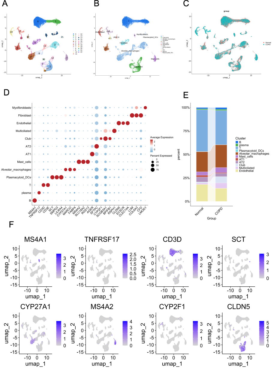 |
Figure 1 UMAP visualization and characterization of single-cell samples. (A) The UMAP plot shows that the single-cell samples can be categorized into 23 distinct clusters. (B) Thirteen cell types are identified based on the expression of specific marker genes. (C) A comparison of the UMAP cluster plots between normal and COPD tissues. (D) The dot plot illustrates the expression of selected marker genes for each identified cell type. (E) The proportions of different cell types within the samples are represented. (F) The UMAP plot highlights the expression profiles of marker genes for the 13 identified cell types. |
Identification of AM Subtypes at Different Developmental Stages Using Dimensionality Reduction Clustering
AM subtypes at various physiological developmental stages were identified through a dimensionality reduction and clustering approach. Following dimensionality reduction, AMs were categorized into 14 distinct subclusters (Figure 2A). Based on differential gene expression patterns, we classified these AM subpopulations into nine functional subtypes (Figure 2B). A detailed transcriptomic profile of these cell populations in both healthy controls and COPD samples is presented in Figure 2C. The marker genes defining the nine subtypes are illustrated in a dot plot (Figure 2D). Quantitative analysis indicated that COPD patients exhibited a higher proportion of specific subtypes, such as senescent AMs, compared to the control group (Figure 2E). The nine identified subtypes within the COPD microenvironment included senescence-associated AMs (Senescent AMs), Fibro AMs (fibroblast-related AMs), MTSS1+ AMs (AMs with high MTSS1 expression), Lipid AMs (lipid metabolism-related AMs), FCER+ AMs (Fcε receptor-positive AMs), SELLENOP AMs (AMs with low expression of SELL and ENOPH1), T-like AMs (T cell-associated AMs), CXCL1+ AMs (AMs with high CXCL1 expression), and IDC1+ AMs (inflammatory AMs marked by IDC1), whose expression patterns are highlighted in the UMAP visualization (Figure 2F).
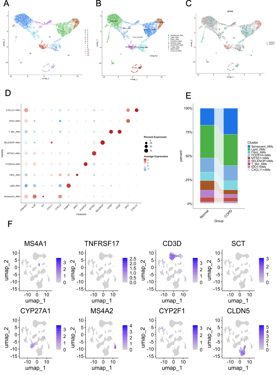 |
Figure 2 Characterization of AM subtypes through UMAP visualization. (A) The UMAP image indicates the presence of 14 distinct groups of AMs. (B) UMAP visualization presents the nine identified subtypes of AMs based on marker gene expression. (C) A comparison of UMAP clustering plots reveals differences between normal tissue and COPD tissue concerning AM subtypes. (D) The dot plot illustrates the marker genes associated with the nine identified AM subtypes. (E) The bar graph displays the proportions of AM subtypes in the COPD group compared to the normal group. (F) UMAP visualization highlights the expression patterns of marker genes for the nine AM subtypes. |
Pseudotime Trajectory Analysis Reveals Functional Heterogeneity and Differentiation Pathways of AM Subtypes in COPD
To investigate the maturation trajectory of AMs in the COPD microenvironment, pseudotime analysis was conducted to simulate their differentiation pathways. The analysis revealed a progression from Lipid_AMs to Senescent_AMs, indicating a dynamic shift towards a Senescent_AMs phenotype within COPD (Figure 3A). These subtypes reflect the functional heterogeneity of AMs in COPD and may play roles in processes such as inflammatory regulation, tissue repair, and immune dysregulation during disease progression. The Monocle2 algorithm was used to establish a pseudotime trajectory for AMs in COPD, simulating their dynamic changes in the disease (Figure 3B). Nine distinct AMs were observed at various time points (Figure 3C). Figure 3D shows the differential genes associated with COPD-related AMs, categorized into four modules based on the trends of gene expression along the pseudotime axis: Module 1 (purple): gradually upregulated with pseudotime, enriched in “regulation of inflammatory response” and “response to lipopolysaccharide,” reflecting the progressive acquisition of the senescent AMs and the pro-inflammatory polarization of AMs during COPD progression. Module 2 (blue): initially increased, then decreased, and later increased again, involving “ribosome biogenesis” and “cytoplasmic translation,” representing the dynamic remodeling of protein synthesis and metabolic stress adaptation in AMs under the chronic irritant milieu of COPD; Module 3 (orange): gradually decreased after a mid-peak, participating in “positive regulation of lipid localization” and “positive regulation of lipid transport,” suggesting the impairment of lipid homeostasis and surfactant clearance capabilities as AMs transition towards a terminal senescent state; and, Module 4 (green): downregulated with pseudotime, enriched in “antigen processing and presentation of exogenous peptide antigen via MHC class II” and “MHC class II protein complex assembly,” indicating the significant decline in immune surveillance and antigen-presenting capacity in senescent AMs. The pseudotime trajectory of the top eight genes (CD300E, CD48, CD93, CSF3R, FCN1, HRH2, S100A12, and VCAN) among the nine different AM subpopulations was determined, and the results were consistent with the heatmap findings (Figure 3E).
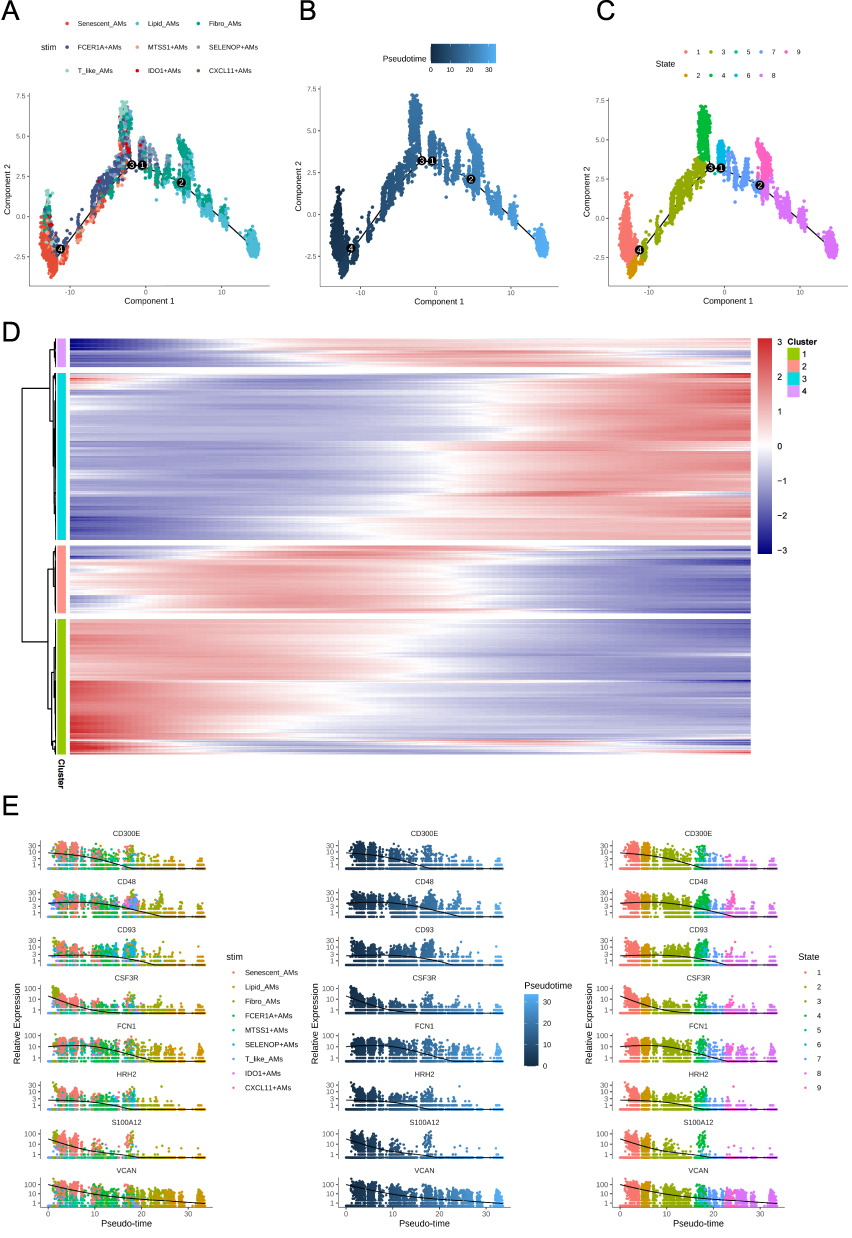 |
Figure 3 Pseudotime analysis. (A) Visualization of each stage of AMs along the pseudotime axis. (B) The developmental trajectory of AMs was plotted using the Monocle2 algorithm. (C) The trajectory plot illustrates the differentiation stages of AMs along the pseudotime axis. (D) Heatmap displaying the enrichment patterns of GO terms along the pseudotime trajectory. The color scale represents enrichment significance from low (blue) to high (red). The top enriched terms are grouped into four color-coded clusters (purple, blue, Orange, green) as indicated. (E) Pseudotime dynamics of the top eight genes among the nine macrophage subpopulations. Each point represents a specific cell, and different colors correspond to different clusters. The vertical axis indicates the expression levels of the genes. |
Alterations in Intercellular Communication Patterns and Signaling Pathways in AMs During COPD
This study utilized single-cell RNA sequencing datasets to analyze the signaling pathways mediated by AMs in COPD tissues, revealing a significant alteration in intercellular communication patterns within the COPD group. Compared to normal lung tissues, both the quantity and quality of intercellular communication patterns decreased in the COPD group (Figure 4A). Notably, connections between Senescent_AMs and other cells were reduced in the COPD state, indicating a change in the functionality of Senescent_AMs and a potential alteration in their cell communication capacity during COPD (Figure 4B). The coordination level of Senescent_AMs within the AM immune system may decrease due to their aging during COPD (Figure 4A and B). The pathway analysis comparing COPD and normal lung tissues revealed differences in cell-cell signaling networks (Figure 4C). The bubble plot of differential ligand-receptor interactions (Figure 4D) highlighted significant alterations (p < 0.05 or p < 0.01) in multiple inflammatory pathways, including reduced signaling in ICAM1-(ITGA4+ITGB1), IL1B-IL1R1, TNFSF12-TNFRSF13C, and CCL family pairs, as well as changes in other cytokine-mediated interactions.
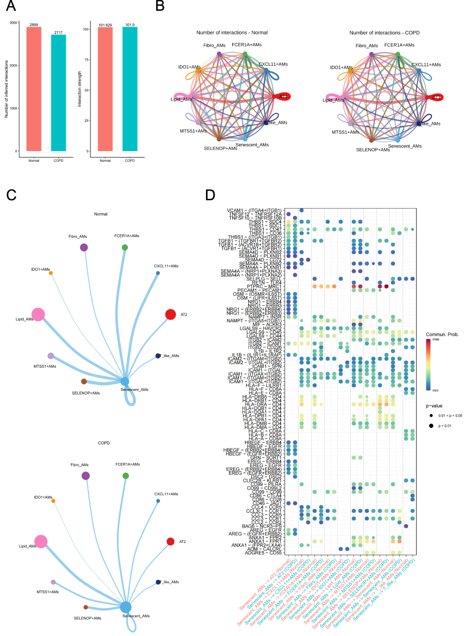 |
Figure 4 Differences in cell communication networks between normal and COPD tissues. (A) Comparison of the quantity and intensity of cell communication between COPD samples and normal tissues. (B) A differential interaction diagram illustrating the quantitative differences between the normal and COPD groups. Thicker lines indicate more frequent interactions between the two cell types. (C) A pathway status diagram showing differences in the quantity and intensity of signaling pathway-related cell communications between COPD and normal tissues. (D) Bubble plot representing significantly altered key ligand-receptor pairs in COPD and normal tissues. |
Network Pharmacology Analysis of Fenugreek: Active Components and Molecular Targets Related to COPD
Network pharmacology investigations revealed that fenugreek contained numerous bioactive components that interacted with various molecular targets. Specifically, the drug active component-target network constructed using Cytoscape showed the relationships between bioactive components and their expected targets, providing a more intuitive understanding of how fenugreek may connect its active components with target genes, thereby clarifying its mode of action. A total of ten bioactive compounds were identified in fenugreek, which interacted with 4746 distinct molecular targets (Figure 5A).
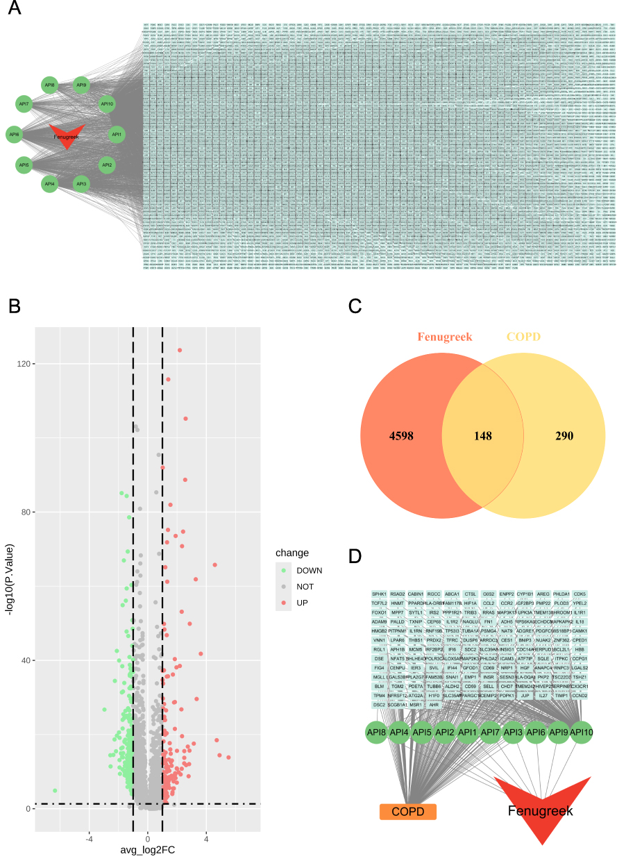 |
Figure 5 Network pharmacology reveals active components and targets of fenugreek related to COPD. (A) The network of “drug-active component-target genes”, where the red arrows represent fenugreek, the green circles represent active components, and the cyan rectangles denote target genes. (B) The volcano plot illustrates the gene expression differences between COPD and normal, highlighting significant DEGs. (C) A Venn diagram depicting the shared targets between fenugreek and COPD. (D) The “drug-active component-target genes-disease” network, with red arrows representing fenugreek, green circles representing active components, cyan rectangles indicating intersecting target genes, and Orange rectangles representing COPD. |
Furthermore, to identify COPD-related genes that reflect the disease-associated transcriptional changes observed in our single-cell analysis—particularly those linked to the prominent increase in senescent AMs—we performed bulk-level differential expression analysis on the lung tissue samples. This analysis identified 438 DEGs between the COPD and control groups, of which 177 were significantly upregulated, and 261 were significantly downregulated (Figure 5B). These genes may be crucial for the onset and development of COPD and represent potential targets for the effects of fenugreek. Additionally, Venn diagrams revealed that there were 148 overlapping genes between fenugreek-related targets and COPD-related DEGs (Figure 5C). To further reveal the potential molecular targets and pathways through which fenugreek affects COPD, we constructed the drug-active component-target genes-disease network based on these overlapping genes. As shown in Figure 5D, fenugreek influenced COPD through a molecular network involving active components and COPD-related targets.
Mechanistic Insights of Fenugreek in Modulating Cytokine-Cytokine Receptor Interactions Related to Oxidative Stress in COPD
To elucidate the mechanisms by which fenugreek influences chronic COPD, we conducted GO and KEGG pathway analyses on the shared targets of fenugreek and COPD. GO enrichment analysis in biological processes demonstrated significant enrichment in the pathways related to oxidative stress response and reactive oxygen species response (Figure 6A). For cellular components, there was a predominant localization of these targets in focal adhesion, endocytic vesicles, and lipid droplets (Figure 6A). For molecular function, these targets were enriched in cytokine binding, protease binding, and growth factor binding (Figure 6A).
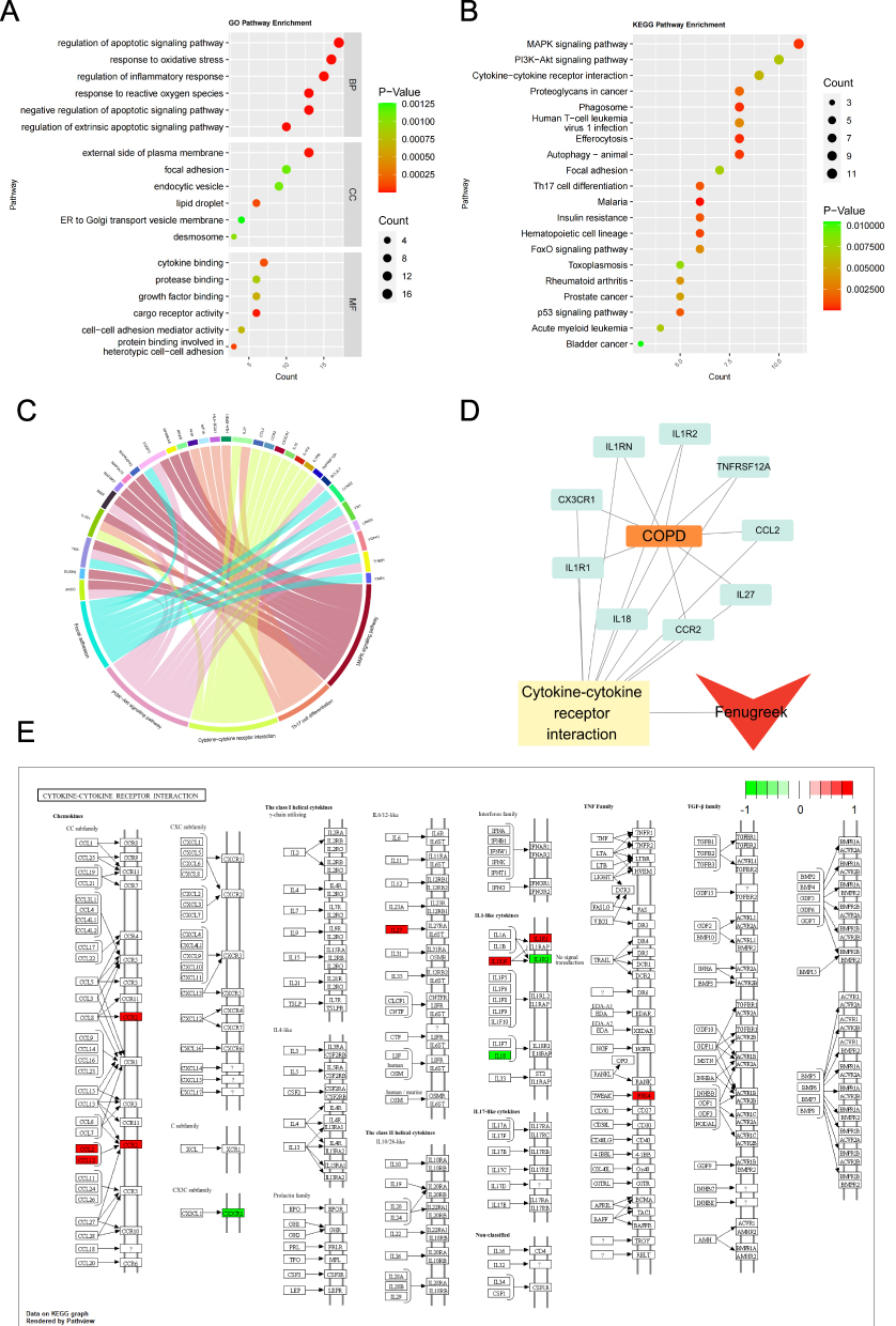 |
Figure 6 Functional analysis of the shared targets of fenugreek and COPD. (A) The top six GO terms in the categories of Biological Process (BP), Molecular Function (MF), and Cellular Component (CC). (B) The top 20 significant KEGG pathways. (C) Chord diagram analysis of five key pathways involved in signal transduction. (D) The “drug-pathway-genes-disease” network, where the red arrow represents fenugreek, pale yellow rectangles denote the Cytokine-cytokine receptor interaction pathway, green rectangles indicate the shared genes, and the Orange rectangle represents COPD. (E) Upregulated genes in the NF-kappa B signaling pathway are highlighted in red. |
A bubble plot illustrates the top 20 pathways with the highest significance, ranked by the degree of gene involvement (Figure 6B). The results highlighted several central signaling axes, including lipid metabolism and atherosclerosis, the MAPK signaling pathway, Th17 cell differentiation, cytokine−cytokine receptor interaction, focal adhesion, and the PI3K−Akt signaling pathway. Moreover, these relationships are also represented in a chord diagram (Figure 6C).
Additionally, we constructed a drug-pathway-genes-disease network, which elucidates the complex relationships between fenugreek, the cytokine−cytokine receptor interaction pathway, COPD pathology, and associated genetic targets (Figure 6D). Finally, the KEGG pathway analysis confirmed that the cytokine−cytokine receptor interaction pathway was crucial for the action of fenugreek (Figure 6E).
Molecular Docking Results
We conducted molecular docking analyses to investigate the interactions between 10 bioactive compounds derived from fenugreek and the targets CCL2, CCR2, IL1R1, IL1RN, IL27, and TNFRSF12A. The binding affinity thresholds were established as follows: < 4.25 kcal/mol (potential binding), < 5.0 kcal/mol (favorable binding), and < 7.0 kcal/mol (robust binding). The calculated binding affinities of fenugreek’s active compounds with these targets were found to be strong (Figure 7A). Notably, stronger binding affinities were observed for diosgenin and CCL2 (−7.50 kcal/mol) (Figure 7B), neotigogenin and CCL2 (−7.20 kcal/mol) (Figure 7C), luteolin and IL1R1 (−9.10 kcal/mol) (Figure 7D), and quercetin and IL1R1 (−9.10 kcal/mol) (Figure 7E). Therefore, molecular docking analysis supports the hypothesis that the effective components of fenugreek could target CCL2 and IL1R1, suggesting a potential mechanism to regulate cytokine signaling and indirectly modulate oxidative stress in COPD.
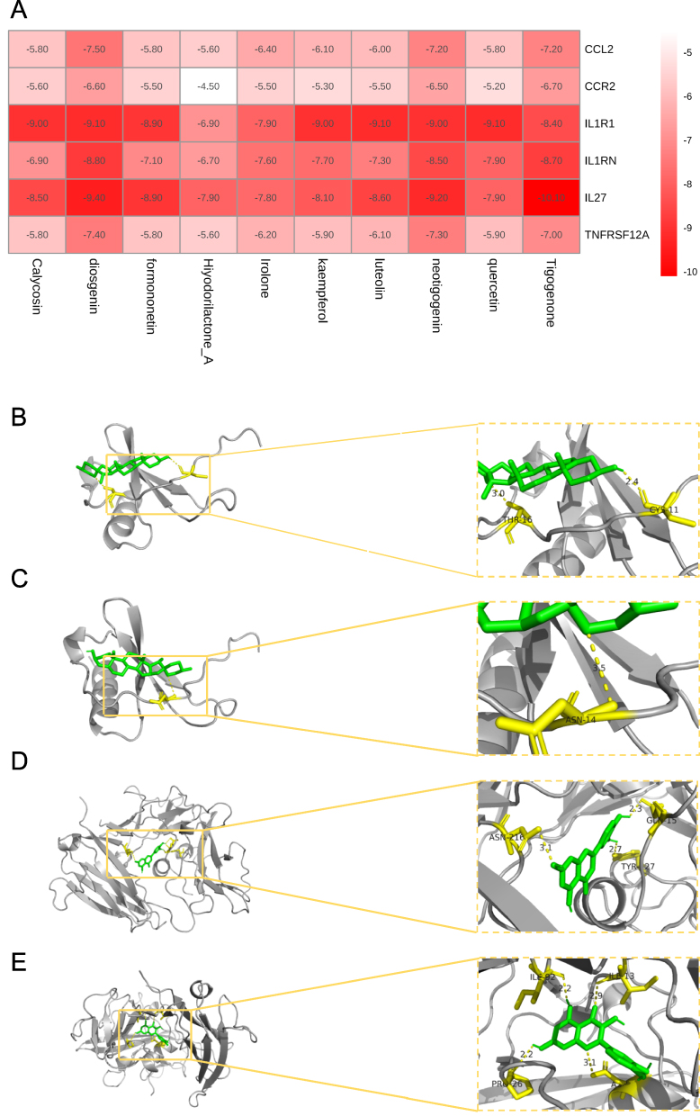 |
Figure 7 Molecular docking results of fenugreek active components and their targets. (A) The heatmap of molecular docking results for small molecule components and COPD-related targets. (B) The docking result of diosgenin with CCL2 (binding affinity: −7.50 kcal/mol). (C) The docking result of neotigogenin with CCL2 (binding affinity: −7.20 kcal/mol). (D) The docking result of luteolin with IL1R1 (binding affinity: −9.10 kcal/mol). (E) The docking result of quercetin with IL1R1 (binding affinity: −9.10 kcal/mol). |
Discussion
This study used network pharmacology and single-cell transcriptome sequencing to generate a robust, data-driven hypothesis and a testable mechanistic framework for how fenugreek, a key component of Hanchuan Zupa Granules, might influence the inflammatory and oxidative stress pathways in COPD. Our integrative analysis highlights the potential pivotal roles of critical targets such as CCL2 and IL1R1 within the cytokine-cytokine receptor interaction pathway. We propose that by targeting these nodes, fenugreek may indirectly mitigate oxidative stress in alveolar cells and inhibit the associated inflammatory cascades. This integrative approach provides a novel and testable hypothesis for the interaction between fenugreek’s bioactive components and COPD pathophysiology, reinforcing the multi-target, multi-component strategy of TCM.
The anti-inflammatory mechanisms hypothesized for fenugreek targeting the CCL2/IL1R1 axis are supported by our molecular docking results, which predict that its active components (eg, diosgenin and isovitexin) can potentially bind to CCL2 and IL1R1 with high affinity, exhibiting favorable binding energies (eg, −7.50 kcal/mol and −9.10 kcal/mol, respectively). This potential binding could inhibit the interaction between the chemokine CCL2 and its receptor IL1R1, thereby potentially suppressing downstream inflammatory signaling pathways such as NF-κB and MAPK activation.24 It is well-established that CCL2 is a critical mediator of macrophage chemotaxis and recruitment, and neutrophils are known to mediate CCL2-induced airway inflammation in COPD.25 Concurrently, IL1R1 serves as a key receptor for IL-1β signaling, whose activation is a potent inducer of NF-κB and MAPK pathways, leading to the upregulation of oxidative stress and exacerbation of tissue damage.26 Based on the co-expression of CCL2+ subpopulations of AMs and the enzymes NOX4 and SOD2 in COPD patients, the CCL2/IL1R1 axis may represent a critical node in the inflammatory-oxidative stress vicious cycle associated with COPD.27 Therefore, we hypothesize that fenugreek might improve the characteristic immune microenvironment dysregulation in COPD by synergistically inhibiting the CCL2/IL1R1 axis and thereby blocking the abnormal communication between macrophages, neutrophils, and other immune cells.7 These predictive insights suggest that modulating the CCL2/IL1R1 axis could be a promising strategy to ameliorate oxidative stress in COPD, aligning with the growing interest in antioxidant therapies for this disease.
Although we did not perform additional protein-protein interaction network analysis to identify hub genes, the selection of the core pathways was based on an unbiased, data-driven approach. GO and KEGG enrichment analyses indicated that the fenugreek-related COPD targets were significantly enriched in biological processes such as “cytokine-mediated signaling” and “oxidative stress response,” with the cytokine-cytokine receptor interaction pathway identified as the core functional pathway. This focus on inflammation and oxidative stress is consistent with their well-established roles as core pathological mechanisms in COPD.28,29 The pathway contains key molecules like CCL2 and IL1R1, which are closely related to alveolar epithelial damage, macrophage polarization, and cellular senescence in COPD.30 Single-cell trajectory analysis revealed a significant increase in the proportion of Senescent_AMs within the COPD microenvironment, with their senescence-associated secretory phenotype amplifying oxidative stress signals through the IL-1β/IL1R1 axis. Our finding that fenugreek components could target and inhibit this axis suggests a putative mechanism to indirectly reduce alveolar epithelial damage. While our convergent evidence strongly implicates these pathways as central, the identified targets likely function within a more complex interactome. Future studies incorporating protein-protein interaction network analysis and experimental validation will be valuable to fully delineate this network of interactions. Collectively, our integrated analysis proposes a testable mechanistic hypothesis wherein fenugreek modulates the COPD microenvironment through “multi-component, multi-target, and multi-pathway” actions centered on the IL1R1/CCL2 axis.
Single-cell technology facilitates the elucidation of the multi-component and multi-target characteristics of TCM. In this study, through single-cell transcriptomic analysis, there were significant alterations in AMs of patients with COPD. The characteristics of co-expression with oxidative stress genes suggest that these AMs may act as an “amplifier” for inflammation-oxidative stress. Intercellular communication analysis revealed that AMs in COPD tissues had limited interactions with other immune cells, indicating a potential immune dysregulation under COPD conditions. Fenugreek may regulate immune homeostasis by repairing CCL2/IL1R1-mediated intercellular signaling pathways (such as ligand-receptor pairs including ICAM1-ITGA4/ITGB1 and TNFSF12-TNFRSF13C). This finding is consistent with recent research aimed at improving COPD through macrophage-epithelial cell interactions.31
This study has several limitations that should be considered. First, the single-cell RNA sequencing analysis, while revealing compelling cellular dynamics, was based on a cohort of 5 patients per group. Although this sample size is common in initial exploratory single-cell studies in the field and was sufficient to identify major cell populations and significant shifts like the increase in senescent AMs, future studies with larger, independent cohorts are warranted to confirm and extend these findings, assess patient heterogeneity, and enhance statistical power for rare cell subtypes. Second, our findings are primarily computational and predictive; experimental validation in in vitro and in vivo models is essential to confirm the proposed mechanisms of fenugreek targeting the IL1R1/CCL2 axis. Finally, the network pharmacology approach relies on existing databases, which may have incomplete annotations.
Conclusion
In conclusion, this integrative computational study provides a hypothetical framework and a series of testable predictions regarding the therapeutic mechanisms of fenugreek in COPD. We propose that fenugreek may modulate inflammatory and oxidative stress pathways, potentially through targeting the IL1R1/CCL2 axis and influencing senescent AMs. The molecular docking results provide a structural rationale for these predicted interactions. Collectively, these findings pave the way for future wet-lab experiments to validate the proposed multi-target actions of fenugreek and to assess its therapeutic potential in modulating the COPD immune microenvironment.
Data Sharing Statement
The data and materials in the current study are available from the corresponding author on reasonable request.
Ethics Approval and Informed Consent
The single-cell RNA sequencing data used in this study were obtained from the publicly available dataset GSE173896 in the Gene Expression Omnibus database. The original study had obtained ethical approval and informed consent from participants. As this is a secondary analysis of publicly available de-identified data, according to Article 32, Items 1 and 2 of the Measures for Ethical Review of Life Science and Medical Research Involving Human Subjects (February 18, 2023, China), this study is exempt from additional ethics review.
Acknowledgments
This study was supported by the Natural Science Foundation of Xinjiang Uygur Autonomous Region (Grant No. 2023D03018) and the Tianshan Talents “Medical and Health High Level Talent Project (Grant No.: TSYC202401B111).
Author Contributions
All authors made a significant contribution to the work reported, whether that is in the conception, study design, execution, acquisition of data, analysis and interpretation, or in all these areas; took part in drafting, revising or critically reviewing the article; gave final approval of the version to be published; have agreed on the journal to which the article has been submitted; and agree to be accountable for all aspects of the work.
Disclosure
The authors report no conflicts of interest in this work.
References
1. Lee Y-M. Chronic obstructive pulmonary disease: respiratory review of 2014. Tuberc Respir Dis. 2014;77(4):155–17. doi:10.4046/trd.2014.77.4.155
2. Chen R, Zhan Y, Lin Z, et al. Effect of YuPingFeng granules on clinical symptoms of stable COPD: study protocol for a multicenter, double-blind, and randomized controlled trial. BMC Complement Med Ther. 2024;24(1):25. doi:10.1186/s12906-023-04271-7
3. Li LC, Han YY, Zhang ZH, et al. Chronic obstructive pulmonary disease treatment and pharmacist-led medication management. Drug Des Devel Ther. 2021;15:111–124. doi:10.2147/DDDT.S286315
4. Liang K, Wang Z, Huang K, et al. New advancements in the study of the regulatory effects of the PI3K-AKT signaling pathway on airway remodeling in chronic obstructive pulmonary disease and interventions using traditional Chinese medicine. Chin Med Pharm Clin. 2024;40(5):103–108. doi:10.13412/j.cnki.zyyl.20230725.004
5. Yu K, Xiao Y. The effects of Hanchuan Zupa Granules on lung function and inflammatory indicators in patients with chronic obstructive pulmonary disease. Mod Diag Treat. 2023;34(09):1295–1297.
6. Pauwels NS, Bracke KR, Dupont LL, et al. Role of IL-1α and the Nlrp3/caspase-1/IL-1β axis in cigarette smoke-induced pulmonary inflammation and COPD. Eur Respir J. 2011;38(5):1019–1028. doi:10.1183/09031936.00158110
7. de Boer WI, Yao H, Rahman I. Future therapeutic treatment of COPD: struggle between oxidants and cytokines. Int J Chron Obstruct Pulmon Dis. 2007;2(3):205–228.
8. Schiffers C, Reynaert NL, Wouters EFM, van der Vliet A. Redox dysregulation in aging and COPD: role of NOX enzymes and implications for antioxidant strategies. Antioxidants. 2021;10(11):1799. doi:10.3390/antiox10111799
9. Hopkins AL. Network pharmacology: the next paradigm in drug discovery. Nat Chem Biol. 2008;4(11):682–690. doi:10.1038/nchembio.118
10. Shao X, Chen Y, Zhang J, et al. Advancing network pharmacology with artificial intelligence: the next paradigm in traditional Chinese medicine. Chin J Nat Med. 2025;23(11):1358–1376. doi:10.1016/S1875-5364(25)60941-1
11. Sauler M, McDonough JE, Adams TS, et al. Characterization of the COPD alveolar niche using single-cell RNA sequencing. Nat Commun. 2022;13(1):494. doi:10.1038/s41467-022-28062-9
12. Blackburn JB, Tufenkjian TS, Liu Y, Nichols DS, Blackwell TS, Richmond BW. A single-cell RNA sequencing atlas of the chronic obstructive pulmonary disease distal lung to predict cell-cell communication. Am J Respir Cell Mol Biol. 2025;72(3):332–335. doi:10.1165/rcmb.2024-0232LE
13. Watanabe N, Fujita Y, Nakayama J, et al. Anomalous epithelial variations and ectopic inflammatory response in chronic obstructive pulmonary disease. Am J Respir Cell Mol Biol. 2022;67(6):708–719. doi:10.1165/rcmb.2021-0555OC
14. Trapnell C, Cacchiarelli D, Grimsby J, et al. The dynamics and regulators of cell fate decisions are revealed by pseudotemporal ordering of single cells. Nat Biotechnol. 2014;32(4):381–386. doi:10.1038/nbt.2859
15. Efremova M, Vento-Tormo M, Teichmann SA, Vento-Tormo R. CellPhoneDB: inferring cell-cell communication from combined expression of multi-subunit ligand-receptor complexes. Nat Protoc. 2020;15(4):1484–1506. doi:10.1038/s41596-020-0292-x
16. Xu X, Zhang W, Huang C, et al. A novel chemometric method for the prediction of human oral bioavailability. Int J Mol Sci. 2012;13(6):6964–6982. doi:10.3390/ijms13066964
17. Shannon P, Markiel A, Ozier O, et al. Cytoscape: a software environment for integrated models of biomolecular interaction networks. Genome Res. 2003;13(11):2498–2504. doi:10.1101/gr.1239303
18. Sherman BT, Hao M, Qiu J, et al. DAVID: a web server for functional enrichment analysis and functional annotation of gene lists (2021 update). Nucleic Acids Res. 2022;50(W1):W216–W221. doi:10.1093/nar/gkac194
19. Forli S, Huey R, Pique ME, Sanner MF, Goodsell DS, Olson AJ. Computational protein-ligand docking and virtual drug screening with the AutoDock suite. Nat Protoc. 2016;11(5):905–919. doi:10.1038/nprot.2016.051
20. De Vita S, Chini MG, Bifulco G, Lauro G. Insights into the ligand binding to Bromodomain-Containing Protein 9 (BRD9): a guide to the selection of potential binders by computational methods. Molecules. 2021;26(23):7192. doi:10.3390/molecules26237192
21. Theerawatanasirikul S, Semkum P, Lueangaramkul V, Chankeeree P, Thangthamniyom N, Lekcharoensuk P. Non-nucleoside inhibitors decrease foot-and-mouth disease virus replication by blocking the viral 3D(pol). Viruses. 2022;15(1):124. doi:10.3390/v15010124
22. Shamsol Azman ANS, Tan JJ, Abdullah MNH, Bahari H, Lim V, Yong YK. Network pharmacology and molecular docking analysis of active compounds in tualang honey against atherosclerosis. Foods. 2023;12(9):1779. doi:10.3390/foods12091779
23. Adams TS, Schupp JC, Poli S, et al. Single-cell RNA-seq reveals ectopic and aberrant lung-resident cell populations in idiopathic pulmonary fibrosis. Sci Adv. 2020;6(28):eaba1983. doi:10.1126/sciadv.aba1983
24. Li Z, Chen L, Wang L. Pharmacological study of the traditional Chinese medicine combination of Ephedra and Ginkgo biloba in the treatment of bronchial asthma and chronic obstructive pulmonary disease under the principle of “treating different diseases with the same method”. Shaanxi Tradit Chin Med. 2021;42(8):1128–1132. doi:10.3969/j.issn.1000-7369.2021.08.035
25. Dong Y, Dong Y, Zhu C, et al. Targeting CCL2-CCR2 signaling pathway alleviates macrophage dysfunction in COPD via PI3K-AKT axis. Cell Commun Signal. 2024;22(1):364. doi:10.1186/s12964-024-01746-z
26. Sarray S, Lamine LB, Dallel M, et al. Association of MMP-2 genes variants with diabetic retinopathy in Tunisian population with type 2 diabetes. J Diabetes Complicat. 2022;36(5):108182. doi:10.1016/j.jdiacomp.2022.108182
27. Wang X, Murugesan P, Zhang P, et al. NADPH oxidase isoforms in COPD patients and acute cigarette smoke-exposed mice: induction of oxidative stress and lung inflammation. Antioxidants. 2022;11(8):1539. doi:10.3390/antiox11081539
28. Zhao W, Bai B, Li H, et al. The role of oxidative stress-related genes in idiopathic pulmonary fibrosis. Sci Rep. 2025;15(1):5954. doi:10.1038/s41598-025-89770-y
29. Xiao X, Guo W, Li N, Chen N, Zhang Q. Identifying KL-6-associated immune cell signatures and key genes in emphysematous COPD. J Inflamm Res. 2025;18:6453–6466. doi:10.2147/jir.S515653
30. Kumar M, Seeger W, Voswinckel R. Senescence-associated secretory phenotype and its possible role in chronic obstructive pulmonary disease. Am J Respir Cell Mol Biol. 2014;51(3):323–333. doi:10.1165/rcmb.2013-0382PS
31. Habermann AC, Gutierrez AJ, Bui LT, et al. Single-cell RNA sequencing reveals profibrotic roles of distinct epithelial and mesenchymal lineages in pulmonary fibrosis. Sci Adv. 2020;6(28):eaba1972. doi:10.1126/sciadv.aba1972
 © 2026 The Author(s). This work is published and licensed by Dove Medical Press Limited. The
full terms of this license are available at https://www.dovepress.com/terms
and incorporate the Creative Commons Attribution
- Non Commercial (unported, 4.0) License.
By accessing the work you hereby accept the Terms. Non-commercial uses of the work are permitted
without any further permission from Dove Medical Press Limited, provided the work is properly
attributed. For permission for commercial use of this work, please see paragraphs 4.2 and 5 of our Terms.
© 2026 The Author(s). This work is published and licensed by Dove Medical Press Limited. The
full terms of this license are available at https://www.dovepress.com/terms
and incorporate the Creative Commons Attribution
- Non Commercial (unported, 4.0) License.
By accessing the work you hereby accept the Terms. Non-commercial uses of the work are permitted
without any further permission from Dove Medical Press Limited, provided the work is properly
attributed. For permission for commercial use of this work, please see paragraphs 4.2 and 5 of our Terms.
Recommended articles
Anti-Inflammatory Effects and Molecular Mechanisms of Shenmai Injection in Treating Acute Pancreatitis: Network Pharmacology Analysis and Experimental Verification
He Y, Hu C, Liu S, Xu M, Liang G, Du D, Liu T, Cai F, Chen Z, Tan Q, Deng L, Xia Q
Drug Design, Development and Therapy 2022, 16:2479-2495
Published Date: 2 August 2022
A Novel Approach Based on Gut Microbiota Analysis and Network Pharmacology to Explain the Mechanisms of Action of Cichorium intybus L. Formula in the Improvement of Hyperuricemic Nephropathy in Rats

Amatjan M, Li N, He P, Zhang B, Mai X, Jiang Q, Xie H, Shao X
Drug Design, Development and Therapy 2023, 17:107-128
Published Date: 20 January 2023
Network Pharmacology and Experimental Validation to Explore That Celastrol Targeting PTEN is the Potential Mechanism of Tripterygium wilfordii (Lév.) Hutch Against IgA Nephropathy

Zhao J, Liu H, Xia M, Chen Q, Wan L, Leng B, Tang C, Chen G, Liu Y, Zhang L, Liu H
Drug Design, Development and Therapy 2023, 17:887-900
Published Date: 23 March 2023
Investigating the Mechanism of Action of Schisandra chinensis Combined with Coenzyme Q10 in the Treatment of Heart Failure Based on PI3K-AKT Pathway

Wen S, Yang K, Bai Y, Wu Y, Liu D, Wu X, Zhang X, Sun J
Drug Design, Development and Therapy 2023, 17:939-957
Published Date: 27 March 2023
Network Pharmacology and Experimental Verification Reveal the Regulatory Mechanism of Chuanbeimu in Treating Chronic Obstructive Pulmonary Disease
Xian M, Xu J, Zheng Y, Zhang L, Zhao J, Chen J, Li S, Lin L, Zhong Y, Yang Z, Xie T, Huang L, Ding Y
International Journal of Chronic Obstructive Pulmonary Disease 2024, 19:799-813
Published Date: 21 March 2024