Back to Journals » Cancer Management and Research » Volume 12
miR-455-3p Functions as a Tumor Suppressor by Restraining Wnt/β-Catenin Signaling via TAZ in Pancreatic Cancer
Authors Zhan T ![]() , Zhu Q, Han Z, Tan J, Liu M
, Zhu Q, Han Z, Tan J, Liu M ![]() , Liu W, Chen W, Chen X, Chen X, Deng J, Tian X, Huang X
, Liu W, Chen W, Chen X, Chen X, Deng J, Tian X, Huang X
Received 23 October 2019
Accepted for publication 12 February 2020
Published 27 February 2020 Volume 2020:12 Pages 1483—1492
DOI https://doi.org/10.2147/CMAR.S235794
Checked for plagiarism Yes
Review by Single anonymous peer review
Peer reviewer comments 2
Editor who approved publication: Professor Rudolph Navari
Ting Zhan, 1,* Qingxi Zhu, 1,* Zheng Han, 1 Jie Tan, 1 Meng Liu, 1 Weijie Liu, 1 Wei Chen, 1 Xiaoli Chen, 1 Xueting Chen, 2 Junsheng Deng, 2 Xia Tian, 1 Xiaodong Huang 1
1Department of Gastroenterology, Wuhan Third Hospital (Tongren Hospital of Wuhan University), Wuhan 430060, People’s Republic of China; 2Department of Gastroenterology, Zhongnan Hospital of Wuhan University, Wuhan 430060, People’s Republic of China
*These authors contributed equally to this work
Correspondence: Xiaodong Huang; Xia Tian Tel +86 1388619054; +86 13871480868
Email [email protected]; [email protected]
Background: Pancreatic cancer (PC) is a highly invasive tumor with a poor prognosis, short overall survival rate and few chemotherapeutic choices. Despite the importance of finding ways to treat pancreatic cancer, the mechanisms of tumor progression have not been fully elucidated. microRNA-455-3p (miR-455-3p) has been reported to play an important role in several cancers, but its function in pancreatic cancer remains unclear.
Methods: To investigate the biological functions, miRNAs mimics or inhibitors were transfected into pancreatic cancer cells. Flow cytometry was used to detect cell apoptosis. Wound healing and Transwell assays were employed to observe cell invasion and migration abilities. The expression of Bcl-2, Bax, caspase-3, E-cadherin, N-cadherin, Snail, β-Catenin, c-Myc and Cyclin D1 were evaluated by qPCR and Western blot.
Results: We confirmed that inhibition of miR-455-3p decreases cell apoptosis and increases cell migration, invasion and EMT of pancreatic cancer, whereas forced overexpression of miR-455-3p has the opposite effect. Furthermore, we demonstrated that the tumor suppression effects of miR-455-3p were partially reversed by TAZ overexpression. In addition, miR-455-3p led to inactivation of Wnt/β-catenin signaling in pancreatic cancer cells, and TAZ overexpression restored the inhibition of Wnt/β-catenin signaling.
Conclusion: Taken together, our data demonstrated that miR-455-3p functions as an important tumor suppressor that suppresses the Wnt/β-catenin signaling pathway via TAZ to inhibit tumor progression in pancreatic cancer. We conclude that the miR-455-3p/TAZ/Wnt axis may be a potential therapeutic target for pancreatic cancer.
Keywords: miR-455-3p, Wnt, apoptosis, metastasis, EMT
Corrigendum for this paper has been published.
Introduction
Pancreatic cancer (PC) is the third most common cause of cancer-related death, and most patients are diagnosed with metastasis due to lack of early symptoms and diagnostic techniques.1 Despite advances in the processes of surgery and chemotherapy, the mortality rate of PC is predicted to increase markedly.2 Given that only a minority of patients diagnosed with PC are eligible for surgical intervention, more and more research is gradually shifting towards identifying the molecular mechanisms of risk factors associated with PC promotion in order to facilitate the discovery of novel targets and agents.
In recent years, microRNA (miRNA) has become one of the main hotspots of research regarding the development and progression of cancer. As a post-transcriptional regulator of gene expression, miRNA is involved in many biological processes, including development, differentiation, proliferation and apoptosis of cells.3,4 Particular miRNAs function either as tumor suppressors or oncogenes, which leads to the abnormal activity of miRNA target genes in different types of cancer, including PC.5 It has been found that miR-455-3p plays an important role in many tumors, such as colorectal cancer, esophageal cancer, lung cancer, breast cancer, and melanoma.6–10 Our previous studies have found that miR-455-3p is downregulated in PC and is involved in regulating proliferation and drug resistance by targeting TAZ.11 Transcriptional co-activator with PDZ-binding motif (TAZ), also known as WW domain-containing transcriptional regulator 1, is a transcriptional activator pervasively induced in several human tumors.12 The expression of TAZ has been found to be increased in many human tumors, and TAZ has been shown to be essential for cancer initiation, progression, and metastasis.13 Given that miR-455-3p can inhibit the expression of TAZ, which is considered to be an oncogene, we infer that miR-455-3p may function as a tumor suppressor via TAZ.
The Wnt signaling pathway is a highly conserved signaling pathway closely related to cell proliferation and differentiation.14 Activated β-catenin, Cyclin D and C-myc are involved in Wnt/β-catenin signaling.15 It has a wide range of biological effects and plays an important role in regulating most biological phenomena, such as ontogenesis, cell differentiation and apoptosis.16 Some studies have found that β-catenin can activate TAZ by binding to β-catenin and TBX5, and some studies have also found that polymerase activity inhibitors can inhibit the activation of TAZ through the Wnt signaling pathway.17 Melucci et al found TAZ and Wnt-related biomarkers may predict clinical outcomes in gastric cancer patients treated with chemotherapy,18 although the role of TAZ/Wnt/β-catenin signaling in PC still remains unknown.
This study aimed to further explore the function of miR-455-3p in human cells and whether it can restrain Wnt/β-catenin signaling via TAZ in pancreatic cancer.
Materials and Methods
Cell Lines and Cell Culture
Human pancreatic cancer cell lines PANC-1 and MIAPaCa-2 were provided by the Cell Bank of Type Culture Collection of the Chinese Academy of Sciences (Shanghai, China). They were cultured in DMEM (Gibco, USA) supplemented with 10% fetal bovine serum (NQBB, USA), 100 mg/mL streptomycin, and 100 IU/mL penicillin (Thermo Fisher Scientific, USA). Cells were incubated in a humidified incubator at 37°C with 5% CO2.
Cell Transfection
Hsa-miR-455-3p mimics (sense: 50-GCAGUCCAUGGGCAUAUACAC-30; antisense: 50-GUAUAUGCCCAUGGACUGCUU-30), hsa-miR-455-3p inhibitors (sense: 50-GUGUAUAUGCCCAUGGACUGC-30), hsa-miR mimics NC (sense: 50-UUCUCCGAACGUGUCACGUTT-30; antisense: 50-ACGUGACACGUUCGGAGAATT-30), and hsa-miR inhibitors NC (50-CAGUACUUUUGUGUAGUACAA-30) were synthesized by Genepharma (Shanghai, China).PANC-1 and MIA paCa2 cells were transfected with miR-455-3p mimics (40 nM), miR-455-3p inhibitors (80 nM), or their NC using Lipofectamine 2000 (Invitrogen, USA), according to the manufacturer’s instructions. The TAZ lentiviral expression vector (lenti-TAZ) were constructed by Shanghai GeneChem (Shanghai, China). The lentiviruses were transfected according to the manufacturer’s instructions.
Analysis of Cell Apoptosis
Pancreatic cancer cells were transfected in 6-well plates. After incubation for 48 hrs, the cells were stained using the Annexin V-PE Detection Kit (BD Biosciences, USA), according to the manufacturer’s protocols. All the samples were analyzed using the FACS Caliber II Sorter and Cell Quest FACS System (BD Biosciences, USA).
Total RNA Extaction and qPCR Analysis
Total RNA from tissues and cell lines was extracted using TRIzol Reagent (Invitrogen, USA), and subsequent synthesis of cDNA was carried out according to the manufacturer’s protocols (TOYOBO, Japan). To evaluate TAZ mRNA levels, the reverse transcription products were analyzed using UltraSYBR Mixture (ComWin Biotech, China) on an ABI StepOne Plus qPCR System (Applied Biosystems, USA). GAPDH mRNA was used as an endogenous control. The expression level of miR-455-3p was evaluated using the same qPCR system. Endogenous U6 small nuclear RNA (snRNA) was used for normalization. The primers for amplifification were as follows: Actin (sense: 50 -CATGTACGTTGCTATCCAGGC-30; antisense: 50 -CTCCTTAATGTCACGCACGAT-30); Bcl (sense: 50 -CTTTGAGTTCGGTGGGGTCA-30; antisense: 50 -GGGCCGTACAGTTCCACAAA-30); Bax (sense: 50 -CATGGGCTGGACATTGGACT-30; antisense: 50 -AAAGTAGGAGAGGAGGCCGT); Caspase3 (sense: 50 -CGGCGCTCTGGTTTTCGTTA-30; antisense: 50-CCGAGA TGTCATTCCAGT GCT); E-Cadherin (sense: 50 -GCTGGACCGAGAGAGTTTCC-30; antisense: 50 -CAAAATCCAAGCCCGTGGTG-30); N-Cadherin (sense: 50 -TGGGAAATGGAAACTTGATGGC-30; antisense: 50 -TGGAAAGCTTCTCACGGCAT); Snail (sense: 50 -AAGATGCACATC CGAAGCCA-30; antisense: 50 -CATTCGGGAGAAGGTCCGAG); β-catenin(sense: 50 -CTGAGGAGCAGCTTCAGTCC-30; antisense: 50 -GGCCATGTCCAACTCCATCA-30); Cyclin D1 (sense: 50 -CCTTGTGGAGCCGGAGC-30; antisense: 50-CACTGGGCCGA AGAGGC); C-Myc (sense: 50 -GCAATGCGTTGCTGGGTTAT-30; antisense: 50 -CGCATCCTTGTCCTGTGAGT); The 2−ΔΔCT method was used to calculate changes in miRNA expression levels.
Western Blot Analysis
Cells were lysed in radioimmunoprecipitation assay (RIPA) buffer containing protease inhibitors (Beyotime, Shanghai, China), and the protein concentration was measured using Pierce BCA Protein Assay Kit (Thermo Fisher Scientific, USA). Soluble lysate was mixed with loading buffer and boiled for 10 min. Proteins were separated by 10% SDS-PAGE, transferred to NC membranes, and then incubated with primary antibodies against Bcl-2 (1:1000, abcam, USA),Bax(1:1000, abcam), caspase-3(1:1000, abcam), E-cadherin (1:1000, CST, Danvers, MA), N-cadherin (1:1000, CST), snail (1:1000, CST), β-Catenin(1:1000, abcam), c-Myc(1:1000, abcam), Cyclin D1(1:1000, abcam) and β-actin (1:1000, Santa Cruz, Delaware, CA) overnight at 4°C. The membranes were further incubated with secondary antibodies (LI-COR Biosciences, USA) for 1 hr at 37°C, and the protein levels were detected by the enhanced chemiluminescence (ECL) Western Blot Analysis Detection System (Amersham, USA).
Wound Healing Assay
Pancreatic cancer cells were transfected in 6-well plates and grown to monolayer confluency. Straight wounds were created in the cell monolayers using a sterile pipette tip. In order to remove the detached cells, the plates were washed twice using phosphate-buffered saline. Cells were then cultured in medium without fetal bovine serum at 37°C. Progression of migration was observed and photographed 24 hrs after wounding. Three random fields were captured and measured for each well.
Transwell Assay
Transwell chambers with a pore size of 8 µm and precoated with Matrigel (BD Biosciences, San Jose, CA, USA) were used to determine cells’ invasion ability. Cells h(5 × 10⁴) were seeded into the upper chambers with 200 μL serum-free medium, and the lower chambers were filled with culture medium containing 10% fetal bovine serum. After 24 hrs of incubation, cells that failed to invade the membrane were removed gently with cotton swabs, while the invaded cells on the bottom were fixed with 4% formaldehyde for 20 min and stained with 0.1% crystal violet for 30 min before being photographed under a microscope (OLYMPUScx31, TOKYO, Japan). Five visual fields were randomly chosen and imaged.
Statistical Analysis
All statistical analyses were conducted using SPSS software and illustration data were performed by GraphPad Prism. The statistical significance was evaluated using Student’s t-test or one-way ANOVA and is presented as the mean ± SD; p < 0.05 was considered statistically significant.
Results
miR-455-3p Contributes to Apoptosis of PC Cells
Having observed that miR-455-3p is downregulated in PC and participates in regulating the proliferation of PC cells,11 we went on functionally characterizing miR-455-3p by studying its impact on PC cell apoptosis. PANC-1 and MIAPaCa-2 cells were transfected with miR-455-3p mimics, miR-455-3p inhibitors or their negative control. Flow cytometry was used to detect cell apoptosis, and we demonstrated that overexpression of miR-455-3p increased the number of apoptotic cells in both PANC1 and MIAPaCa2, and inhibition of miR-455-3p reduced the number of apoptotic cells (Figure 1A and B). To further explore the effect of miR-455-3p on apoptosis of pancreatic cancer cells, we detected the expression of apoptosis-related gene Bcl-2, Bax and Caspase-3 by qPCR and Western blotting. The results showed that overexpression of miR-455-3p increased the expression of Bcl-2 and decreased the expression of Bax and Caspase-3, whereas inhibition of miR-455-3p had the opposite effect (Figure 1C–F). These data indicate that miR-455-3p promotes apoptosis of PC cells by regulating apoptosis-related proteins.
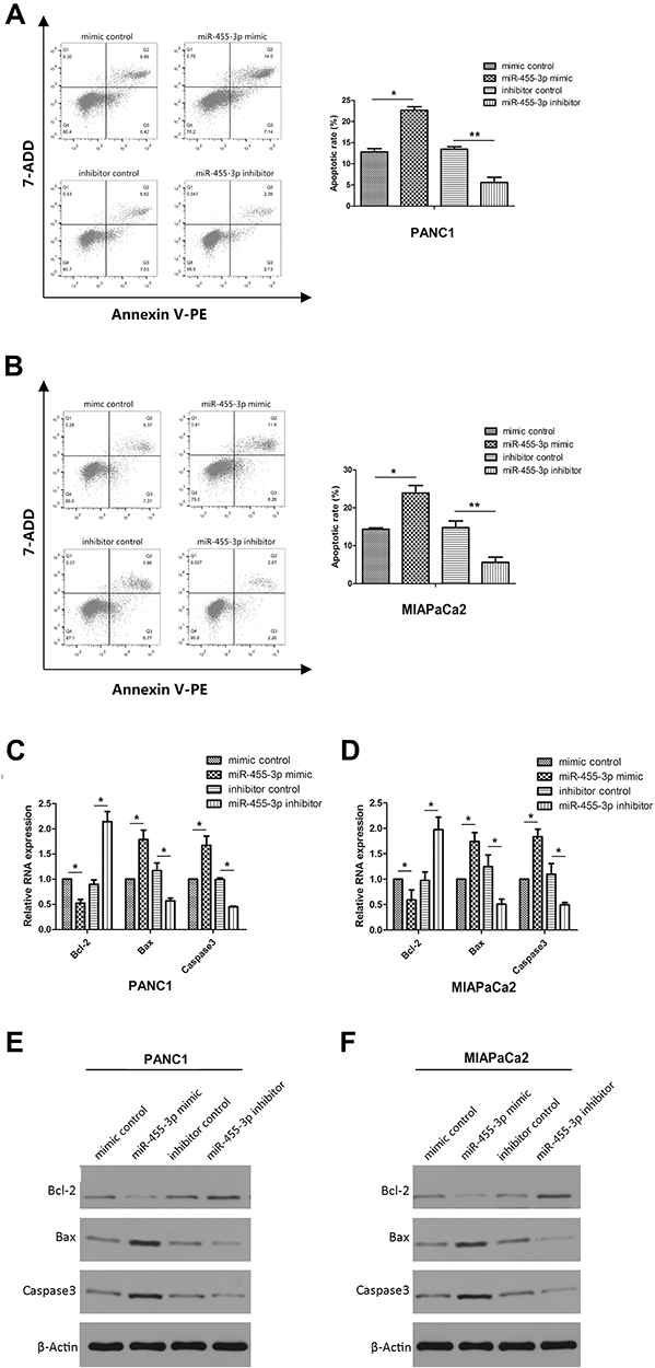 |
Figure 1 MiR-455-3p contributes to apoptosis in pancreatic cancer cell lines. (A, B) Flow cytometry assessment of apoptosis in PANC1 and MIAPaCa2 cells transfected with miR-455-3p mimics or inhibitors. The total events shown in the lower right-hand and upper right-hand quadrants are apoptotic cells. (C, D) qPCR analysis of the mRNA expression of Bcl-2,Bax and Caspase-3 in pancreatic cancer cells transfected with miR-455-3p mimics or inhibitors. (E, F) Expression of Bcl-2, Bax and Caspase-3 in pancreatic cancer cells transfected with miR-455-3p mimics or inhibitors was detected by Western Blot. β-actin is used as loading control. All data are presented as the mean ± SD. *p<0.05, **p < 0.01, miR-455-3p mimics vs mimic control group, miR-455-3p inhibitors vs inhibitors control group. |
miR-455-3p Inhibits the EMT-Mediated Metastasis of PC Cells
Wound healing assay and Transwell assay were carried out to investigate the effect of miR-455-3p on metastasis of pancreatic cancer. Similarly, PANC-1 and MIAPaCa-2 cells were transfected with miR-455-3p mimics, miR-455-3p inhibitors or their negative control. Wound healing assays demonstrated that overexpression of miR-455-3p significantly weakened migratory capability, while the inhibition of miR-455-3p enhanced the migratory capability of PANC-1 and MIAPaCa-2 cells (Figure 2A and B). The Matrigel invasion assay results showed the same trend: overexpression of miR-455-3p reduced the capacity of cell invasion and the inhibition of miR-455-3p increased the capacity of cell invasion (Figure 2C and D). In order to investigate whether miR-455-3p is involved in EMT-mediated metastasis, we performed qPCR and Western blotting to detect the expression of EMT-related proteins E-cadherin, N-cadherin and snail in PC cells. The results show that overexpression of miR-455-3p increased the expression of E-cadherin but decreased that of N-cadherin and snail. Conversely, the inhibition of miR-455-3p decreased the expression of E-cadherin but increased that of N-cadherin and snail (Figure 2E–H). These results support the conclusion that miR-455-3p suppresses the metastasis of PC by mediating EMT.
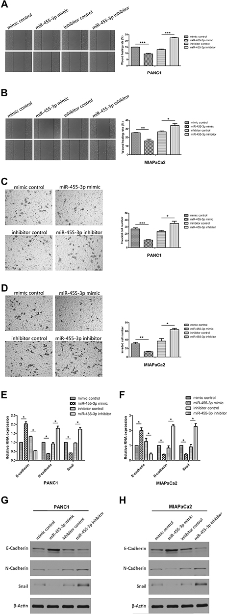 |
Figure 2 MiR-455-3p inhibits EMT-mediated metastasis in pancreatic cancer cells. (A, B) Wound healing assays of PANC-1 and MIAPaCa-2 after transfection with miR-455-3p mimics or inhibitors. Representative images depict the beginning (t = 0 h) and end (t = 24 h) of the recording. (C, D) Transwell assay of PANC-1 and MIAPaCa-2 cells transfected with miR-455-3p mimics or inhibitors. Cell invasion was analyzed 24 h after seeding in Transwells. (E, F) qPCR analysis of the mRNA expression of E-cadherin, N-cadherin and Snail in PANC1 and MIAPaCa2 cells transfected with miR-455-3p mimics or inhibitors. (G, H)Western blot analysis of the protein levels of E-cadherin, N-cadherin and Snail in PANC1 and MIAPaCa2 cells transfected with miR-455-3p mimics or inhibitors. β-actin is used as loading control. All data are presented as the mean ± SD. *p <0.05, **p <0.01, ***p < 0.001, miR-455-3 p mimics vs mimic control group, miR-455-3p inhibitors vs inhibitors control group. |
miR-455-3p Functions as a Tumor Suppressor via Targeting of TAZ
Our previous studies have confirmed that TAZ is a new direct downstream target of miR-455-3p,11 but whether miR-455-3p can exert its anti-cancer effect by targeting TAZ is still unclear. Thus, we detected cell apoptosis and EMT-mediated metastasis of PC cells, which were transfected with miR-455-3p mimics or miR-455-3p mimics plus TAZ expression vector. We confirmed that overexpression of miR-455-3 promoted apoptosis and inhibited migration, invasion and EMT in both PANC-1 and MIAPaCa-2 cells, whereas overexpression of TAZ reversed the anti-cancer effect of miR-455-3p (Figure 3A–N). Taken together, these data clearly suggest that miR-455-3p is not only involved in the proliferation and drug resistance of PC, but is also closely related to apoptosis, migration, invasion and EMT via targeting of TAZ.
 |
Figure 3 MiR-455-3p participates in cell apoptosis, migration, invasion and EMT via targeting TAZ. (A, B) Flow cytometry assessment of apoptosis in PANC1 and MIAPaCa2 cells transfected with miR-455-3p mimic or miR-455-3p mimic plus TAZ expression vector. (C, D) Expression of Bcl-2, Bax and Caspase-3 in pancreatic cancer cells transfected with miR-455-3p mimic or miR-455-3p mimic plus TAZ expression vector was detected by qPCR. (E, F) Western blot analysis of Bcl-2,Bax and Caspase-3 in PANC1 and MIAPaCa2 cells transfected with miR-455-3p mimic or miR-455-3p mimic plus TAZ expression vector. β-actin is used as loading control. (G, H) The wound healing assay of PANC1 and MIAPaCa2 cells transfected with miR-455-3p mimic or miR-455-3p mimic plus TAZ expression vector. (I, J) The Transwell assay of PANC1 and MIAPaCa2 cells transfected with miR-455-3p mimic or miR-455-3p mimic plus TAZ expression vector. (K, L) Expression of E-cadherin, N-cadherin and Snail in pancreatic cancer cells transfected with miR-455-3p mimic or miR-455-3p mimic plus TAZ expression vector was detected by qPCR. (M, N) Western blot analysis of the protein levels of E-cadherin, N-cadherin and Snail in PANC1 and MIAPaCa2 cells transfected with miR-455-3p mimics or miR-455-3p mimic plus TAZ expression vector. β-actin is used as loading control. All data are presented as the mean ± SD. *p <0.05, **p < 0.01, *** p < 0.001 ?miR-455-3p mimics vs mimic control group, miR-455-3p mimics vs miR-455-3p mimics+TAZ group. |
miR-455-3p Restrains Wnt/β-Catenin Signaling via TAZ
The Wnt/β-catenin signaling pathway is involved in the development and progression of many human cancers. Wnt/β-catenin signaling promotes tumor proliferation and progression by activating the downstream genes β-catenin, Cyclin D and C-myc.15 To determine whether miR-455-3p acts as a tumor suppressor by restraining Wnt signaling via TAZ in PC, β-catenin, Cyclin D and C-myc were analyzed in PC cells transfected with miR-455-3p mimics or miR-455-3p mimics plus TAZ expression vector. As shown in Figure 4, overexpression of miR-455-3p suppressed the endogenous expression of β-catenin, Cyclin D and C-myc, but when cells were transfected with miR-455-3p mimics plus TAZ expression vector, the inhibitory effect of miR-455-3p failed to be exhibited. Together, these results demonstrated that miR-455-3p promotes apoptosis and suppresses migration, invasion and EMT by targeting TAZ and consequently deactivating the Wnt/β-catenin signaling in PC.
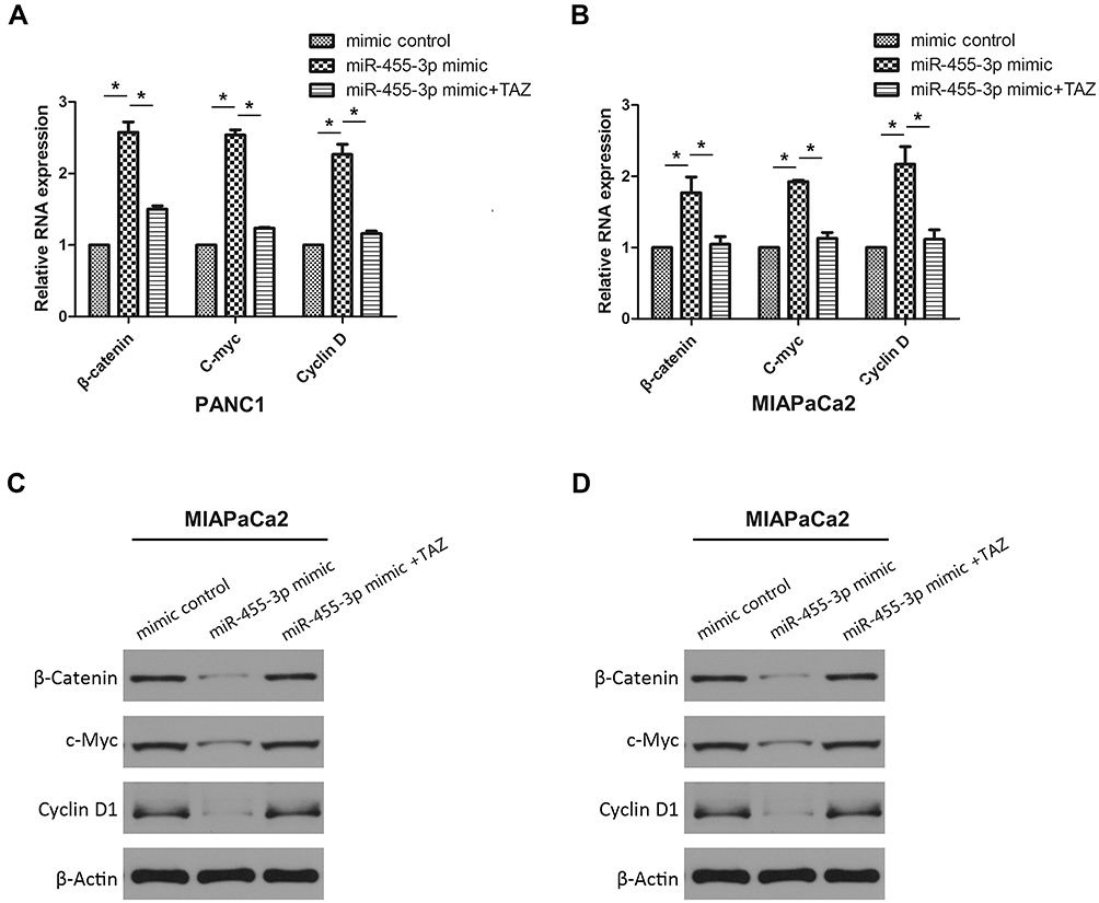 |
Figure 4 miR-455-3p restrains Wnt/β-catenin signaling via TAZ. (A, B) Expression of β-catenin, Cyclin D and C-myc in pancreatic cancer cells transfected with miR-455-3p mimic or miR-455-3p mimic plus TAZ expression vector was detected by qPCR. (C, D) Western blot analysis of β-catenin, Cyclin D and C-myc in PANC1 and MIAPaCa2 cells transfected with miR-455-3p mimic or miR-455-3p mimic plus TAZ expression vector β-actin is used as loading control vs miR-455-3p mimics/mimic control group, miR-455-3p mimics/miR-455-3p mimics+TAZ group. *p <0.05, miR-455-3p mimics vs mimic control group, miR-455-3p inhibitors vs inhibitors control group. |
Discussion
Micro-RNAs (miRNAs) are noncoding, single-stranded RNAs of around 22 nucleotides involved in the pathogenic mechanisms of carcinogenesis and metastasis.19 Based on the different genes targeted by different miRNAs in various types of cells, miRNAs may function as either oncogenes or tumor suppressors.20 Recently, the roles of miRNAs in the progression of PC are becoming recognized and a lot of work has focused on understanding how miRNAs regulate target genes in PC initiation, progression, metastasis, and relapse in addition to drug response and resistance in order to improve diagnosis, predict prognosis, and develop new therapeutic strategies.21 MicroRNA-455 is a family of miRNAs that are strongly associated with many biological processes.22–26 Specifically, accumulating evidence suggests that miR-455-3p acts as a tumor suppressor in several cancers including colorectal cancer, esophageal cancer, lung cancer, breast cancer and melanoma,6–10 suggesting that miR-455-3p might exert important effects on PC. Our previous studies have found that miR-455-3p is down-regulated in pancreatic cancer and is involved in regulating proliferation and drug resistance via targeting TAZ. However, the other functions associated with cancer progression and the potential mechanism of miR-455-3p in pancreatic cancer have not been explored.
In this study, we demonstrated that miR-455-3p contributes to cell apoptosis and inhibits cell migration, invasion and EMT in PC by directly suppressing TAZ expression and consequently inactivating Wnt/β-catenin signaling. We used flow cytometry to identify the effect on cell apoptosis in PC cells. The results showed that miR-455-3p overexpression prompted apoptosis and inhibited migration, invasion and EMT of PC cells, whereas miR-455-3p downregulation significantly inhibited cell apoptosis and prompted migration, invasion and EMT in both PANC-1 and MIAPaCa-2 cells. Furthermore, qPCR and Western Blot Analysis demonstrated that miR-455-3p promotes the apoptosis of pancreatic cancer cells by regulating apoptosis-related proteins Bcl-2, Bax and Caspase-3. An analysis of wound healing assay and Transwell assay revealed that miR-455-3p suppressed migration and invasion in pancreatic cancer cells. EMT is a biological process that plays a key role in the progression of many cancers, including in metastasis.27 Our research confirmed that miR-455-3p suppressed the EMT of PC. These data indicate that miR-455-3p functions as a tumor suppressor in PC.
TAZ is an important functional component of the Hippo pathway, a pathway associated with tumorigenesis.28 It has been widely accepted that TAZ acts as a potent oncogene in multiple cell types, including PC.29 In this paper, by transfecting pancreatic cancer cells with miR-455-3p mimics or miR-455-3p mimics plus TAZ expression vector, we confirmed that miR-455-3p exerts its anti-cancer effect through TAZ. The Wnt/β-catenin pathway has long been associated with tumorigenesis, tumor plasticity, and cancer stem cells, and high expression levels of the Wnt pathway has been associated with poor survival rates.30 Moreover, some studies have demonstrated that Wnt signaling is closely related to TAZ.17,18,31 β-catenin is a main member of the Wnt signaling pathway, and Cyclin D and C-myc are the major downstream effectors of this pathway.32 Therefore, we examined the expression of β-catenin, Cyclin D and C-myc. We found that overexpression of miR-455-3p suppressed β-catenin, Cyclin D and C-myc in cells, and TAZ reversed the inhibitory effect of miR-455-3p on the Wnt pathway. These findings suggest that miR-455-3p functions as a tumor suppressor by restraining Wnt/β-catenin signaling via TAZ in PC.
In summary, we provide convincing evidence that miR-455-3p directly targets TAZ to regulate cell apoptosis, migration, invasion and EMT in PC. Additionally,our study elucidates a novel mechanism by which miR-455-3p suppresses PC development by deactivating Wnt/β-catenin signaling via TAZ. We suggest that these findings may provide a promising target in the treatment of PC.
Author Contributions
All authors contributed to data analysis, drafting and revising the article, gave final approval of the version to be published, and agree to be accountable for all aspects of the work.
Funding
This study was financially supported from the Natural Science Foundation of Hubei Province in China 2018CFB338 (TZ), the Wuhan Health and Family Planning Commission Medical Research Project WX19Q40 (TZ), the Health Commission of Hubei Province scientific research project WJ2019H387 (TZ), the Central Guidance Local Science and Technology Development Special of Hubei Province 2019ZYYD067 (XH), and the Wuhan Health and Family Planning Commission Medical Research Project WX17C09 (QZ).
Disclosure
The authors declare no competing financial or non-financial interests.
References
1. Siegel RL, Miller KD, Jemal A. Cancer statistics, 2019. CA Cancer J Clin. 2019;69(1):7–34. doi:10.3322/caac.v69.1
2. Torres C, Grippo PJ. Pancreatic cancer subtypes: a roadmap for precision medicine. Ann Med. 2018;50(4):277–287. doi:10.1080/07853890.2018.1453168
3. Tiwari A, Mukherjee B, Dixit M. MicroRNA key to angiogenesis regulation: miRNA biology and therapy. Curr Cancer Drug Targets. 2018;18(3):266–277. doi:10.2174/1568009617666170630142725
4. Wang Y, Wang L, Chen C, et al. New insights into the regulatory role of microRNA in tumor angiogenesis and clinical implications. Mol Cancer. 2018;17(1):22. doi:10.1186/s12943-018-0766-4
5. Ebrahimi S, Hosseini M, Ghasemi F, et al. Circulating microRNAs as potential diagnostic, prognostic and therapeutic targets in pancreatic cancer. Curr Pharm Des. 2016;22(42):6444–6450. doi:10.2174/1381612822666160817095047
6. Zheng J, Lin Z, Zhang L, et al. MicroRNA-455-3p inhibits tumor cell proliferation and induces apoptosis in HCT116 human colon cancer cells. Med Sci Monit. 2016;22:4431–4437. doi:10.12659/MSM.898452
7. Liu A, Zhu J, Wu G, et al. Antagonizing miR-455-3p inhibits chemoresistance and aggressiveness in esophageal squamous cell carcinoma. Mol Cancer. 2017;16(1):106. doi:10.1186/s12943-017-0669-9
8. Gao X, Zhao H, Diao C, et al. miR-455-3p serves as prognostic factor and regulates the proliferation and migration of non-small cell lung cancer through targeting HOXB5. Biochem Biophys Res Commun. 2018;495(1):1074–1080. doi:10.1016/j.bbrc.2017.11.123
9. Li Z, Meng Q, Pan A, et al. MicroRNA-455-3p promotes invasion and migration in triple negative breast cancer by targeting tumor suppressor EI24. Oncotarget. 2017;8(12):19455–19466. doi:10.18632/oncotarget.14307
10. Chai L, Kang X-J, Sun -Z-Z, et al. MiR-497-5p, miR-195-5p and miR-455-3p function as tumor suppressors by targeting hTERT in melanoma A375 cells. Cancer Manag Res. 2018;10:989–1003. doi:10.2147/CMAR
11. Zhan T, Huang X, Tian X, et al. Downregulation of MicroRNA-455-3p links to proliferation and drug resistance of pancreatic cancer cells via targeting TAZ. Mol Ther Nucleic Acids. 2018;10:215–226. doi:10.1016/j.omtn.2017.12.002
12. Zanconato F, Cordenonsi M, Piccolo S. YAP and TAZ: a signalling hub of the tumour microenvironment. Nat Rev Cancer. 2019;19(8):454–464. doi:10.1038/s41568-019-0168-y
13. Zanconato F, Cordenonsi M, Piccolo S. YAP/TAZ at the roots of cancer. Cancer Cell. 2016;29(6):783–803. doi:10.1016/j.ccell.2016.05.005
14. El-Sahli S, Xie Y, Wang L, et al. Wnt signaling in cancer metabolism and immunity. Cancers (Basel). 2019;11(7):904. doi:10.3390/cancers11070904
15. Ghosh N, Hossain U, Mandal A, et al. The Wnt signaling pathway: a potential therapeutic target against cancer. Ann N Y Acad Sci. 2019;1443(1):54–74. doi:10.1111/nyas.2019.1443.issue-1
16. Tsao CM, Yan M-D, Shih Y-L, et al. SOX1 functions as a tumor suppressor by antagonizing the WNT/beta-catenin signaling pathway in hepatocellular carcinoma. Hepatology. 2012;56(6):2277–2287. doi:10.1002/hep.25933
17. Hansen CG, Moroishi T, Guan KL. YAP and TAZ: a nexus for Hippo signaling and beyond. Trends Cell Biol. 2015;25(9):499–513. doi:10.1016/j.tcb.2015.05.002
18. Melucci E, Casini B, Ronchetti L, et al. Expression of the Hippo transducer TAZ in association with WNT pathway mutations impacts survival outcomes in advanced gastric cancer patients treated with first-line chemotherapy. J Transl Med. 2018;16(1):22. doi:10.1186/s12967-018-1385-y
19. Brunetti O, Russo A, Scarpa A, et al. MicroRNA in pancreatic adenocarcinoma: predictive/prognostic biomarkers or therapeutic targets? Oncotarget. 2015;6(27):23323–23341. doi:10.18632/oncotarget.v6i27
20. Iorio MV, Croce CM. Causes and consequences of microRNA dysregulation. Cancer J. 2012;18(3):215–222. doi:10.1097/PPO.0b013e318250c001
21. Price C, Chen J. MicroRNAs in cancer biology and therapy: current status and perspectives. Genes Dis. 2014;1(1):53–63. doi:10.1016/j.gendis.2014.06.004
22. Qin L, Zhang Y, Lin J, et al. MicroRNA-455 regulates migration and invasion of human hepatocellular carcinoma by targeting Runx2. Oncol Rep. 2016;36(6):3325–3332. doi:10.3892/or.2016.5139
23. Zhang H, Guan M, Townsend KL, et al. MicroRNA-455 regulates brown adipogenesis via a novel HIF1an-AMPK-PGC1alpha signaling network. EMBO Rep. 2015;16(10):1378–1393. doi:10.15252/embr.201540837
24. Hong J, Zhou W, Wang X. Involvement of miR-455 in the protective effect of H2S against chemical hypoxia-induced injury in BEAS-2B cells. Pathol Res Pract. 2018;214(11):1804–1810. doi:10.1016/j.prp.2018.08.008
25. Arai T, Kojima S, Yamada Y, et al. Pirin: a potential novel therapeutic target for castration-resistant prostate cancer regulated by miR-455-5p. Mol Oncol. 2019;13(2):322–337. doi:10.1002/mol2.2019.13.issue-2
26. Yamada Y, Arai T, Kojima S, et al. Anti-tumor roles of both strands of the miR-455 duplex: their targets SKA1 and SKA3 are involved in the pathogenesis of renal cell carcinoma. Oncotarget. 2018;9(42):26638–26658. doi:10.18632/oncotarget.v9i42
27. Thiery JP, Acloque H, Huang RYJ, et al. Epithelial-mesenchymal transitions in development and disease. Cell. 2009;139(5):871–890. doi:10.1016/j.cell.2009.11.007
28. Misra JR, Irvine KD. The Hippo signaling network and its biological functions. Annu Rev Genet. 2018;52:65–87. doi:10.1146/annurev-genet-120417-031621
29. Moroishi T, Hansen CG, Guan KL. The emerging roles of YAP and TAZ in cancer. Nat Rev Cancer. 2015;15(2):73–79. doi:10.1038/nrc3876
30. Lopez-Knowles E, Zardawi SJ, McNeil CM, et al. Cytoplasmic localization of beta-catenin is a marker of poor outcome in breast cancer patients. Cancer Epidemiol Biomarkers Prev. 2010;19(1):301–309. doi:10.1158/1055-9965.EPI-09-0741
31. Wang C, Han X, Zhou Z, et al. Wnt3a activates the WNT-YAP/TAZ pathway to sustain CDX2 expression in bovine trophoblast stem cells. DNA Cell Biol. 2019;38(5):410–422. doi:10.1089/dna.2018.4458
32. Zhang Y, Sun M, Chen Y, et al. MiR-519b-3p inhibits the proliferation and invasion in colorectal cancer via modulating the uMtCK/Wnt signaling pathway. Front Pharmacol. 2019;10:741. doi:10.3389/fphar.2019.00741
 © 2020 The Author(s). This work is published and licensed by Dove Medical Press Limited. The
full terms of this license are available at https://www.dovepress.com/terms
and incorporate the Creative Commons Attribution
- Non Commercial (unported, 3.0) License.
By accessing the work you hereby accept the Terms. Non-commercial uses of the work are permitted
without any further permission from Dove Medical Press Limited, provided the work is properly
attributed. For permission for commercial use of this work, please see paragraphs 4.2 and 5 of our Terms.
© 2020 The Author(s). This work is published and licensed by Dove Medical Press Limited. The
full terms of this license are available at https://www.dovepress.com/terms
and incorporate the Creative Commons Attribution
- Non Commercial (unported, 3.0) License.
By accessing the work you hereby accept the Terms. Non-commercial uses of the work are permitted
without any further permission from Dove Medical Press Limited, provided the work is properly
attributed. For permission for commercial use of this work, please see paragraphs 4.2 and 5 of our Terms.