
Back to Journals » Clinical Interventions in Aging » Volume 21
Long-Term Surgical Outcomes and Influential Factors of Subthalamic Nucleus Deep Brain Stimulation for Dyskinesia in Parkinson’s Disease: A 3-Year Longitudinal Cohort Study
Authors Wang S  , Xue T, Ma R, Shi L, Zhu G, Jiang Y, Yang A, Meng F, Zhang J
Received 30 January 2026
Accepted for publication 15 May 2026
Published 22 May 2026 Volume 2026:21 600031
DOI https://doi.org/10.2147/CIA.S600031
Checked for plagiarism Yes
Review by Single anonymous peer review
Peer reviewer comments 5
Editor who approved publication: Dr Maddalena Illario
Shu Wang,1 Tao Xue,1 Ruoyu Ma,1 Lin Shi,1 Guanyu Zhu,1 Yin Jiang,2,3 Anchao Yang,1 Fangang Meng,1– 3 Jianguo Zhang1– 3
1Department of Neurosurgery, Beijing Tiantan Hospital, Capital Medical University, Beijing, People’s Republic of China; 2Beijing Neurosurgical Institute, Beijing Tiantan Hospital, Capital Medical University, Beijing, People’s Republic of China; 3Beijing Key Laboratory of Neurostimulation, Beijing, People’s Republic of China
Correspondence: Jianguo Zhang, Department of Neurosurgery, Beijing Tiantan Hospital, Capital Medical University, Beijing, People’s Republic of China, Email [email protected] Shu Wang, Department of Neurosurgery, Beijing Tiantan Hospital, Capital Medical University, Beijing, People’s Republic of China, Email [email protected]
Background: Subthalamic deep brain stimulation (STN-DBS) has emerged for Parkinson’s disease (PD), but its long-term effects on levodopa-induced dyskinesia (LID) remain poorly understood.
Objective: To assess the long-term LID outcomes and prognostic factors of STN-DBS.
Methods: A single-blind longitudinal cohort study was conducted in evaluating 84 PD patients with LID (mean age 61.89 years; 46.4% female; mean disease duration 10.30 years; and mean baseline levodopa-equivalent dose 854.16 mg/day) who underwent STN-DBS at Beijing Tiantan Hospital, Capital Medical University between 2019 and 2021. Assessments at baseline, 1-year (short-term), and 3-year (long-term) regarding motor symptoms, quality of life, neuropsychological status, and cognitive function were analyzed. Primary outcomes focused on LID symptoms (Unified Dyskinesia Rating Scale [UDysRS]). Multivariable linear regression identified prognostic factors.
Results: At 1-year, the UDysRS score improved significantly (74.4% reduction, P< 0.001), with sustained but diminished benefits at 3-year (64.9% reduction vs baseline, P< 0.001; 36.9% decline vs 1 year, P= 0.012). The time and functional impact of LID also improved initially (62.5% and 64.3% reduction) but worsened over time (38.8% and 33.3% decline). Motor function and quality of life showed similar trends, while neuropsychological symptoms improved stably even after long-term follow-up; and cognitive function remained unchanged. Multivariable regression identified diphasic dyskinesia as a negative prognostic factor (short-term std.β=− 0 .296; long-term std.β=− 0 .239), whereas a higher levodopa-equivalent dose (short-term std.β= 0.275; long-term std.β= 0.261) and greater levodopa responsiveness (short-term std.β= 0.215; long-term std.β= 0.216) predicted better short- and long-term results. A longer disease duration correlated with worse long-term outcomes (std.β=− 0 .212).
Conclusion: STN-DBS was associated with significant long-term improvements in LID, although the effectiveness gradually declined. The identified prognostic factors help in patient selection and counseling.
Keywords: Parkinson’s disease, dyskinesia, movement disorders, deep brain stimulation, neuromodulation
Introduction
Parkinson’s disease (PD), the second most common neurodegenerative disorder, affects more than 10 million individuals1,2 and imposes a significant burden on patients and healthcare systems globally.3,4 While levodopa remains the cornerstone of symptomatic treatment,2,5 its long-term use is frequently complicated by levodopa-induced dyskinesia (LID),6 a debilitating motor complication that affects approximately 50%–80% of patients with PD after 5–10 years of therapy initiation, causing involuntary movements.7 Symptoms such as LID severely impair quality of life, limit therapeutic efficacy, and represent a major challenge in PD management.8,9 Subthalamic nucleus deep brain stimulation (STN-DBS) has emerged as an effective treatment for advanced PD10–12 and has demonstrated robust efficacy in alleviating motor fluctuations and LID.2,12–14 However, while the short-term benefits of STN-DBS are well documented, evidence of its long-term dyskinesia outcomes for patients with PD is limited,15–17 and critical knowledge gaps persist regarding its prognostic factors.15,18,19
The antidyskinetic effect of STN-DBS is thought to be mediated both indirectly, by enabling substantial reductions in dopaminergic medication,20,21 and directly, through modulation of pathological basal ganglia oscillatory activity.22,23 However, existing studies on STN-DBS for LID have focused predominantly on short-term outcomes (≤ 12 months).15,21,24 Although these studies demonstrated significant improvements in motor symptoms and a reduction in LID, the durability of these benefits over extended periods remains understudied.15,24 While some studies have suggested that the therapeutic effects remain,25,26 others have raised concerns that STN-DBS may lose effectiveness for LID over long-term follow-up due to the progressive nature of PD, neurostimulation-related adaptations, and increasing levodopa dosage again,16,27,28 necessitating longitudinal evaluation to inform clinical decision-making. In addition, while several prior studies have explored the long-term effects of STN-DBS on LID, the heterogeneity in assessment scales used to evaluate dyskinesia limits comparability.16,25–28 Crucially, no large-scale studies have systematically analyzed the influential factors of both short- and long-term LID outcomes following STN-DBS,10,18 and clinicians lack reliable evidence to optimize patient selection and postoperative counseling.18 Furthermore, while STN-DBS is known to improve motor symptoms, its long-term effects on neuropsychological outcomes and cognitive function, particularly in patients with preexisting LID, remain poorly characterized.6,21,24 Current evidence is also constrained by methodological limitations, including small sample sizes, unblinded outcome assessments, and heterogeneous evaluation protocols, which compromise the generalizability and reliability of findings.15,20,28
To address these knowledge gaps, we conducted a rigorous retrospective cohort study with extended follow-up (1-year and 3-year intervals) in 84 consecutive PD patients with medically refractory LID who underwent STN-DBS. Using validated standardized rating scales (Unified Dyskinesia Rating Scale [UDysRS]24) with single-blind assessment and analyses (observer-blinded) to minimize rater bias,29 we comprehensively evaluated longitudinal trajectories of LID symptoms, motor symptoms, quality of life neuropsychological status, and cognitive function and investigated potential prognostic factors that may influence short- and long-term dyskinesia outcomes. By combining a robust study design with a substantial sample size, this research offers valuable insights into the outcomes of STN-DBS for patients with PD and LID and identifies key prognostic factors, thereby contributing to informed clinical decision-making and future practice improvements.16
Methods
Patients and Eligibility Criteria
This single blind longitudinal retrospective cohort study analyzed prospectively collected clinical data that were entered into a structured database at the time of patient visits.14 All patients with PD and LID who underwent STN-DBS at Beijing Tiantan Hospital, Capital Medical University, from January 2019 to December 2021 were consecutively screened and evaluated. Patients were included in this study if they met the following conditions: (1) diagnosed with idiopathic PD according to the Movement Disorders Society (MDS) Clinical Diagnostic Criteria30 and the UK Brain criteria31 with the presence of disabling LID symptoms (with typical clinical symptoms such as levodopa-induced involuntary chorea/choreoathetoid movements6 and a total score greater than 0 on the UDysRS24) not optimized with anti-PD medications, confirmed by at least two qualified senior movement disorder specialists; (2) received bilateral STN-DBS between January 2019 and December 2021 at Beijing Tiantan Hospital, Capital Medical University; and (3) without dementia, severe psychiatric disorders, severe brain atrophy or cerebral ischemic lesions, or systemic diseases that interfered with the surgery. The exclusion criteria were (1) patients without written informed consent; (2) patients with major missing baseline or follow-up data; or (3) patients who finished less than 3 years of follow-up. Patients with missing data or insufficient follow-up were contacted first for further review; those unreachable due to changed contact details were classified as lost to follow-up. The data collection process was finished by December 2024.
Surgical Procedure
The surgical procedure for DBS implantation has been previously described in detail in our previous studies.32,33 On the basis of the patients’ symptoms, all included patients were bilaterally implanted with electrodes during the same surgery. In general, on the day of surgery, patients were mounted with a Leksell-G stereotactic frame (Elekta Instrument AB, Stockholm, Sweden) for a 0.625-mm consecutive CT scan (Revolution CT Scanner, GE Healthcare, Chicago, Illinois), which was then merged with preoperative 3-T MR images (MAGNETOM Verio MRI Scanner, Siemens, Berlin, Germany) to visually target the STN coordinates and trajectory. After that, the stimulation electrodes (Medtronic-3389, Medtronic, Minneapolis, Minnesota; or PINS-L301, PINS Medical, Beijing, China; with the same designs as quadripolar contacts, length of 1.5 mm, spaced 0.5 mm apart) were implemented with microelectrode recordings and intraoperative high-frequency macrostimulation testing for further target refinement under local anesthesia. An implantable pulse generator (IPG; Activa RC, Medtronic, Minneapolis, Minnesota; or Model G102R, PINS, PINS Medical, Beijing, China) was then implanted and connected with the intracranial leads through subcutaneous extension wires positioned in the left subclavian region on the same day under general anesthesia.
A postoperative CT scan was performed to confirm the lead position and to evaluate potential surgical complications. The IPG was initiated approximately 1 month later to avoid the microthalamotomy effect.29 Patients were tested in unipolar mode first (with lead contact as the cathode [-] and IPG as the anode [+]). In the case of intolerable side effects, it was switched into bipolar mode (different contacts at the same electrode as the cathode and anode). A specialized multidisciplinary team was responsible for the programming and further adjustments in the follow-ups as needed to achieve the best optimization of therapeutic and adverse effects.
Clinical Assessment
All patient baseline clinical records, including sex, age at surgery, disease duration, and age at onset (those who had an age at onset < 40 years were defined as having young-onset PD34,35), were recorded. In addition, LID and PD phenotypes were further classified by experienced movement specialists. The LID symptoms were categorized as peak-dose dyskinesia, diphasic dyskinesia, or off-period dystonia according to the relationships between their occurrence time and levodopa treatment as well as their typical symptoms.6,8,36 One or several different types of LID can occur in the same patient.8,36 Motor subtypes of PD were classified as tremor dominant (TD), postural instability/gait difficulty (PIGD), or indeterminate on the basis of the MDS-sponsored revision of the Unified Parkinson’s Disease Rating Scale (MDS-UPDRS; TD/PIGD score ratio ≥ 1.15 for TD, ratio ≤ 0.90 for PIGD, and others for indeterminate).37,38
All postoperative evaluations were performed by independent, trained experienced neuropsychologists who were blinded to the patients’ baseline clinical data, stimulation parameters, and the specific aims of the study (observer-blinded design). Patients were evaluated with validated standardized Chinese versions of assessment scales at (preoperative) baseline and each year postoperatively. The outcomes of short-term (1 year) and long-term (3 years) follow-up were collected for analysis. For motor evaluations, movement disorder specialists evaluate the clinically defined “off-medicine” and “on-medication” conditions. The “off-medication” state was evaluated after at least a 12-hour period without medication, whereas the “on-medication” state was assessed approximately one hour after medication intake, when its benefits were at the peak.12,18,39 The levodopa responsiveness (%) was calculated for the included patients as a percentage of part III of the MDS-UPDRS “off-medicine” score minus the “on-medicine” score divided by the “off-medicine” score at baseline.40 Postoperative outcomes were assessed during a “stimulation-on” phase after a minimum of one week of continuous stimulation with no parameter adjustments under the abovementioned “off-medicine” and “on-medication” conditions.18,39
Surgical Outcomes
The primary outcome of this study was dyskinesia outcomes, which were assessed by the UDysRS (maximum 104 points, higher scores for more severe symptoms of dyskinesia).41,42 We also calculated patients’ time spent with LID on the basis of the sum of the MDS-UPDRS item 4.1 (total hours with dyskinesia other than off-period dystonia) and the MDS-UPDRS item 4.6 (total hours with off-period dystonia) and evaluated the functional impact of LID on the basis of the MDS-UPDRS item 4.2 (scores ranging from 0--4, with higher scores indicating a more severe impact of LID on the patient’s daily function).43
We also evaluated other motor outcomes, quality of life, neuropsychological status, and cognitive function as secondary outcomes. Assessments for motor outcomes included part III (motor examination) of the MDS-UPDRS (MDS-UPDRS-III; maximum 132 points, higher scores for more severe motor symptoms) in the “off-medicine” and “on-medicine” conditions,43,44 part II (daily living) of the MDS-UPDRS (MDS-UPDRS-II; maximum 52 points, higher scores for more severe impact on daily living activities),43,44 part IV (motor complications) of the MDS-UPDRS (MDS-UPDRS-IV; maximum 24 points, higher scores for more severe motor complications),43,44 the Hoehn & Yahr scale (stages 1–5, higher stages for more severe impairment) in the “off-medicine” condition,45 and the levodopa equivalent daily dose (LEDD, calculated by the latest 2023 MDS conversion formulae).46
The quality of life of the patients was evaluated via the Parkinson Disease Questionnaire-39 (PDQ-39; maximum 156 points, higher scores for worse health-related quality of life).47 In addition, neuropsychological status was assessed by part I (nonmotor experiences) of the MDS-UPDRS (MDS-UPDRS-I; maximum 52 points, higher scores for more severe nonmotor symptoms),43,44 the Hamilton Anxiety Scale (HAM-A; maximum 56 points, higher scores for more severe anxiety symptoms)48 and the Hamilton Depression Scale (HAM-D; maximum 76 points, higher scores for more severe depression symptoms),49 and cognitive function was evaluated by the Chinese Mini-Mental Status Exam (CMMS; maximum 30 points; lower scores for possible cognitive impairment)50 and the Montreal Cognitive Assessment (MoCA; maximum 30 points; lower scores for possible cognitive impairment). Any adverse events (AEs), including those related to the surgery, stimulation, or device, and their prognoses were also recorded.
Statistical Analysis
The assumption of a normal distribution was tested with the Kolmogorov‒Smirnov test, and Levene’s test was used for homogeneity of variance. On the basis of the variable types and distributions, means (± standard deviations, SDs) or numbers (percentages) were used for descriptive statistics. Given the potential clinical relevance of all variables included in the regression model, we employed complete-case analysis (listwise deletion) rather than data imputation to maintain analytical integrity and avoid potential bias from estimated values.
A series of one-way repeated-measures ANOVAs (or Friedman test, for ordinal variables such as Hoehn & Yahr scales), including time as a within-subjects factor (3 levels: baseline [preoperative], 1 year postoperative [short-term follow-up], and 3 years postoperative [long-term follow-up]), with post hoc pairwise comparisons adjusted by Bonferroni correction, was performed to analyze whether there were significant differences among the different periods. Furthermore, to identify potential influential factors for short-term (1 year) and long-term (3 years) dyskinesia outcomes, multivariable linear regression was performed. Sphericity was tested with Mauchly’s test; when violated, the Greenhouse-Geisser correction was applied. As our dataset was balanced across the three time points, repeated-measures ANOVA was appropriate; however, future analyses could consider linear mixed-effects models to handle potential missing data and intra-subject variability more flexibly.
All observations per independent variable (EPV) were confirmed with an adequate sample size (all EPVs ≥ 10). Univariable linear regression was performed first, and those variables with P < 0.10 were then entered into the multivariable linear regression (backward). The model-correlated independent variables were excluded to avoid multicollinearity. The β coefficient and its 95% confidence interval (CI), as well as the standardized β coefficient, were reported for the independent factors. Sensitivity analysis was performed by switching the regression model into enter method. Collinearity was assessed using variance inflation factors (VIF); all VIF values were <5, confirming the absence of harmful multicollinearity. Model diagnostics included inspection of residual plots and normality tests.
All the statistical tests were two-tailed, and P (or Bonferroni correction adjusted P) < 0.05 was regarded as statistically significant. All the statistical analyses and figures were generated via SPSS (IBM, Armonk, New York; version 27) and GraphPad Prism (GraphPad Software, San Diego, California; version 10).
Results
Baseline Characteristics
A total of 105 patients who were diagnosed with PD and disabling LID and who underwent bilateral STN-DBS surgery within the enrollment period were retrieved and evaluated for eligibility. After the exclusion of 21 patients because of a lack of informed consent (n = 3), missing baseline data (n = 5), and less than 3 years of follow-up (n = 13; 10 patients with missing follow-up data, 1 patient who died with no relationship with the surgery, and 2 patients who were explanted from the IPG due to infection and finished less than 3 years of follow-up), 84 patients (39 females, 46.4%) were ultimately included in the analysis. All included patients were confirmed to have stimulation electrodes correctly placed in the bilateral STN through neuroimaging. No known surgical-related mortality or morbidity was reported during the follow-up period. Figure 1 shows the details of the selection and inclusion of the patients.
 |
Figure 1 Flow chart of the study and selection and inclusion of patients. Abbreviations: PD, Parkinson’s disease; LID, levodopa-induced dyskinesia; STN-DBS, subthalamic nucleus deep brain stimulation. |
The baseline clinical characteristics of the included patients are described in Table 1 and baseline assessments are shown in Table 2. After a mean disease duration of 10.30 ± 3.02 years, these patients underwent surgery at a mean age of 61.89 ± 6.69 years. The mean age at PD onset was 51.59 ± 6.82 years, and 15.5% (13) of the patients were considered to have young-onset PD. For LID symptoms, 79.8% (67), 21.4% (18), and 26.2% (22) of the patients experienced peak-dose dyskinesia, diphasic dyskinesia, and off-period dystonia, respectively. The baseline mean score of the UDysRS scale was 26.73 ± 17.02, the mean duration of LID was 5.36 ± 2.73 hours, and the mean score of the functional impact of LID was 2.44 ± 0.95. With respect to other motor symptoms of PD, 25.0%, 61.9%, and 13.1% of the patients were considered TD, PIGD, and indeterminate phenotypes, respectively. The mean scores of the MDS-UPDRS-III (off-medicine), MDS-UPDRS-III (on-medicine), MDS-UPDRS-II, and MDS-UPDRS-IV were 52.65 ± 14.84, 22.63 ± 8.81, 20.12 ± 5.14, and 8.38 ± 3.89, respectively. In addition, 7.1% [6], 21.4% [18], 67.9% [57], and 3.6% [3] of patients were assessed with Hoehn & Yahr stages 2, 2.5, 3, and 4, respectively. The mean LEDD was 854.16 ± 387.24. In addition, other baseline assessments suggested mean scores of 62.95 ± 25.68 for the PDQ-39 for quality of life; 15.14 ± 4.39, 17.27 ± 8.93, and 16.18 ± 8.51 for the MDS-UPDRS-I, HAM-A, and HAM-D for neuropsychological status; and 27.10 ± 2.05 and 26.30 ± 2.07 for the CMMS and MoCA for cognitive function, respectively. No severe psychiatric symptoms or cognitive decline were observed in the current cohort.
 |
Table 1 Baseline Demographic and Clinical Characteristics of the Included Patients (n = 84) with Parkinson’s Disease (PD) and Levodopa-Induced Dyskinesia (LID) Who Underwent Subthalamic Nucleus Deep Brain Stimulation (STN-DBS) |
 |
Table 2 Short (1 Year) and Long (3 Years) Term Dyskinesia Outcome, Other Motor Outcomes, Quality of Life, Neuropsychological Outcomes, and Cognitive Functions of the Included Patients (n = 84) with Parkinson’s Disease (PD) and Levodopa-Induced Dyskinesia (LID) Who Underwent Subthalamic Nucleus Deep Brain Stimulation (STN-DBS) |
Surgical Outcomes
The surgical outcomes are shown in Table 2 and are illustrated in Figure 2 (for the scores of the original scales) and Figure 3 (for the relative changes). Overall, there were significant differences among the baseline, 1-year, and 3-year follow-ups regarding dyskinesia, motor, neuropsychological outcomes, and quality of life (all P < 0.001), while cognitive functions were not significantly changed (all P > 0.05). The UDysRS, time spent with LID, and functional impact of LID improved significantly by 74.4%, 62.5%, and 64.3%, respectively, from baseline to short-term follow-up (all P < 0.001), and the improvement remained significant (all P < 0.001) after long-term follow-up (improved 64.9%, 48.0%, and 52.5% from baseline for the UDysRS, LID time, and LID impact, respectively). However, these benefits declined significantly from short- to long-term follow-up by 36.9% (P = 0.012), 38.8% (P = 0.007), and 33.3% (P < 0.001) in the scores/hours of UDysRS, LID time, and LID impact, respectively.
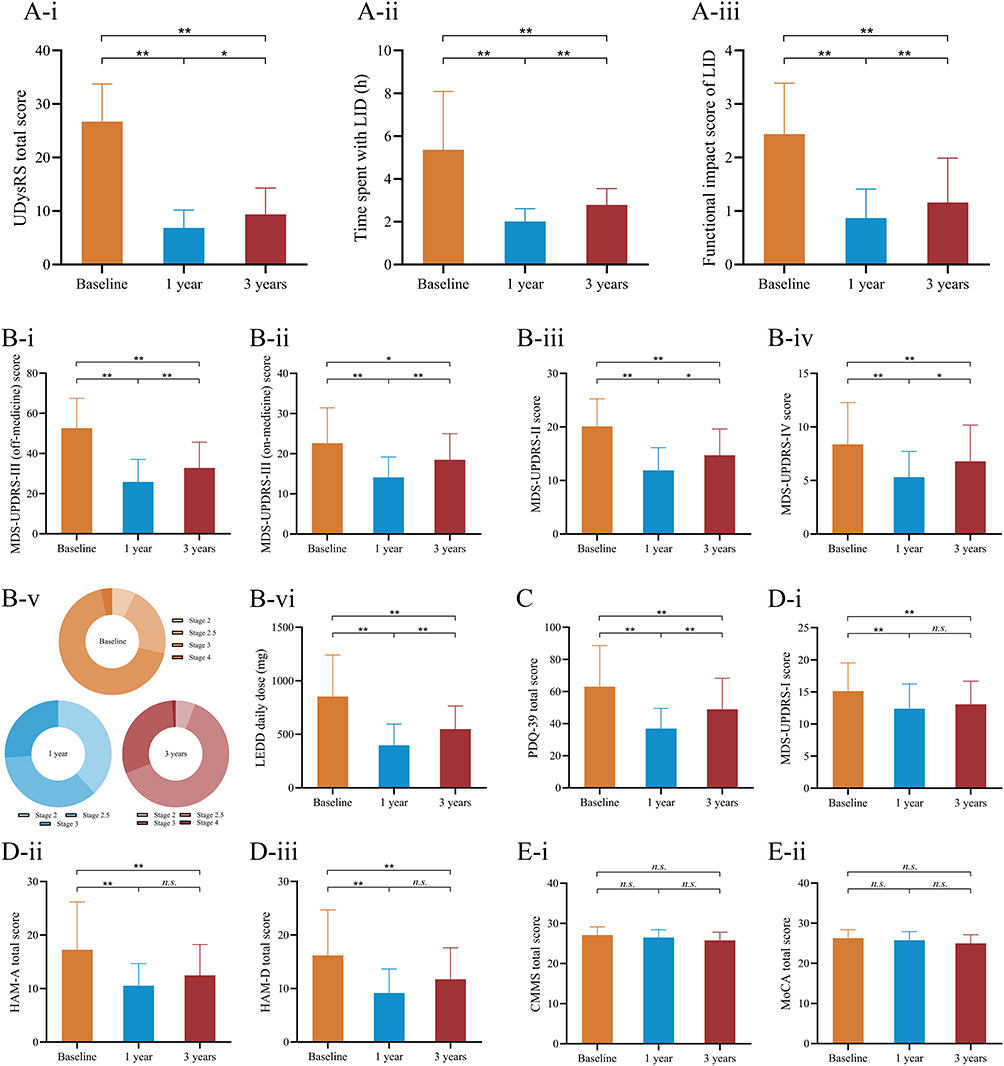 |
Figure 2 Short-term (1 year) and long-term (3 years) dyskinesia outcomes, other motor outcomes, quality of life, neuropsychological outcomes, and cognitive functions of the included patients (n = 84) with Parkinson’s disease (PD) and levodopa-induced dyskinesia (LID) who underwent subthalamic nucleus deep brain stimulation (STN-DBS). (A) Dyskinesia outcomes such as UDysRS (A-i), time spent with LID (MDS-UPDRS-4.1), hours (A-ii), and functional impact of LID (MDS-UPDRS-4.2; A-iii); (B) Other motor outcomes such as MDS-UPDRS-III (off-medicine; (B-i)), MDS-UPDRS-III (on-medicine; (B-ii)), MDS-UPDRS-II (daily living, (B-iii)), MDS-UPDRS-IV (motor complications; (B-iv)), Hoehn & Yahr (off-medicine; (B-v)), and LEDD, mg (B-vi); (C) Quality of life as PDQ-39; (D) Neuropsychological outcomes such as MDS-UPDRS-I (nonmotor experiences; (D-i), HAM-A (D-ii), and HAM-D (D-iii); and (E) Cognitive function as CMMS (E-i) and MoCA (E-ii). *P < 0.05 (one-way repeated-measures ANOVA, with post hoc pairwise comparisons adjusted by Bonferroni correction); **P < 0.01 (one-way repeated-measures ANOVA, with post hoc pairwise comparisons adjusted by Bonferroni correction). Abbreviations: n.s., not significant. PD, Parkinson’s disease; LID, levodopa-induced dyskinesia; STN-DBS, subthalamic nucleus deep brain stimulation; UDysRS, Unified Dyskinesia Rating Scale; MDS-UPDRS, Movement Disorder Society-sponsored revision of the Unified Parkinson’s Disease Rating Scale; LEDD, levodopa-equivalent daily dose; PDQ-39, Parkinson Disease Questionnaire-39; HAM-A, Hamilton Anxiety Rating Scale; HAM-D, Hamilton Depression Rating Scale; CMMS, Chinese Mini-Mental Status Examination; MoCA, Montreal Cognitive Assessment. |
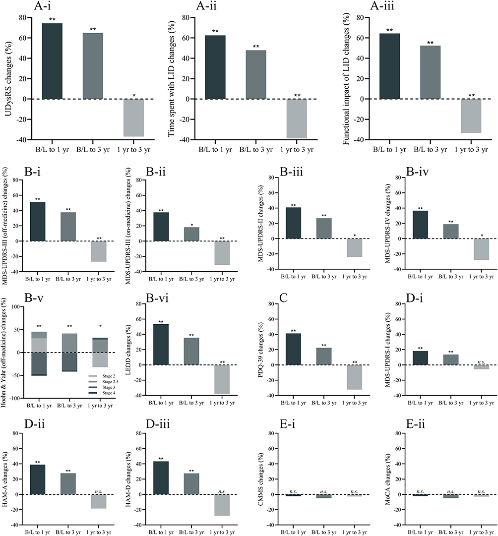 |
Figure 3 Relative changes (%) from baseline to short (1 year) term, baseline to long (3 years) term, and short (1 year) term to long (3 years) term dyskinesia outcomes; other motor outcomes; quality of life; neuropsychological outcomes; and cognitive functions of the included patients (n = 84) with Parkinson’s disease (PD) and levodopa-induced dyskinesia (LID) who underwent subthalamic nucleus deep brain stimulation (STN-DBS). (A) Dyskinesia outcomes such as UDysRS (A-i), time spent with LID (MDS-UPDRS-4.1), hours (A-ii), and functional impact of LID (MDS-UPDRS-4.2; (A-iii)); (B) Other motor outcomes such as MDS-UPDRS-III (off-medicine; (B-i)), MDS-UPDRS-III (on-medicine; (B-ii)), MDS-UPDRS-II (daily living, (B-iii)), MDS-UPDRS-IV (motor complications; (B-iv)), Hoehn & Yahr (off-medicine; (B-v)), and LEDD, mg (B-vi); (C) Quality of life as PDQ-39; (D) Neuropsychological outcomes such as MDS-UPDRS-I (nonmotor experiences; (D-i)), HAM-A (D-ii), and HAM-D (D-iii); and (E) Cognitive function as CMMS (E-i) and MoCA (E-ii). *P < 0.05 (one-way repeated-measures ANOVA or Friedman test, as appropriate, with post hoc pairwise comparisons adjusted by Bonferroni correction); **P < 0.01 (one-way repeated-measures ANOVA or Friedman test, as appropriate, with post hoc pairwise comparisons adjusted by Bonferroni correction). Abbreviations: n.s., not significant. PD, Parkinson’s disease; LID, levodopa-induced dyskinesia; STN-DBS, subthalamic nucleus deep brain stimulation; B/L, baseline; UDysRS, Unified Dyskinesia Rating Scale; MDS-UPDRS, Movement Disorder Society-sponsored revision of the Unified Parkinson’s Disease Rating Scale; LEDD, levodopa-equivalent daily dose; PDQ-39, Parkinson Disease Questionnaire-39; HAM-A, Hamilton Anxiety Rating Scale; HAM-D, Hamilton Depression Rating Scale; CMMS, Chinese Mini-Mental Status Examination; MoCA, Montreal Cognitive Assessment. |
Similar findings were also revealed regarding other motor outcomes. While MDS-UPDRS-III (off-medicine), MDS-UPDRS-III (on-medicine), MDS-UPDRS-II, MDS-UPDRS-IV, and LEDD suggested significant improvements in both short-term (by 50.9%, 37.7%, 41.0%, 36.6%, and 53.5%, respectively) and long-term (37.7%, 18.2%, 26.8%, 19.0%, and 35.7%, respectively) follow-up, their effects also worsened significantly by 27.0%, 31.4%, 24.0%, 27.9%, and 38.4% in the scores (all P < 0.05), respectively. The Hoehn & Yahr stages also significantly improved from baseline to 1-year (P < 0.001) and 3-year (P = 0.004) follow-up, with also diminished benefits with time (P = 0.049). A post‑hoc analysis revealed a significant positive correlation between the increase in LEDD and the worsening of the UDysRS total score from 1 year to 3 years (r = 0.34, P = 0.002; Pearson’s correlation), suggesting that the requirement for higher levodopa doses partially contributes to the attenuation of antidyskinetic benefit over time.
In terms of quality of life, the PDQ-39 score significantly improved by 41.3% (P < 0.001) from baseline at the short-term follow-up, and after the score decreased by 22.4% (P = 0.005), it improved by 22.4% (P < 0.001) after long-term follow-up. The outcomes of neuropsychological status revealed significant improvements in nonmotor symptoms of PD (assessed by the MDS-UPDRS-I), anxiety symptoms (assessed by the HAM-A), and depression symptoms (assessed by the HAM-D) at both short-term (improved by 18.2%, 39.0%, and 43.3%, respectively) and long-term (improved by 13.6%, 27.7%, and 27.5%, respectively) follow-ups (all P < 0.05), and short- and long-term outcomes were comparable (all P > 0.05). With respect to cognitive function (assessed by the CMMS and MoCA), no significant differences were revealed among baseline and different follow-up periods (all P > 0.05).
All AEs are reported in Supplementary Table S1. During the follow-up period, the most common surgery-related AE was infection (3.6%, n = 3), whereas the most common stimulation-related AEs were dyskinesia (6.0%, n = 5) and insomnia (6.0%, n = 5). All AEs were successfully resolved; infections were managed with surgical debridement and, in two cases, IPG explantation, while stimulation induced AEs were controlled through reprogramming. One patient experienced a small intracerebral hemorrhage that resolved without sequelae. No surgical mortality or permanent morbidity occurred. Patients who underwent IPG explantation were not included in the 3-year efficacy analysis due to incomplete follow up.
Prognostic Factors
Univariate analyses revealed the following preoperative clinical factors that potentially influence short-term dyskinesia outcomes: LID type, LEDD, and levodopa responsiveness (Supplementary Table S2). After multivariable linear regression models were adjusted, having diphasic dyskinesia (adjusted β = −16.932, 95% CI: −28.331–5.533, standardized β = −0.296, P = 0.004) was a negative factor for short-term dyskinesia outcomes, whereas a higher LEDD (adjusted β = 0.017, 95% CI: 0.005 to 0.029, standardized β = 0.275, P = 0.008) and greater levodopa responsiveness (adjusted β = 0.227, 95% CI: 0.016 to 0.438, standardized β = 0.215, P = 0.035) at baseline were independent positive prognostic factors (Table 3).
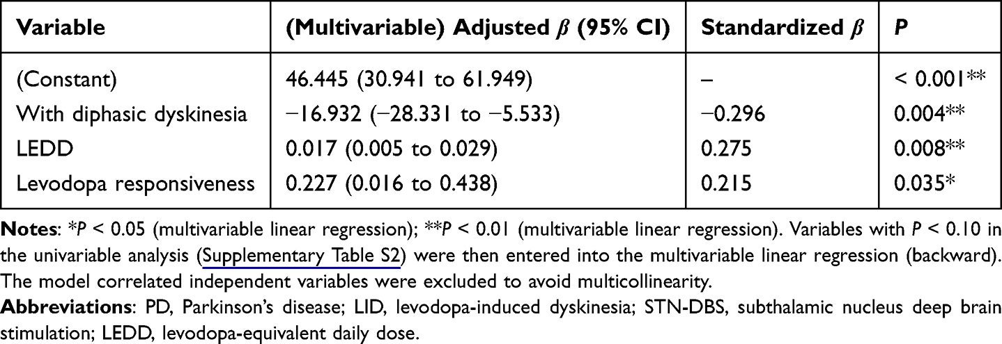 |
Table 3 Multivariable Linear Regression for Influential Factors of Short (1 Year) Term Dyskinesia Outcome for the Included Patients (n = 84) with Parkinson’s Disease (PD) and Levodopa-Induced Dyskinesia (LID) Who Underwent Subthalamic Nucleus Deep Brain Stimulation (STN-DBS) |
With respect to long-term follow-up, disease duration, LID type, LEDD, and levodopa responsiveness were identified as potential factors in the univariable analyses (Supplementary Table S3). A multivariate linear regression model confirmed that longer disease duration (adjusted β = −1.012, 95% CI: −1.995 to −0.029, standardized β = −0.212, P = 0.044) and diphasic dyskinesia (adjusted β = −13.607, 95% CI: −24.763 to −2.451, standardized β = −0.239, P = 0.017) were associated with worse dyskinesia outcomes, whereas higher LEDD (adjusted β = 0.016, 95% CI: 0.004 to 0.028, standardized β = 0.261, P = 0.009) and greater levodopa responsiveness (adjusted β = 0.226, 95% CI: 0.011 to 0.441, standardized β = 0.216, P = 0.040) were associated with better dyskinesia outcomes (Table 4). Collinearity diagnostics revealed VIF values ranging from 1.03 to 1.41. Sensitivity analyses using forced entry models yielded consistent results.
 |
Table 4 Multivariable Linear Regression for Influential Factors of Long (3 Years) Term Dyskinesia Outcome for the Included Patients (n = 84) with Parkinson’s Disease (PD) and Levodopa-Induced Dyskinesia (LID) Who Underwent Subthalamic Nucleus Deep Brain Stimulation (STN-DBS) |
Discussion
The UDysRS provides a comprehensive method for evaluating dyskinesia symptoms in PD patients with excellent clinical properties;41,51 it has been recommended by the MDS and widely verified among different PD groups and for assessing surgical outcomes.24,52,53 Juhász et al analyzed 71 patients with PD and LID who underwent bilateral STN-DBS and revealed that the total UDysRS score significantly decreased from 38.0 ± 17.8 to 10.8 ± 13.0 (improved by approximately 71.6%) at 1 year after follow-up and suggested that UDysRS could reliably detect surgical improvements.24 Fan et al revealed that STN-DBS significantly improved the UDysRS score by 60.73 ± 40.29% after a mean 21.60 ± 8.79 months of follow-up in 20 PD patients with preoperative LID.21 A cohort study with a longer follow-up period suggested that dystonia and peak-dose dyskinesia could be improved even after 5 years of STN-DBS in 33 patients, as evaluated by the UPDRS part IV and the CAPSIT-PD scales.54 This study documented 74.4% and 64.9% dyskinesia improvement at 1-year and 3-year follow-ups respectively in PD patients with LID, utilizing standardized UDysRS assessments to fill an important research gap in longitudinal STN-DBS outcomes. In summary, our results align with those of prior studies suggesting STN-DBS as a cornerstone therapy for patients with PD and preexisting LID in mitigating dyskinesia and motor fluctuations for a long-term period.
However, the observed decline in benefits over time, as evidenced by a 36.9% rebound in dyskinesia outcomes, echoes concerns raised by Limousin et al16 and others regarding the progressive nature of PD, adaptations of the stimulation, and increasing levodopa dosage again.16,27,28,55,56 Compared with the 5-year outcomes reported by Simonin et al54 and the randomized comparison by Follett et al57 our 3-year data show a comparable trend of early, substantial benefit with subsequent partial decline, underscoring the need for continuous multidisciplinary management. Notably, in addition to the improvements in dyskinesia outcomes over time, scores on the MDS-UPDRS-III scale worsened faster under on-medicine conditions than under off-medicine conditions (31.4% vs 27.0%, respectively), even with a 38.4% increase in LEDD. This may reflect that the resurgence of levodopa requirements could only partially explain the decline in dyskinesia outcomes. Another potential cause could be the degeneration of nondopaminergic pathways, such as the cholinergic system at the midbrain, forebrain, and cortical level,58,59 in the development of levodopa refractory or stimulation-resistant symptoms with the progression of PD.16 This highlights the complex interplay between disease progression, neurostimulation adaptation, and pharmacological management.16,27,28
Furthermore, the sustained improvements in anxiety and depression scores underscore the stable psychological benefits of STN-DBS, which are likely mediated through motor symptom alleviation and increased psychosocial functioning. However, although current study suggested non statistical significance in changes of cognitive function, these results must be interpreted with caution, as the CMMS and MoCA are screening tools that are susceptible to ceiling effects and may not detect subtle declines in specific cognitive domains such as verbal fluency, executive function, or working memory.60,61 The absence of a significant change in these scores should therefore not be taken as definitive evidence of cognitive preservation. Dedicated neuropsychological batteries in future studies are essential to fully elucidate the cognitive effects of chronic STN DBS. Previous studies presented inconsistent findings regarding the cognitive effects of DBS in PD patients, with some studies reporting stable cognitive function14,62,63 while others observe mild cognitive decline (especially long-term memory, verbal fluency, and phonemic fluency).64,65 These discrepancies may be attributed to differences in assessment tools, potential ceiling effects, natural age-related cognitive deterioration, and the potential pathological changes associated with PD.55,66 A prospective controlled study is still needed to better understand the cognitive effects of DBS for PD, which can be a future research direction.
Diphasic dyskinesia was identified as a negative prognostic marker for both short- and long-term dyskinesia outcomes. Managing medication for diphasic dyskinesia can be difficult because simply reducing the dose of levodopa may not be sufficient to alleviate the symptoms caused by fluctuations in drug concentration and changes in dopamine receptor sensitivity.67,68 Therefore, for patients with diphasic dyskinesia, fractionation of the levodopa dosage and combination with other types of medications, such as COMT inhibitors, MAO-B inhibitors, dopamine agonists, and apomorphine, should be carefully considered after STN-DBS surgery.5,8,67,69 In addition, long disease duration was also identified as a negative factor of long-term dyskinesia outcomes, which might be related to the long-term reduction in dopaminergic receptor sensitivity.6 Kim et al also suggested that the disease duration at surgery was longer in patients whose diphasic dyskinesia persisted after STN-DBS than in those whose symptoms disappeared.70 They also suggested that younger patients at onset had worse outcomes,70 which was not observed in the present study. This might be related to the fact that the present study included only a limited number of patients with young-onset PD (15.5%). Conversely, higher baseline LEDD and levodopa responsiveness emerged as positive predictors. Patients with higher baseline LEDDs tend to have greater postoperative decreases in medications, thus resulting in better remission of LID.24 However, it should be reiterated that our analysis tracked total LEDD rather than the adjustments of individual medication classes (eg., amantadine, dopamine agonists) due to unavailable data, which limits the ability to disentangle the specific contribution of each drug to dyskinesia improvement; therefore, future studies should systematically record and analyze medication class specific changes to refine prognostic models.6,8,67,68 Given the potential influence of medication class adjustments on dyskinesia outcomes,6,8,67,68 future studies should implement comprehensive medication class tracking to better characterize their effects on individual patient outcomes. Levodopa responsiveness is also regarded as one of the most important indicators of motor outcomes in PD patients after STN-DBS,13,18,19 which was also revealed in current findings on dyskinesia outcomes.
Furthermore, emerging evidence suggests that globus pallidus interna (GPi)-DBS may offer superior long-term dyskinesia control compared with STN-DBS,15,16,20,28 potentially due to its direct modulation of pallidal output pathways implicated in hyperkinetic movement generation.15,20 In contrast, STN-DBS primarily reduces dyskinesia indirectly by enabling significant levodopa dose reduction,20,21 although some studies have also suggested additional direct antidyskinetic effects.22,23 However, comparative meta-analyses remain inconclusive,15,16,20,28,71 with heterogeneity in patient selection and outcome measures complicating cross-study interpretations. The RCT by Follett et al comparing STN-DBS and GPi-DBS revealed similar improvements in motor function and dyskinesia at 24 months,57 but longer follow-up periods are still warranted in the future. In addition, the activation of contact localization and tissue connectivity could influence dyskinesia outcomes,72,73 suggesting that further refinement of stimulation subregions of the STN may further optimize dyskinesia outcomes.22,23 For example, Li et al suggested potential direct suppression of LID reported for areas above the STN.22 Furthermore, closed-loop adaptive DBS systems, leveraging real-time biomarkers such as dyskinesia-associated gamma oscillations, could also hold promise for further improvements in dyskinesia outcomes and sustaining therapeutic benefits.74–76 Future studies integrating target-specific neuroimaging, adaptive algorithms, and mechanistic biomarkers are critical to resolve these controversies and advance personalized neuromodulation strategies.
Limitations
While our study has advanced in the field, several limitations warrant acknowledgment. First, potential selection bias and residual confounding inherent to the observational design may influence the magnitude of the observed associations. Second, attrition caused by missing data or incomplete follow up could influence outcome interpretations; patients who underwent device explantation were not included, which may overestimate the apparent durability of benefit. Complete-case analysis, while transparent, may introduce bias if excluded patients differed systematically from those retained; future intention-to-treat analyses with sensitivity analyses employing multiple imputation methods are advisable in future confirmatory studies. Also, the use of complete-case repeated-measures ANOVA, while valid for our balanced dataset, could be complemented by mixed-effects models in future multicenter studies with missing data. Third, the single-center nature of the study may limit generalizability to other clinical settings with different patient demographics or surgical protocols. Fourth, the absence of a control group prevents direct comparison with natural disease progression under best medical therapy or with other surgical options such as GPi-DBS; therefore, the observed changes should be interpreted as longitudinal associations in a real-world surgical cohort rather than as definitive causal effects of STN-DBS. Additionally, detailed information on active contact locations, chronic stimulation parameters (pulse width, frequency, amplitude), and medication subclasses was not systematically analyzed, which limits our ability to investigate the mechanisms underlying individual differences in dyskinesia outcomes. Future prospective studies integrating imaging-based contact localization and pharmacological profiling are needed. Finally, the current follow-up period is limited to 3 years. Future multicenter, prospective studies with extended follow-up (>5 years) and randomized designs are needed to validate our prognostic models and explore the interplay between stimulation parameters, genetic subtypes, and disease progression.
Conclusions
In this longitudinal cohort, STN-DBS was associated with significant improvements in dyskinesia outcomes, motor function, quality of life, and neuropsychological status over a 3-year period. However, the gradual decline in the therapeutic effects on LID and motor outcomes, thereby worsening quality of life, should also be considered. Furthermore, several key factors, such as LID type, LEDD, levodopa responsiveness, and disease duration, were identified as influencing dyskinesia outcomes. This could improve patient selection for DBS surgery and better address patient counseling. Given the observational, single center design, future randomized controlled trials with extended follow up are warranted to confirm these findings.
Abbreviations
PD, Parkinson’s disease; LID, levodopa-induced dyskinesia; STN-DBS, subthalamic nucleus deep brain stimulation; SD, standard deviation; UDysRS, Unified Dyskinesia Rating Scale; MDS-UPDRS, the Movement Disorder Society-sponsored revision of the Unified Parkinson’s Disease Rating Scale; LEDD, levodopa-equivalent daily dose; TD, tremor dominant; PIGD, postural instability/gait difficulty.
Data Sharing Statement
The data that support the findings of this study are included in the article/Supplementary Material.
Ethics Approval and Consent to Participate
All participants in this study were previously informed and have provided written consent for the surgical procedures as well as for the collection and use of their anonymized data for scientific research purposes. The study has received approval from the institutional review board of Beijing Tiantan Hospital, Capital Medical University (No. KY 2020-030-02) on September 14, 2020. This study was conducted in compliance with the 1964 Helsinki Declaration and its latest 2024 revision, reported in accordance with the STROBE Checklist.
Acknowledgments
Dr. Shu Wang receives certification (ID: CNWANSH710626346-01) to score and administer the Montreal Cognitive Assessment (MoCA); and would like to thank the MoCA team.
Author Contributions
All authors made a significant contribution to the work reported, whether that is in the conception, study design, execution, acquisition of data, analysis and interpretation, or in all these areas; took part in drafting, revising or critically reviewing the article; gave final approval of the version to be published; have agreed on the journal to which the article has been submitted; and agree to be accountable for all aspects of the work.
Funding
This study was supported by Beijing Natural Science Foundation (7264268), National Natural Science Foundation of China (81830033, 81971070) and National Key Research and Development Program of China (2022YFC2405100, 2016YFC0105900). The funders played no role in the design and conduct of the study; the collection, management, analysis, and interpretation of the data; the writing, review, or approval of the manuscript; or the decision to submit the manuscript for publication.
Disclosure
The authors declare that they have no conflicts of interest in this work.
References
1. Ben-Shlomo Y, Darweesh S, Llibre-Guerra J, Marras C, San Luciano M, Tanner C. The epidemiology of Parkinson’s disease. Lancet. 2024;403(10423):283–16. doi:10.1016/s0140-6736(23)01419-8
2. Tanner CM, Ostrem JL. Parkinson’s Disease. N Engl J Med. 2024;391(5):442–452. doi:10.1056/NEJMra2401857
3. GBD 2021 Nervous System Disorders Collaborators. Global, regional, and national burden of disorders affecting the nervous system, 1990–2021: a systematic analysis for the Global Burden of Disease Study 2021. Lancet Neurol. 2024;23(4):344–381. doi:10.1016/s1474-4422(24)00038-3
4. Wang S, Jiang Y, Yang A, Meng F, Zhang J. The Expanding Burden of Neurodegenerative Diseases: an Unmet Medical and Social Need. Aging Dis. 2024;2024:1071. doi:10.14336/ad.2024.1071
5. Foltynie T, Bruno V, Fox S, Kühn AA, Lindop F, Lees AJ. Medical, surgical, and physical treatments for Parkinson’s disease. Lancet. 2024;403(10423):305–324. doi:10.1016/s0140-6736(23)01429-0
6. Espay AJ, Morgante F, Merola A, et al. Levodopa-induced dyskinesia in Parkinson disease: current and evolving concepts. Ann Neurol. 2018;84(6):797–811. doi:10.1002/ana.25364
7. Aradi SD, Hauser RA. Medical Management and Prevention of Motor Complications in Parkinson’s Disease. Neurotherapeutics. 2020;17(4):1339–1365. doi:10.1007/s13311-020-00889-4
8. Calabresi P, Di Filippo M, Ghiglieri V, Tambasco N, Picconi B. Levodopa-induced dyskinesias in patients with Parkinson’s disease: filling the bench-to-bedside gap. Lancet Neurol. 2010;9(11):1106–1117. doi:10.1016/s1474-4422(10)70218-0
9. Manson A, Stirpe P, Schrag A. Levodopa-induced-dyskinesias clinical features, incidence, risk factors, management and impact on quality of life. J Parkinsons Dis. 2012;2(3):189–198. doi:10.3233/jpd-2012-120103
10. Hariz M, Blomstedt P. Deep brain stimulation for Parkinson’s disease. J Intern Med. 2022;292(5):764–778. doi:10.1111/joim.13541
11. Meng F, Hu W, Wang S, et al. Utilization, surgical populations, centers, coverages, regional balance, and their influential factors of deep brain stimulation for Parkinson’s disease: a large-scale multicenter cross-sectional study from 1997 to 2021. Int J Surg. 2023;109(11):3322–3336. doi:10.1097/js9.0000000000000603
12. Krack P, Batir A, Van Blercom N, et al. Five-year follow-up of bilateral stimulation of the subthalamic nucleus in advanced Parkinson’s disease. N Engl J Med. 2003;349(20):1925–1934. doi:10.1056/NEJMoa035275
13. Deuschl G, Antonini A, Costa J, et al. European Academy of Neurology/Movement Disorder Society-European Section Guideline on the Treatment of Parkinson’s Disease: i. Invasive Therapies. Mov Disord. 2022;37(7):1360–1374. doi:10.1002/mds.29066
14. Wang S, Hu W, Gao Y, et al. Deep brain stimulation surgical timing, outcomes, and prognostic factors in patients with Parkinson’s disease: a Chinese retrospective multicenter cohort study. PLoS Med. 2025;22(8):e1004670. doi:10.1371/journal.pmed.1004670
15. Lachenmayer ML, Mürset M, Antih N, et al. Subthalamic and pallidal deep brain stimulation for Parkinson’s disease-meta-analysis of outcomes. NPJ Parkinsons Dis. 2021;7(1):77. doi:10.1038/s41531-021-00223-5
16. Limousin P, Foltynie T. Long-term outcomes of deep brain stimulation in Parkinson disease. Nat Rev Neurol. 2019;15(4):234–242. doi:10.1038/s41582-019-0145-9
17. Ramirez-Zamora A, Ostrem JL. Globus Pallidus Interna or Subthalamic Nucleus Deep Brain Stimulation for Parkinson Disease: a Review. JAMA Neurol. 2018;75(3):367–372. doi:10.1001/jamaneurol.2017.4321
18. Cavallieri F, Fraix V, Bove F, et al. Predictors of Long-Term Outcome of Subthalamic Stimulation in Parkinson Disease. Ann Neurol. 2021;89(3):587–597. doi:10.1002/ana.25994
19. Rughani A, Schwalb JM, Sidiropoulos C, et al. Congress of Neurological Surgeons Systematic Review and Evidence-Based Guideline on Subthalamic Nucleus and Globus Pallidus Internus Deep Brain Stimulation for the Treatment of Patients With Parkinson’s Disease: executive Summary. Neurosurgery. 2018;82(6):753–756. doi:10.1093/neuros/nyy037
20. Zhang J, Li J, Chen F, et al. STN versus GPi deep brain stimulation for dyskinesia improvement in advanced Parkinson’s disease: a meta-analysis of randomized controlled trials. Clin Neurol Neurosurg. 2021;201:106450. doi:10.1016/j.clineuro.2020.106450
21. Fan SY, Wang KL, Hu W, et al. Pallidal versus subthalamic nucleus deep brain stimulation for levodopa-induced dyskinesia. Ann Clin Transl Neurol. 2020;7(1):59–68. doi:10.1002/acn3.50961
22. Li J, Mei S, Jia X, Zhang Y. Evaluation of the Direct Effect of Bilateral Deep Brain Stimulation of the Subthalamic Nucleus on Levodopa-Induced On-Dyskinesia in Parkinson’s Disease. Front Neurol. 2021;12:595741. doi:10.3389/fneur.2021.595741
23. Oyama G, Foote KD, Jacobson CE, et al. GPi and STN deep brain stimulation can suppress dyskinesia in Parkinson’s disease. Parkinsonism Relat Disord. 2012;18(7):814–818. doi:10.1016/j.parkreldis.2012.03.022
24. Juhász A, Deli G, Aschermann Z, et al. How Efficient Is Subthalamic Deep Brain Stimulation in Reducing Dyskinesia in Parkinson’s Disease? Eur Neurol. 2017;77(5–6):281–287. doi:10.1159/000455208
25. Lezcano E, Gómez-Esteban JC, Tijero B, et al. Long-term impact on quality of life of subthalamic nucleus stimulation in Parkinson’s disease. J Neurol. 2016;263(5):895–905. doi:10.1007/s00415-016-8077-4
26. Merola A, Rizzi L, Zibetti M, et al. Medical therapy and subthalamic deep brain stimulation in advanced Parkinson’s disease: a different long-term outcome? J Neurol Neurosurg Psychiatry. 2014;85(5):552–559. doi:10.1136/jnnp-2013-305271
27. Mainardi M, Ciprietti D, Pilleri M, et al. Deep brain stimulation of globus pallidus internus and subthalamic nucleus in Parkinson’s disease: a multicenter, retrospective study of efficacy and safety. Neurol Sci. 2024;45(1):177–185. doi:10.1007/s10072-023-06999-z
28. Huhn M, Prewett M, Rossignol J, Dunbar GL. Comparison of the Long-Term Efficacy of Targeting the Subthalamic Nucleus Versus the Globus Pallidus Interna for Deep Brain Stimulation Treatment of Motor Dysfunction in Patients With Parkinson’s Disease: a Meta-Analysis Study. Parkinsons Dis. 2024;2024:5157873. doi:10.1155/padi/5157873
29. Wang S, Wang X, Zhao M, et al. Long-term efficacy and cognitive effects of voltage-based deep brain stimulation for drug-resistant essential tremor. Clin Neurol Neurosurg. 2020;194:105940. doi:10.1016/j.clineuro.2020.105940
30. Postuma RB, Berg D, Stern M, et al. MDS clinical diagnostic criteria for Parkinson’s disease. Mov Disord. 2015;30(12):1591–1601. doi:10.1002/mds.26424
31. Tolosa E, Garrido A, Scholz SW, Poewe W. Challenges in the diagnosis of Parkinson’s disease. Lancet Neurol. 2021;20(5):385–397. doi:10.1016/s1474-4422(21)00030-2
32. Liu HG, Ma Y, Zhang K, et al. Subthalamic deep brain stimulation with a new device in Parkinson’s disease: an open-label trial. Neuromodulation. 2013;16(3):212–218. doi:10.1111/ner.12050
33. Wang S, Fan S, Gan Y, et al. Efficacy and safety of combined deep brain stimulation with capsulotomy for comorbid motor and psychiatric symptoms in Tourette’s syndrome: experience and evidence. Asian J Psychiatr. 2024;94:103960. doi:10.1016/j.ajp.2024.103960
34. Post B, van den Heuvel L, van Prooije T, van Ruissen X, van de Warrenburg B, Nonnekes J. Young Onset Parkinson’s Disease: a Modern and Tailored Approach. J Parkinsons Dis. 2020;10(s1):S29–S36. doi:10.3233/jpd-202135
35. Mehanna R, Jankovic J. Young-onset Parkinson’s disease: its unique features and their impact on quality of life. Parkinsonism Relat Disord. 2019;65:39–48. doi:10.1016/j.parkreldis.2019.06.001
36. Zesiewicz TA, Sullivan KL, Hauser RA. Levodopa-induced dyskinesia in Parkinson’s disease: epidemiology, etiology, and treatment. Curr Neurol Neurosci Rep. 2007;7(4):302–310. doi:10.1007/s11910-007-0046-y
37. Stebbins GT, Goetz CG, Burn DJ, Jankovic J, Khoo TK, Tilley BC. How to identify tremor dominant and postural instability/gait difficulty groups with the movement disorder society unified Parkinson’s disease rating scale: comparison with the unified Parkinson’s disease rating scale. Mov Disord. 2013;28(5):668–670. doi:10.1002/mds.25383
38. Nutt JG. Motor subtype in Parkinson’s disease: different disorders or different stages of disease? Mov Disord. 2016;31(7):957–961. doi:10.1002/mds.26657
39. Jost ST, Aloui S, Evans J, et al. Neurostimulation for Advanced Parkinson Disease and Quality of Life at 5 Years: a Nonrandomized Controlled Trial. JAMA Network Open. 2024;7(1):e2352177. doi:10.1001/jamanetworkopen.2023.52177
40. Smeding HM, Speelman JD, Huizenga HM, Schuurman PR, Schmand B. Predictors of cognitive and psychosocial outcome after STN DBS in Parkinson’s Disease. J Neurol Neurosurg Psychiatry. 2011;82(7):754–760. doi:10.1136/jnnp.2007.140012
41. Goetz CG, Nutt JG, Stebbins GT. The Unified Dyskinesia Rating Scale: presentation and clinimetric profile. Mov Disord. 2008;23(16):2398–2403. doi:10.1002/mds.22341
42. Goetz CG, Nutt JG, Stebbins GT, Chmura TA. Teaching program for the Unified Dyskinesia Rating Scale. Mov Disord. 2009;24(9):1296–1298. doi:10.1002/mds.22563
43. Goetz CG, Tilley BC, Shaftman SR, et al. Movement Disorder Society-sponsored revision of the Unified Parkinson’s Disease Rating Scale (MDS-UPDRS): scale presentation and clinimetric testing results. Mov Disord. 2008;23(15):2129–2170. doi:10.1002/mds.22340
44. Martinez-Martin P, Rodriguez-Blazquez C, Alvarez-Sanchez M, et al. Expanded and independent validation of the Movement Disorder Society-Unified Parkinson’s Disease Rating Scale (MDS-UPDRS). J Neurol. 2013;260(1):228–236. doi:10.1007/s00415-012-6624-1
45. Goetz CG, Poewe W, Rascol O, et al. Movement Disorder Society Task Force report on the Hoehn and Yahr staging scale: status and recommendations. Mov Disord. 2004;19(9):1020–1028. doi:10.1002/mds.20213
46. Jost ST, Kaldenbach MA, Antonini A, et al. Levodopa Dose Equivalency in Parkinson’s Disease: updated Systematic Review and Proposals. Mov Disord. 2023;38(7):1236–1252. doi:10.1002/mds.29410
47. Hagell P, Nygren C. The 39 item Parkinson’s disease questionnaire (PDQ-39) revisited: implications for evidence based medicine. J Neurol Neurosurg Psychiatry. 2007;78(11):1191–1198. doi:10.1136/jnnp.2006.111161
48. Leentjens AF, Dujardin K, Marsh L, Richard IH, Starkstein SE, Martinez-Martin P. Anxiety rating scales in Parkinson’s disease: a validation study of the Hamilton anxiety rating scale, the Beck anxiety inventory, and the Hospital Anxiety and Depression Scale. Mov Disord. 2011;26(3):407–415. doi:10.1002/mds.23184
49. Torbey E, Pachana NA, Dissanayaka NN. Depression rating scales in Parkinson’s disease: a critical review updating recent literature. J Affect Disord. 2015;184:216–224. doi:10.1016/j.jad.2015.05.059
50. Katzman R, Zhang MY, Ouang Ya Q, et al. A Chinese version of the Mini-Mental State Examination; impact of illiteracy in a Shanghai dementia survey. J Clin Epidemiol. 1988;41(10):971–978. doi:10.1016/0895-4356(88)90034-0
51. Luo S, Liu Y, Teresi JA, Stebbins GT, Goetz CG. Differential item functioning in the Unified Dyskinesia Rating Scale (UDysRS). Mov Disord. 2017;32(8):1244–1249. doi:10.1002/mds.27058
52. Goetz CG, Stebbins GT, Theeuwes A, et al. Temporal stability of the Unified Dyskinesia Rating Scale. Mov Disord. 2011;26(14):2556–2559. doi:10.1002/mds.23931
53. Colosimo C, Martínez-Martín P, Fabbrini G, et al. Task force report on scales to assess dyskinesia in Parkinson’s disease: critique and recommendations. Mov Disord. 2010;25(9):1131–1142. doi:10.1002/mds.23072
54. Simonin C, Tir M, Devos D, et al. Reduced levodopa-induced complications after 5 years of subthalamic stimulation in Parkinson’s disease: a second honeymoon. J Neurol. 2009;256(10):1736–1741. doi:10.1007/s00415-009-5195-2
55. Morris HR, Spillantini MG, Sue CM, Williams-Gray CH. The pathogenesis of Parkinson’s disease. Lancet. 2024;403(10423):293–304. doi:10.1016/s0140-6736(23)01478-2
56. Leite Silva ABR, Gonçalves de Oliveira RW, Diógenes GP, et al. Premotor, nonmotor and motor symptoms of Parkinson’s Disease: a new clinical state of the art. Ageing Res Rev. 2023;84:101834. doi:10.1016/j.arr.2022.101834
57. Follett KA, Weaver FM, Stern M, et al. Pallidal versus subthalamic deep-brain stimulation for Parkinson’s disease. N Engl J Med. 2010;362(22):2077–2091. doi:10.1056/NEJMoa0907083
58. Bohnen NI, Yarnall AJ, Weil RS, et al. Cholinergic system changes in Parkinson’s disease: emerging therapeutic approaches. Lancet Neurol. 2022;21(4):381–392. doi:10.1016/s1474-4422(21)00377-x
59. Lim EW, Aarsland D, Ffytche D, et al. Amyloid-β and Parkinson’s disease. J Neurol. 2019;266(11):2605–2619. doi:10.1007/s00415-018-9100-8
60. Hoops S, Nazem S, Siderowf AD, et al. Validity of the MoCA and MMSE in the detection of MCI and dementia in Parkinson disease. Neurology. 2009;73(21):1738–1745. doi:10.1212/WNL.0b013e3181c34b47
61. Jia X, Wang Z, Huang F, et al. A comparison of the Mini-Mental State Examination (MMSE) with the Montreal Cognitive Assessment (MoCA) for mild cognitive impairment screening in Chinese middle-aged and older population: a cross-sectional study. BMC Psychiatry. 2021;21(1):485. doi:10.1186/s12888-021-03495-6
62. Boel JA, Odekerken VJ, Schmand BA, et al. Cognitive and psychiatric outcome 3 years after globus pallidus pars interna or subthalamic nucleus deep brain stimulation for Parkinson’s disease. Parkinsonism Relat Disord. 2016;33:90–95. doi:10.1016/j.parkreldis.2016.09.018
63. Georgiev D, Mencinger M, Rajnar R, et al. Long-term effect of bilateral STN-DBS on non-motor symptoms in Parkinson’s disease: a four-year observational, prospective study. Parkinsonism Relat Disord. 2021;89:13–16. doi:10.1016/j.parkreldis.2021.06.017
64. Combs HL, Folley BS, Berry DT, et al. Cognition and Depression Following Deep Brain Stimulation of the Subthalamic Nucleus and Globus Pallidus Pars Internus in Parkinson’s Disease: a Meta-Analysis. Neuropsychol Rev. 2015;25(4):439–454. doi:10.1007/s11065-015-9302-0
65. Bucur M, Papagno C. Deep Brain Stimulation in Parkinson Disease: a Meta-analysis of the Long-term Neuropsychological Outcomes. Neuropsychol Rev. 2023;33(2):307–346. doi:10.1007/s11065-022-09540-9
66. Bezdicek O, Biundo R, Boelema S, et al. Neuropsychological Tests of Memory, Visuospatial, and Language Function in Parkinson’s Disease: review, Critique, and Recommendations. Mov Disord. 2025;40(5):795–806. doi:10.1002/mds.30166
67. Vijayakumar D, Jankovic J. Drug-Induced Dyskinesia, Part 1: treatment of Levodopa-Induced Dyskinesia. Drugs. 2016;76(7):759–777. doi:10.1007/s40265-016-0566-3
68. Gottwald MD, Aminoff MJ. Therapies for dopaminergic-induced dyskinesias in Parkinson disease. Ann Neurol. 2011;69(6):919–927. doi:10.1002/ana.22423
69. Wang S, Jiang Y, Yang A, Meng F, Zhang J. Continuous subcutaneous levodopa-carbidopa infusion for Parkinson’s disease. Lancet Neurol. 2024;23(9):856. doi:10.1016/s1474-4422(24)00273-4
70. Kim A, Kim HJ, Kim A, et al. Bilateral subthalamic nucleus deep brain stimulation is an effective treatment for diphasic dyskinesia. Eur J Neurol. 2021;28(5):1574–1580. doi:10.1111/ene.14756
71. Mansouri A, Taslimi S, Badhiwala JH, et al. Deep brain stimulation for Parkinson’s disease: meta-analysis of results of randomized trials at varying lengths of follow-up. J Neurosurg. 2018;128(4):1199–1213. doi:10.3171/2016.11.Jns16715
72. Garcia-Garcia D, Guridi J, Toledo JB, Alegre M, Obeso JA, Rodríguez-Oroz MC. Stimulation sites in the subthalamic nucleus and clinical improvement in Parkinson’s disease: a new approach for active contact localization. J Neurosurg. 2016;125(5):1068–1079. doi:10.3171/2015.9.Jns15868
73. Gadot R, Vanegas Arroyave N, Dang H, et al. Association of clinical outcomes and connectivity in awake versus asleep deep brain stimulation for Parkinson disease. J Neurosurg. 2023;138(4):1016–1027. doi:10.3171/2022.6.Jns212904
74. Wang S, Zhu G, Shi L, et al. Closed-Loop Adaptive Deep Brain Stimulation in Parkinson’s Disease: procedures to Achieve It and Future Perspectives. J Parkinsons Dis. 2023;13(4):453–471. doi:10.3233/jpd-225053
75. Olaru M, Cernera S, Hahn A, et al. Motor network gamma oscillations in chronic home recordings predict dyskinesia in Parkinson’s disease. Brain. 2024;147(6):2038–2052. doi:10.1093/brain/awae004
76. Swann NC, de Hemptinne C, Thompson MC, et al. Adaptive deep brain stimulation for Parkinson’s disease using motor cortex sensing. J Neural Eng. 2018;15(4):046006. doi:10.1088/1741-2552/aabc9b
 © 2026 The Author(s). This work is published and licensed by Dove Medical Press Limited. The
full terms of this license are available at https://www.dovepress.com/terms
and incorporate the Creative Commons Attribution
- Non Commercial (unported, 4.0) License.
By accessing the work you hereby accept the Terms. Non-commercial uses of the work are permitted
without any further permission from Dove Medical Press Limited, provided the work is properly
attributed. For permission for commercial use of this work, please see paragraphs 4.2 and 5 of our Terms.
© 2026 The Author(s). This work is published and licensed by Dove Medical Press Limited. The
full terms of this license are available at https://www.dovepress.com/terms
and incorporate the Creative Commons Attribution
- Non Commercial (unported, 4.0) License.
By accessing the work you hereby accept the Terms. Non-commercial uses of the work are permitted
without any further permission from Dove Medical Press Limited, provided the work is properly
attributed. For permission for commercial use of this work, please see paragraphs 4.2 and 5 of our Terms.
Recommended articles

Detection Analysis of Perioperative Plasma and CSF Reveals Risk Biomarkers of Postoperative Delirium of Parkinson’s Disease Patients Undergoing Deep Brain Stimulation of the Subthalamic Nuclei
Zhou Y, Ma Y, Yu C, Chen Y, Ding J, Yu J, Zhou R, Wang X, Fan T, Shi C
Clinical Interventions in Aging 2022, 17:1739-1749
Published Date: 30 November 2022
Functional Connectivity and Anxiety Improvement After Subthalamic Nucleus Deep Brain Stimulation in Parkinson’s Disease
Chang B, Mei J, Ni C, Niu C
Clinical Interventions in Aging 2023, 18:1437-1445
Published Date: 29 August 2023
Updates on Parkinson’s Disease
Bai H, Ma W, Zhu L, Lu Y, Fan J, Chen M, Huang C
Neuropsychiatric Disease and Treatment 2025, 21:1945-1953
Published Date: 4 September 2025
Apathy and Reduced Voluntary Activity in Older Adults with Parkinson’s Disease: Mechanisms, Clinical Assessment, and Rehabilitation Implications
Kong J, Yang Z, Liu Y, Chen W
Clinical Interventions in Aging 2026, 21:620247
Published Date: 9 July 2026