
Back to Journals » Clinical Interventions in Aging » Volume 21
Apathy and Reduced Voluntary Activity in Older Adults with Parkinson’s Disease: Mechanisms, Clinical Assessment, and Rehabilitation Implications
Authors Kong J  , Yang Z, Liu Y, Chen W
Received 27 April 2026
Accepted for publication 25 June 2026
Published 9 July 2026 Volume 2026:21 620247
DOI https://doi.org/10.2147/CIA.S620247
Checked for plagiarism Yes
Review by Single anonymous peer review
Peer reviewer comments 4
Editor who approved publication: Dr Maddalena Illario
Jianda Kong,1,2 Zizheng Yang,1,2 Yang Liu,1,2 Wei Chen1,2
1School of Physical Education, Hebei Normal University, Shijiazhuang, Hebei, People’s Republic of China; 2Key Laboratory of Measurement and Evaluation in Exercise Bioinformation of Hebei Province, Shijiazhuang, Hebei, People’s Republic of China
Correspondence: Wei Chen, School of Physical Education, Hebei Normal University, Shijiazhuang, Hebei, People’s Republic of China, Email [email protected]
Abstract: Apathy in Parkinson’s disease is characterized by reduced self-initiated, goal-directed behaviour and may overlap clinically with bradykinesia, akinesia, freezing of gait, fatigue, depression, and executive dysfunction. This narrative Review argues that reduced voluntary activity in Parkinson’s disease reflects two interacting but partially dissociable domains: motor-execution deficits mediated mainly by nigrostriatal cortico-basal ganglia circuits, and motivational-initiation deficits mediated by mesolimbic, prefrontal-striatal, limbic, and non-dopaminergic modulatory systems. We integrate clinical, neuroimaging, electrophysiological, computational, and preclinical evidence to explain how disrupted dopaminergic signalling, striatal D1/D2 pathway imbalance, prefrontal effort valuation, noradrenergic arousal, serotonergic and cholinergic dysfunction, and glutamatergic control may influence the transition from motor capacity to self-initiated behaviour. We also review validated clinical tools for assessing apathy and discuss how apathy should be distinguished from depression, fatigue, cognitive impairment, normal aging, and advanced motor disability. The proposed framework helps explain why dopaminergic therapy, behavioural rehabilitation, and conventional neuromodulation may improve motor performance without consistently restoring spontaneous initiative or rehabilitation engagement. Future work should prioritize validated motivational phenotyping, PD-specific rehabilitation trials, careful differentiation between established and emerging interventions, and clinically feasible strategies for improving self-initiated daily activity.
Keywords: Parkinson’s disease, apathy, voluntary activity, rehabilitation, motivational phenotyping
Introduction
Reduced daily activity in Parkinson’s disease (PD) is often attributed to bradykinesia, rigidity, gait impairment, or disability. However, many patients retain measurable motor capacity yet show reduced spontaneous engagement in daily activities, social participation, and rehabilitation. This discrepancy indicates that reduced voluntary activity cannot be explained by motor execution alone.1 Apathy is a core non-motor symptom defined by a multidimensional reduction in goal-directed behaviour across emotional, cognitive, and behavioural domains.2 As an independent syndrome, apathy is a major barrier to engagement in physical activity and rehabilitation among patients with PD. Apathy has been linked to dysfunction of ventral striatal, mesolimbic dopaminergic, prefrontal, and cingulate circuits, but its clinical expression is also shaped by executive control, fatigue, mood, arousal, and non-dopaminergic neurotransmitter systems. Therefore, apathy should be treated as a multidimensional syndrome rather than a simple downstream consequence of dopamine depletion or motor disability.3,4 This creates a central clinical dilemma: some patients retain measurable motor capacity, yet show markedly reduced spontaneous participation in daily activities and rehabilitation. Such reduced activity may reflect not only motor slowness or impaired action initiation, but also diminished initiative, effort valuation, and willingness to engage. The motor motivation hypothesis does not deny the contribution of impaired motor control; rather, it suggests that reduced voluntary activity in PD may also reflect abnormal cost-benefit computation of motor effort, especially when patients must internally initiate and sustain goal-directed actions.5 Evidence further indicates that motor reduction in PD is induced by motivational factors and is associated with dopaminergic degeneration in the striatum.6
The neurobiological basis of this dilemma lies in the multisystem pathophysiology of PD. Progressive loss of dopaminergic neurons in the substantia nigra pars compacta (SNc) and resulting dysfunction in the basal ganglia (BG)-thalamus-cortex motor loop are core features of the PD motor phenotype.7 Nevertheless, the processes underlying apathy and motivational impairment are more complex. Apathy and motivational impairment involve mesolimbic dopamine dysfunction, particularly VTA–NAc projections that support value estimation, effort-cost computation, temporal discounting, and behavioural activation.8,9 Moreover, a broader motivational network involving the prefrontal cortex (PFC), such as the anterior cingulate cortex (ACC), orbitofrontal cortex (OFC), amygdala, and hippocampus, also becomes dysregulated, contributing to motivational impairments, such as abnormal effort discounting and impaired reward learning.10,11
However, current interventions have significant limitations when addressing apathy and motivational impairment. Dopamine replacement therapies, such as levodopa (L-DOPA), can alleviate bradykinesia and improve motor performance, but they do not necessarily restore physiological phasic dopaminergic signalling within mesolimbic and prefrontal-striatal circuits. This may partly explain their variable and often incomplete effects on intrinsic motivation and rehabilitation engagement.12,13 Behavioural and rehabilitation interventions, while driving neuroplasticity, are hindered by the initiation barriers and poor adherence associated with apathy.14 Deep brain stimulation can improve motor circuit output, but its effects on apathy may be limited by a mismatch between conventional motor targets and motivational networks.15
This Review addresses four questions: first, how should apathy be clinically distinguished from motor-execution deficits, depression, fatigue, cognitive impairment, medication effects, and normal aging; second, which motivational-initiation circuits are implicated in PD-related apathy; third, why motor-directed therapies may not reliably restore spontaneous initiative; and fourth, how assessment and rehabilitation strategies should be adapted when reduced activity reflects motivational as well as motor mechanisms.
The added value of this Review is not to restate the general neurobiology of PD apathy, but to connect mechanistic evidence with clinical assessment and rehabilitation planning through a dual-domain framework that separates, but does not isolate, motivational initiation from motor execution. Rather than treating these literatures as parallel descriptions, this Review synthesizes them around three critical contrasts: motor capacity versus spontaneous initiative, dopaminergic availability versus behaviourally timed dopaminergic signalling, and improvement in clinical motor performance versus improvement in real-world participation. This structure is used to identify where evidence is convergent, where it remains indirect or hypothesis-generating, and where clinical assessment or rehabilitation trials are still insufficient.
Review Scope and Literature Selection
This article is a structured narrative review rather than a systematic review or meta-analysis. We searched PubMed, Web of Science, Scopus, and Google Scholar for English-language peer-reviewed articles from database inception to May 2026. Priority was given to literature published from 2000 onward, but earlier seminal articles were included when they provided foundational definitions, clinical scales, or basal ganglia models relevant to the topic. Search concepts included combinations of “Parkinson’s disease”, “apathy”, “motivation”, “voluntary activity”, “reduced activity”, “effort-based decision-making”, “bradykinesia”, “akinesia”, “freezing of gait”, “depression”, “fatigue”, “cognitive impairment”, “aging”, “frailty”, “dopamine”, “ventral striatum”, “nucleus accumbens”, “prefrontal cortex”, “anterior cingulate cortex”, “functional MRI”, “PET”, “CSF biomarkers”, “rehabilitation”, “exercise”, “deep brain stimulation”, “optogenetics”, and “chemogenetics.”
Articles were included if they addressed at least one of the following domains: clinical definition or assessment of apathy in PD; differentiation of apathy from motor disability, depression, fatigue, cognitive impairment, or aging-related inactivity; human neuroimaging, electrophysiological, or biomarker evidence related to motivational impairment in PD; pharmacological, behavioural, rehabilitation, or neuromodulatory interventions relevant to apathy or reduced voluntary activity; or preclinical studies clarifying motivational or motor circuit mechanisms with relevance to PD. We excluded articles that focused exclusively on unrelated neurological or psychiatric disorders, non-PD motor disability, basic molecular pathology without relevance to apathy or voluntary activity, or experimental technologies without clear relevance to motivational or motor circuit mechanisms.
Evidence was prioritized according to directness and clinical relevance. Human PD studies, validated clinical assessment studies, PD-specific rehabilitation studies, and PD intervention studies were treated as the most directly relevant evidence. Human studies in other neurodegenerative disorders, aging studies, computational models, and animal studies were used only when they clarified mechanisms not directly testable in humans. Evidence from non-PD or preclinical models is explicitly interpreted as indirect or hypothesis-generating rather than as established evidence for PD-related apathy. Because the goal was conceptual synthesis and clinical interpretation rather than pooled effect estimation, no formal meta-analysis, risk-of-bias scoring, or PRISMA-style systematic selection was performed. The final synthesis included 245 cited sources, including clinical studies, assessment studies, neuroimaging and electrophysiological reports, intervention studies, computational work, and preclinical evidence.
Conceptual and Clinical Framework
PD is characterized by nigrostriatal dopaminergic degeneration and α-synuclein pathology, but reduced voluntary activity cannot be explained by nigrostriatal motor dysfunction alone.16 Its clinical expression reflects broader multisystem involvement, including mesolimbic, prefrontal-striatal, limbic, noradrenergic, serotonergic, cholinergic, and glutamatergic systems.17–19 This broader view is essential because apathy and reduced self-initiated activity may persist even when motor capacity is partially preserved or improved by treatment.17 At the motor-system level, loss of dopaminergic input from the substantia nigra pars compacta (SNc) to the dorsal striatum disrupts cortico-basal ganglia-thalamo-cortical motor loops and contributes to bradykinesia, akinesia, freezing of gait, and impaired action release.20,21 Pathological beta-band synchronization is discussed here mainly as an electrophysiological marker of motor-circuit dysfunction and motor inflexibility, not as a direct explanation for apathy.22–24 Dopaminergic therapy and STN-DBS can modulate beta-frequency activity, and beta suppression has been associated with motor improvement, supporting its use here as a motor-circuit marker rather than as a primary mechanism of apathy.25 This distinction supports the central argument of the Review: reduced voluntary activity in PD may arise from motor-execution deficits, motivational-initiation deficits, executive-control problems, or their interaction.17,19
Clinical Relevance, Prevalence, and Functional Impact of Apathy in PD
Apathy is one of the most clinically relevant non-motor symptoms of Parkinson’s disease (PD), but its reported prevalence varies substantially across studies because of differences in disease stage, cognitive status, medication state, informant involvement, diagnostic criteria, and assessment instruments. In the Norwegian ParkWest cohort of drug-naïve patients with incident PD, apathy was already present in 22.9% of patients, indicating that motivational impairment may occur early and is not simply a consequence of advanced motor disability or long-term dopaminergic treatment.26 Across the disease course, apathy is increasingly recognized as a multidimensional syndrome involving cognitive, emotional, and behavioural components, and its prevalence appears to increase with disease progression.2 Therefore, apathy should be considered a core neuropsychiatric feature of PD rather than a secondary psychological reaction to motor impairment alone.17
The clinical importance of apathy lies not only in symptom frequency but also in its functional consequences. Apathy may reduce spontaneous physical activity, weaken medication and rehabilitation adherence, impair social participation, increase caregiver burden, and worsen health-related quality of life.17 Clinical studies further suggest that apathy, particularly executive apathy, is associated with reduced functional autonomy and poorer perceived quality of life in PD.27 Because reduced voluntary activity in PD can affect not only motor performance but also daily autonomy and interpersonal functioning, assessment should include outcomes that capture social participation and real-world functioning, such as PD-specific social functioning measures.28 Recent clinically oriented reviews also emphasize that apathy may represent a distinct non-motor subtype or phenotype in PD and that its assessment remains challenging because motivational, cognitive, affective, and motor contributors often overlap.19 Thus, identifying apathy has direct implications for prognosis, rehabilitation planning, caregiver support, and interpretation of treatment response.
Beyond the Single DA Model: Multi-Transmitters and Network Vulnerabilities
Although degeneration of nigrostriatal dopaminergic pathways is central to the motor phenotype of PD, apathy and motivational impairment cannot be explained by dopamine loss alone. Noradrenergic, serotonergic, cholinergic, glutamatergic, and fronto-striatal network abnormalities may directly influence arousal, reward sensitivity, attention, cognitive control, fatigue, and effort mobilization, thereby shaping the clinical expression of apathy.29 Degeneration of the noradrenergic system, particularly the locus coeruleus (LC), is associated with attention/arousal, hypotension, and fatigue; serotonergic dysfunction in the dorsal raphe nucleus (5-hydroxytryptamine, 5-HT) is related to depression, anxiety, impulse control disorders, and several tremor phenotypes; cholinergic damage (in the basal forebrain/striatum) is involved in gait freezing, postural instability, and cognitive decline.30 At the cortical network level, the PFC and parietal-frontal executive networks display decreased functional connectivity and task regulation abilities early in the disease, explaining why PD patients display cognitive flexibility deficits, working memory impairments, and motivational control disorders in the early stages.31 Thus, PD is a systemic brain disease defined by multi-transmitter and multi-network vulnerabilities, with the BG circuit functioning as a central hub.
Clinical Spectrum of PD
Clinically, the motor symptoms of PD include bradykinesia, rigidity, resting tremor, and postural instability, but non-motor symptoms, such as olfactory dysfunction, constipation, REM sleep behaviour disorder, pain, mood and motivational disorders, cognitive decline, and fatigue are present throughout the disease course and significantly impact quality of life.1,29 Importantly, apathy is not only a psychological response to motor difficulties, but has been associated with dysfunction in the VS-mesolimbic DA system and the PFC-cingulate loop, serving as a core non-motor dimension that influences a patient’s ability to initiate and sustain spontaneous activities and rehabilitation training. Moreover, apathy may persist despite measurable improvement in motor performance, indicating that motivational engagement and motor execution can show partially dissociable treatment responses.3,4 The VS plays an important role in apathy and motivational impairment. For instance, a non-human primate study reported that the cortical-BG circuit involving the VS is associated with different motivational disorders, including food motivation loss, stereotypical behaviours, and apathy. These circuits, involving the PFC, cingulate cortex, and VS, reveal that apathy and motivational impairment not only stem from dysfunction of the dopaminergic system, but may also involve specific cortical-BG circuit imbalances.3 In addition, positron emission tomography (PET) scans have shown that PD patients’ apathy symptoms are closely associated with the loss of DA and LC projections in the VS, ACC, and other limbic system regions. These findings further support the view that apathy and motivational impairment represent core non-motor dimensions of PD, highlighting its correlation with dopaminergic system dysfunction and activity levels in specific brain regions.4 This clarifies the clinical paradox in which patients may retain the ability to move but lack the willingness to do so, as improved motor performance does not necessarily translate into sustained participation in daily activities and rehabilitation. The motor motivation hypothesis should therefore be interpreted as a complementary framework rather than a replacement for motor-control accounts. In PD, reduced voluntary activity may result from impaired motor execution, impaired motivational initiation, executive-control deficits, or their interaction; however, apathy specifically refers to reduced self-initiated goal-directed behaviour and should not be conflated with akinesia, freezing of gait, or response inhibition deficits.5
Conceptual Boundaries: Apathy, Motor Execution, and Executive Dysfunction
A central conceptual requirement is to distinguish apathy from motor-execution and executive-control deficits.32–34 Apathy refers to reduced self-initiated, voluntary, goal-directed behaviour, including diminished initiative, curiosity, enthusiasm, motivation, and spontaneous engagement.32,33,35 By contrast, bradykinesia refers to slowness of movement and progressive reduction in movement speed or amplitude, while parkinsonism is clinically defined by bradykinesia in combination with rest tremor or rigidity.36,37 Akinesia refers to difficulty initiating voluntary movement, freezing of gait refers to a brief, episodic absence or marked reduction of forward progression of the feet despite the intention to walk, and response inhibition deficits refer to impaired suppression of ongoing or prepotent responses within fronto-basal ganglia control systems.38–41 These conditions may converge clinically as reduced activity, but they do not represent the same syndrome.13,19,33 For example, freezing of gait may involve a failure to execute or release a motor programme despite preserved intention, whereas apathy may involve reduced spontaneous initiative even when basic motor capacity is relatively preserved.13,38 Therefore, this review uses “reduced voluntary activity” as a broad clinical outcome and “apathy” as a specific motivational syndrome. The proposed framework does not claim that motor blockade causes apathy; rather, it argues that motivational-initiation deficits and motor-execution deficits can interact to produce global impoverishment of movement in PD.2,13 The conceptual distinctions among these clinically overlapping but mechanistically different domains are summarized in Table 1.
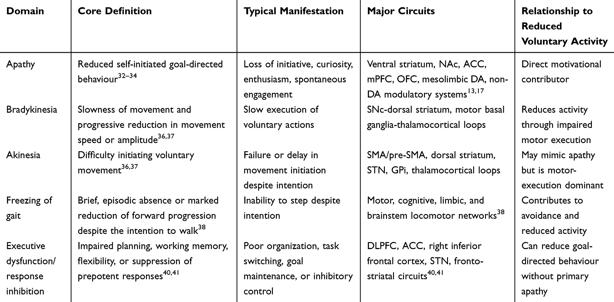 |
Table 1 Conceptual Distinction Between Apathy, Motor-Execution Deficits, and Executive-Control Deficits in PD |
Differential Diagnosis: Apathy, Depression, Fatigue, Cognitive Impairment, and Aging
Clinically, apathy should be distinguished from depression, fatigue, cognitive impairment, anhedonia, sleep disturbance, medication effects, normal aging, frailty, and advanced motor disability, because all of these conditions can reduce daily activity and rehabilitation engagement. Apathy is primarily defined by reduced self-initiated, goal-directed behaviour and diminished initiative, whereas depression is typically characterized by persistent low mood, guilt, hopelessness, negative self-evaluation, and affective distress.17 This distinction is clinically important because apathy and depression may overlap but are not equivalent syndromes in PD. Factor-analytic evidence supports the separability of apathy, depression, anxiety, and fatigue in PD, indicating that apathy should not be treated merely as a subcomponent of depression or fatigue.42 Earlier clinical work also showed that apathy and depression can be dissociated in PD, supporting the need for separate clinical screening rather than relying on mood scales alone.43
Fatigue should also be separated from apathy. Fatigue reflects subjective exhaustion, reduced energy, or increased perceived effort, and may limit activity even when motivation is partly preserved. By contrast, apathy refers to reduced initiation, interest, or goal-directed engagement, even when the patient has sufficient physical capacity to act.44 Cognitive impairment can reduce goal-directed behaviour through deficits in planning, working memory, cognitive flexibility, or goal maintenance, but this does not necessarily imply primary motivational loss. Therefore, cognitive screening should accompany apathy assessment, especially in older adults and patients with advanced PD.45,46
Normal aging and frailty further complicate clinical interpretation. Older adults may show reduced physical activity because of reduced physiological reserve, sarcopenia, comorbidity, sensory impairment, social isolation, or age-related changes in affective and motivational decision-making. These factors may resemble apathy but do not necessarily indicate PD-specific motivational-initiation failure.47,48 In PD, however, reduced spontaneous activity is more strongly linked to disease-specific disruption of internally generated action, basal ganglia output, mesolimbic-prefrontal circuits, dopaminergic and non-dopaminergic modulatory systems, and motor-execution impairment.19 Normal aging may also modify the clinical expression and treatment response of PD-related apathy. Aging-related frailty, reduced cognitive reserve, comorbidity, sensory impairment, pain, sleep disturbance, and reduced social support can lower exercise tolerance, increase perceived effort, reduce adherence, and increase dependence on external cueing, caregiver-supported routines, and environmental structuring.47 Age-related changes in affective and motivational decision-making circuits may further alter reward sensitivity, effort valuation, risk perception, and willingness to initiate behaviour, thereby interacting with PD-related mesolimbic and fronto-basal ganglia dysfunction.48 Therefore, in older adults with PD, reduced voluntary activity should be interpreted as the product of disease-specific motivational and motor dysfunction interacting with aging-related vulnerability, rather than as either normal aging or PD pathology alone. Clinically, the most useful approach is therefore not to ask whether reduced activity is “motor” or “motivational” in a binary sense, but to determine whether the dominant contributor is motor disability, motivational-initiation failure, fatigue, depression, cognitive impairment, aging-related frailty, or a mixed phenotype. Table 2 outlines Clinical differentiation of apathy from overlapping causes of reduced activity in older adults with Parkinson’s disease.
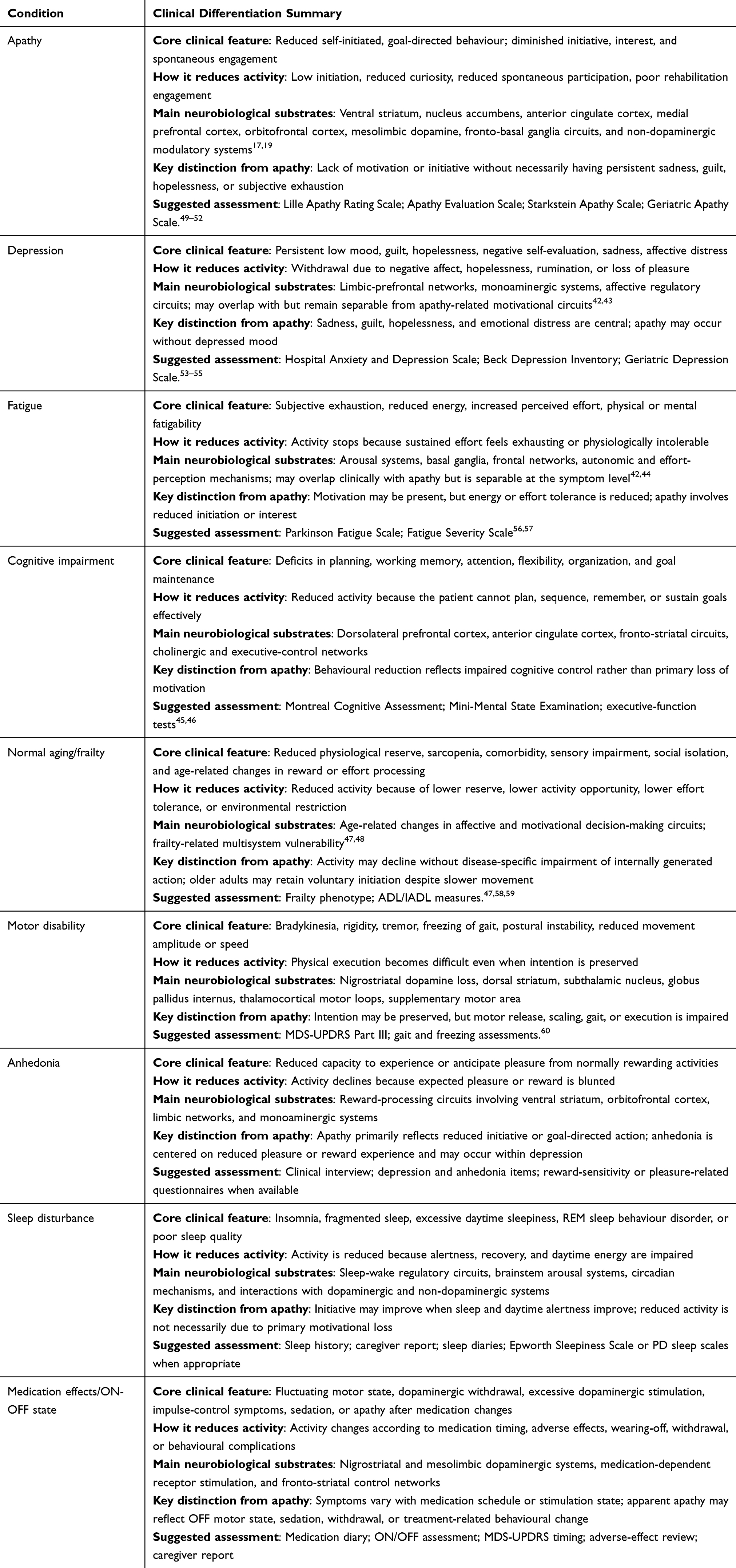 |
Table 2 Clinical Differentiation of Apathy from Overlapping Causes of Reduced Activity in Older Adults with Parkinson’s Disease |
Clinical Assessment Tools and Motivational Phenotyping
Assessment of apathy in PD should not rely on a single global impression of reduced activity. A clinically useful approach should combine apathy-specific scales, mood and fatigue measures, cognitive screening, motor assessment, informant reports, and objective activity or participation outcomes. The Lille Apathy Rating Scale (LARS) is particularly relevant because it was developed as a structured interview for detecting and quantifying apathy and was validated in PD. It also supports screening and severity classification and has been reported to distinguish apathy from depression.49 The Apathy Evaluation Scale and the Starkstein Apathy Scale are also widely used in clinical and research settings, although their sensitivity to different multidimensional apathy profiles may vary across populations and study designs.50,51 More recently, the Geriatric Apathy Scale was developed and validated to characterize multidimensional apathy profiles in older neurodegenerative populations, which may be useful when evaluating apathy in older adults with PD and overlapping cognitive, affective, and functional impairments.52
In PD rehabilitation studies, apathy assessment should be paired with measures that distinguish motor capacity from actual daily engagement. Motor impairment can be evaluated with instruments such as the Movement Disorder Society-sponsored revision of the Unified Parkinson’s Disease Rating Scale Part III, but motor scales alone do not determine whether patients spontaneously initiate activity in daily life.60 Functional autonomy and quality-of-life measures are therefore important because executive apathy and reduced functional autonomy have been associated with worse perceived quality of life in PD.27 Social participation should also be assessed, because reduced voluntary activity may affect interpersonal functioning even when conventional motor outcomes improve. The Parkinson’s Disease Social Functioning Scale was developed as a PD-specific instrument for assessing social functioning and may help evaluate whether motor or motivational improvement translates into meaningful daily and social participation.28 Wearable activity monitoring, activity diaries, caregiver reports, and adherence records can further complement scale-based assessment by capturing real-world activity initiation and persistence.61 This combined strategy can help determine whether a patient fails to move mainly because movement execution is impaired, because motivation is reduced, because fatigue, depression, or cognitive impairment interferes with engagement, or because these mechanisms coexist.
Patient heterogeneity should also be considered during motivational phenotyping. Apathy expression may vary according to age, sex, disease duration, cognitive status, motor phenotype, medication state, comorbid depression or fatigue, and social context. Older patients may show overlapping effects of frailty, cognitive decline, and reduced opportunity for activity, whereas sex-related differences may influence symptom reporting, social roles, caregiver support, and vulnerability to mood or motivational symptoms. Therefore, apathy assessment should not rely on a single scale score but should be interpreted in relation to demographic, clinical, and contextual factors.
Biomarkers and Imaging-Assisted Phenotyping
Emerging biomarker approaches may support motivational phenotyping in PD, although none is currently sufficient for routine diagnosis of PD-related apathy. Functional MRI can characterize altered connectivity within prefrontal-striatal, limbic, salience, default-mode, and executive-control networks. Resting-state fMRI evidence indicates that PD-related apathy is associated with reduced frontostriatal functional connectivity, particularly involving limbic striatal and frontal territories.62 More broadly, resting-state fMRI studies in PD suggest that distributed functional connectivity changes may contribute to motor, cognitive, and non-motor heterogeneity, supporting the use of network-based imaging as a research tool for patient phenotyping.63 PET and SPECT can provide complementary information by assessing dopaminergic and non-dopaminergic neurotransmitter systems; for example, PET evidence in de novo PD suggests that serotonergic degeneration may contribute to apathy, anxiety, and depression, reinforcing the need to avoid a purely dopaminergic explanation of motivational impairment.18
CSF and blood-based biomarkers may help characterize disease biology, neurodegenerative burden, or molecular subtype, but their apathy-specific diagnostic value remains limited. Candidate markers include α-synuclein-related measures, neurofilament light chain, tau-related markers, inflammatory markers, lysosomal and mitochondrial markers, and neurotransmitter-related metabolites.64,65 Neurofilament light chain has been investigated as a marker of neurodegeneration and cognitive impairment in PD, but it should be interpreted as a nonspecific marker of neuronal injury rather than a direct biomarker of apathy.66 α-Synuclein seed amplification assays may identify molecular heterogeneity in PD and improve biological classification, but current evidence does not establish them as markers of motivational impairment or rehabilitation engagement.67 Therefore, imaging and fluid biomarkers should currently be treated as research tools that may support multidimensional phenotyping, not as stand-alone clinical tests for PD-related apathy.
Mechanistic Framework: Coupled but Dissociable Motivational and Motor Systems
The following mechanistic synthesis distinguishes direct evidence from human PD studies from indirect evidence derived from animal models, non-PD disorders, computational models, and general motivational neuroscience. Where evidence is indirect, the mechanisms are presented as hypothesis-generating rather than established explanations of PD apathy.
Distributed Circuits for Motivational Initiation and Effort-Based Action Selection
Motivation depends on distributed cortico-striato-limbic circuits rather than a single reward centre. Within this network, mesolimbic dopamine signalling contributes to reward prediction, incentive salience, effort valuation, and action invigoration through interactions with prefrontal, striatal, and limbic circuits. The following sections examine how interactions between motivational-initiation and motor-execution circuits contribute to apathy and reduced voluntary activity in PD (Figure 1).
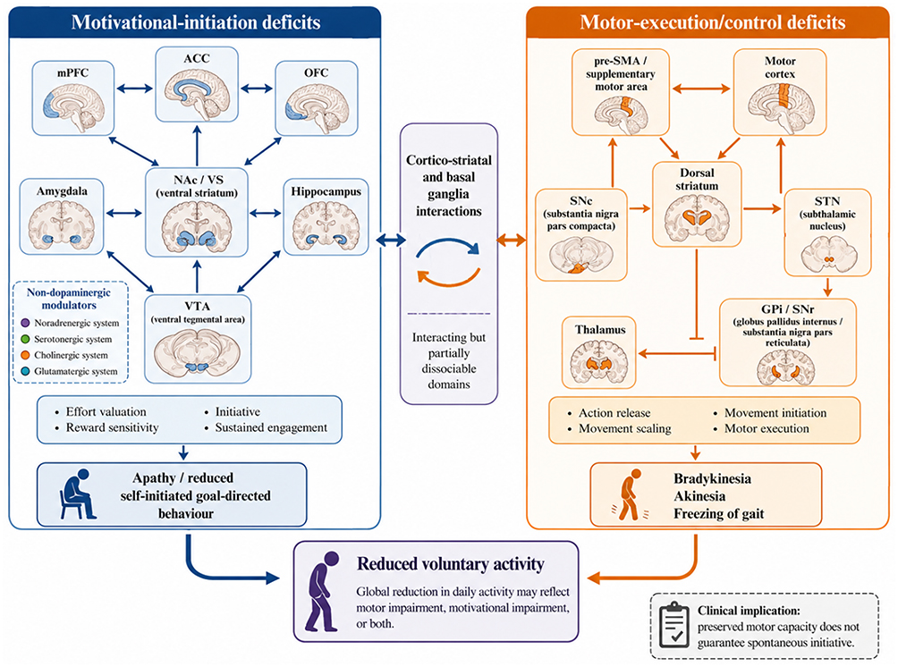 |
Figure 1 Dual-domain framework of reduced voluntary activity in Parkinson’s disease. This figure summarizes the proposed distinction and interaction between motivational-initiation deficits and motor-execution/control deficits in Parkinson’s disease (PD). The blue left panel represents motivational-initiation mechanisms. Apathy is conceptualized as a disorder of reduced self-initiated goal-directed behaviour involving mesolimbic, prefrontal-striatal, and limbic circuits, including the ventral tegmental area (VTA), nucleus accumbens/ventral striatum (NAc/VS), anterior cingulate cortex (ACC), medial prefrontal cortex (mPFC), orbitofrontal cortex (OFC), amygdala, and hippocampus. These circuits, together with non-dopaminergic modulatory systems, including noradrenergic, serotonergic, cholinergic, and glutamatergic systems, contribute to effort valuation, reward sensitivity, initiative, and sustained engagement. The Orange right panel represents motor-execution/control mechanisms. Motor impairment is conceptualized as dysfunction of nigrostriatal and cortico-basal ganglia-thalamo-cortical motor circuits, including the substantia nigra pars compacta (SNc), dorsal striatum, subthalamic nucleus (STN), globus pallidus internus/substantia nigra pars reticulata (GPi/SNr), thalamus, supplementary motor area/pre-supplementary motor area (SMA/pre-SMA), and motor cortex. Dysfunction within this domain contributes to impaired action release, movement scaling, movement initiation, and motor execution, manifesting clinically as bradykinesia, akinesia, and freezing of gait. The central box indicates that motivational-initiation and motor-execution/control domains are interacting but partially dissociable through cortico-striatal and basal ganglia networks. Blue elements denote motivational-initiation circuits and outcomes; orange elements denote motor-execution/control circuits and outcomes; purple elements denote convergent clinical consequences. Arrows indicate functional interactions or directional circuit influence rather than strict anatomical one-to-one projections. The bottom convergence box emphasizes that reduced voluntary activity may reflect motivational impairment, motor impairment, or both. Clinically, preserved motor capacity does not guarantee spontaneous initiative or sustained daily participation. |
Mesolimbic Dopamine Signalling in Incentive Salience, Effort Valuation, and Action Invigoration
The mesolimbic dopamine system is a key component of motivational control, with projections from the VTA to the NAc supporting reward prediction, incentive salience, effort allocation, and action invigoration.68,69 This pathway adaptively modulates motivational mechanisms across multiple timescales. On the one hand, VTA dopaminergic neurons generate phasic discharges at the millisecond to second level, transmitting reward prediction errors and cue-salience information to the NAc, which subsequently updates value-action mappings and alters behavioural strategies;70,71 on the other hand, its tonic activity provides a background level of motivational drive, setting the vigor and persistence of actions during goal pursuit.8
At the information processing level, the NAc does not simply receive upstream signals but integrates multi-dimensional motivational information, particularly assessing effort, reward expectations, and environmental uncertainty.72 This structure is functionally differentiated, with the shell region involved in integrating environmental context and emotional valence, while the core region is more involved in action initiation and goal-directed behaviour regulation.73 At the microcircuit level, medium spiny neurons (MSNs) within the NAc, which express different DA receptor subtypes, play complementary roles in motivational regulation. Importantly, D1-MSNs (direct pathway) facilitate action initiation guided by high-value cues, while D2-MSNs (indirect pathway) contribute to inhibitory modulation related to costs, risks, and distractions.74,75 The dynamic balance between these two pathways coordinates key parameters of motivational decision-making, such as action initiation, effort investment, and behavioural persistence.74,75
Thus, the VTA–NAc pathway should be viewed as part of a broader motivational control system that links value estimation, effort costs, and temporal discounting to behavioural selection and persistence.
Distributed Cortico-Striato-Limbic Networks Supporting Motivational Control
The neural basis of motivation expands far beyond the isolated mesolimbic DA system. Motivational behaviour emerges from an extensively distributed and hierarchically organized network. This network includes PFC subregions and NAc–thalamocortical pathways that support cost-benefit computation during motivated behaviour.10 The anterior cingulate cortex, particularly the dorsal ACC (dACC), is considered a key region for computing the expected value of control.11 During motivational decision-making, the dACC contributes to the integration of expected reward, cognitive effort, and action costs, thereby helping determine whether an individual should persist and how much effort should be allocated.76 Clinical evidence has further suggested that dysfunction of the dACC is closely associated with pathological motivational loss, such as apathy in frontotemporal dementia, in which a key mechanism may involve excessive sensitivity to effort costs.77
The OFC contributes to integrating value, risk, and uncertainty, and adaptively updates preference for options, providing the neural basis for value-cost trade-offs in decision-making.78 Evidence suggests that the OFC, via its subjective value representation function, contributes to non-normative decisions under uncertainty, and in diseases, such as addiction, its ability to integrate utility prediction errors with the ventral striatum is impaired, implying a dysfunction in the motivational computation mechanisms at the circuit level.79 Through interactions with prefrontal and sensorimotor cortical inputs, the dorsal striatum supports the transition from goal-directed action to habitual responding during learning. This process may reduce cognitive demands but can also constrain flexible motivational control when fronto-striatal regulation is impaired. This shift decreases cognitive load in decision-making, providing the neural basis for sustaining long-term behavioural compliance.80 In addition, core nodes in the limbic system provide motivational behaviour with emotional and contextual information. The amygdala alters the strategy/prevention threshold by assigning emotional valence to environmental cues, impacting sensitivity to negative costs and potential threats.81 By encoding precise contextual and temporal information, the hippocampus and parahippocampal circuits enable specific rewards to trigger various behavioural tendencies across different environments, supporting the adaptive linkage between motivational behaviour and specific contexts.82
In this architecture, the NAc functions as the central hub, receiving and combining inputs from the PFC (value and cost computation), amygdala (emotional valence), hippocampus (contextual information), and VTA (dopaminergic modulatory signals).83 After initial integration in the NAc, information is relayed back to the cortex via thalamic relay stations, forming a closed-loop regulatory system, establishing a multi-node network with the NAc as the centre and feedback to the cortex and thalamus.84,85
In conclusion, motivation emerges from distributed cortico-striato-limbic computations rather than from a single reward centre. The NAc integrates dopaminergic, prefrontal, amygdalar, and hippocampal inputs, while thalamocortical feedback supports the updating of action value, effort cost, contextual relevance, and behavioural persistence. This network-level view provides a more testable framework for understanding motivational impairment in PD. Together, these distributed circuits allow motivational behaviour to be shaped by intrinsic value, effort cost, contextual memory, emotional salience, and learned action rules, thereby producing adaptive and executable behavioural strategies.
Coupled but Dissociable Motor-Execution and Motivational-Initiation Circuits
Voluntary movement in PD should be understood as the product of at least two interacting but dissociable processes: motivational initiation, which determines whether an action is worth selecting and sustaining, and motor execution, which determines whether the selected action can be released and performed efficiently. The former relies strongly on mesolimbic and prefrontal-striatal circuits, including the VTA, nucleus accumbens, anterior cingulate cortex, orbitofrontal cortex, and related limbic structures; the latter depends more heavily on nigrostriatal and motor cortico-basal ganglia circuits involving the SNc, dorsal striatum, supplementary motor areas, STN, GPi, and thalamocortical projections.10
The motor circuit fine-tunes action sequences via the cortex-BG-thalamus-cortex loop. The pre-supplementary motor area (pre-SMA) is crucial for updating goal-directed actions according to behavioural consequences, functioning as a core node for flexible motor control.86 Motivational circuits influence whether an action is selected, energized, and sustained, whereas motor circuits determine whether the selected action can be executed efficiently. Mesolimbic dopamine contributes to reward prediction, incentive salience, and effort allocation, thereby modulating the likelihood that a patient will initiate or persist in an action. This modulatory role should be distinguished from the direct control of motor programme release within nigrostriatal and motor basal ganglia circuits.87 A defining pathological feature of PD is the progressive loss of dopaminergic neurons in the SNc, leading to marked dopamine depletion in the dorsal striatum, which directly impairs the release of motor programmes and contributes to bradykinesia, akinesia, and freezing of gait.7
However, the pathological mechanism in PD expands beyond the SNc. As the disease progresses, dysfunction in the ventral DA pathway from the VTA to the NAc and the functional connectivity and synaptic plasticity of PFC-NAc glutamatergic projections may also emerge.88 This may impair motivational computation, reflected in delayed value learning, excessive effort discounting, and insensitivity to positive feedback, contributing to a low motivational state characteristic of apathy.89 Current evidence is consistent with this interpretation. A study of non-treated early PD patients reported that their willingness to exert grip strength was significantly lower than that of healthy controls, especially at low reward levels. This reduction in effort expenditure was directly associated with the loss of DA transporter (DAT) in the shell of the NAc.6 These findings suggest that accumbal dopamine, particularly within ventral striatal subregions, is more closely related to effort-reward trade-offs and motivational invigoration than to motor execution per se. Thus, reduced effort expenditure in PD should be interpreted as a motivational-circuit abnormality that may coexist with, but should be distinguished from, motor execution deficits.
This dual-domain framework may help explain two clinically relevant observations in PD: (1) motor symptoms and motivational deficits commonly coexist because both are part of the DA-dependent system of the BG network, and the SNc and VTA are structurally and functionally tightly related, with pathological processes potentially affecting both systems to different degrees.90 (2) Motor symptoms and motivational deficits have comparatively independent pathological bases and treatment responses, with severe DA depletion in the dorsal striatum being the primary factor in motor dysfunction, while motivational deficits are more closely associated with dysfunction in ventral striatal–prefrontal networks, involving a more complex balance of DA and glutamatergic signals with variable responses to DA replacement therapy.91 Computational neuroscience evidence has further suggested that DA therapy has dual effects on cognitive and motivational functions: under non-treated conditions, DA deficiency in the DS causes decreased cognitive flexibility and difficulty updating working memory; after medication, the drug may over-augment executive signals in the ventral or DS, impairing cognitive control in situations requiring inhibition of interference, but potentially promoting feedback-based learning.92 This creates a core clinical challenge: although L-DOPA can substantially alleviate bradykinesia and improve motor execution, it does not reliably restore spontaneous initiative and sustained engagement in daily activities or rehabilitation.13 When the phasic DA encoding of the motivational circuit and the top-down modulation from the PFC are not concurrently rebuilt, enhancements in motor function may not translate into maintained participation and adherence to daily activities and rehabilitation.13 Hence, interventions for PD-related apathy should move beyond simple dopaminergic replacement and consider circuit-level interactions among mesolimbic, prefrontal, striatal, and non-dopaminergic modulatory systems.
DA Signal Encoding of Motivation: Concentration and Pattern Dimensions
Dopamine influences motivation not only through its overall availability, but also through the timing, spatial spread, and receptor-level decoding of its signals. These features determine how reward prediction, effort cost, and action invigoration are translated into behaviour. The core mechanism by which DA encodes motivation in PD is summarized in Figure 2.
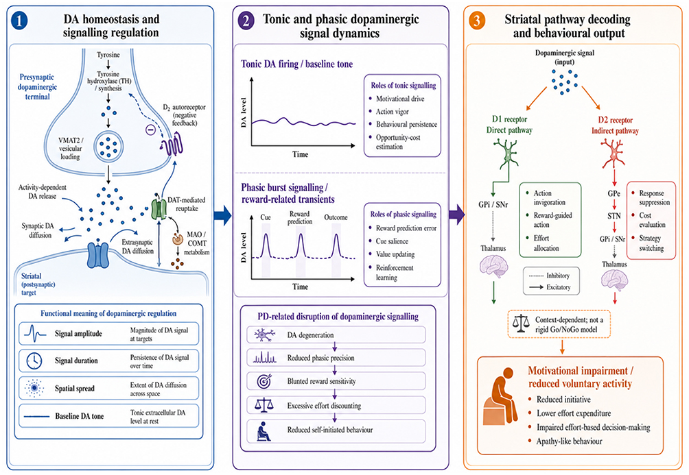 |
Figure 2 Dopaminergic signal dynamics underlying motivational impairment in Parkinson’s disease. This schematic summarizes three interrelated dimensions through which dopaminergic signalling may influence motivational control and reduced voluntary activity in Parkinson’s disease (PD). Panel 1 illustrates dopamine (DA) homeostasis and signalling regulation at the presynaptic dopaminergic terminal and striatal postsynaptic target. DA signalling depends on tyrosine hydroxylase (TH)-dependent synthesis, vesicular loading through vesicular monoamine transporter 2 (VMAT2), activity-dependent release, synaptic and extrasynaptic diffusion, dopamine transporter (DAT)-mediated reuptake, monoamine oxidase/catechol-O-methyltransferase (MAO/COMT)-dependent metabolism, and D2 autoreceptor-mediated negative feedback. These mechanisms jointly determine the amplitude, duration, spatial spread, and baseline tone of DA signals. Panel 2 illustrates tonic and phasic dopaminergic signal dynamics. Tonic DA firing and baseline extracellular DA tone support motivational drive, action vigor, behavioural persistence, and opportunity-cost estimation. Phasic burst signalling and reward-related DA transients contribute to reward prediction error, cue salience, value updating, and reinforcement learning. In PD, dopaminergic degeneration and impaired phasic precision may blunt reward sensitivity, increase effort discounting, delay value learning, and reduce self-initiated behaviour. Panel 3 illustrates striatal pathway decoding and behavioural output. Dopaminergic signals are decoded through partially opponent but context-dependent D1/direct and D2/indirect pathway mechanisms. D1-mediated mechanisms generally support action invigoration, reward-guided action, and effort allocation, whereas D2-mediated mechanisms contribute to response suppression, cost evaluation, and strategy switching. This distinction should not be interpreted as a rigid binary Go/NoGo model. In PD, reduced DA availability and disrupted dopaminergic signal dynamics may disturb D1–D2 pathway balance, contributing to motivational impairment, reduced voluntary activity, reduced initiative, lower effort expenditure, impaired effort-based decision-making, and apathy-like behaviour. Arrows indicate functional regulation or circuit influence; dashed inhibitory lines indicate suppressive or indirect pathway effects. Abbreviations: DA, dopamine; DAT, dopamine transporter; GPi, globus pallidus internus; MAO/COMT, monoamine oxidase/catechol-O-methyltransferase; SNr, substantia nigra pars reticulata; STN, subthalamic nucleus; TH, tyrosine hydroxylase; VMAT2, vesicular monoamine transporter 2. |
Dynamic Homeostasis of DA Signals
Evidence suggests that apathy and motivational impairment in PD may partly reflect insufficient DA signalling in the brain.6,93 Nevertheless, this is an oversimplification of its pathophysiological mechanisms. This view neglects the precise dynamic modulation of DA signals in both spatiotemporal dimensions and neural circuits. In reality, DA is not merely a static concentration variable, but is part of a dynamic homeostasis system composed of multiple interrelated stages, including synthesis, vesicular loading, activity-dependent release, volume transmission diffusion, reuptake, and enzymatic metabolism.94
Within this system, tyrosine hydroxylase (TH) is the rate-limiting enzyme for DA synthesis, with its activity precisely modulated by phosphorylation and feedback inhibition, collectively regulating the rate of DA synthesis.95 Vesicular monoamine transporter 2 (VMAT2) is responsible for loading cytoplasmic DA into synaptic vesicles, and its functional level directly influences the size of the DA pool available for release at the presynaptic terminal. Activity-dependent vesicular exocytosis generates a transient DA peak at synapses, which then diffuses via volume transmission to form a broad DA gradient, modulating neuronal network activity in a non-synaptic manner.96
Efficient signal termination and resetting depend on a robust clearing process. DAT clears dopamine from the extracellular space by reuptake into presynaptic terminals, whereas presynaptic D2 autoreceptors inhibit further dopamine release through negative feedback. Together, these mechanisms help preserve the temporal and spatial fidelity of dopaminergic signalling.97 Ultimately, monoamine oxidase (MAO) and catechol-O-methyltransferase (COMT) set core time constants for DA metabolism, with COMT being particularly important for DA clearance in brain areas, such as the PFC, where DAT expression is low.98 Any disruption in these stages of the homeostasis system significantly modifies the trajectory of DA receptor occupancy over time and ultimately distorts the value-effort shaping at the behavioural level. For instance, a decrease in DAT function may enhance extracellular DA concentration and its lifespan, amplifying background dopaminergic tone, but blurring core phasic differences, impairing reward prediction error-based learning.99 In contrast, a reduction in vesicular release probability or depletion of the vesicular DA pool (as may occur because of α-Syn pathology) reduces responses to high-frequency events (eg, unexpected rewards), decreasing the signal-to-noise ratio of positive feedback.100
Thus, DA dysfunction in PD is not only a quantitative deficiency, but an integrative imbalance in the dynamic homeostasis system at numerous nodes. Degeneration in the SNc/VTA, together with the failure of compensatory plasticity in the cortical-striatal-thalamic network, may create a system that cannot maintain stable tonic background drive (manifested as common apathy and decreased vigor) and lacks precise phasic encoding (resulting in delayed value learning and insensitivity to immediate feedback).101 This dynamic homeostasis framework also provides insight into the heterogeneity of PD clinical presentations and treatment responses. The reason why L-DOPA demonstrates highly heterogeneous motivational effects across patients or even within the same patient in various task contexts is that, at various pathological stages, the specific components of the individual DA homeostasis system (such as synthesis capacity, vesicular loading, DAT function) and their compensatory levels vary.6 Nevertheless, simply replenishing DA precursors may correct the absolute deficiency in concentration, but does not necessarily rebuild the natural dynamic release pattern that matches behavioural demands.102 DA release has complex dynamic features, including baseline and phasic modes. Baseline release provides sustained neural modulation, while phasic release is tightly related to reward prediction errors and reinforcement learning.103 Moreover, supplementation with DA precursors such as L-DOPA increases DA synthesis, but this supplementation is typically continuous and may not cue natural phasic release patterns. For instance, L-DOPA supplementation may cause maintained DA elevation, which can influence behavioural flexibility and decision-making.104 Hence, although DA precursor supplementation can increase DA concentration, to restore natural, behaviour-matched dopaminergic dynamics, more precise modulation strategies may be required.
Tonic and Phasic Firing Patterns of DA Neurons
Dopaminergic neurons convey motivational information and shape behaviour via two firing patterns with distinct timescales: tonic firing, which is continuous, low-frequency, and comparatively regular, and phasic bursts, which occur on a millisecond to hundred-millisecond scale. Tonic firing is responsible for sustaining the baseline concentration of DA in the BG and other target brain regions. This continuous signal establishes a background level of motivational drive by impacting the average occupancy of DA receptors and encoding the average reward rate in the environment, ie, the opportunity cost.8 Higher tonic DA levels commonly signal a higher perceived potential reward in the environment, which decreases the threshold for initiating actions and enhances behavioural persistence, making the individual more willing to start and maintain goal-directed behaviours.105
Complementing this is phasic bursting, a high-frequency clustered discharge that occurs on a millisecond-to-hundred-millisecond scale. These transient signal peaks are responsible for encoding reward prediction errors—ie, the difference between actual and expected rewards—into critical brain regions, such as the NAc and PFC, and they convey motivational salience.106,107 Phasic signals are central to reinforcement learning and instantaneous decision-making. They enable the rapid updating of mappings among value, cost, and strategy, thereby determining whether an individual should expend greater effort, disengage decisively, or pursue the next most valuable option.108
Importantly, these two modes do not operate independently; they form a dynamic equilibrium system. Based on Grace’s tonic/phasic model,109 the maintained tonic DA level in the extracellular space exerts baseline inhibitory control over phasic DA release via autoreceptors (eg, D2 short receptors) situated on the terminals and cell bodies of DA neurons. Hence, optimal motivational behaviour depends on the precise balance between tonic background and phasic events. In PD, this delicate balance is profoundly disturbed. Multiple pathological alterations at the membrane and network levels cause sparse and phase-incorrect phasic bursts. These include dysfunctions of hyperpolarization-activated cyclic nucleotide-gated channels (HCN) and small-conductance calcium-activated potassium channels (SK), which disturb the intrinsic pacing and firing patterns of neurons;110 enhanced pathological β-oscillations (~13–30 Hz) in downstream BG circuits, leading to excessive synchrony and interference with information processing;111 and impaired top-down coupling from the cortex to the striatum, making it difficult for DA neurons to receive the precise instructions from the PFC to trigger phasic bursts.112 Thus, motivational signalling may shift from event-linked phasic control toward a less precise tonic-dominant state that is insufficient for fine-grained reinforcement learning and effort-based decision-making. This may contribute to a range of clinically observed motivational deficits, particularly, a significant reduction in sensitivity to positive feedback and novel cues, an abnormally steep effort discounting curve (ie, abandoning behaviours because of small increases in cost), and a decrease in the learning rate for value (slower learning).113 Such disruption of tonic-phasic dopaminergic balance may contribute to motivational impairment in PD, particularly by reducing sensitivity to positive feedback, cue salience, and effort-based action selection. However, this mechanism should be considered alongside non-dopaminergic and executive-control contributions to apathy.
This framework may also explain why oral DA replacement therapies (eg, L-DOPA) elevate tonic DA concentrations in a pulsatile manner, which can improve motor function, but may not fully restore precise phasic coding needed for millisecond-second decision-making. These drugs non-physiologically raise tonic DA levels, which may promote overall action vigor to some extent, but they cannot reconstruct the precise temporal sequencing and effectiveness of phasic signal transmission, resulting in limited and heterogeneous effects in reinforcement learning and fine-tuned motivational decision-making.
Role of D1/D2 Receptor-Mediated Direct and Indirect Pathways
In the core structure of the BG, the ultimate integration of motivational signals and behavioural transformation relies on two major types of MSNs and their functionally antagonistic microcircuits. D1- and D2-receptor-expressing medium spiny neurons contribute to action selection through partially opponent and context-dependent effects on striatal output. Although often simplified as “Go” and “NoGo” pathways, their roles in motivation, learning, and effort allocation are dynamic rather than strictly binary.114,115
D1R (Direct Pathway): The D1-MSNs → globus pallidus internus (GPi)/substantia nigra pars reticulata (SNr) inhibitory projections serve to initiate motivational behaviour. D1R is coupled to the guanine nucleotide-binding protein G(olf) subunit alpha (Gαolf), and its activation enhances intracellular cyclic adenosine monophosphate (cAMP) levels and protein kinase A (PKA) activity, which increases neuronal excitability and promotes long-term potentiation (LTP) and synaptic plasticity processes, thereby facilitating action initiation.116 In behavioural decision-making, this pathway is preferentially recruited in response to high-value or high-certainty rewards, significantly enhancing the likelihood and intensity of action implementation, thereby functioning as a “Go” signal.117 D2R (Indirect Pathway): The D2-MSNs project via the external globus pallidus (GPe) and subthalamic nucleus (STN) to the GPi or SNr, and their main function is to inhibit behaviour, functioning as a “brake”.118 D2R is coupled to the guanine nucleotide-binding protein G(i/o) subunit alpha (Gαi/o), and its activation suppresses the cAMP-PKA signalling pathway, producing sustained inhibitory modulation on neuronal activity.119 This pathway adaptively evaluates the costs, potential risks, and environmental distractions of behaviour, raising the response threshold for behavioural termination or strategy switching.120 Its function can be regarded as a “NoGo” signal, aimed at inhibiting low-effort or potentially harmful behaviours.
Under physiological conditions, there is a dynamic balance between the phasic gain of the D1 pathway and the tonic inhibitory background of the D2 pathway, improving value-effort decision functions. Positive prediction errors and reward-predictive cues can preferentially facilitate D1-mediated plasticity and action invigoration, whereas negative outcomes, conflict, or increased effort costs may engage D2-mediated mechanisms that bias action suppression or strategy switching. These effects, however, are task-dependent and should not be interpreted as a rigid one-to-one mapping between receptor subtype and behavioural outcome.121 Nevertheless, during PD progression, widespread DA depletion and disrupted signal timing impair this balance. Because of differences in receptor expression and signalling processes, the net effect of DA deficiency causes relative overactivation of the indirect pathway (NoGo) while significantly inhibiting the function of the direct pathway (Go).122
This imbalance at the circuit level may contribute to characteristic learning and decision-making biases. In unmedicated PD, reduced dopaminergic tone may impair learning from positive feedback and reduce action invigoration, particularly in tasks that require reward-guided updating. Conversely, relatively preserved or less dopaminergically dependent mechanisms for learning from negative feedback may contribute to better performance in some punishment- or avoidance-based learning contexts, although this pattern varies across tasks, disease stage, medication state, and cognitive profile.120 This response tendency is also revealed in reaction time regulation: when on medication, DA supplementation improves Go learning, helping patients learn more effectively from rewards in tasks requiring faster responses; while off medication, they may perform relatively better in NoGo learning tasks, where delayed responses adapt to task demands.123 DA modulation of higher cognitive functions follows an inverted U-shaped curve, with its ultimate behavioural effects highly dependent on the specific task demands and the baseline DA levels in the involved brain regions (eg, PFC and striatal subregions).124 This clarifies the complex and highly individual differences in the effectiveness of DA replacement therapies: dopaminergic medication may shift neural signals to more optimal levels at certain circuit nodes, but it may also excessively increase signals at other nodes, causing new cognitive biases or non-motor symptoms.
Overall, the neurobiological basis of PD apathy and motivational impairment is far from a simple neurotransmitter deficiency. One major contributor is DA depletion, which disrupts the balance between direct and indirect striatal pathways and impairs Go/NoGo regulation within the behavioural selection system.
Progressive Dysfunction of Motivational-Initiation Circuits in PD
Motivational impairment in PD is better understood as a progressive dysfunction of interacting mesolimbic, prefrontal, striatal, and non-dopaminergic systems rather than as an abrupt collapse of a single motivational circuit. Although PD progression is heterogeneous, a staged framework may help organize how motivational-initiation circuits could become progressively vulnerable across disease stages. This framework should be interpreted as a hypothesis-generating model rather than a confirmed sequence of damage in individual patients (such as the VTA, SNc, NAc) at different pathological stages and their corresponding motivational deficits (Figure 3).
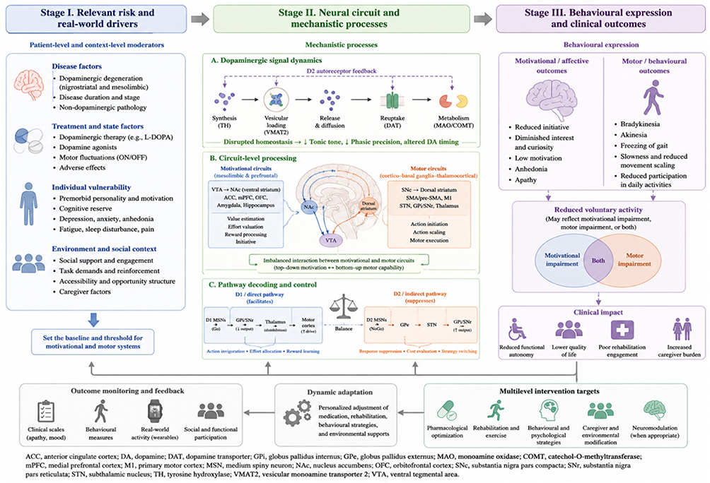 |
Figure 3 Proposed hypothesis-generating framework linking patient-level drivers, neural circuit mechanisms, behavioural outcomes, and intervention targets in PD-related apathy and reduced voluntary activity. This figure presents a proposed, hypothesis-generating framework for understanding reduced voluntary activity in Parkinson’s disease (PD) as the result of interacting patient-level drivers, neural circuit mechanisms, behavioural expression, and modifiable intervention targets. Stage I summarizes relevant risk and real-world drivers that may set the baseline threshold for motivational and motor systems. These include disease-related factors, such as nigrostriatal and mesolimbic dopaminergic degeneration, disease duration, disease stage, and non-dopaminergic pathology; treatment and state factors, including L-DOPA exposure, dopamine agonist use, ON/OFF fluctuations, and adverse effects; individual vulnerability factors, including premorbid motivation, cognitive reserve, depression, anxiety, anhedonia, fatigue, sleep disturbance, and pain; and environmental or social-context factors, including caregiver support, task demands, reinforcement, accessibility, and opportunity structure. Stage II illustrates neural circuit and mechanistic processes that may contribute to motivational and motor impairment. Dopaminergic signal dynamics include tyrosine hydroxylase (TH)-dependent synthesis, vesicular loading through vesicular monoamine transporter 2 (VMAT2), activity-dependent release and diffusion, dopamine transporter (DAT)-mediated reuptake, monoamine oxidase/catechol-O-methyltransferase (MAO/COMT)-dependent metabolism, and D2 autoreceptor-mediated feedback. Disruption of these processes may alter tonic dopaminergic tone, phasic precision, and behaviourally timed dopaminergic signalling. Circuit-level processing involves partially dissociable but interacting motivational circuits, including the ventral tegmental area (VTA), nucleus accumbens/ventral striatum (NAc/VS), anterior cingulate cortex (ACC), medial prefrontal cortex (mPFC), orbitofrontal cortex (OFC), amygdala, and hippocampus, and motor circuits, including the substantia nigra pars compacta (SNc), dorsal striatum, supplementary motor area/pre-supplementary motor area (SMA/pre-SMA), primary motor cortex (M1), subthalamic nucleus (STN), globus pallidus internus/substantia nigra pars reticulata (GPi/SNr), and thalamus. D1/direct and D2/indirect pathway decoding is represented as a context-dependent balance between action invigoration, effort allocation, reward learning, response suppression, cost evaluation, and strategy switching, rather than as a rigid binary Go/NoGo model. Stage III shows how these mechanisms may converge into behavioural expression and clinical outcomes. Motivational or affective outcomes include reduced initiative, diminished interest and curiosity, low motivation, anhedonia, and apathy. Motor or behavioural outcomes include bradykinesia, akinesia, freezing of gait, slowness, reduced movement scaling, and reduced participation in daily activities. Reduced voluntary activity may therefore reflect predominant motivational impairment, predominant motor impairment, or both. Clinically, this can lead to reduced functional autonomy, poorer quality of life, poor rehabilitation engagement, and increased caregiver burden. The lower panels emphasize that assessment and management should be iterative. Outcome monitoring should include clinical scales for apathy and mood, behavioural measures, real-world activity monitoring, and social or functional participation outcomes. Dynamic adaptation refers to personalized adjustment of medication, rehabilitation, behavioural strategies, and environmental supports according to response and tolerability. Multilevel intervention targets include pharmacological optimization, rehabilitation and exercise, behavioural and psychological strategies, caregiver and environmental modification, and neuromodulation when clinically appropriate. The figure is intended to organize current evidence and clinical reasoning; it should not be interpreted as a confirmed linear disease-staging model. Arrows indicate hypothesized directional or feedback relationships. Blue elements indicate patient-level and motivational factors, green elements indicate mechanistic processes, purple elements indicate behavioural and clinical outcomes, and grey lower panels indicate monitoring, adaptation, and intervention targets. |
Temporal Progression
PD pathology may show partly ordered spatial and temporal patterns, but substantial heterogeneity exists across patients; therefore, motivational-circuit involvement should not be described as evolving in uniform or clearly demarcated stages. This process is not confined to the nigrostriatal system but may progressively involve mesolimbic dopamine pathways, prefrontal-striatal networks, and non-dopaminergic modulatory systems.
In the preclinical stage, pathological changes first affect the medullary motor nuclei and olfactory nuclei, then progress in a caudal-to-rostral direction along the brainstem.125 Although prominent motor symptoms are absent at this stage, subtle functional changes in motivational signalling may occur; however, prodromal PD remains a probabilistic research construct, and direct evidence for early VTA dopaminergic dysfunction before clinical diagnosis remains limited.18,126 Compared with SNc dopaminergic neurons, VTA dopaminergic neurons may show relative vulnerability or resilience depending on disease stage, model system, and pathological burden. Therefore, motivational symptoms should not be attributed to a uniform or linear degeneration of the VTA-NAc pathway alone.127 However, subtle dysfunction within VTA-related motivational signalling may contribute to early reductions in reward responsiveness and motivational drive, manifesting as reduced responsiveness to reward stimuli, although it has not yet reached the clinically recognizable level of apathy.128,129 As the pathological process progresses to the early clinical stage, neurodegeneration significantly affects the substantia nigra pars compacta, leading to a sharp decline in striatal DA concentrations, and classical motor symptoms emerge. Notably, the dysfunction of the VTA-NAc pathway further intensifies at this stage.69,130 In MPTP-treated primate models, alterations within the VTA-NAc pathway have been associated with apathy-like behaviours, suggesting that mesolimbic dysfunction may contribute to motivational impairment beyond classical nigrostriatal motor deficits.131
Selective damage to SNc DA neurons (without significant VTA loss) can induce motivational and emotional deficits, suggesting that motivational disorders may represent core impairments in PD rather than merely secondary psychological responses to motor symptoms.132 In the middle and late stages of the disease, pathological changes extensively affect the limbic system and prefrontal cortex (PFC). At this stage, significant iron accumulation may appear in the VTA, suggesting a possible decline in compensatory capacity.133 Reduced functional connectivity within prefrontal-striatal loops may impair goal-directed control, especially when compensatory prefrontal mechanisms become insufficient. Functional neuroimaging studies reveal that PD patients show compensatory activation of the PFC when processing positive feedback, but this compensatory mechanism fails when feedback information loses clear meaning, highlighting striatal dysfunction.134
From a computational neuroscience perspective, the depletion of DA leads to a significant reduction in reward prediction error signals, thereby impairing behaviour optimization based on reinforcement learning.135 Although dopaminergic medications may partially restore Go learning and accelerate responses driven by rewards, they may simultaneously impair performance in tasks requiring cognitive flexibility, reflecting the inverted U-shaped regulation of PFC function by DA.91,136 In advanced PD, patients exhibit the triple motivational deficit syndrome described by Levy and Dubois, consisting of emotional motivational deficits (due to limbic circuit damage), cognitive motivational deficits (due to associative circuit dysfunction), and self-activation deficits (due to widespread basal ganglia output failure).33 This network-level dysfunction may help explain why some patients retain the ability to execute externally cued actions but lose the internal drive to initiate and maintain goal-directed behaviour, reflecting severe impairment of motivational-initiation systems.2
System-Level Consequences of Motivational-Circuit Dysfunction
The neurodegenerative process of PD may progressively disrupt midbrain-limbic and nigrostriatal dopaminergic systems, resulting in network-level impairments in motivational control.
VTA DA Dysfunction and Motivational Drive
Progressive dysfunction of VTA DA neurons may disrupt neural processes supporting motivated behaviour. Although PD’s motivational defects have traditionally been thought to originate from VTA degeneration, carefully designed animal models reveal a more complex picture: selective damage to SNc DA neurons is sufficient to induce significant motivational and emotional deficits, independent of motor impairments.132 However, the VTA-NAc pathway appears to play an important role.131 Dysfunction in this pathway leads to a lack of necessary motivational drive when facing reward opportunities, manifesting as a typical reduction in goal-directed behaviour, severely affecting patient participation in motor activities and daily life quality.9
Dysfunction of NAc Impairs Reward Processing and Motivational Expression
As the integration centre of the motivational circuit, dysfunction in the NAc may disrupt multiple aspects of reward processing. Structural neuroimaging studies show that PD patients with apathy exhibit significant reductions in grey matter volume in the NAc, and this change, along with the atrophy of core nodes in the executive and reward pathways, forms the neurobiological basis for apathy.137 The midbrain-limbic DA system’s role in motivation is far from merely transmitting rewards. This system finely regulates several dimensions of motivation, including behavioural activation, effort expenditure, sustained task participation, and tool learning, rather than mediating primary appetites or hunger.9 In PD pathology, NAc dysfunction manifests as reduced sensitivity to positive feedback, inaccurate reward prediction error encoding, and impaired reward-based learning processes.138 Notably, excessive activation of the VTA-NAc pathway leads to abnormal processing of loss and punishment signals, resulting in decision-making defects. This mechanism may be closely related to behavioural abnormalities triggered by PD DA replacement therapies.138
Prefrontal-Striatal Dysfunction and Executive Contributions to Reduced Goal-Directed Behaviour
Prefrontal-striatal dysfunction contributes to reduced goal-directed behaviour in PD, but this contribution should be separated from apathy per se.33,139 Dorsolateral prefrontal circuits are more closely related to working memory, planning, cognitive flexibility, and action selection, whereas medial prefrontal and anterior cingulate circuits are more directly involved in effort valuation, initiative, and the expected value of control.11,140,141 Dysfunction in these systems may reduce daily activity by impairing planning and self-monitoring, by increasing the perceived cost of effort, or by weakening spontaneous initiation.13,33,142 Therefore, prefrontal-basal ganglia abnormalities should be interpreted as a set of interacting executive and motivational mechanisms rather than as a single global collapse of goal-directed behaviour.17,33
Non-Dopaminergic Contributions to Apathy and Motivational Impairment
Although dopamine is central to effort valuation and action invigoration, apathy in PD should not be reduced to dopaminergic failure alone.13,17,19 Noradrenergic degeneration, particularly involving the locus coeruleus, may impair arousal, attentional readiness, fatigue regulation, and effort mobilization, thereby reducing the energetic state required for self-initiated behaviour.4,143,144 Serotonergic dysfunction may contribute through mood regulation, reward sensitivity, behavioural inhibition, and the overlap between depression and apathy.18,145,146 Cholinergic degeneration, involving basal forebrain and brainstem cholinergic systems, may weaken attention, cognitive control, gait-cognition coupling, and the ability to maintain goal-directed behaviour over time.147–150 In addition, glutamatergic projections from the prefrontal cortex to the striatum and nucleus accumbens are essential for translating goals, expected value, and action costs into behavioural selection.13,142,151,152
These systems may influence apathy in at least two ways. First, they may directly alter motivational states by changing arousal, reward responsiveness, fatigue, and attentional control.18,143,149 Second, they may interact with dopaminergic circuits through compensatory or maladaptive network changes, thereby modifying how dopamine depletion is expressed behaviourally.17,142,145 This explains why some patients show prominent apathy despite partial motor responsiveness to dopaminergic treatment, and why dopaminergic replacement alone may fail to restore initiative, sustained participation, or rehabilitation adherence.13,148,153 A more complete framework should therefore treat dopamine as a major but not exclusive component of motivational impairment in PD.2,17,19
Critical Synthesis: What the Mechanistic Evidence Does and Does Not Show
Taken together, the mechanistic evidence supports a convergent but not fully settled interpretation of PD-related apathy. Human clinical and neuroimaging studies consistently implicate ventral striatal, mesolimbic, prefrontal-striatal, cingulate, and non-dopaminergic systems in motivational impairment, whereas motor-execution deficits are more closely linked to nigrostriatal and motor cortico-basal ganglia-thalamo-cortical dysfunction.17,20 This supports the dual-domain framework proposed in this Review. However, the current evidence does not justify a simple one-to-one mapping between a single neurotransmitter system, a single brain region, and apathy. Apathy in PD is better interpreted as a network-level syndrome in which motivational valuation, executive control, arousal, affective state, and motor release interact.2,19
The strongest pattern across the literature is the clinical dissociation between capacity and initiative. Dopaminergic therapy, motor rehabilitation, or DBS may improve measurable motor performance, yet spontaneous engagement, daily activity, and rehabilitation adherence may remain impaired.17,27 This pattern suggests that restoring the ability to execute movement is not equivalent to restoring the motivation to initiate and sustain movement. The most clinically relevant question is therefore not whether reduced activity is purely motor or purely motivational, but which mechanism is dominant in a given patient and how these mechanisms interact over time.
Several knowledge gaps remain. First, most studies are cross-sectional and cannot determine whether motivational impairment precedes, follows, or develops in parallel with motor-execution decline. Second, evidence linking specific dopaminergic signal dynamics, such as tonic-phasic imbalance or D1/D2 pathway decoding, to human PD apathy remains partly inferential. Third, animal and computational models clarify causal circuit principles but cannot be directly translated into clinical phenotypes without human validation. Fourth, few studies combine apathy-specific scales, motor measures, cognitive assessment, neuroimaging, and real-world activity monitoring in the same cohort. Future studies should therefore use longitudinal, multimodal designs to test whether motivational phenotypes predict rehabilitation engagement, daily participation, and treatment response beyond standard motor severity.19,28
Clinical Interventions and Rehabilitation Implications: Evidence, Limitations, and Readiness
The “Double-Edged Sword” Effect of Dopaminergic Replacement Therapy
Asymmetry of Treatment Efficacy
Dopamine substitution therapy, particularly L-DOPA-based regimens, has long been regarded as a cornerstone of PD treatment.154 Nevertheless, accumulated clinical evidence has suggested significant asymmetry in its effectiveness in improving motor symptoms versus non-motor symptoms, especially motivational deficits.155,156
L-DOPA increases dopaminergic stimulation within the nigrostriatal pathway and improves core motor symptoms such as bradykinesia, rigidity, and tremor.156 Nevertheless, these motor benefits do not necessarily translate into proportional improvements in motivation. Prospective evidence suggests that although L-DOPA can improve motor function, apathy and motivational deficits may persist, fluctuate, or even worsen during chronic dopaminergic therapy in some patients. This therapeutic dissociation is evident even in early PD. In particular, early-stage PD patients with emotional apathy may show longer disease duration and more severe motor impairment, with their apathy levels negatively associated with cognitive dysfunction.157 Importantly, even treatments particularly designed for non-motor symptoms face challenges. For instance, the rotigotine transdermal patch in clinical trials failed to significantly improve self-reported apathy symptoms in PD patients, but clearly demonstrated positive effects on motor symptoms and daily living capabilities.158
This asymmetry in effectiveness stems from the unique neurobiological properties of the PD motivational circuit. The midbrain-limbic system and PFC need more precisely tuned DA regulation. While L-DOPA can elevate overall brain DA levels, it may not fully restore physiological phasic signal transmission and region-specific modulation within these circuits.159 In addition, the occurrence of DA dysregulation syndrome highlights the potential risks of substitution therapy. Some patients show alternating or coexisting impulse-control disorders and motivational deficits after DA treatment, implying an imbalance in system regulation.160 Longitudinal neuroimaging evidence provides structural support for understanding these limitations. The development of apathy symptoms in PD patients is closely associated with atrophy in specific brain regions, including the bilateral NAc, insula, ACC, and prefrontal regions. Moreover, enhanced white matter hyperintensities, particularly in the frontal lobes, are associated with worsening apathy.161 These structural changes involve networks far beyond the DA system, explaining why a simple dopaminergic replacement therapy is insufficient to comprehensively improve motivational symptoms. Individual differences in treatment response stem partly from the involvement of non-dopamine neurotransmitter systems. The appearance of DA agonist withdrawal syndrome—manifested as anxiety, depression, and a complete loss of motivation—highlights the complex crosstalk between the DA system and other monoaminergic systems (such as the LC and serotonergic systems).162 In addition, PD-associated depressive symptoms may separately or synergistically trigger motivational deficits, further complicating treatment.163
Overall, the limitations of dopaminergic replacement therapy in controlling motivational deficits highlight the need for more precise neuromodulatory strategies. The primary bottleneck of current treatment strategies lies in the inability to restore physiological dopaminergic dynamics within mesolimbic and prefrontal circuits while preserving motor benefits. Future treatment development should test whether multimodal strategies that combine dopaminergic optimization, non-dopaminergic targets, behavioural activation, and individualized rehabilitation can improve motivational outcomes beyond motor performance alone, including the co-regulation of non-dopaminergic systems and precise modulation of specific neural circuits. The marked asymmetry between improvements in motor and motivational symptoms seen in dopaminergic replacement therapy suggests the inherent differences in how various neural circuits modulate DA signals. Recognizing the neurobiological processes behind this context-dependent effects is crucial for developing more effective treatments for apathy and motivational impairment.
Pulsatile Stimulation and Homeostatic Imbalance
The core dilemma of dopaminergic replacement therapy lies in its attempt to reproduce the fine-tuned modulation of the endogenous DA system via non-physiological pharmacological interventions. Standard L-DOPA treatment produces pulsatile DA receptor stimulation, which, while improving motor symptoms in the short term, inevitably disturbs the remaining DA system’s homeostatic balance, contributing to complex iatrogenic consequences.
Under physiological conditions, nigrostriatal dopaminergic neurons release DA via maintained tonic firing and precise phasic bursts, sustaining a comparatively stable DA concentration in the striatum to determine continuous moderate receptor activation.164,165 Nevertheless, in the context of ongoing DA depletion in PD, the intermittent, peak-concentration pulsatile stimulation produced by standard L-DOPA treatment disrupts this precisely modulated environment.12 This non-physiological stimulation pattern disturbs the normal functioning of the BG network via several processes, for example, it drives abnormal adaptation of the signalling pathways in the striatal MSNs, including cAMP signalling, changes in phosphorylation states of dopamine- and cAMP-regulated phosphoprotein (DARPP-32), and alterations in gene expression; it disturbs the firing patterns of the BG-thalamocortical loops, promoting pathological oscillatory activity; and ultimately, it induces abnormal synaptic plasticity, forming a molecular environment that underlies persistent motor and behavioural complications.166
One major consequence of pulsatile DA stimulation is the development of L-DOPA-related motor complications, including end-of-dose wearing off, on-off fluctuations, and dyskinesias. Extensive preclinical and clinical evidence demonstrates that pulsatile DA receptor stimulation is a core factor in the occurrence and progression of these motor complications.167 More complex is the fact that this non-physiological stimulation also impacts motivational and reward-processing systems. DA receptor agonist therapy increases the risk of impulsive control disorders by 2 to 3.5 times, manifesting as pathological gambling, compulsive shopping, binge eating, and hypersexuality.168 These behavioural abnormalities reflect dysfunction of dopaminergic reward processing. Under pulsatile stimulation, limbic and PFC circuits may support abnormal reinforcement learning, leading to decreased sensitivity to natural rewards and heightened sensitivity to drug-associated cues.
Although L-DOPA can improve motor function, its pulsatile features may not fully restore the normal temporal precision and signal stability of the motivational system. Physiological DA release encodes reward prediction errors via precise phasic signals to guide reinforcement learning and behavioural adaptation.169 Nevertheless, medication-induced non-physiological DA fluctuations disturb this intricate system’s normal operation.170 It is noteworthy that, unlike physiological DA release, which mainly happens in the synaptic cleft, DA generated by L-DOPA conversion diffuses widely via non-synaptic volume transmission, forming abnormal DA peaks. This aberrant form of neurotransmission is thought to trigger behavioural sensitization, functioning as the neurobiological basis for dyskinesia and potentially causing the formation of impulsive control disorders.166
In response to the limitations of pulsatile stimulation, the concept of continuous DA stimulation has emerged. This therapeutic strategy is based on the assumption that continuous, stable DA receptor stimulation can prevent the neuroplastic abnormalities driven by pulsatile stimulation, thereby reducing motor and behavioural complications.12 In MPTP-treated primate models and PD patients, long-acting or continuous DA infusion has shown potential to decrease the risk of motor complications. Nevertheless, achieving ideal continuous DA stimulation remains challenging, particularly in developing long-acting oral L-DOPA formulations that provide stable clinical benefit while preventing motor complications.167 Current continuous infusion approaches, such as subcutaneous apomorphine pumps171 or duodenal L-DOPA gels,172 although able to provide more stable DA stimulation, are limited in widespread use by operational complexity and patient acceptability.
Overall, while the pulsatile stimulation produced by standard dopaminergic replacement therapy has important value in managing motor symptoms, it may also cause homeostatic dysregulation of the DA system, contributing to complex motor and non-motor complications. Recognizing the neurobiological processes behind this “double-edged sword” effect is crucial for developing treatment strategies that more closely align with the physiological features of PD. Future therapeutic strategies may require more refined, continuous, and physiologically compatible approaches to DA modulation.
Limitations of Metabolism and Signalling Pathways
Long-term dopaminergic replacement therapy triggers a series of complex adaptive changes at the molecular and system levels, which not only limit treatment effectiveness, but may also interact with disease-related pathological processes. While L-DOPA increases synaptic DA availability, its metabolic properties and interference with signalling pathways constitute significant limitations of the therapy.
Long-term L-DOPA treatment impacts the self-regulatory capacity of the DA system. Animal models have shown that significant stimulant exposure can cause decreased DA levels in the striatum, reduced TH protein concentrations, and a reduction in DA transporter (DAT) numbers.173 As indirect evidence for dopaminergic transporter plasticity, long-term methylphenidate treatment has been shown to enhance striatal DAT availability in ADHD patients (24% increase in caudate, putamen, and VS).174 Increased cytosolic DA generated by L-DOPA metabolism may disrupt dopaminergic homeostasis in the NAc and contribute to adaptive changes in DA, TH, and DAT levels.175 This metabolic adaptation may disrupt dopaminergic homeostasis and alter PFC–BG signalling, thereby affecting the normal modulation of cognitive function.176
DAT plays a central role in regulating the spatiotemporal dynamics of DA neurotransmission by mediating the reuptake of extracellular dopamine into presynaptic neurons, thereby precisely modulating signal transmission.177 Nevertheless, DAT is not only the core target for therapeutic drugs, but also the main target for stimulant abuse.178 Long-term L-DOPA treatment-driven adaptive modifications in DAT function cause abnormal fluctuations in extracellular DA concentrations, reducing the spatiotemporal fidelity of DA signals.179,180 This loss of signal fidelity directly impacts PFC-dependent executive functions, including decision-making, behavioural control, and reinforcement learning abilities, causing abnormal processing of reward signals, hindered value evaluation, and reduced persistence in goal-directed behaviour.179,180
The metabolic fluctuations of L-DOPA treatment directly trigger abnormal activation of intracellular signalling cascades. In unilateral PD mouse models, motor symptoms appear at the end of the first week of treatment, associated with L-DOPA-driven ΔFosB changes. In addition, L-DOPA drives ERK1/2 activation in dopamine-depleted striata.181 These abnormal signalling pathways not only induce the onset of motor complications, but also contribute to persistent impairments in motivational and cognitive functions. Chronic DA signal fluctuations drive persistent transcriptional changes in striatal neurons, establishing a molecular environment that may contribute to motor complications and behavioural abnormalities.182 The limitations of metabolism and signalling pathways are especially evident in DA dysregulation syndrome. This syndrome, an uncommon complication of PD treatment, is defined by addictive behaviours and excessive use of dopaminergic medications.183 Long-term drug exposure causes metabolic adaptations that modify the sensitivity of the limbic DA system to reward processing, leading patients to show exaggerated responses to drug-associated cues while displaying a decreased response to natural rewards.184 This neuroadaptive change forms the pathological foundation for the coexistence of motivational deficits and impulsive control disorders, implying dysfunction in the DA system under chronic pharmacological intervention.
Clinical Bottlenecks of Behavioural Rehabilitation Interventions
Effectiveness Evidence and Adherence Dilemmas
Behavioural and rehabilitative interventions are core components of integrated PD management and have shown neuroprotective and neuroplasticity-promoting potential in both theoretical and empirical studies. Nevertheless, the major challenge in this field is the gap between the objective effectiveness of exercise rehabilitation and the clinical practice of overcoming motivational deficits, a major symptom inherent to PD, which impairs patient participation and long-term adherence.
Randomized controlled trials provide compelling evidence of the neuroprotective effects of exercise interventions. Recent reviews further emphasize that exercise-based rehabilitation in PD can improve motor function, physical capacity, and participation-related outcomes, although the optimal exercise prescription and its effects on non-motor symptoms require further clarification.185 The Park-in-Shape trial showed that 6 months of aerobic exercise (home-based stationary bicycle training) significantly increased functional connectivity between the anterior putamen and sensorimotor cortex, enhanced cognitive control, and decreased brain atrophy.186 More importantly, long-term observational evidence indicates that sustaining high levels of regular physical activity was significantly associated with a better clinical course for PD patients. For instance, a 5-year follow-up of 237 early-stage PD patients reported that maintained physical activity levels were closely associated with slower deterioration in postural stability, daily living abilities, and processing speed.187 Mechanistically, exercise-driven neuroplasticity involves numerous pathways. Animal experiments have shown that voluntary wheel running enhances DA release in the striatum, a mechanism induced by brain-derived neurotrophic factor, independent of the cholinergic system.188 Moreover, exercise training reshapes brain network functional connectivity, notably the default mode network and fronto-executive networks, which are prone to dysfunction during aging.189
Despite the clear theoretical advantages and practical value of exercise rehabilitation, motivational deficits—one of the most general neuropsychiatric symptoms in PD—constitute a major obstacle to rehabilitation interventions. Motivational deficits are defined as a loss of drive for goal-directed behaviour, and this multidimensional syndrome includes cognitive, emotional, and behavioural components. It is the most prevalent neuropsychiatric characteristic of PD.2 The key issue is that sustained adherence to exercise requires both sufficient motivation and systematic management of individual barriers.14 Behaviour-change approaches targeting self-efficacy, goal regulation, and behavioural barriers may therefore represent important strategies to support sustained exercise engagement in people with PD, although their direct effects on apathy remain insufficiently established.190 Motivational deficits make it difficult for patients to initiate and maintain rehabilitation behaviours, even if they cognitively understand the importance of exercise. This lack of motivation is tightly tied to disruptions in effort-based decision-making mechanisms. Research demonstrates that motivational deficits in PD patients are associated with interruptions in effort-based decision-making mechanisms. Nevertheless, this effect has a separable pattern from the DA depletion-driven dysfunction, with motivational deficits characteristically involving enhanced rejection of low-reward options, suggesting an impaired incentive effect of action rewards. Conversely, DA mainly increases responses to high-effort, high-reward options without modifying the characteristic response patterns of motivational deficits.13 This finding has important clinical implications: motivational deficits in PD should not be attributed solely to depletion of midbrain-limbic DA pathways, and non-dopaminergic strategies may represent important therapeutic targets.
The adherence dilemma in behavioural interventions stems from numerous factors. From a psychological perspective, motivation may mediate the relationship between psychological needs and exercise adherence. Satisfying patients’ exercise-related psychological needs may enhance motivation and support sustained exercise behaviour. As motivation becomes internalized, patients may set clearer goals and sustain or adapt home-based rehabilitation activities over time.191 From a neurobiological perspective, motivational deficits may stem from neurotransmitter imbalances, BG dysfunction, or alterations in emotion and reward processing, all of which may reduce the neural drive required for rehabilitation tasks. Even if they cognitively understand the importance of rehabilitation, they may fail to translate this understanding into sustained behavioural engagement.192,193
Overall, while behavioural and rehabilitation interventions are undoubtedly valuable in PD control, their clinical effectiveness is profoundly constrained by motivational deficits, a major symptom of the disease. Future research must investigate the neurobiological mechanisms of motivational circuits and develop novel intervention strategies that directly target these circuits to overcome intrinsic motivational barriers and better realize the therapeutic potential of rehabilitation interventions. Figure 4 summarizes a clinically oriented pathway for phenotyping reduced voluntary activity, selecting mechanism-matched interventions, translating gains into daily participation, and refining long-term management.
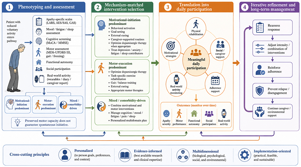 |
Figure 4 Clinical pathway for phenotyping reduced voluntary activity and tailoring rehabilitation in Parkinson’s disease. This figure illustrates a clinically oriented pathway for assessing and managing reduced voluntary activity in Parkinson’s disease (PD). Step 1 emphasizes multidomain phenotyping and assessment. Patients with reduced voluntary activity should be evaluated using apathy-specific scales, mood, fatigue and sleep assessment, cognitive screening, motor assessment, functional autonomy measures, social participation outcomes, and real-world activity monitoring through wearables, diaries, or caregiver reports. This assessment is intended to classify patients into motivational-initiation predominant, motor-execution predominant, or mixed/comorbidity-driven phenotypes. The key clinical principle is that preserved motor capacity does not guarantee spontaneous initiative. Step 2 summarizes mechanism-matched intervention selection. When motivational-initiation deficits predominate, rehabilitation may require behavioural activation, goal setting, external cueing, caregiver-supported routines, reinforcement strategies, optimization of dopaminergic therapy when appropriate, and treatment of depression, anxiety, fatigue, pain, or sleep contributors. When motor-execution deficits predominate, intervention should emphasize dopaminergic optimization, task-specific exercise rehabilitation, gait and balance training, external cueing, and appropriate motor therapies. Mixed or comorbidity-driven phenotypes require combined motivational and motor interventions together with management of cognitive, affective, fatigue, pain, sleep, and environmental contributors. Step 3 shows translation into meaningful daily participation. Rehabilitation should not be evaluated only by improvement in motor scores or clinic-based performance. Outcomes should also include apathy severity, motor performance, functional autonomy, social participation, adherence, and real-world activity. Motivational strategies, physical rehabilitation, caregiver or social support, adherence support, and real-world monitoring may help translate gains into daily participation. Step 4 illustrates iterative refinement and long-term management. Treatment response should be reassessed over time, intervention intensity or combinations should be adjusted, adherence should be reinforced, relapse or disengagement should be prevented, and caregiver or environmental support should be continued when needed. The pathway is personalized, evidence-informed, multidimensional, and implementation-oriented. It is intended as a practical clinical framework rather than a validated treatment algorithm. |
Rehabilitation studies should distinguish improvement in motor capacity from improvement in self-initiated daily activity. Outcome assessment should therefore include not only MDS-UPDRS motor scores, gait speed, balance, or exercise capacity, but also measures of functional autonomy, social participation, adherence, time spent in voluntary activity, and patient- or caregiver-reported initiation.60 Functional autonomy and quality-of-life measures are particularly relevant because apathy and reduced functional autonomy have been associated with poorer perceived quality of life in PD.27 PD-specific social functioning instruments can further test whether motor improvement translates into meaningful daily and social participation, rather than only better performance under clinical testing conditions.28 In older patients, treatment response should also be interpreted in light of frailty, fall concern, comorbidity, cognitive reserve, and social support. These factors may limit exercise tolerance and adherence even when motor capacity improves, and perceived barriers such as fear of falling or low outcome expectations can reduce exercise participation in PD.194 Rehabilitation plans for older adults with PD-related apathy may therefore require lower initial task demands, structured progression, external cueing, caregiver-supported scheduling, and repeated reinforcement of functional goals.
The Initiation Problem
A core challenge in PD rehabilitation interventions is the initiation problem: how to facilitate initial participation in rehabilitation when patients have impaired motivational systems. This problem is rooted in the neurobiological features of PD, representing a core bottleneck between theory and practice.
Motivational deficits, a prominent non-motor symptom of PD, involve a multidimensional reduction in emotional, cognitive, and behavioural dimensions of goal-directed behaviour. The prevalence of motivational deficits in PD patients varies extensively, implying the lack of consistency between current diagnostic standards and assessment tools.195,196 In addition, current treatment strategies commonly control symptoms without halting disease progression, making early diagnosis and effective intervention critical unmet requirements in PD care.197 This highlights the foundational value of improving doctor-patient communication and symptom education to overcome the initiation problem. Hence, healthcare professionals face a dual challenge in identifying and managing motivational deficits: the absence of standardized diagnostic mechanisms and the limited evidence for specific interventions targeting motivational deficits.
Empirical evidence has suggested significant variability in exercise participation among PD patients. One study reported that nearly half of PD patients reported less than 3 hours of exercise per week, categorizing them as low-activity individuals. Importantly, these patients were more likely to decrease, rather than start or sustain, regular exercise post-diagnosis compared to high-activity individuals.198 Low-activity patients reported approximately twice as many barriers as high-activity patients, with three core factors being particularly prominent: lack of external incentives, fatigue, and depression.198 These findings confirm that successful exercise interventions must particularly address these barriers, rather than simply providing generic exercise advice.198
One potentially useful but still indirect approach to the initiation problem is the application of contingency-management principles. This behavioural intervention approach comprehensively connects tangible rewards with target behaviours, providing clear external drives for individuals with impaired motivational systems.199,200 Evidence for contingency management comes mainly from non-PD behavioural medicine and addiction settings; therefore, its relevance to PD rehabilitation should be treated as indirect and requires PD-specific testing. When patients were given an opportunity to win rewards for submitting drug-negative samples, their treatment adherence and duration of abstinence improved significantly.201 The utilization of this principle in PD rehabilitation involves integrating immediate, tangible positive reinforcement with the completion of rehabilitation tasks, thereby compensating for an impaired internal motivational system. Embedding rehabilitation activities in emotionally meaningful and socially valuable environments is another promising way to overcome the initiation problem. Research on community choirs provides empirical support for this: patients with neurodegenerative diseases who engaged in group music activities showed several benefits, including enhanced confidence, improved mood, and increased motivation.202 Studies show that well-implemented gamification principles can significantly increase patient participation motivation and behavioural persistence in neurorehabilitation.203
From a neurobiological perspective, these strategies may compensate for impaired motivational systems via different pathways: external rewards may indirectly increase motivational signals by activating brain regions, such as the NAc;204 emotionally rich environments may modulate participation motivation via the limbic system;205 social interaction may improve behavioural drive through neurotransmitter systems, such as oxytocin.206
Target Misalignment in Invasive Neuromodulation
Benefits and Limitations of DBS Therapy
Deep brain stimulation (DBS) is an established therapy for selected motor symptoms of advanced PD, but it should not be presented as a primary treatment for apathy. Its relevance to PD-related apathy lies mainly in treatment interpretation: DBS can improve motor output while producing variable, neutral, or adverse effects on motivation, depending on target, electrode location, stimulation parameters, medication reduction, and patient vulnerability.
DBS alters abnormal activity in the BG circuits via high-frequency electrical stimulation, providing reliable symptom control for advanced PD patients. High-frequency stimulation of the subthalamic nucleus (STN) can suppress abnormal beta-band oscillations in the local area, which are closely associated with PD-related motor bradykinesia.207 Evidence has shown that the suppression of beta activity after STN stimulation is positively correlated with improvements in motor performance, providing direct physiological evidence for the mechanism of DBS.25 Randomized controlled trials further confirm the clinical value of DBS. Research on advanced PD patients confirms that integrating STN-DBS with medication therapy yields greater improvements in quality of life and motor symptoms compared to medication alone, with average enhancements of 9.5 points on the 39-item Parkinson’s Disease Questionnaire (PDQ-39) and 19.6 points on the Unified Parkinson’s Disease Rating Scale, Part III (UPDRS-III).208
Despite its significant effectiveness in motor control, DBS shows highly variable and unpredictable effects on non-motor symptoms, such as motivation and mood disorders. The anatomical location of the electrode contact points is clearly associated with emotional responses: left-sided STN stimulation improves mood better than right-sided stimulation, while right-sided DBS-driven positive emotional enhancement is associated with more medial and dorsal contact point locations.209 STN-DBS may also be associated with exacerbation of motivational deficits. Meta-analytic evidence shows that, compared to pre-surgical conditions and medication therapy alone, STN-DBS significantly increases motivational-deficit scores after surgery, independent of reductions in dopaminergic medications.15 This finding suggests that DBS may impact motivation-related neural circuits via direct or indirect processes.
The limitations of DBS in interventions for motivational disorders are rooted in its inherent target misalignment. Motivational deficits in PD are mainly associated with dysfunction in the prefrontal-limbic system, particularly the motivational-processing network composed of the dorsally located ACC, medial OFC (mOFC), and the ventral striatum (VS). These areas are modulated by the midbrain DA system arising from the VTA.210 In contrast, traditional DBS targets primarily focus on the motor circuit. While STN and the globus pallidus internus (GPi) are core nodes in the BG motor pathway, they are anatomically and functionally separated from the core brain regions involved in motivation and emotion processing, such as the NAc, PFC, and the limbic system.211,212 This target difference implies that conventional DBS primarily modulates motor circuitry and may not reliably engage the motivational networks most closely linked to apathy.211,212 Functional imaging evidence supports this view, showing that subcortical DBS stimulation-driven temporary depressive moods are associated with modifications in the structure of the midbrain-limbic cortical system, revealing the relative independence of motor circuits from emotional circuits.213 Moreover, STN and GPi as DBS targets are equally effective in tremor control, but may differ in their effects on non-motor symptoms. Systematic reviews display no significant difference between the two targets in tremor suppression.214 Nevertheless, no significant difference has been reported between STN-DBS and GPi-DBS in terms of cognitive and psychiatric symptoms.215
Overall, while DBS represents a significant achievement in neuromodulation technology for PD treatment, its limited effects on motivational disorders highlight the constraints of current neuromodulation technologies. Recognizing the neurobiological mechanisms of target misalignment is crucial for developing more integrated and precise PD treatment strategies. Future neuromodulation research should carefully evaluate whether individualized targeting or adaptive stimulation can preserve motor benefits while minimizing adverse motivational and affective outcomes.
Precision and Reversibility Limitations of DBS Technology
DBS shows significant therapeutic effects in controlling PD motor symptoms, but its inherent limitations in precision and reversibility remain key factors limiting its use in interventions for motivational disorders. These technical challenges directly impact DBS’s ability to adapt to dynamic changes in non-motor symptoms, particularly motivational states.
Once the DBS electrode is implanted, its spatial position becomes fixed, which establishes a significant technical bottleneck in clinical practice. In open-loop DBS, the stimulation parameters, including duration, amplitude, and pulse frequency, remain constant despite fluctuations in disease state, and cannot adaptively adjust to changes in the patient’s motivation state or other non-motor symptoms.216 More importantly, the precise position of the electrode within the STN is closely associated with post-surgical changes in motivation state. Individualised network analysis using 7T MRI has shown that patients who develop motivational deficits after surgery have more effective contact points located within the STN, notably in regions with high cortical projections, while the motor projection density is significantly lower.217 This finding offers an anatomical basis for DBS-driven motivational deficits, highlighting the significance of electrode placement precision in non-motor outcomes. Another major challenge is the irreversibility of DBS therapy. Unlike pharmacological treatments, DBS needs permanent electrode implantation and connection to a neurostimulator, and modifications or termination of treatment are not as flexible as altering drug doses.218 This irreversibility can pose significant clinical risks in certain situations. Patients with PD may develop a rare but potentially life-threatening complication—DBS withdrawal syndrome—needing urgent intervention to maintain or rescue stimulation. When patients face intolerable cognitive, emotional, or behavioural side effects, the inability to swiftly alter or withdraw treatment becomes a significant consideration in clinical decision-making.219
DBS can also produce chronic neuropsychiatric or stimulation-related side effects, and these effects are closely related to electrode location, stimulation target, and stimulation parameters. Systematic reviews indicate that thalamic stimulation is associated with fatigue, whereas STN stimulation is more commonly associated with emotional side effects, including depression and suicidal ideation. Higher stimulation voltage has also been associated with more severe post-operative depression.220 The side-effect profile differs across stimulation targets. Studies of essential tremor have reported that stimulation of the thalamic ventral intermediate nucleus is commonly associated with dysarthria, whereas posterior subthalamic area stimulation is more commonly associated with gait disturbance. Bilateral DBS is associated with more side effects than unilateral DBS, and adjustment of active contacts to optimize stimulation of the ventral intermediate nucleus or posterior subthalamic area may help reduce stimulation-related adverse effects.221 These findings highlight the limited ability of conventional DBS to adapt to dynamic changes in motivational and affective states. Although DBS can improve selected PD motor symptoms, STN-DBS has been associated with adverse effects on mood and cognition, including apathy symptoms that may emerge or worsen after surgery.222 This limitation is particularly prominent in the context of fluctuating non-motor symptoms. Anxiety, depression, fatigue, inner unrest, pain, concentration difficulties, and dizziness are known to fluctuate with motor symptoms and are more frequent and severe during off periods than during on periods.223 Although DBS can improve selected motor symptoms in PD, its effects on mood, motivation, and apathy remain variable and difficult to predict.224 Recent evidence also suggests that patient-level factors, including purine intake, may influence motor prognosis after STN-DBS, further supporting individualized preoperative and postoperative phenotyping when interpreting DBS response.225 Adaptive DBS may help address some of these limitations by adjusting stimulation amplitude according to relevant neural activity. A long-term study of home-based adaptive deep brain stimulation (aDBS) has shown that it is well tolerated, effective, and safe in PD patients previously receiving continuous DBS.226
Overall, limitations in precision, adaptability, and reversibility constrain the use of DBS for motivational symptoms in PD. Although adaptive stimulation and individualized targeting may improve flexibility, future neuromodulation approaches should not only sustain motor symptom control but also enable more precise modulation of non-motor symptoms, particularly motivational states.
Summary
Current interventions for PD-related apathy and reduced voluntary activity face several important limitations, including limited circuit specificity, incomplete restoration of physiological dopaminergic dynamics, variable effects on non-motor symptoms, and adherence barriers in behavioural rehabilitation. An integrative analysis of current dopaminergic replacement therapies, behavioural rehabilitation interventions, and invasive neuromodulation suggests a series of shared core issues. The lack of specific modulation for neural circuits remains a fundamental limitation. While DA replacement therapy can enhance overall brain DA levels, it cannot precisely target the mesolimbic motivational circuits.159 Although DBS can precisely modulate the BG motor circuits, its targets are anatomically misaligned with the core brain regions involved in motivation and emotional processing.217 This lack of circuit specificity causes significant dissociation between treatment effects on motor and motivational dimensions. Moreover, the lack of dynamic steady-state modulation limits long-term efficacy. Pulsatile stimulation from DA replacement disrupts the fine modulation of the endogenous DA system, driving metabolic and signalling pathway dysfunction.166 Traditional DBS’s fixed parameters cannot adapt to physiological fluctuations in motivational states, and behavioural interventions are restricted by the adherence dilemma triggered by motivational deficits.192 In addition, invasive or systemic side effects restrict therapeutic benefits. DA replacement therapy is associated with the risk of motor complications and behavioural abnormalities;168 DBS implantation’s irreversibility brings the risk of neuropsychiatric side effects;220 and high-intensity behavioural interventions are commonly difficult to implement in patients with motivational deficits because of initiation barriers.198 Neuroimaging evidence further supports the structural basis for these limitations: the development of apathy symptoms is closely associated with atrophy in specific brain regions and white matter damage, which involve networks beyond the regulatory reach of current interventions.161
Overall, the numerous limitations of current intervention paradigms in specificity, dynamics, and safety highlight the need for more circuit-specific, adaptive, and individualized intervention strategies. Future work should focus on developing interventions that can target motivational circuits more precisely, provide on-demand dynamic regulation, and reduce invasiveness. Preliminary advances in adaptive DBS technology226 offer proof of concept for this direction, but obtaining precise, non-invasive modulation of the motivational circuits still faces significant challenges. Only via interdisciplinary collaboration, combining neural circuit knowledge, advanced modulation technologies, and individualised treatment strategies, can the current treatment dilemmas for PD-related apathy and reduced voluntary activity be overcome. The limitations, mechanisms, and bottlenecks of PD-related apathy and reduced voluntary activity interventions are summarized in Table 3.
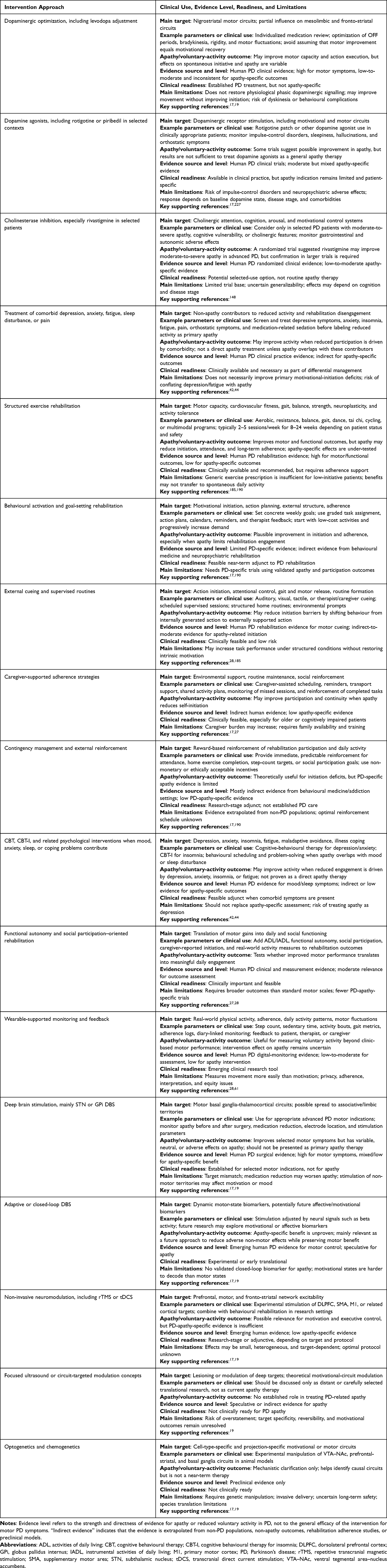 |
Table 3 Clinical and Preclinical Intervention Approaches for PD-Related Apathy and Reduced Voluntary Activity: Evidence Level and Clinical Readiness |
Critical Synthesis of Intervention Evidence
The intervention literature reveals a consistent mismatch between motor efficacy and motivational efficacy. Dopaminergic therapy is clinically established for motor symptoms, but its effects on apathy and spontaneous initiative are variable. Although dopaminergic stimulation may improve apathy in selected contexts, such as D2/D3 receptor stimulation with piribedil, this does not mean that dopaminergic treatment provides a general or reliable solution for PD-related apathy.17,227 This does not mean that dopamine is irrelevant to motivation; rather, it indicates that increasing dopaminergic availability does not necessarily restore the behaviourally timed, circuit-specific signalling required for effort valuation, reward learning, and self-initiated action. Similarly, structured exercise and rehabilitation can improve motor capacity and functional performance in PD, but apathetic patients may fail to initiate, attend, or sustain participation unless the intervention also addresses motivational and environmental barriers.17,228
The evidence base is therefore uneven. Pharmacological and DBS approaches have stronger evidence for motor outcomes than for apathy-specific outcomes, although selected pharmacological studies, such as rivastigmine in apathetic but dementia- and depression-free PD patients, suggest that non-dopaminergic treatment strategies may deserve further testing in defined subgroups.17,148 Behavioural activation, cueing, goal setting, caregiver-supported routines, and external reinforcement are clinically plausible and low risk, but require more PD-specific trials using validated apathy outcomes and real-world participation measures. Evidence extrapolated from non-PD behavioural medicine, other neurodegenerative diseases, or preclinical circuit studies should be labelled as indirect. The practical implication is that apathy treatment should not be framed as a single-intervention problem. It should be framed as a phenotyping and implementation problem: clinicians must identify whether reduced activity is driven primarily by motor execution, motivational initiation, fatigue, depression, cognitive impairment, environmental barriers, or mixed mechanisms.
The main research gap is the lack of trials designed around motivational phenotypes rather than motor diagnosis alone. Future rehabilitation studies should stratify patients by apathy severity, depression and fatigue status, cognitive profile, motor severity, and real-world activity level. Outcomes should include not only MDS-UPDRS motor scores or gait performance, but also rehabilitation adherence, functional autonomy, social participation, caregiver-reported initiation, and wearable or diary-based daily activity.27,28,60,61 This would allow the field to determine whether an intervention improves movement capacity, spontaneous initiative, or both.
Preclinical Tools and Distant Translational Concepts
Optogenetic and chemogenetic approaches are discussed here only as preclinical tools for causal circuit dissection. They should not be interpreted as near-term therapeutic options for PD-related apathy or reduced voluntary activity. Their primary value lies in testing how defined neuronal populations, projection-specific pathways, and stimulation patterns contribute to motivational initiation, effort valuation, reward learning, action invigoration, and motor execution. Therefore, this section discusses these approaches as experimental tools that clarify circuit principles and may inform future clinically feasible neuromodulation, rather than as directly translatable treatment paradigms.
Optogenetics as a Causal Tool for Dissecting Motivational and Motor Circuits
Optogenetics provides a powerful preclinical method for testing causal relationships between neuronal activity and behaviour. By introducing light-sensitive proteins into selected neuronal populations and delivering temporally precise light stimulation, optogenetics enables millisecond-level manipulation of circuit activity in vivo.229 In the context of PD-related apathy, its value is mechanistic rather than therapeutic: it can help determine whether specific dopaminergic, striatal, prefrontal, or limbic circuit elements are sufficient or necessary for motivational initiation, effort allocation, and reward-guided action. More broadly, modern optogenetic approaches allow genetically targeted and temporally precise control of selected cells within heterogeneous biological tissues, but their application remains primarily experimental in the context of deep-brain motivational circuits.230
This approach is particularly relevant for dissecting mesolimbic motivational circuits. Optogenetic studies have shown that temporally specific activation of VTA dopaminergic neurons can influence reward prediction, reinforcement learning, and motivated behaviour.231 Temporally precise dopamine-neuron stimulation can also provide causal evidence linking dopamine prediction-error-like signals to cue-reward learning.232 Input- and projection-specific work further shows that distinct VTA circuits can generate different motivational signatures, including reward- and aversion-related responses.233 These findings are relevant to PD-related apathy because they help separate circuit mechanisms of motivational initiation from mechanisms of motor execution. In addition, systematic input-output mapping of VTA dopamine neurons has clarified the circuit architecture through which VTA dopamine neurons integrate diverse inputs and influence motivated behaviour via distinct projections.234
Optogenetics has also been useful for dissecting PD-relevant motor circuitry in animal models. For example, optogenetic manipulation of defined basal ganglia and cortical circuit elements has been used to deconstruct parkinsonian motor circuitry in freely moving rodents.235 This supports the value of optogenetics as a causal circuit tool, but it does not imply clinical readiness for treating apathy in PD.
Despite this mechanistic value, optogenetics faces major translational barriers. These include the need for safe and targeted gene delivery, long-term control of opsin expression, invasive light-delivery procedures, limited tissue penetration, immune and inflammatory risks, and uncertain long-term safety in chronically ill patients. Reviews of human translational barriers emphasize that opsin-based therapeutic applications require reliable gene delivery, cell-type specificity, adequate illumination of target tissue, and careful management of safety concerns.236 Visible light penetration remains a core limitation for deep-brain applications; although upconversion nanoparticle systems and related approaches may allow near-infrared-triggered optogenetic stimulation in experimental models, these strategies remain preclinical.237 Similarly, ultrasound-linked light-delivery strategies may reduce dependence on implanted optical fibres in animal models, but they should not be interpreted as evidence that optogenetics is ready for clinical treatment of PD-related apathy.238
Thus, optogenetics should be presented as a causal circuit-mapping tool. Its contribution to this review is to identify which neuronal populations, projection-defined pathways, and stimulation patterns may regulate motivation, effort valuation, reward learning, and action selection. It should not be framed as a practical treatment for PD-related apathy or reduced voluntary activity.
Chemogenetics as a Tool for Prolonged Circuit Modulation
Chemogenetics, particularly designer receptors exclusively activated by designer drugs (DREADDs), provides a complementary preclinical approach for ligand-based modulation of defined neuronal populations. DREADDs were originally developed as engineered G-protein-coupled receptors activated by otherwise pharmacologically selective ligands, allowing remote modulation of defined cell populations.239 Unlike optogenetics, which relies on external light delivery, DREADD-based approaches use engineered receptors activated by administered ligands to modulate neuronal activity over longer timescales.240 This light-free approach avoids optical-fibre implantation and is useful for testing how sustained activation or inhibition of selected neuronal populations alters motivational state, reward processing, and behavioural persistence in animal models.241
For PD-related apathy, chemogenetics is relevant because it can help test how prolonged modulation of defined mesolimbic, prefrontal-striatal, and basal ganglia circuit elements influences motivational behaviour. DREADD-based methods have been widely discussed as tools for modulating dopamine signalling and behaviour in experimental models, while also having important limitations related to ligand specificity, receptor expression, and behavioural interpretation.242 Such studies can help identify circuit mechanisms that may later inform clinically feasible interventions, but the evidence remains preclinical and should not be extrapolated directly to patient treatment.
Chemogenetics also has important limitations. DREADD effects depend on ligand administration, receptor expression, ligand pharmacokinetics, receptor occupancy, metabolism, and inter-individual variability. The early assumption that clozapine-N-oxide was inert has been challenged by evidence that CNO can be converted to clozapine, which may contribute to DREADD occupancy and activation.243 Pharmacokinetic and pharmacodynamic comparisons of clozapine-N-oxide, clozapine, and compound 21 further show that ligand selection can substantially influence the onset, duration, and interpretation of DREADD-based effects.244 Newer ligands such as deschloroclozapine may improve potency and pharmacokinetic properties in experimental settings, including non-human primates, but these advances do not remove the major clinical barriers of gene delivery, receptor-expression control, reversibility, and long-term safety.245
Chemogenetics also has lower temporal precision than optogenetics. Because DREADD-based modulation depends on systemic or local ligand administration, onset and offset usually occur over minutes to hours rather than milliseconds. This limits its ability to reproduce rapid phasic dopamine signals, reward prediction errors, or real-time action-initiation dynamics. Therefore, chemogenetics is better suited for testing how sustained changes in defined neuronal populations influence motivational state over longer timescales, rather than for modelling rapid motivational switching or behaviourally timed dopaminergic signalling.240
Overall, optogenetics and chemogenetics are valuable because they allow causal testing of motivational and motor circuit mechanisms in animal models. Their relevance to PD-related apathy is mainly conceptual and mechanistic: they can clarify how VTA–NAc, prefrontal-striatal, basal ganglia, and non-dopaminergic modulatory circuits contribute to self-initiated behaviour. However, current clinical priorities should remain focused on validated motivational phenotyping, PD-specific rehabilitation trials, behavioural strategies that improve initiation and adherence, and careful evaluation of existing pharmacological and neuromodulatory approaches. Circuit-specific genetic technologies should be described as distant translational concepts, not as near-term therapies for PD-related apathy.
Limitations and Future Directions
This Review has several limitations. First, it is a structured narrative review rather than a systematic review or meta-analysis; therefore, evidence selection and weighting necessarily involved expert judgment. Second, published studies use heterogeneous apathy definitions, assessment scales, medication states, disease stages, cognitive profiles, and activity outcomes, which limits direct comparison across studies. Third, several circuit-level interpretations rely on indirect evidence from neuroimaging, computational models, preclinical studies, or non-PD disorders and should therefore be interpreted as hypothesis-generating rather than established causal evidence for PD-related apathy.
Future work should prioritize PD-specific longitudinal studies and intervention trials that use validated apathy outcomes, objective activity monitoring, functional autonomy and social participation measures, and clearly defined motivational phenotypes. Trials should distinguish motor capacity from self-initiated daily participation, stratify patients by depression, fatigue, cognition, frailty, medication state, and motor phenotype, and test whether tailored behavioural, rehabilitation, pharmacological, or neuromodulatory strategies improve real-world engagement rather than motor performance alone.
Conclusions, Clinical Implications, and Research Priorities
Apathy in PD is clinically distinct from motor impairment, although the two frequently coexist and interact. Reduced voluntary activity may arise from impaired motor execution, impaired motivational initiation, executive dysfunction, fatigue, depression, cognitive impairment, normal aging, or combinations of these factors. The central clinical implication is that preserved motor capacity does not guarantee spontaneous initiative, rehabilitation adherence, or meaningful daily participation.
Routine assessment should therefore include apathy-specific instruments, mood and fatigue scales, cognitive screening, motor assessment, informant input, and measures of functional autonomy, social participation, and real-world activity. Rehabilitation planning should move beyond generic exercise prescription and identify whether the major barrier is motor execution, motivational initiation, fatigue, depression, executive dysfunction, environmental support, or adherence. Interventions should then be tailored through structured cueing, goal setting, behavioural activation, caregiver-supported routines, external reinforcement, and management of treatable comorbid contributors.
Future research should prioritize PD-specific trials that use validated apathy outcomes and distinguish motivational improvement from motor improvement. Mechanistic studies should separate human PD evidence from preclinical and non-PD evidence, and staged circuit models should be treated as hypothesis-generating. Neuromodulatory approaches, wearable measures, digital phenotyping, and circuit-informed biomarkers may support future research, but their role in treating PD-related apathy remains investigational. The immediate priority is not speculative circuit control, but clinically grounded motivational phenotyping and rehabilitation strategies that improve self-initiated daily activity.
Funding
This research was funded by the National Natural Science Foundation of China (32071171), Science and Technology Program of Hebei (22555702K), and Hebei Provincial Department of Education Postgraduate Innovation Funding Program (CXZZBS2025123).
Disclosure
The authors report no conflicts of interest in this work.
References
1. Bloem BR, Okun MS, Klein C. Parkinson’s disease. Lancet. 2021;397(10291):2284–41. doi:10.1016/S0140-6736(21)00218-X
2. Béreau M, Van Waes V, Servant M, Magnin E, Tatu L, Anheim M. Apathy in Parkinson’s disease: clinical patterns and neurobiological basis. Cells. 2023;12(12):1599. doi:10.3390/cells12121599
3. Sgambato-Faure V, Worbe Y, Epinat J, Féger J, Tremblay L. Cortico-basal ganglia circuits involved in different motivation disorders in non-human primates. Brain Struct Funct. 2016;221:345–364. doi:10.1007/s00429-014-0911-9
4. Remy P, Doder M, Lees A, Turjanski N, Brooks D. Depression in Parkinson’s disease: loss of dopamine and noradrenaline innervation in the limbic system. Brain. 2005;128:1314–1322. doi:10.1093/brain/awh445
5. Mazzoni P, Hristova A, Krakauer JW. Why don’t we move faster? Parkinson’s disease, movement vigor, and implicit motivation. J Neurosci. 2007;27:7105–7116. doi:10.1523/JNEUROSCI.0264-07.2007
6. Banwinkler M, Dzialas V, Rigoux L, et al. Putaminal dopamine modulates movement motivation in Parkinson’s disease. Brain. 2024;147(10):3352–3357. doi:10.1093/brain/awae214
7. Dauer W, Przedborski S. Parkinson’s disease: mechanisms and models. Neuron. 2003;39:889–909. doi:10.1016/s0896-6273(03)00568-3
8. Niv Y, Daw ND, Joel D, Dayan P. Tonic dopamine: opportunity costs and the control of response vigor. Psychopharmacology. 2007;191:507–520. doi:10.1007/s00213-006-0502-4
9. Salamone JD, Correa M. The mysterious motivational functions of mesolimbic dopamine. Neuron. 2012;76(3):470–485. doi:10.1016/j.neuron.2012.10.021
10. Haber SN, Knutson B. The reward circuit: linking primate anatomy and human imaging. Neuropsychopharmacology. 2010;35:4–26. doi:10.1038/npp.2009.129
11. Shenhav A, Botvinick MM, Cohen JD. The expected value of control: an integrative theory of anterior cingulate cortex function. Neuron. 2013;79:217–240. doi:10.1016/j.neuron.2013.07.007
12. Olanow CW, Obeso JA, Stocchi F. Continuous dopamine-receptor treatment of Parkinson’s disease: scientific rationale and clinical implications. Lancet Neurol. 2006;5:677–687. doi:10.1016/S1474-4422(06)70521-X
13. Le Heron C, Plant O, Manohar S, et al. Distinct effects of apathy and dopamine on effort-based decision-making in Parkinson’s disease. Brain. 2018;141:1455–1469. doi:10.1093/brain/awy110
14. Schootemeijer S, van der Kolk NM, Ellis T, et al. Barriers and motivators to engage in exercise for persons with Parkinson’s disease. J Parkinsons Dis. 2020;10:1293–1299. doi:10.3233/JPD-202247
15. Zoon TJC, van Rooijen G, Balm GMFC, et al. Apathy induced by subthalamic nucleus deep brain stimulation in Parkinson’s disease: a meta-analysis. Mov Disord. 2021;36:317–326. doi:10.1002/mds.28390
16. Kalia LV, Lang AE. Parkinson’s disease. Lancet. 2015;386:896–912. doi:10.1016/S0140-6736(14)61393-3
17. Pagonabarraga J, Kulisevsky J, Strafella AP, Krack P. Apathy in Parkinson’s disease: clinical features, neural substrates, diagnosis, and treatment. Lancet Neurol. 2015;14:518–531. doi:10.1016/S1474-4422(15)00019-8
18. Maillet A, Krack P, Lhommée E, et al. The prominent role of serotonergic degeneration in apathy, anxiety and depression in de novo Parkinson’s disease. Brain. 2016;139:2486–2502. doi:10.1093/brain/aww162
19. De Waele S, Cras P, Crosiers D. Apathy in Parkinson’s disease: defining the park apathy subtype. Brain Sci. 2022;12(7):923. doi:10.3390/brainsci12070923
20. Obeso JA, Rodríguez-Oroz MC, Rodríguez M, et al. Pathophysiology of the basal ganglia in Parkinson’s disease. Trends Neurosci. 2000;23:S8–19. doi:10.1016/s1471-1931(00)00028-8
21. Galvan A, Wichmann T. Pathophysiology of parkinsonism. Clin Neurophysiol. 2008;119:1459–1474. doi:10.1016/j.clinph.2008.03.017
22. Hu B, Wang X, Lu S, Ying X. A study of bidirectional control of Parkinson’s beta oscillations by basal ganglia, Chaos. Solitons Fractals. 2025;195:116267. doi:10.1016/j.chaos.2025.116267
23. Jenkinson N, Brown P. New insights into the relationship between dopamine, beta oscillations and motor function. Trends Neurosci. 2011;34(12):611–618. doi:10.1016/j.tins.2011.09.003
24. Neumann W-J, Degen K, Schneider G-H, et al. Subthalamic synchronized oscillatory activity correlates with motor impairment in patients with Parkinson’s disease. Mov Disord. 2016;31:1748–1751. doi:10.1002/mds.26759
25. Kühn AA, Kempf F, Brücke C, et al. High-frequency stimulation of the subthalamic nucleus suppresses oscillatory beta activity in patients with Parkinson’s disease in parallel with improvement in motor performance. J Neurosci. 2008;28:6165–6173. doi:10.1523/JNEUROSCI.0282-08.2008
26. Pedersen KF, Alves G, Brønnick K, Aarsland D, Tysnes O-B, Larsen JP. Apathy in drug-naïve patients with incident Parkinson’s disease: the Norwegian ParkWest study. J Neurol. 2010;257:217–223. doi:10.1007/s00415-009-5297-x
27. D’Iorio A, Vitale C, Piscopo F, et al. Impact of anxiety, apathy and reduced functional autonomy on perceived quality of life in Parkinson’s disease. Parkinsonism Relat Disord. 2017;43:114–117. doi:10.1016/j.parkreldis.2017.08.003
28. Su F-T, Tai C-H, Tan C-H, Hwang W-J, Yu R-L. The development of the social functioning scale for patients with Parkinson’s disease. J Parkinsons Dis. 2020;10(3):1143–1151. doi:10.3233/JPD-201930
29. Chaudhuri KR, Healy DG, Schapira AHV. National institute for clinical excellence, non-motor symptoms of Parkinson’s disease: diagnosis and management. Lancet Neurol. 2006;5:235–245. doi:10.1016/S1474-4422(06)70373-8
30. Devilbiss DM. Consequences of tuning network function by tonic and phasic locus coeruleus output and stress: regulating detection and discrimination of peripheral stimuli. Brain Res. 2019;1709:16–27. doi:10.1016/j.brainres.2018.06.015
31. Friedman NP, Robbins TW. The role of prefrontal cortex in cognitive control and executive function. Neuropsychopharmacology. 2022;47:72–89. doi:10.1038/s41386-021-01132-0
32. Marin RS. Apathy: a neuropsychiatric syndrome. J Neuropsychiatry Clin Neurosci. 1991;3:243–254. doi:10.1176/jnp.3.3.243
33. Levy R, Dubois B. Apathy and the functional anatomy of the prefrontal cortex-basal ganglia circuits. Cereb Cortex. 2006;16:916–928. doi:10.1093/cercor/bhj043
34. Robert P, Onyike CU, Leentjens AFG, et al. Proposed diagnostic criteria for apathy in Alzheimer’s disease and other neuropsychiatric disorders. Eur Psychiatry. 2009;24:98–104. doi:10.1016/j.eurpsy.2008.09.001
35. Miller DS, Robert P, Ereshefsky L, et al. Diagnostic criteria for apathy in neurocognitive disorders. Alzheimers Dement. 2021;17:1892–1904. doi:10.1002/alz.12358
36. Postuma RB, Berg D, Stern M, et al. MDS clinical diagnostic criteria for Parkinson’s disease. Mov Disord. 2015;30(12):1591–1601. doi:10.1002/mds.26424
37. Bologna M, Paparella G, Fasano A, Hallett M, Berardelli A. Evolving concepts on bradykinesia. Brain. 2020;143:727–750. doi:10.1093/brain/awz344
38. Nutt JG, Bloem BR, Giladi N, Hallett M, Horak FB, Nieuwboer A. Freezing of gait: moving forward on a mysterious clinical phenomenon. Lancet Neurol. 2011;10:734–744. doi:10.1016/S1474-4422(11)70143-0
39. Gauggel S, Rieger M, Feghoff T-A. Inhibition of ongoing responses in patients with Parkinson’s disease. J Neurol Neurosurg Psychiatry. 2004;75(4):539–544. doi:10.1136/jnnp.2003.016469
40. Aron AR, Robbins TW, Poldrack RA. Inhibition and the right inferior frontal cortex: one decade on. Trends Cognit Sci. 2014;18(4):177–185. doi:10.1016/j.tics.2013.12.003
41. Manza P, Amandola M, Tatineni V, Li C-SR, Leung H-C. Response inhibition in Parkinson’s disease: a meta-analysis of dopaminergic medication and disease duration effects. NPJ Parkinsons Dis. 2017;3:23. doi:10.1038/s41531-017-0024-2
42. Ineichen C, Baumann-Vogel H. Deconstructing apathy in Parkinson’s disease: challenges in isolating core components of apathy from depression, anxiety, and fatigue. Front Neurol. 2021;12:720921. doi:10.3389/fneur.2021.720921
43. Kirsch-Darrow L, Fernandez HF, Marsiske M, Okun MS, Bowers D. Dissociating apathy and depression in Parkinson disease. Neurology. 2006;67(1):33–38. doi:10.1212/01.wnl.0000230572.07791.22
44. Lazcano-Ocampo C, Wan YM, Van wamelen DJ, et al. Identifying and responding to fatigue and apathy in Parkinson’s disease: a review of current practice. Expert Rev Neurother. 2020;20(5):477–495. doi:10.1080/14737175.2020.1752669
45. Nasreddine ZS, Phillips NA, Bédirian V, et al. The montreal cognitive assessment, MoCA: a brief screening tool for mild cognitive impairment. J Am Geriatr Soc. 2005;53(4):695–699. doi:10.1111/j.1532-5415.2005.53221.x
46. Folstein MF, Folstein SE, McHugh PR. Mini-mental state”. A practical method for grading the cognitive state of patients for the clinician. J Psychiatr Res. 1975;12:189–198. doi:10.1016/0022-3956(75)90026-6
47. Fried LP, Tangen CM, Walston J, et al. Cardiovascular health study collaborative research group, frailty in older adults: evidence for a phenotype. J Gerontol a Biol Sci Med Sci. 2001;56(3):M146–156. doi:10.1093/gerona/56.3.m146
48. Samanez-Larkin GR, Knutson B. Decision making in the ageing brain: changes in affective and motivational circuits. Nat Rev Neurosci. 2015;16:278–289. doi:10.1038/nrn3917
49. Sockeel P, Dujardin K, Devos D, Denève C, Destée A, Defebvre L. The Lille apathy rating scale (LARS), a new instrument for detecting and quantifying apathy: validation in Parkinson’s disease. J Neurol Neurosurg Psychiatry. 2006;77:579–584. doi:10.1136/jnnp.2005.075929
50. Marin RS, Biedrzycki RC, Firinciogullari S. Reliability and validity of the apathy evaluation scale. Psychiatry Res. 1991;38:143–162. doi:10.1016/0165-1781(91)90040-v
51. Starkstein SE, Mayberg HS, Preziosi TJ, Andrezejewski P, Leiguarda R, Robinson RG. Reliability, validity, and clinical correlates of apathy in Parkinson’s disease. J Neuropsychiatry Clin Neurosci. 1992;4:134–139. doi:10.1176/jnp.4.2.134
52. Yi H-J, Tan C-H, Hong W-P, Yu R-L. Development and validation of the geriatric apathy scale: examining multi-dimensional apathy profiles in a neurodegenerative population with cultural considerations. Asian J Psychiatr. 2024;93:103924. doi:10.1016/j.ajp.2024.103924
53. Zigmond AS, Snaith RP. The hospital anxiety and depression scale. Acta Psychiatr Scand. 1983;67:361–370. doi:10.1111/j.1600-0447.1983.tb09716.x
54. Beck AT, Ward CH, Mendelson M, Mock J, Erbaugh J. An inventory for measuring depression. Arch Gen Psychiatry. 1961;4:561–571. doi:10.1001/archpsyc.1961.01710120031004
55. Yesavage JA, Brink TL, Rose TL, et al. Development and validation of a geriatric depression screening scale: a preliminary report. J Psychiatr Res. 1982;17:37–49. doi:10.1016/0022-3956(82)90033-4
56. Brown RG, Dittner A, Findley L, Wessely SC. The Parkinson fatigue scale. Parkinsonism Relat Disord. 2005;11(1):49–55. doi:10.1016/j.parkreldis.2004.07.007
57. Krupp LB, LaRocca NG, Muir-Nash J, Steinberg AD. The fatigue severity scale. Application to patients with multiple sclerosis and systemic lupus erythematosus. Arch Neurol. 1989;46:1121–1123. doi:10.1001/archneur.1989.00520460115022
58. Katz S, Ford AB, Moskowitz RW, Jackson BA, Jaffe MW. Studies of illness in the aged. the index of adl: a standardized measure of biological and psychosocial function. JAMA. 1963;185:914–919. doi:10.1001/jama.1963.03060120024016
59. Lawton MP, Brody EM. Assessment of older people: self-maintaining and instrumental activities of daily living. Gerontologist. 1969;9(3 Part 1):179–186. doi:10.1093/geront/9.3_Part_1.179
60. Goetz CG, Tilley BC, Shaftman SR, et al. Movement disorder society UPDRS revision task force, movement disorder society-sponsored revision of the unified Parkinson’s disease rating scale (MDS-UPDRS): scale presentation and clinimetric testing results. Mov Disord. 2008;23:2129–2170. doi:10.1002/mds.22340
61. Paul SS, Ellis TD, Dibble LE, et al. Obtaining reliable estimates of ambulatory physical activity in people with Parkinson’s disease. J Parkinsons Dis. 2016;6:301–305. doi:10.3233/JPD-160791
62. Baggio HC, Segura B, Garrido-Millan JL, et al. Resting-state frontostriatal functional connectivity in Parkinson’s disease-related apathy. Mov Disord. 2015;30:671–679. doi:10.1002/mds.26137
63. Tessitore A, Cirillo M, De Micco R. Functional connectivity signatures of Parkinson’s disease. J Parkinsons Dis. 2019;9(4):637–652. doi:10.3233/JPD-191592
64. Parnetti L, Gaetani L, Eusebi P, et al. CSF and blood biomarkers for Parkinson’s disease. Lancet Neurol. 2019;18:573–586. doi:10.1016/S1474-4422(19)30024-9
65. Kwon EH, Tennagels S, Gold R, Gerwert K, Beyer L, Tönges L. Update on CSF biomarkers in Parkinson’s disease. Biomolecules. 2022;12:329. doi:10.3390/biom12020329
66. Aamodt WW, Waligorska T, Shen J, et al. Neurofilament light chain as a biomarker for cognitive decline in Parkinson disease. Mov Disord. 2021;36:2945–2950. doi:10.1002/mds.28779
67. Siderowf A, Concha-Marambio L, Lafontant D-E, et al. Parkinson’s progression markers initiative, assessment of heterogeneity among participants in the Parkinson’s progression markers initiative cohort using α-synuclein seed amplification: a cross-sectional study. Lancet Neurol. 2023;22:407–417. doi:10.1016/S1474-4422(23)00109-6
68. Russo SJ, Nestler EJ. The brain reward circuitry in mood disorders. Nat Rev Neurosci. 2013;14:609–625. doi:10.1038/nrn3381
69. Hou G, Hao M, Duan J, Han M-H. The formation and function of the VTA dopamine system. Int J Mol Sci. 2024;25(7):3875. doi:10.3390/ijms25073875
70. Hart AS, Rutledge RB, Glimcher PW, Phillips PEM. Phasic dopamine release in the rat nucleus accumbens symmetrically encodes a reward prediction error term. J Neurosci. 2014;34:698–704. doi:10.1523/JNEUROSCI.2489-13.2014
71. Bromberg-Martin ES, Matsumoto M, Hikosaka O. Dopamine in motivational control: rewarding, aversive, and alerting. Neuron. 2010;68:815–834. doi:10.1016/j.neuron.2010.11.022
72. Salamone JD, Correa M. The neurobiology of activational aspects of motivation: exertion of effort, effort-based decision making, and the role of dopamine. Annu Rev Psychol. 2024;75:1–32. doi:10.1146/annurev-psych-020223-012208
73. Ambroggi F, Ghazizadeh A, Nicola SM, Fields HL. Roles of nucleus accumbens core and shell in incentive-cue responding and behavioral inhibition. J Neurosci. 2011;31(18):6820–6830. doi:10.1523/JNEUROSCI.6491-10.2011
74. Walle R, Petitbon A, Fois GR, et al. Nucleus accumbens D1- and D2-expressing neurons control the balance between feeding and activity-mediated energy expenditure. Nat Commun. 2024;15(1):2543. doi:10.1038/s41467-024-46874-9
75. Frank MJ. Adaptive cost-benefit control fueled by striatal dopamine. Annu Rev Neurosci. 2025;48(1):1–22. doi:10.1146/annurev-neuro-112723-025228
76. Touroutoglou A, Andreano JM, Adebayo M, Lyons S, Barrett LF. Motivation in the service of allostasis: the role of anterior mid cingulate cortex. Adv Motiv Sci. 2019;6:1–25. doi:10.1016/bs.adms.2018.09.002
77. Le Bouc R, Borderies N, Carle G, et al. Effort avoidance as a core mechanism of apathy in frontotemporal dementia. Brain. 2023;146(2):712–726. doi:10.1093/brain/awac427
78. Barrus MM, Hosking JG, Cocker PJ, Winstanley CA. Inactivation of the orbitofrontal cortex reduces irrational choice on a rodent Betting Task. Neuroscience. 2017;345:38–48. doi:10.1016/j.neuroscience.2016.02.028
79. Konova AB, Ceceli AO, Horga G, Moeller SJ, Alia-Klein N, Goldstein RZ. Reduced neural encoding of utility prediction errors in cocaine addiction. Neuron. 2023;111(24):4058–4070.e6. doi:10.1016/j.neuron.2023.09.015
80. Kim S, Bobeica I, Gamo NJ, Arnsten AFT, Lee D. Effects of α-2A adrenergic receptor agonist on time and risk preference in primates. Psychopharmacology. 2012;219:363–375. doi:10.1007/s00213-011-2520-0
81. Smith DM, Torregrossa MM. Valence encoding in the amygdala influences motivated behavior. Behav Brain Res. 2021;411:113370. doi:10.1016/j.bbr.2021.113370
82. Sesack SR, Grace AA. Cortico-Basal Ganglia reward network: microcircuitry. Neuropsychopharmacology. 2010;35:27–47. doi:10.1038/npp.2009.93
83. Bayassi-Jakowicka M, Lietzau G, Czuba E, Steliga A, Waśkow M, Kowiański P. Neuroplasticity and multilevel system of connections determine the integrative role of nucleus accumbens in the brain reward system. Int J Mol Sci. 2021;22:9806. doi:10.3390/ijms22189806
84. De Groote A, de Kerchove d’Exaerde A. Thalamo-nucleus accumbens projections in motivated behaviors and addiction. Front Syst Neurosci. 2021;15:711350. doi:10.3389/fnsys.2021.711350
85. Ikemoto S, Yang C, Tan A. Basal ganglia circuit loops, dopamine and motivation: a review and enquiry. Behav Brain Res. 2015;290:17–31. doi:10.1016/j.bbr.2015.04.018
86. Gremel CM, Costa RM. Premotor cortex is critical for goal-directed actions. Front Comput Neurosci. 2013;7:110. doi:10.3389/fncom.2013.00110
87. Le Heron C, Morris L-A, Manohar S. Understanding disrupted motivation in Parkinson’s disease through a value-based decision-making lens. Trends Neurosci. 2025;48:297–311. doi:10.1016/j.tins.2025.02.008
88. Schultz W. Reward functions of the basal ganglia. J Neural Transm. 2016;123:679–693. doi:10.1007/s00702-016-1510-0
89. Schapira AHV, Chaudhuri KR, Jenner P. Non-motor features of Parkinson disease. Nat Rev Neurosci. 2017;18:435–450. doi:10.1038/nrn.2017.62
90. Da Cunha C, Boschen SL, Gómez-A A, et al. Toward sophisticated basal ganglia neuromodulation: review on basal ganglia deep brain stimulation. Neurosci Biobehav Rev. 2015;58:186–210. doi:10.1016/j.neubiorev.2015.02.003
91. Cools R. Dopaminergic modulation of cognitive function-implications for L-DOPA treatment in Parkinson’s disease. Neurosci Biobehav Rev. 2006;30:1–23. doi:10.1016/j.neubiorev.2005.03.024
92. Moustafa AA, Sherman SJ, Frank MJ. A dopaminergic basis for working memory, learning and attentional shifting in Parkinsonism. Neuropsychologia. 2008;46:3144–3156. doi:10.1016/j.neuropsychologia.2008.07.011
93. Ramesh S, Arachchige ASPM. Depletion of dopamine in Parkinson’s disease and relevant therapeutic options: a review of the literature. AIMS Neurosci. 2023;10:200–231. doi:10.3934/Neuroscience.2023017
94. Pannu A, Goyal RK. The potential role of dopamine pathways in the pathophysiology of depression: current advances and future aspects. CNS Neurol Disord Drug Targets. 2025;24:340–352. doi:10.2174/0118715273357909241126064951
95. Kaushik P, Gorin F, Vali S. Dynamics of tyrosine hydroxylase mediated regulation of dopamine synthesis. J Comput Neurosci. 2007;22:147–160. doi:10.1007/s10827-006-0004-8
96. Fuxe K, Borroto-Escuela DO, Romero-Fernandez W, Zhang W-B, Agnati LF. Volume transmission and its different forms in the central nervous system. Chin J Integr Med. 2013;19:323–329. doi:10.1007/s11655-013-1455-1
97. Fuxe K, Rivera A, Jacobsen KX, et al. Dynamics of volume transmission in the brain. Focus on catecholamine and opioid peptide communication and the role of uncoupling protein 2. J Neural Transm. 2005;112:65–76. doi:10.1007/s00702-004-0158-3
98. Käenmäki M, Tammimäki A, Myöhänen T, et al. Quantitative role of COMT in dopamine clearance in the prefrontal cortex of freely moving mice. J Neurochem. 2010;114:1745–1755. doi:10.1111/j.1471-4159.2010.06889.x
99. Dürst CD, Wiegert JS, Schulze C, Helassa N, Török K, Oertner TG. Vesicular release probability sets the strength of individual Schaffer collateral synapses. Nat Commun. 2022;13:6126. doi:10.1038/s41467-022-33565-6
100. Cabin DE, Shimazu K, Murphy D, et al. Synaptic vesicle depletion correlates with attenuated synaptic responses to prolonged repetitive stimulation in mice lacking alpha-synuclein. J Neurosci. 2002;22:8797–8807. doi:10.1523/JNEUROSCI.22-20-08797.2002
101. Silberstein RB, Pipingas A, Farrow M, Levy F, Stough CK. Dopaminergic modulation of default mode network brain functional connectivity in attention deficit hyperactivity disorder. Brain Behav. 2016;6:e00582. doi:10.1002/brb3.582
102. Cai X, Liu C, Tsutsui-Kimura I, et al. Dopamine dynamics are dispensable for movement but promote reward responses. Nature. 2024;635(8038):406–414. doi:10.1038/s41586-024-08038-z
103. Sippy T, Tritsch NX. Unraveling the dynamics of dopamine release and its actions on target cells. Trends Neurosci. 2023;46:228–239. doi:10.1016/j.tins.2022.12.005
104. Chakroun K, Wiehler A, Wagner B, et al. Dopamine regulates decision thresholds in human reinforcement learning in males. Nat Commun. 2023;14:5369. doi:10.1038/s41467-023-41130-y
105. Michely J, Viswanathan S, Hauser TU, Delker L, Dolan RJ, Grefkes C. The role of dopamine in dynamic effort-reward integration. Neuropsychopharmacology. 2020;45:1448–1453. doi:10.1038/s41386-020-0669-0
106. Schultz W, Dayan P, Montague PR. A neural substrate of prediction and reward. Science. 1997;275(5306):1593–1599. doi:10.1126/science.275.5306.1593
107. Schultz W. Dopamine signals for reward value and risk: basic and recent data. Behav Brain Funct. 2010;6:24. doi:10.1186/1744-9081-6-24
108. Berridge KC, Robinson TE. What is the role of dopamine in reward: hedonic impact, reward learning, or incentive salience? Brain Res Brain Res Rev. 1998;28:309–369. doi:10.1016/s0165-0173(98)00019-8
109. Grace AA. The tonic/phasic model of dopamine system regulation and its implications for understanding alcohol and psychostimulant craving. Addiction. 2000;95 Suppl 2:S119–128. doi:10.1080/09652140050111690
110. Stocker M. Ca(2+)-activated K+ channels: molecular determinants and function of the SK family. Nat Rev Neurosci. 2004;5:758–770. doi:10.1038/nrn1516
111. Brittain J-S, Brown P. Oscillations and the basal ganglia: motor control and beyond. Neuroimage. 2014;85(Pt 2):637–647. doi:10.1016/j.neuroimage.2013.05.084
112. Cohen JD, Braver TS, Brown JW. Computational perspectives on dopamine function in prefrontal cortex. Curr Opin Neurobiol. 2002;12:223–229. doi:10.1016/s0959-4388(02)00314-8
113. Cools R, Barker RA, Sahakian BJ, Robbins TW. Enhanced or impaired cognitive function in Parkinson’s disease as a function of dopaminergic medication and task demands. Cereb Cortex. 2001;11:1136–1143. doi:10.1093/cercor/11.12.1136
114. Locke TM, Soden ME, Miller SM, et al. Dopamine D1 receptor-positive neurons in the lateral nucleus of the cerebellum contribute to cognitive behavior. Biol Psychiatry. 2018;84:401–412. doi:10.1016/j.biopsych.2018.01.019
115. Daws SE, Konradi C. Expression and function of dopamine receptors in the developing medial frontal cortex and striatum of the rat. Neuroscience. 2011;199:501–514. doi:10.1016/j.neuroscience.2011.10.004
116. Jones-Tabah J, Mohammad H, Paulus EG, Clarke PBS, Hébert TE. The signaling and pharmacology of the dopamine D1 receptor. Front Cell Neurosci. 2021;15:806618. doi:10.3389/fncel.2021.806618
117. Grima LL, Panayi MC, Härmson O, et al. Nucleus accumbens D1-receptors regulate and focus transitions to reward-seeking action. Neuropsychopharmacology. 2022;47:1721–1731. doi:10.1038/s41386-022-01312-6
118. Gerfen CR. Segregation of D1 and D2 dopamine receptors in the striatal direct and indirect pathways: an historical perspective. Front Synaptic Neurosci. 2022;14:1002960. doi:10.3389/fnsyn.2022.1002960
119. Ferraiolo M, Hermans E. The complex molecular pharmacology of the dopamine D2 receptor: implications for pramipexole, ropinirole, and rotigotine. Pharmacol Ther. 2023;245:108392. doi:10.1016/j.pharmthera.2023.108392
120. Frank MJ, Seeberger LC, O’reilly RC. By carrot or by stick: cognitive reinforcement learning in parkinsonism. Science. 2004;306(5703):1940–1943. doi:10.1126/science.1102941
121. Schultz W. Dopamine reward prediction error coding. Dialogues Clin Neurosci. 2016;18(1):23–32. doi:10.31887/DCNS.2016.18.1/wschultz
122. Stoker TB, Greenland JC, eds. Parkinson’s Disease: Pathogenesis and Clinical Aspects. Brisbane (AU): Codon Publications; 2018. Available from: http://www.ncbi.nlm.nih.gov/books/NBK536721/.
123. Moustafa AA, Cohen MX, Sherman SJ, Frank MJ. A role for dopamine in temporal decision making and reward maximization in parkinsonism. J Neurosci. 2008;28:12294–12304. doi:10.1523/JNEUROSCI.3116-08.2008
124. Cools R, D’Esposito M. Inverted-U-shaped dopamine actions on human working memory and cognitive control. Biol Psychiatry. 2011;69:e113–125. doi:10.1016/j.biopsych.2011.03.028
125. Braak H, Del Tredici K, Rüb U, de Vos RAI, Jansen Steur ENH, Braak E. Staging of brain pathology related to sporadic Parkinson’s disease. Neurobiol Aging. 2003;24:197–211. doi:10.1016/s0197-4580(02)00065-9
126. Heinzel S, Berg D, Gasser T, Chen H, Yao C, Postuma RB. MDS task force on the definition of Parkinson’s disease, update of the MDS research criteria for prodromal Parkinson’s disease. Mov Disord. 2019;34:1464–1470. doi:10.1002/mds.27802
127. Phan L, Miller D, Gopinath A, et al. Parkinson’s paradox: alpha-synuclein’s selective strike on SNc dopamine neurons over VTA. NPJ Parkinsons Dis. 2025;11:207. doi:10.1038/s41531-025-01055-3
128. Ferraro S, Demichelis G, Medina Carrion JP, et al. Dysfunctional mesocorticolimbic circuitry in cluster headache. J Headache Pain. 2025;26:121. doi:10.1186/s10194-025-02017-z
129. Song N, Liu Z, Gao Y, Lu S, Yang S, Yuan C. NAc-DBS corrects depression-like behaviors in CUMS mouse model via disinhibition of DA neurons in the VTA. Mol Psychiatry. 2024;29:1550–1566. doi:10.1038/s41380-024-02476-x
130. Flores-Ponce X, Velasco I. Dopaminergic neuron metabolism: relevance for understanding Parkinson’s disease. Metabolomics. 2024;20:116. doi:10.1007/s11306-024-02181-4
131. Brown CA, Campbell MC, Karimi M, et al. Dopamine pathway loss in nucleus accumbens and ventral tegmental area predicts apathetic behavior in MPTP-lesioned monkeys. Exp Neurol. 2012;236:190–197. doi:10.1016/j.expneurol.2012.04.025
132. Drui G, Carnicella S, Carcenac C, et al. Loss of dopaminergic nigrostriatal neurons accounts for the motivational and affective deficits in Parkinson’s disease. Mol Psychiatry. 2014;19:358–367. doi:10.1038/mp.2013.3
133. Zhang D, Yao J, Sun J, et al. Iron accumulation in the ventral tegmental area in Parkinson’s disease. Front Aging Neurosci. 2023;15:1187684. doi:10.3389/fnagi.2023.1187684
134. Keitz M, Koerts J, Kortekaas R, Renken R, de Jong BM, Leenders KL. Prefrontal cortex and striatal activation by feedback in Parkinson’s disease. Brain Res. 2008;1236:225–233. doi:10.1016/j.brainres.2008.07.110
135. Pessiglione M, Seymour B, Flandin G, Dolan RJ, Frith CD. Dopamine-dependent prediction errors underpin reward-seeking behaviour in humans. Nature. 2006;442(7106):1042–1045. doi:10.1038/nature05051
136. Cools R, Barker RA, Sahakian BJ, Robbins TW. Mechanisms of cognitive set flexibility in Parkinson’s disease. Brain. 2001;124:2503–2512. doi:10.1093/brain/124.12.2503
137. Martinez-Horta S, Sampedro F, Pagonabarraga J, et al. Non-demented Parkinson’s disease patients with apathy show decreased grey matter volume in key executive and reward-related nodes. Brain Imaging Behav. 2017;11:1334–1342. doi:10.1007/s11682-016-9607-5
138. Verharen JPH, de Jong JW, Roelofs TJM, et al. A neuronal mechanism underlying decision-making deficits during hyperdopaminergic states. Nat Commun. 2018;9:731. doi:10.1038/s41467-018-03087-1
139. Meyer A, Zimmermann R, Gschwandtner U, et al. Apathy in Parkinson’s disease is related to executive function, gender and age but not to depression. Front Aging Neurosci. 2014;6:350. doi:10.3389/fnagi.2014.00350
140. Zgaljardic DJ, Borod JC, Foldi NS, Mattis P. A review of the cognitive and behavioral sequelae of Parkinson’s disease: relationship to frontostriatal circuitry. Cogn Behav Neurol. 2003;16:193–210. doi:10.1097/00146965-200312000-00001
141. Leh SE, Petrides M, Strafella AP. The neural circuitry of executive functions in healthy subjects and Parkinson’s disease. Neuropsychopharmacology. 2010;35:70–85. doi:10.1038/npp.2009.88
142. Morris L-A, Harrison SJ, Melzer TR, et al. Altered nucleus accumbens functional connectivity precedes apathy in Parkinson’s disease. Brain. 2023;146:2739–2752. doi:10.1093/brain/awad113
143. Hezemans FH, Wolpe N, O’Callaghan C, et al. Noradrenergic deficits contribute to apathy in Parkinson’s disease through the precision of expected outcomes. PLoS Comput Biol. 2022;18:e1010079. doi:10.1371/journal.pcbi.1010079
144. Ye R, O’Callaghan C, Rua C, et al. Locus coeruleus integrity from 7 T MRI relates to apathy and cognition in Parkinsonian disorders. Mov Disord. 2022;37:1663–1672. doi:10.1002/mds.29072
145. Maillet A, Météreau E, Tremblay L, et al. Serotonergic and dopaminergic lesions underlying Parkinsonian neuropsychiatric signs. Mov Disord. 2021;36:2888–2900. doi:10.1002/mds.28722
146. Prange S, Metereau E, Maillet A, et al. Limbic serotonergic plasticity contributes to the compensation of apathy in early Parkinson’s disease. Mov Disord. 2022;37:1211–1221. doi:10.1002/mds.28971
147. Müller MLTM, Bohnen NI. Cholinergic dysfunction in Parkinson’s disease. Curr Neurol Neurosci Rep. 2013;13:377. doi:10.1007/s11910-013-0377-9
148. Devos D, Moreau C, Maltête D, et al. Rivastigmine in apathetic but dementia and depression-free patients with Parkinson’s disease: a double-blind, placebo-controlled, randomised clinical trial. J Neurol Neurosurg Psychiatry. 2014;85:668–674. doi:10.1136/jnnp-2013-306439
149. Pasquini J, Brooks DJ, Pavese N. The cholinergic brain in Parkinson’s disease. Mov Disord Clin Pract. 2021;8:1012–1026. doi:10.1002/mdc3.13319
150. Bohnen NI, Yarnall AJ, Weil RS, et al. Cholinergic system changes in Parkinson’s disease: emerging therapeutic approaches. Lancet Neurol. 2022;21:381–392. doi:10.1016/S1474-4422(21)00377-X
151. Floresco SB, Tse MTL, Ghods-Sharifi S. Dopaminergic and glutamatergic regulation of effort- and delay-based decision making. Neuropsychopharmacology. 2008;33:1966–1979. doi:10.1038/sj.npp.1301565
152. Bailey MR, Simpson EH, Balsam PD. Neural substrates underlying effort, time, and risk-based decision making in motivated behavior. Neurobiol Learn Mem. 2016;133:233–256. doi:10.1016/j.nlm.2016.07.015
153. Maher S, Donlon E, Mullane G, Walsh R, Lynch T, Fearon C. Treatment of apathy in Parkinson’s disease and implications for underlying pathophysiology. J Clin Med. 2024;13:2216. doi:10.3390/jcm13082216
154. Riederer P, Strobel S, Nagatsu T, et al. Levodopa treatment: impacts and mechanisms throughout Parkinson’s disease progression. J Neural Transm. 2025;132:743–779. doi:10.1007/s00702-025-02893-4
155. Sako W, Kogo Y, Koebis M, et al. Comparative efficacy and safety of adjunctive drugs to levodopa for fluctuating Parkinson’s disease - network meta-analysis. NPJ Parkinsons Dis. 2023;9:143. doi:10.1038/s41531-023-00589-8
156. Funkiewiez A, Ardouin C, Cools R, et al. Effects of levodopa and subthalamic nucleus stimulation on cognitive and affective functioning in Parkinson’s disease. Mov Disord. 2006;21:1656–1662. doi:10.1002/mds.21029
157. D’Iorio A, Maggi G, Vitale C, Trojano L, Santangelo G. Pure apathy” and cognitive dysfunctions in Parkinson’s disease: a meta-analytic study. Neurosci Biobehav Rev. 2018;94:1–10. doi:10.1016/j.neubiorev.2018.08.004
158. Hauser RA, Slawek J, Barone P, et al. Evaluation of rotigotine transdermal patch for the treatment of apathy and motor symptoms in Parkinson’s disease. BMC Neurol. 2016;16:90. doi:10.1186/s12883-016-0610-7
159. Thobois S, Ardouin C, Lhommée E, et al. Non-motor dopamine withdrawal syndrome after surgery for Parkinson’s disease: predictors and underlying mesolimbic denervation. Brain. 2010;133:1111–1127. doi:10.1093/brain/awq032
160. O’Sullivan SS, Evans AH, Lees AJ. Dopamine dysregulation syndrome: an overview of its epidemiology, mechanisms and management. CNS Drugs. 2009;23:157–170. doi:10.2165/00023210-200923020-00005
161. Moqadam R, Azizi H, Brzezinski-Rittner A, et al. Apathy progression is associated with brain atrophy and white matter damage in Parkinson’s disease. Brain Commun. 2025;7:fcaf355. doi:10.1093/braincomms/fcaf355
162. Nirenberg MJ. Dopamine agonist withdrawal syndrome: implications for patient care. Drugs Aging. 2013;30:587–592. doi:10.1007/s40266-013-0090-z
163. Richard IH. Depression in Parkinson’s disease. Curr Treat Options Neurol. 2000;85:263–274. doi:10.1007/s11940-000-0008-z
164. Grace AA. Phasic versus tonic dopamine release and the modulation of dopamine system responsivity: a hypothesis for the etiology of schizophrenia. Neuroscience. 1991;41:1–24. doi:10.1016/0306-4522(91)90196-u
165. Dreyer JK, Herrik KF, Berg RW, Hounsgaard JD. Influence of phasic and tonic dopamine release on receptor activation. J Neurosci. 2010;30(42):14273–14283. doi:10.1523/JNEUROSCI.1894-10.2010
166. Calabresi P, Ghiglieri V, Mazzocchetti P, Corbelli I, Picconi B. Levodopa-induced plasticity: a double-edged sword in Parkinson’s disease? Philos Trans R Soc Lond B Biol Sci. 2015;370(1672):20140184. doi:10.1098/rstb.2014.0184
167. Stocchi F, Olanow CW. Continuous dopaminergic stimulation in early and advanced Parkinson’s disease. Neurology. 2004;62(1_suppl_1):S56–63. doi:10.1212/wnl.62.1_suppl_1.s56
168. Weintraub D, Koester J, Potenza MN, et al. Impulse control disorders in Parkinson disease: a cross-sectional study of 3090 patients. Arch Neurol. 2010;67(5):589–595. doi:10.1001/archneurol.2010.65
169. Bayer HM, Glimcher PW. Midbrain dopamine neurons encode a quantitative reward prediction error signal. Neuron. 2005;47(1):129–141. doi:10.1016/j.neuron.2005.05.020
170. Voon V, Fernagut P-O, Wickens J, et al. Chronic dopaminergic stimulation in Parkinson’s disease: from dyskinesias to impulse control disorders. Lancet Neurol. 2009;8:1140–1149. doi:10.1016/S1474-4422(09)70287-X
171. Phokaewvarangkul O, Anan C, Phimpha A, Chaudhuri KR, van Laar T, Bhidayasiri R. Early factors for predicting discontinuation to subcutaneous apomorphine infusion in Parkinson’s disease: a prospective analysis of the Thai apomorphine registry. Parkinsonism Relat Disord. 2021;91:146–151. doi:10.1016/j.parkreldis.2021.09.022
172. Fernandez HH, Standaert DG, Hauser RA, et al. Levodopa-carbidopa intestinal gel in advanced Parkinson’s disease: final 12-month, open-label results. Mov Disord. 2015;30:500–509. doi:10.1002/mds.26123
173. Krasnova IN, Ladenheim B, Hodges AB, Volkow ND, Cadet JL, Asanuma M. Chronic methamphetamine administration causes differential regulation of transcription factors in the rat midbrain. PLoS One. 2011;6(4):e19179. doi:10.1371/journal.pone.0019179
174. Wang G-J, Volkow ND, Wigal T, et al. Long-term stimulant treatment affects brain dopamine transporter level in patients with attention deficit hyperactive disorder. PLoS One. 2013;8(5):e63023. doi:10.1371/journal.pone.0063023
175. Thomas DM, Francescutti-Verbeem DM, Kuhn DM. Increases in cytoplasmic dopamine compromise the normal resistance of the nucleus accumbens to methamphetamine neurotoxicity. J Neurochem. 2009;109:1745–1755. doi:10.1111/j.1471-4159.2009.06094.x
176. Nieoullon A. Dopamine and the regulation of cognition and attention. Prog Neurobiol. 2002;67:53–83. doi:10.1016/s0301-0082(02)00011-4
177. Vaughan RA, Foster JD. Mechanisms of dopamine transporter regulation in normal and disease states. Trends Pharmacol Sci. 2013;34(9):489–496. doi:10.1016/j.tips.2013.07.005
178. Zhu J, Reith MEA. Role of the dopamine transporter in the action of psychostimulants, nicotine, and other drugs of abuse. CNS Neurol Disord Drug Targets. 2008;7(5):393–409. doi:10.2174/187152708786927877
179. Thanvi BR, Lo TCN. Long term motor complications of levodopa: clinical features, mechanisms, and management strategies. Postgrad Med J. 2004;80:452–458. doi:10.1136/pgmj.2003.013912
180. Sharp ME, Foerde K, Daw ND, Shohamy D. Dopamine selectively remediates “model-based” reward learning: a computational approach. Brain. 2016;139:355–364. doi:10.1093/brain/awv347
181. Pavón N, Martín AB, Mendialdua A, Moratalla R. ERK phosphorylation and FosB expression are associated with L-DOPA-induced dyskinesia in hemiparkinsonian mice. Biol Psychiatry. 2006;59:64–74. doi:10.1016/j.biopsych.2005.05.044
182. Figge DA, Eskow Jaunarajs KL, Standaert DG. Dynamic DNA methylation regulates levodopa-induced dyskinesia. J Neurosci. 2016;36(24):6514–6524. doi:10.1523/JNEUROSCI.0683-16.2016
183. Warren N, O’Gorman C, Lehn A, Siskind D. Dopamine dysregulation syndrome in Parkinson’s disease: a systematic review of published cases. J Neurol Neurosurg Psychiatry. 2017;88:1060–1064. doi:10.1136/jnnp-2017-315985
184. Robinson TE, Berridge KC. Review. The incentive sensitization theory of addiction: some current issues. Philos Trans R Soc Lond B Biol Sci. 2008;363(1507):3137–3146. doi:10.1098/rstb.2008.0093
185. McGinley JL, Nakayama Y. Exercise for people with Parkinson’s disease: updates and future considerations. Phys Ther Res. 2024;27:67–75. doi:10.1298/ptr.R0030
186. Johansson ME, Cameron IGM, Van der Kolk NM, et al. Aerobic exercise alters brain function and structure in Parkinson’s disease: a randomized controlled trial. Ann Neurol. 2022;91:203–216. doi:10.1002/ana.26291
187. Tsukita K, Sakamaki-Tsukita H, Takahashi R. Long-term effect of regular physical activity and exercise habits in patients with early Parkinson disease. Neurology. 2022;98:e859–e871. doi:10.1212/WNL.0000000000013218
188. Bastioli G, Arnold JC, Mancini M, et al. Voluntary exercise boosts striatal dopamine release: evidence for the necessary and sufficient role of BDNF. J Neurosci. 2022;42(23):4725–4736. doi:10.1523/JNEUROSCI.2273-21.2022
189. Voss MW, Prakash RS, Erickson KI, et al. Plasticity of brain networks in a randomized intervention trial of exercise training in older adults. Front Aging Neurosci. 2010;2:32. doi:10.3389/fnagi.2010.00032
190. Ahern L, Timmons S, Lamb SE, McCullagh R. A systematic review of behaviour change interventions to improve exercise self-efficacy and adherence in people with Parkinson’s disease using the theoretical domains framework. J Frailty Sarcopenia Falls. 2024;9:66–68. doi:10.22540/JFSF-09-066
191. Yang Z, Huang H, Xu L, Gao Y, Zhang C, Wang A. From need to action: decoding the role of motivation in adherence to home-based cardiac rehabilitation exercise in chronic heart failure patients. BMC Nurs. 2024;23(1):913. doi:10.1186/s12912-024-02617-1
192. Le Heron C, Holroyd CB, Salamone J, Husain M. Brain mechanisms underlying apathy. J Neurol Neurosurg Psychiatry. 2019;90(3):302–312. doi:10.1136/jnnp-2018-318265
193. Adam R, Leff A, Sinha N, et al. Dopamine reverses reward insensitivity in apathy following globus pallidus lesions. Cortex. 2013;49(5):1292–1303. doi:10.1016/j.cortex.2012.04.013
194. Ellis T, Boudreau JK, DeAngelis TR, et al. Barriers to exercise in people with Parkinson disease. Phys Ther. 2013;93(5):628–636. doi:10.2522/ptj.20120279
195. Gu L, Zhang P, Wang Y, Zhu Y, Li H, Shu H. Prevalence, contributing factors, and clinical impact of apathy in Parkinson’s disease. J Psychiatr Res. 2025;191:382–392. doi:10.1016/j.jpsychires.2025.09.031
196. Mele B, Goodarzi Z, Hanson HM, Holroyd-Leduc J. Barriers and facilitators to diagnosing and managing apathy in Parkinson’s disease: a qualitative study. BMC Neurol. 2019;19(1):101. doi:10.1186/s12883-019-1329-z
197. Hopfner F, Höglinger G, Trenkwalder C;. Definition and diagnosis of Parkinson’s disease: guideline “Parkinson’s disease” of the German Society of neurology. J Neurol. 2024;271:7102–7119. doi:10.1007/s00415-024-12687-5
198. Afshari M, Yang A, Bega D. Motivators and barriers to exercise in Parkinson’s disease. J Parkinsons Dis. 2017;7:703–711. doi:10.3233/JPD-171173
199. Walter KN, Petry NM. Motivation and contingency management treatments for substance use disorders. Curr Top Behav Neurosci. 2016;27:569–581. doi:10.1007/7854_2015_374
200. Coughlin LN, Zhang L, Frost MC, et al. Contingency management for substance use disorders in the U.S. Veterans health administration: 2018-2022. J Subst Use Addict Treat. 2024;163:209400. doi:10.1016/j.josat.2024.209400
201. Petry NM, Alessi SM, Ledgerwood DM. A randomized trial of contingency management delivered by community therapists. J Consult Clin Psychol. 2012;80:286–298. doi:10.1037/a0026826
202. Tamplin J, Baker FA, Jones B, Way A, Lee S. Stroke a Chord”: the effect of singing in a community choir on mood and social engagement for people living with aphasia following a stroke. NeuroRehabilitation. 2013;32:929–941. doi:10.3233/NRE-130916
203. Lohse K, Shirzad N, Verster A, Hodges N, Van der Loos HFM. Video games and rehabilitation: using design principles to enhance engagement in physical therapy. J Neurol Phys Ther. 2013;37:166–175. doi:10.1097/NPT.0000000000000017
204. Keysers C, Kaas JH, Gazzola V. Somatosensation in social perception. Nat Rev Neurosci. 2010;11:417–428. doi:10.1038/nrn2833
205. Kalin NH, Larson C, Shelton SE, Davidson RJ. Asymmetric frontal brain activity, cortisol, and behavior associated with fearful temperament in rhesus monkeys. Behav Neurosci. 1998;112:286–292. doi:10.1037//0735-7044.112.2.286
206. Petersson M, Uvnäs-Moberg K. Interactions of oxytocin and dopamine-effects on behavior in health and disease. Biomedicines. 2024;12:2440. doi:10.3390/biomedicines12112440
207. Xu Z, Duan W, Yuan S, et al. Deep brain stimulation alleviates Parkinsonian motor deficits through desynchronizing GABA release in mice. Nat Commun. 2025;16:3726. doi:10.1038/s41467-025-59113-6
208. Deuschl G, Schade-Brittinger C, Krack P, et al. German Parkinson study group, neurostimulation section, a randomized trial of deep-brain stimulation for Parkinson’s disease. N Engl J Med. 2006;355:896–908. doi:10.1056/NEJMoa060281
209. Campbell MC, Black KJ, Weaver PM, et al. Mood response to deep brain stimulation of the subthalamic nucleus in Parkinson’s disease. J Neuropsychiatry Clin Neurosci. 2012;24:28–36. doi:10.1176/appi.neuropsych.11030060
210. Dujardin K, Sgambato V. Neuropsychiatric disorders in parkinson’s disease: what do we know about the role of dopaminergic and non-dopaminergic systems? Front Neurosci. 2020;14:25. doi:10.3389/fnins.2020.00025
211. Alexander GE, Crutcher MD, DeLong MR. Basal ganglia-thalamocortical circuits: parallel substrates for motor, oculomotor, “prefrontal” and “limbic” functions. Prog Brain Res. 1990;85:119–146.
212. Herrington TM, Cheng JJ, Eskandar EN. Mechanisms of deep brain stimulation. J Neurophysiol. 2016;115:19–38. doi:10.1152/jn.00281.2015
213. Stefurak T, Mikulis D, Mayberg H, et al. Deep brain stimulation for Parkinson’s disease dissociates mood and motor circuits: a functional MRI case study. Mov Disord. 2003;18:1508–1516. doi:10.1002/mds.10593
214. Wong JK, Cauraugh JH, Ho KWD, et al. STN vs. GPi deep brain stimulation for tremor suppression in Parkinson disease: a systematic review and meta-analysis. Parkinsonism Relat Disord. 2019;58:56–62. doi:10.1016/j.parkreldis.2018.08.017
215. Wang J-W, Zhang Y-Q, Zhang X-H, Wang Y-P, Li J-P, Li Y-J. Cognitive and psychiatric effects of STN versus GPi deep brain stimulation in Parkinson’s disease: a meta-analysis of randomized controlled trials. PLoS One. 2016;11:e0156721. doi:10.1371/journal.pone.0156721
216. Parastarfeizabadi M, Kouzani AZ. Advances in closed-loop deep brain stimulation devices. J Neuroeng Rehabil. 2017;14:79. doi:10.1186/s12984-017-0295-1
217. Zoon TJC, Mathiopoulou V, van Rooijen G, et al. Apathy following deep brain stimulation in Parkinson’s disease visualized by 7-Tesla MRI subthalamic network analysis. Brain Stimul. 2023;16:1289–1291. doi:10.1016/j.brs.2023.08.013
218. Okun MS. Deep-brain stimulation for Parkinson’s disease. N Engl J Med. 2012;367:1529–1538. doi:10.1056/NEJMct1208070
219. Miocinovic S, Ostrem JL, Okun MS, et al. Recommendations for deep brain stimulation device management during a pandemic. J Parkinsons Dis. 2020;10:903–910. doi:10.3233/JPD-202072
220. Zarzycki MZ, Domitrz I. Stimulation-induced side effects after deep brain stimulation - a systematic review. Acta Neuropsychiatr. 2020;32:57–64. doi:10.1017/neu.2019.35
221. Kim MJ, Chang KW, Park SH, Chang WS, Jung HH, Chang JW. Stimulation-induced side effects of deep brain stimulation in the ventralis intermedius and posterior subthalamic area for essential tremor. Front Neurol. 2021;12:678592. doi:10.3389/fneur.2021.678592
222. Vachez YM, Creed MC. Deep brain stimulation of the subthalamic nucleus modulates reward-related behavior: a systematic review. Front Hum Neurosci. 2020;14:578564. doi:10.3389/fnhum.2020.578564
223. Lee HM, Koh S-B. Many faces of Parkinson’s disease: non-motor symptoms of parkinson’s disease. J Mov Disord. 2015;8:92–97. doi:10.14802/jmd.15003
224. Accolla EA, Pollo C. Mood effects after deep brain stimulation for Parkinson’s disease: an update. Front Neurol. 2019;10:617. doi:10.3389/fneur.2019.00617
225. Chang B, Jiang Y, Feng C, Li B, Mei J, Niu C. Effect of purine diet on prognosis of deep brain stimulation for Parkinson’s disease. Food Science and Human Wellness. 2025;14:9250066. doi:10.26599/FSHW.2024.9250066
226. Bronte-Stewart HM, Beudel M, Ostrem JL, et al. ADAPT-PD investigators, long-term personalized adaptive deep brain stimulation in Parkinson disease: a nonrandomized clinical trial. JAMA Neurol. 2025;82:1171–1180. doi:10.1001/jamaneurol.2025.2781
227. Thobois S, Lhommée E, Klinger H, et al. Parkinsonian apathy responds to dopaminergic stimulation of D2/D3 receptors with piribedil. Brain. 2013;136:1568–1577. doi:10.1093/brain/awt067
228. Radder DLM, Lígia Silva de Lima A, Domingos J, et al. Physiotherapy in Parkinson’s disease: a meta-analysis of present treatment modalities. Neurorehabil Neural Repair. 2020;34:871–880. doi:10.1177/1545968320952799
229. Boyden ES, Zhang F, Bamberg E, Nagel G, Deisseroth K. Millisecond-timescale, genetically targeted optical control of neural activity. Nat Neurosci. 2005;8:1263–1268. doi:10.1038/nn1525
230. Emiliani V, Entcheva E, Hedrich R, et al. Optogenetics for light control of biological systems. Nat Rev Meth Primers. 2022;2:55. doi:10.1038/s43586-022-00136-4
231. Tsai H-C, Zhang F, Adamantidis A, et al. Phasic firing in dopaminergic neurons is sufficient for behavioral conditioning. Science. 2009;324:1080–1084. doi:10.1126/science.1168878
232. Steinberg EE, Keiflin R, Boivin JR, Witten IB, Deisseroth K, Janak PH. A causal link between prediction errors, dopamine neurons and learning. Nat Neurosci. 2013;16:966–973. doi:10.1038/nn.3413
233. Lammel S, Lim BK, Ran C, et al. Input-specific control of reward and aversion in the ventral tegmental area. Nature. 2012;491:212–217. doi:10.1038/nature11527
234. Beier KT, Steinberg EE, DeLoach KE, et al. Circuit architecture of VTA dopamine neurons revealed by systematic input-output mapping. Cell. 2015;162:622–634. doi:10.1016/j.cell.2015.07.015
235. Gradinaru V, Mogri M, Thompson KR, Henderson JM, Deisseroth K. Optical deconstruction of parkinsonian neural circuitry. Science. 2009;324:354–359. doi:10.1126/science.1167093
236. Shen Y, Campbell RE, Côté DC, Paquet M-E. Challenges for therapeutic applications of opsin-based optogenetic tools in humans. Front Neural Circuits. 2020;14:41. doi:10.3389/fncir.2020.00041
237. Chen S, Weitemier AZ, Zeng X, et al. Near-infrared deep brain stimulation via upconversion nanoparticle-mediated optogenetics. Science. 2018;359:679–684. doi:10.1126/science.aaq1144
238. Wu X, Zhu X, Chong P, et al. Sono-optogenetics facilitated by a circulation-delivered rechargeable light source for minimally invasive optogenetics. Proc Natl Acad Sci U S A. 2019;116(52):26332–26342. doi:10.1073/pnas.1914387116
239. Armbruster BN, Li X, Pausch MH, Herlitze S, Roth BL. Evolving the lock to fit the key to create a family of G protein-coupled receptors potently activated by an inert ligand. Proc Natl Acad Sci U S A. 2007;104(12):5163–5168. doi:10.1073/pnas.0700293104
240. Roth BL. DREADDs for Neuroscientists. Neuron. 2016;89(4):683–694. doi:10.1016/j.neuron.2016.01.040
241. Sternson SM, Roth BL. Chemogenetic tools to interrogate brain functions. Annu Rev Neurosci. 2014;37(1):387–407. doi:10.1146/annurev-neuro-071013-014048
242. Runegaard AH, Fitzpatrick CM, Woldbye DPD, Andreasen JT, Sørensen AT, Gether U. Modulating dopamine signaling and behavior with chemogenetics: concepts, progress, and challenges. Pharmacol Rev. 2019;71(2):123–156. doi:10.1124/pr.117.013995
243. Gomez JL, Bonaventura J, Lesniak W, et al. Chemogenetics revealed: DREADD occupancy and activation via converted clozapine. Science. 2017;357(6350):503–507. doi:10.1126/science.aan2475
244. Jendryka M, Palchaudhuri M, Ursu D, et al. Pharmacokinetic and pharmacodynamic actions of clozapine-N-oxide, clozapine, and compound 21 in DREADD-based chemogenetics in mice. Sci Rep. 2019;9(1):4522. doi:10.1038/s41598-019-41088-2
245. Nagai Y, Miyakawa N, Takuwa H, et al. Deschloroclozapine, a potent and selective chemogenetic actuator enables rapid neuronal and behavioral modulations in mice and monkeys. Nat Neurosci. 2020;23(9):1157–1167. doi:10.1038/s41593-020-0661-3
 © 2026 The Author(s). This work is published and licensed by Dove Medical Press Limited. The
full terms of this license are available at https://www.dovepress.com/terms
and incorporate the Creative Commons Attribution
- Non Commercial (unported, 4.0) License.
By accessing the work you hereby accept the Terms. Non-commercial uses of the work are permitted
without any further permission from Dove Medical Press Limited, provided the work is properly
attributed. For permission for commercial use of this work, please see paragraphs 4.2 and 5 of our Terms.
© 2026 The Author(s). This work is published and licensed by Dove Medical Press Limited. The
full terms of this license are available at https://www.dovepress.com/terms
and incorporate the Creative Commons Attribution
- Non Commercial (unported, 4.0) License.
By accessing the work you hereby accept the Terms. Non-commercial uses of the work are permitted
without any further permission from Dove Medical Press Limited, provided the work is properly
attributed. For permission for commercial use of this work, please see paragraphs 4.2 and 5 of our Terms.
Recommended articles
Morin Mitigates Methamphetamine-Induced Neurotoxicity: Effects on Motor and Cognitive Function
Anyanwu GE, Umeano AV, Ojiakor VO, Katchy AU, Anyanwu CN, Fakorede S
Journal of Experimental Pharmacology 2025, 17:307-321
Published Date: 11 June 2025
Long-Term Surgical Outcomes and Influential Factors of Subthalamic Nucleus Deep Brain Stimulation for Dyskinesia in Parkinson’s Disease: A 3-Year Longitudinal Cohort Study
Wang S, Xue T, Ma R, Shi L, Zhu G, Jiang Y, Yang A, Meng F, Zhang J
Clinical Interventions in Aging 2026, 21:600031
Published Date: 22 May 2026