Back to Journals » Journal of Inflammation Research » Volume 16
IL-34 and Immune Checkpoint Inhibitors Resistant through Macrophages: Pan-Cancer Overview
Authors Safi M, Fadhl AS, Algabri YA ![]() , Zhang C
, Zhang C
Received 15 November 2022
Accepted for publication 10 January 2023
Published 21 March 2023 Volume 2023:16 Pages 1209—1212
DOI https://doi.org/10.2147/JIR.S397749
Checked for plagiarism Yes
Review by Single anonymous peer review
Peer reviewer comments 2
Editor who approved publication: Dr Adam Bachstetter
Mohammed Safi,1,* Al-shaebi Fadhl,1,* Yousif A Algabri,2 Caiqing Zhang1
1Department of Respiratory Diseases, Shandong Second Provincial General Hospital, Shandong University, Jinan, Shandong, People’s Republic of China; 2Department of Biomedical Engineering, School of Control Science and Engineering, Shandong University, Jinan, Shandong, People’s Republic of China
*These authors contributed equally to this work
Correspondence: Caiqing Zhang, Department of respiratory, Shandong Second Provincial General Hospital, Shandong University, Jinan, Shandong, 250023, People’s Republic of China, Email [email protected] Al-shaebi Fadhl, Email [email protected]
Utilizing immune checkpoint inhibitors (ICIs) to induce therapeutic anti-tumor activity is a promising strategy. strategy. 1 However, malignancies often develop immunological resistance, limiting the clinical benefits of these approaches. approaches. 2 Several studies have indicated several molecular and cellular processes of immunological resistance. Immunosuppression in the local tumor microenvironment (TME) and epigenetic alterations in tumor cells that reduce interferon-gamma (IFN-γ) signaling constitute two major mechanisms involved in tumor progression. 3 In the context of TME, macrophages are the most common kind of immune cell found in solid tumors, accounting for as much as fifty percent of the total mass of the tumor. tumor. 4 In response to chemotactic chemicals made in the cancer microenvironment, macrophages (monocyte-derived tumor-associated macrophages, or TAMs) are sent from the blood to the tumor tissue. Infiltration of macrophages into and around the tumor nest is a characteristic that is considered to be one of the most crucial. These interactions between macrophages, tumor cells, and the stromal milieu contribute to the phenotypical polarization of macrophages linked with tumors. The process through which macrophages generate different functional phenotypes in response to certain stimuli and signals arising from their local environment is referred to as “polarization.” “polarization”. Macrophages are capable of polarization into conventionally activated (M1) and alternatively activated (M2) subpopulations. M1-phenotype macrophages are tumor-resistant, whereas M2-phenotype macrophages possess various tumor-promoting activities, including immunosuppression, angiogenesis, and neovascularization. The tumor microenvironment (TME) is frequently generated and continuously maintained by biomarkers secreted by the tumor tissues, and enrichment with immunosuppressive cells, like M2-biased tumor-associated macrophages (TAMs), has been suggested to play crucial roles in innate and acquired ICI resistance. resistance. 6
Recently, a large humanized model study revealed that interleukin IL-34 dysregulation could lead to resistance to the recently approved immune checkpoint inhibitors ICI, and IL-34 has come to be recognized as a pivotal molecule in regulating the maintenance, expansion, and activity of M2-biased TAMs that exhibit heightened immunosuppressive capabilities.7 Figure 1. By single-cell analysis of the IL-34 expressions in 208 single-cell datasets (https://ngdc.cncb.ac.cn/cancerscem/index) with 638,341 cells, we showed that the highest expressions were associated with glioblastoma (GBM), lung adenocarcinoma (LUAD), and pancreatic ductal adenocarcinoma (PDAC). Figure 2A; Supplementary Figure 1. In the context of ICIs resistance, cancer’s dysregulation of IL-34 showed how important M2-biased TAMS is in various cancer tissues. Pan-cancer analysis between IL-34 and M2 was conducted and uncovered that IL-34 has a positive correlation with infiltration of M2 among 30 cancer tissues with the highest positive association in prostatic adenocarcinoma (PRAD), testicular germ cell tumors (TCGT) and stomach adenocarcinoma (STAD) and negative correlation in low-grade gliomas (LGG). Figure 2B. Further analysis of the correlation difference in IL-34 expression between M1 and M2 with the same cancer types revealed a higher median value with M2 than M1 (0.33 versus 0.27) (P = 0.002). Figure 2C. Supplementary Methods and Materials. As a result, this may lead us to suggest that higher IL-34 expressions are closely related to M2-biased macrophage infiltration and decreased frequencies of the M1 type, and consequently, a higher possibility of resistance to ICI therapy. According to pan-cancer clinical correspondence data, cancer cells release IL-34 to create chemoprotective niches through the paracrine recruitment of M2-polarized TAMs with an elevated protumorigenic phenotype after treatment with ICIs. Therefore, new evidence for a novel targeted biomarker might be investigated by investigating the possibility that IL-34 plays a role in establishing therapeutic resistance to tumor ICIs.8 However, the molecular processes that underlie these discrepancies must be addressed in future research. In sum, it is conceivable that IL-34 might be exploited as a new therapeutic to improve the effectiveness of ICIs in treating a variety of cancers. However, more rigorous preclinical and clinical research are required to collaborate these findings.
 |
Figure 1 IL-34 expression status and response status in the context of immune checkpoint inhibitors. According to the literature, cases with high IL-34 expression are associated with macrophage M2 infiltration and lower frequencies of M1-type macrophages. Higher response status and decreased tumor tissue are associated with low or depleted IL-34 expression. Created with BioRender.com. |
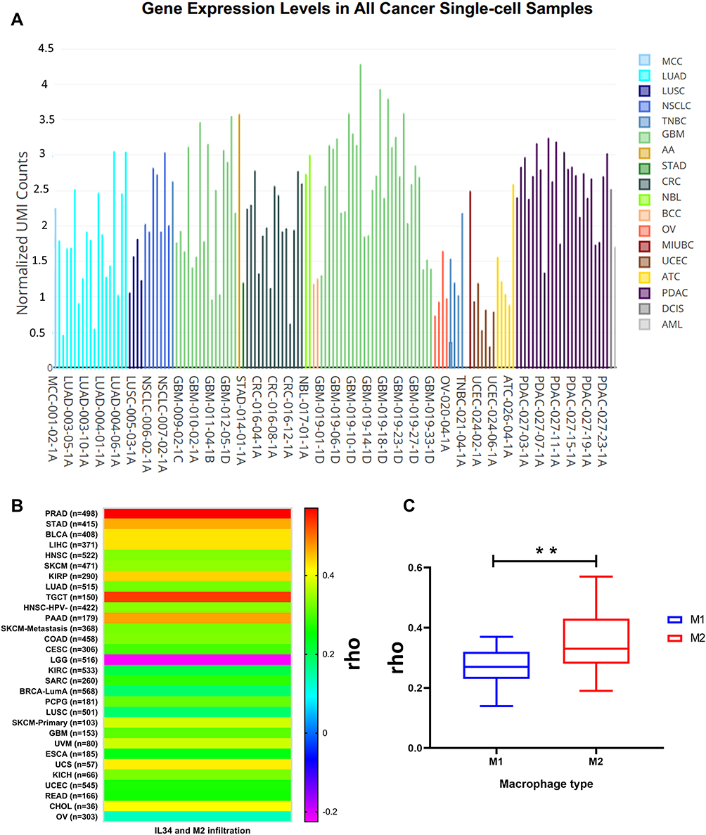 |
Figure 2 (A) Pan-cancer single-cell expression view among 638,341 cells. (B) Pan-cancer analysis for IL-34 expression and M2 associations by Spearman correlation test (P <0.05). (C) An Independent test result of matched correlations values analysis (23 for each) between M1 and M2 macrophages. **(P = 0.002). Abbreviations: MCC, Merkel cell carcinoma; LUAD, Lung adenocarcinoma; LUSC, Lung squamous cancer; NSCLC, non-small cell lung cancer; TNBC, Triple-negative breast cancer; GBM, Glioblastoma; AA, Anaplastic astrocytoma; STAD, Stomach adenocarcinoma; CRC, Colorectal cancer; NBL, Neuroendocrine tumor; BCC, Basal cell carcinoma; OV, Ovarian carcinoma; MIUBC, Muscle-invasive urothelial bladder cancer; UCEC, Uterine cancer- endometrial carcinoma; ATC, Anaplastic thyroid cancer; PDAC, Pancreatic adenocarcinoma; DCIS, Breast ductal carcinoma in situ; AML, Acute myeloid leukemia. |
Data Sharing Statement
We used https://portal.gdc.cancer.gov/, http://ualcan.path.uab.edu/, and http://timer.cistrome.org/ for expression association. Also available in Supplementary Materials and Methods and Supplementary Figure 1.
Author Contributions
All authors made a significant contribution to the work reported, whether that is in the conception, study design, execution, acquisition of data, analysis, and interpretation, or in all these areas; took part in drafting, revising, or critically reviewing the article; gave final approval of the version to be published; have agreed on the journal to which the article has been submitted, and agree to be accountable for all aspects of the work.
Funding
This research was funded by the National Natural Science Foundation of China, Grant/Award Number: 62271292.
Disclosure
The authors report no conflicts of interest in this work.
References
1. Huang AC, Zappasodi R. A decade of checkpoint blockade immunotherapy in melanoma: understanding the molecular basis for immune sensitivity and resistance. Nat Immunol. 2022;23(5):660–670. doi:10.1038/s41590-022-01141-1
2. O’Donnell JS, Long GV, Scolyer RA, Teng MWL, Smyth MJ. Resistance to PD1/PDL1 checkpoint inhibition. Cancer Treat Rev. 2017;52:71–81. doi:10.1016/j.ctrv.2016.11.007
3. Jorgovanovic D, Song M, Wang L, Zhang Y. Roles of IFN-γ in tumor progression and regression: a review. Biomarker Res. 2020;8(1):1–16. doi:10.1186/s40364-020-00228-x
4. Monteleone G, Maresca C, Colella M, et al. Targeting IL-34/MCSF-1R axis in colon cancer. Front Immunol. 2022;13. doi:10.3389/fimmu.2022.917955
5. DeNardo DG, Ruffell B. Macrophages as regulators of tumour immunity and immunotherapy. Nat Rev Immunol. 2019;19(6):369–382. doi:10.1038/s41577-019-0127-6
6. Baghdadi M, Wada H, Nakanishi S, et al. Chemotherapy-induced IL34 enhances immunosuppression by tumor-associated macrophages and mediates survival of chemoresistant lung cancer cells an importance of IL34 in cancer chemoresistance. Cancer Res. 2016;76(20):6030–6042. doi:10.1158/0008-5472.CAN-16-1170
7. Hama N, Kobayashi T, Han N, et al. Interleukin-34 limits the therapeutic effects of immune checkpoint blockade. Iscience. 2020;23(10):101584. doi:10.1016/j.isci.2020.101584
8. Fares CM, Van allen EM, Drake CG, Allison JP, Hu-Lieskovan S. Mechanisms of resistance to immune checkpoint blockade: why does checkpoint inhibitor immunotherapy not work for all patients? Am Soc Clin Oncol Educ Book. 2019;39:147–164. doi:10.1200/EDBK_240837
 © 2023 The Author(s). This work is published and licensed by Dove Medical Press Limited. The
full terms of this license are available at https://www.dovepress.com/terms
and incorporate the Creative Commons Attribution
- Non Commercial (unported, 3.0) License.
By accessing the work you hereby accept the Terms. Non-commercial uses of the work are permitted
without any further permission from Dove Medical Press Limited, provided the work is properly
attributed. For permission for commercial use of this work, please see paragraphs 4.2 and 5 of our Terms.
© 2023 The Author(s). This work is published and licensed by Dove Medical Press Limited. The
full terms of this license are available at https://www.dovepress.com/terms
and incorporate the Creative Commons Attribution
- Non Commercial (unported, 3.0) License.
By accessing the work you hereby accept the Terms. Non-commercial uses of the work are permitted
without any further permission from Dove Medical Press Limited, provided the work is properly
attributed. For permission for commercial use of this work, please see paragraphs 4.2 and 5 of our Terms.