Back to Journals » International Journal of General Medicine » Volume 16
Identification and Validation of Anoikis-Related Signatures for Predicting Prognosis in Lung Adenocarcinoma with Machine Learning
Authors Wang Q ![]() , Sun N, Zhang M
, Sun N, Zhang M
Received 10 March 2023
Accepted for publication 4 May 2023
Published 16 May 2023 Volume 2023:16 Pages 1833—1844
DOI https://doi.org/10.2147/IJGM.S409006
Checked for plagiarism Yes
Review by Single anonymous peer review
Peer reviewer comments 2
Editor who approved publication: Dr Scott Fraser
Qilong Wang,1,2 Nannan Sun,3 Mingzhi Zhang1
1Department of Oncology, The First Affiliated Hospital of Zhengzhou University, Zhengzhou, Henan, People’s Republic of China; 2The Academy of Medical Science of Zhengzhou University, Zhengzhou University, Zhengzhou, Henan, People’s Republic of China; 3Department of Hematology, The First Affiliated Hospital of Zhengzhou University, Zhengzhou, Henan, People’s Republic of China
Correspondence: Mingzhi Zhang, Department of Oncology, The First Affiliated Hospital of Zhengzhou University, No. 1 Jianshe Road, Zhengzhou, Henan, 450052, People’s Republic of China, Email [email protected]
Background: Lung adenocarcinoma (LUAD) is an aggressive cancer that has an extremely poor prognosis. As well as facilitating the detachment of cancer cells from the primary tumor site, anoikis plays an important role in cancer metastasis. Few studies to date, however, have examined the role of anoikis in LUAD, in patient prognosis.
Methods: A total of 316 anoikis-related genes (ANRGs) integrated from Genecards and Harmonizome portals. LUAD transcriptome data were retrieved from the Genotype-Tissue Expression Project (GEO) and The Cancer Genome Atlas (TCGA). Anoikis-related prognostic genes (ANRGs) were primarily screened by univariate Cox regression. All ANRGs were included in the Least Absolute Shrinkage and Selection Operator (LASSO) Cox regression model to construct the powerful prognostic signature. This signature was validated and assessed using the Kaplan-Meier method as well as univariate and multivariate Cox regression analyses. Anoikis-related regulators of risk score were identified using a XG-boost machine learning model. The expression of ITGB4 protein was examined in a ZhengZhou University (ZZU) tissue cohort by immunohistochemistry, and the potential mechanisms of action of ITGB4 in LUAD were explored by GO, KEGG, and ingenuity pathway analyses and by GSEA.
Results: A risk score signature was constructed based on eight ANRGs, with high risk scores found to closely correlate with unfavorable clinical features. ITGB4 expression may be associated with 5-year over survival, with immunohistochemistry showed that the expression of ITGB4 was higher in LUAD than in nontumor tissues. Enrichment analysis suggested that ITGB4 may promote LUAD development by targeting E2F, MYC, and oxidative phosphorylation signaling pathways.
Conclusion: Our anoikis-related signature from RNA-seq data may be a novel prognostic biomarker in patients with LUAD. It may help physicians develop personalized LUAD treatments in clinical practice. Moreover, ITGB4 may affect the development of LUAD through the oxidative phosphorylation pathway.
Keywords: anoikis, lung adenocarcinoma, ITGB4, machine learning, prognosis
Introduction
Lung cancer is currently the second most common cancer in China, accounting for the highest rate of cancer deaths.1,2 Lung adenocarcinoma (LUAD) is the most frequent type of lung cancer, with advanced lung adenocarcinoma being prone to metastasize, resulting in poor patient prognosis. Methods of treating LUAD include surgery, radiotherapy, chemotherapy, targeted therapy, and treatment with immune checkpoint inhibitors. Tumor recurrence and metastasis are the most frequent causes of treatment failure and death. Multiple genes and genetic pathways are thought to be involved in the development of tumor metastases.
Anoikis is a specialized form of programmed cell death that plays important roles in body development, tissue self-balance, disease occurrence and tumor metastasis.3 During the process of tumor metastasis, tumor cells that adhere to the extracellular matrix and other cells detach from their surroundings, and survive in the circulation through paracrine and paracrine mechanisms. These cells resist apoptosis, and subsequently regain the ability to attach to other sites in the body, resulting in tumor spread, metastasis and invasion.4–6 Resistance to anoikis during tumor metastasis allows tumor cells to spread through the circulatory system to distant organs.7 Studies on a variety of tumors have shown that resistance to anoikis can predict the effects of treatment and patient prognosis. For example, anoikis-related genes (ANRGs) were found to play a critical role in the progression of cancer and metastatic cascades.8,9 To date, however, clinical prognostic models based on genes associated with anoikis have not been established in LUAD. The process of development of LUADs may be better understood by investigating the mechanisms of action of ANRGs.
A total of 316 anoikis-related genes (ANRGs) were integrated from the GeneCard database10 (http://www.genecards.org/) and Harmonizome portals,11(Table S1). The present study analyzed alterations of ANRGs in LUAD, showing that these alterations were prognostic of patient outcomes. Our focus is on transcriptomics and somatic mutations are the focus of our next phase of research, so they are described briefly in this article. In addition, XG-boost was used to identify the most important genes associated with LUAD related death. Because these findings suggested that ITGB4 was an oncogene associated with the progression of LUAD, the levels of expression of ITGB4 was analyzed in an LUAD tissue microarray.
Materials and Methods
Datasets
RNA-sequencing transcriptome data for LUAD were obtained from The Gene Expression Omnibus (GEO, http://www.ncbi.nlm.nih.gov/geo/GSE31210) and The Cancer Genome Atlas (TCGA, http://portal.gdc.cancer.gov/)12 The RNA sequence transcriptome profiles and clinicopathological data from 535 LUAD tissue samples and 59 normal tissue samples were collected from the LUAD cohort in the TCGA (TCGA-LUAD), and data on 226 tumor and 20 non-tumor tissue from the GSE31210 dataset were obtained through the GEO database.
Pathway Enrichment Analysis
Correlations between the levels of expression and functions of eight anoikis regulators were evaluated by Spearman correlation analysis, with interplay between regulators estimated using the String database tool (https://string-db.org). A complete evaluation of Gene characteristics were evaluated using Metascape (http://metascape.org). Targetable mechanisms of action of anoikis regulators in LUAD were evaluated by Gene Set Enrichment Analysis (GSEA), Gene Ontology (GO) and the Kyoto Encyclopedia of Genes and Genomes (KEGG).
Construction and Validation of the Risk Score
Factors highly prognostic of survival were evaluated using a Cox regression model of the least absolute shrinkage and selection operators (LASSO), which selected eight regulators of anoikis. These eight genes were analyzed by Cox regression analysis, with a P-value <0.2 set as the cutoff value to avoid omissions. The risk score for each patient in the training (TCGA) and validation (GSE31210) datasets was calculated using the formula:
in which coefi represents the coefficient and xi represents the expression value of each selected anokis regulator.
Machine Learning Algorithm
The mechanism by which anoikis regulators affected the risk scores was determined using an additive explanation method with the XG-Boost machine learning model. This machine learning algorithm assembles potential prediction models to yield a fine-tuned predictive result. Because the algorithm involved parallel, distributed, out-of-core, and cache-aware computing, it was more than ten times faster than popular machine learning (ML) and deep learning models.
Tissue Samples and Immunohistochemistry
Samples of LUAD tumors and adjacent normal tissue were collected and assessed. These samples were embedded in paraffin, dewazed, separate and hydrated according to standard protocols. The tissue samples were mounted on slides, heated in Tris-EDTA buffer, and blocked by overnight incubation with 5% gout serum. The samples were subsequently incubated overnight with primary anti- ITGB4 antibody (Proteintech, Cat No.21738-1-AP, Wuhan, Hubei, China), followed by incubation with secondary antibody. Slides were viewed by light microscopy, and immunohistochemical staining was analyzed semiquantitatively by two independent, experienced pathologists. Each sample was scored according to the percentage of positive cells, with 1+ indicating no staining, 2+ indicating weak staining, 3+ indicating moderate staining, and 4+ indicating strong staining. All experiments with human samples were approved by the Ethics Committee of the First Affiliated Hospital of Zhengzhou University. This study abode by the Declaration of Helsinki and good clinical practice guidelines. Written informed consent to participate was waived for the use of the clinical samples as retrospective samples were used in this study. All the patients’ information was made anonymous in this study.
Statistical Analysis
All statistical analyses were performed using the R statistical software package (3.5.3). Continuous variables in two groups were compared using Student’s t-tests (unpaired, two-tailed), whereas comparisons in three or more groups were performed using one-way analysis of variance (ANOVA) and Kruskal–Wallis tests. The relationships between the patterns of expression of anoikis regulators and scientific features were analyzed by Chi-square tests. Survival was assessed using the Kaplan–Meier method, with survival as a function of each ectopically expressed gene compared by Log rank tests. The relationships between patient variables and prognosis were evaluated by univariate and multivariable Cox regression analyses. P-values were corrected for multiple comparisons using the Benjamini–Hochberg (BH) method and used to predict medical responses to immune checkpoint blockade. In all statistical analyses, P <0.05 was considered statistically significant.
Results
Identification of Anoikis-Related Genes Associated with Prognosis
The levels of expression of anoikis-related genes were obtained from the TCGA dataset, with the differences in the expression of these genes in LUAD tumor and pericarcinomous tissue samples analyzed by principal component analysis (Figure 1A). The levels of expression of 311 of these genes differed significantly in tumor and adjacent normal tissue samples, with heatmaps showing the same trend (Figure 1B). Volcano plots showed that the expression of 28 DEGs were significantly higher and the levels of 10 significantly lower in LUAD than in normal tissues (Figure 1C). Further univariate regression analysis also showed that 15 of the 28 genes significantly upregulated in LUAD were closely associated with overall survival (p < 0.01) in both the training (TCGA) and validation (GSE31210) cohorts (Figure 1D). Among all the 557 patients, 86 experienced anoikis-related genes mutations with a frequency of 15.44%, indicating a high frequency of somatic mutations of the anoikis-related gene in LUAD (Figure S1).
 |
Figure 1 Characteristics and differences of anoikis-related regulators in LUAD. (A) Principal component analysis of tumor and non-tumor tissue. (B) Heatmap showing the differential expression of anoikis-related regulators. (C) Volcano plot of differentially expressed genes (DEGs) in LUAD. (D) Effects of 15 anoikis-related genes on overall survival in the TCGA and GEO datasets. |
Prognostic Value of Regulators Related to Anoikis and the Construction of a Risk Score Model
The 28 significantly differentially expressed anoikis-related regulators and the 15 anoikis-related genes that had a significant effect on the prognosis of patients with LUAD, were used to develop a prognostic risk model. A LASSO Cox regression model using the minimum criteria to determine whether anoikis modification could accurately predict outcomes in individual patients with LUAD was found to include the PLK1, SHC1, ITGB4, ANGPTL4, SLCO1B3, SPIB, KL and DAPK2 genes (Figure 2A and B). The ANRG risk score for each patient with LUAD in the training (TCGA) and validation (GSE31210) datasets was calculated using the formula: ANRG risk score = 0.075503×PLK1 +0.002077×SHC1 +0.017577×ITGB4 +0.012239×ANGPTL4 +0.013272×SLCO1B3 −0.01686×SPIB −0.01809×KL −0.09847×DAPK2. In addition, Kaplan-Meier analysis showed that higher risk scores in both the training (TCGA) and validation (GSE31210) datasets were associated with poorer OS (Figure 2C and D), indicating a close relationship between ANRG risk score and the prognosis of patients with LUAD.
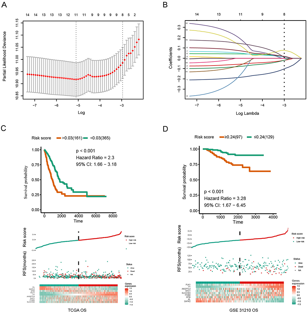 |
Figure 2 Identification of an anoikis-related prognostic signature. (A) Identification of eight anoikis-related genes associated with prognosis of patients with LUAD by LASSO analysis with 10-fold cross validation. (B) Coefficient profile plots of eight prognostic anoikis-related genes. (C and D) Kaplan-Meier analyses showing differences in overall survival in subgroups of patients with LUAD. |
Correlation of Anoikis Risk Scores with Clinical Characteristics
The optimal anoikis risk score cutoff values for patients with LUAD was calculated, and patients were divided into those with a low and high anoikis scores (Figure 3A). Correlations between anoikis scores and various clinicopathological characteristics were subsequently analyzed in patients with LUAD. ANRG risk scores differed significantly between between groups, with patients in the TCGA dataset stratified by PFS, tumor status, pathologic stage and OS (Figure 3B–E). Univariate and multivariable Cox regression analyses of the TCGA data identified ANRG risk scores as an independent prognostic factor associated with the outcome of patients with LUAD (Figure 3B), with anoikis risk scores significantly correlated with several clinical characteristics and patient prognosis.
 |
Figure 3 Association between ITGB4 expression and key characteristics of patients with LUAD. (A): Relationships between ANRGs risk scores and characteristics of patients with LUAD. (B–E): Relationships between ANRGs risk scores and medical characteristics of patients with LUAD. ****p<0.0001. |
Importance of Anoikis-Related Regulators to Risk Scores Determined by Machine Learning
The XGBoost algorithm was used to construct a classifier that could recognize the outstanding contributors to risk score. Evaluation by Shapley additive explanatory values (SHAP) showed that the four leading regulators were DAPK2, ITGB4, PLK1, and SHC1 (Figures 4A). The scores of all samples were summed and their features were sorted, with risk scores showing that DAPK2 and ITGB4 were the most important regulators (Figures 4B). Previous studies have suggested that DAPK2 contributes to the process of autophagy. Because less is known about ITGB4, the expression of ITGB4 was further analyzed and verified.
 |
Figure 4 Contributions of anoikis regulators to machine learning risk scores. (A): Significance of anoikis regulators on chance scores using the LASSO algorithm. (B): Individual SHAP values of PLK1, ALDH2, PANX1, NR2C2, TLR4, and OUTLIN. |
Expression of ITGB4 Protein in Two Cohorts of Tissue Samples
IHC analysis of the ZZU-cohort obtained from the First Affiliated Hospital of Zhengzhou University was performed to analyze the levels of ITGB4 protein in representative LUAD and peritumor tissues (Figure 5A). The IHC score of ITGB4 was significantly higher in LUAD than in paired normal lung tissues (p < 0.01, Figure 5B and C). Moreover, ITGB4 expression level differed significantly between groups, with the TCGA dataset stratified by OS, tumor status, and T stage (Figure 5D–F). These results confirmed at the protein level that ITGB4 is upregulated in LUAD.
 |
Figure 5 Immunohistochemical (IHC) analysis of components of the anoikis-related prognosis signature in LUAD and normal lung tissue. (A): Level of expression of ITGB4 in LUAD and adjacent normal lung tissue. (B): Statistical analysis of the differences in scores. (C): IHC images with different ratings. (D): Correlation between the expression of ITGB4 and overall survival. (E): Relationship between ITGB4 expression level and T stage. (F): Relationship between ITGB4 expression level and tumor stage. 1+ indicating no staining, 2+ indicating weak staining, 3+ indicating moderate staining, and 4+ indicating strong staining.*p<0.05, ****p<0.0001. |
Functional Enrichment Analysis with ITGB4
GO and KEGG analyses were also performed to further elucidate the role of ITGB4 in LUAD (Figure 6A and B). These analyses showed that ITGB4 participated in the cell cycle and energy metabolism, including in four pathways that play an important role in common pathway analysis: HALLMARK E2F TARGETS, HALLMARK MYC TARGETS V1, HALLMARK MYC TARGETS V2, HALLMARK OXIDATIVE PHOSPHORYLATION (Figure 6C). GSEA performed to identify the signals differentially activated in groups with high versus low ITGB4 expression (Figure 6D–F) showed that the dataset was highly enriched in genes involved in the four above pathways, further confirming that ITGB4 may be involved in the development of LUAD.
 |
Figure 6 ITGB4-related gene enrichment analysis. (A): GO term enrichment analysis of ITGB4-binding genes. (B): KEGG term enrichment analysis of ITGB4-binding genes. (C): Ingenuity analysis of the ITGB4-related pathway. (D): GSEA analysis of the E2F TARGETS pathway. (E): GSEA analysis of the MYC TARGETS V1 and V2 pathways. (F): GSEA analysis of the HALLMARK OXIDATIVE PHOSPHORYLATION pathway. |
Discussion
The rapid development of LUAD and the high incidence of distant metastases have made it difficult to improve patient survival using a single route or treatment. Construction of prediction models for transfer-related genes can provide a more effective method for early intervention. The predictive ability of these models is enhanced by the use of multiple biomarkers.
The present study found that a robust risk score could be generated using eight genes: PLK1, SHC1, ITGB4, ANGPTL4, SLCO1B3, SPIB, and DAPK2. Genes related to anoikis were found to be significantly correlated with several clinical features of glioblastoma.13 Moreover, anoikis-related signatures can be used to stratify risks in patients with endometrial carcinoma and predict their survival outcomes.14 Anoikis-related genes were also found to play a carcinogenic role in patients with clear cell renal cell carcinoma, with a model integrating multiple genes related to this disease found to predict the risk of anoikis.8 A seven-gene signature and nomogram was found to help select personalized treatment for patients with head and neck squamous cell carcinoma (HNSCC),15 and decision curve analysis (DCA), was used to develop a nomogram for selecting treatment strategies in patients with low-grade glioma based on their clinicopathological features.9
Upregulation of the TGFb/SMAD pathway in non-small cell lung cancer (NSCLC) was found to correlate with the role of PLK1,16 with SHC1 found to play a carcinogenic role in lung cancer.17 ANGPTL4 was found to affect the energy metabolism of NSCLC cells,18 with CtSLCO1B3 playing an important role in the development and/or progression of NSCLC.19 The ability of SPIB to provide prognostic information in pan-cancer has been assessed based on mutational load, microsatellite instability, immune cell infiltration, and prognosis.20 Klotho (KL) has been shown to regulate autophagy,21 with several steps of the autophagic process involving DAPK2.22
Higher ITGB4 expression has been associated with tumor aggressiveness and metastasis, including in patients with prostate cancer,23 NSCLC,24 hepatocellular carcinoma,25 cervical cancer,26 HNSCC27 and colon cancer.28 In addition, high ITGB4 level has been associated with a poorer prognosis in patients with LUAD.24,29 ITGB4 is a signaling protein that promotes intracellular signaling and protects the integrity of epithelial cells.30 Moreover, bioinformatics analysis showed that ITGB4 acts as a pan-cancer oncogene in 33 different types of human tumors.31 ITGB4 overexpression has been associated with venous invasion and decreased overall survival in patients with NSCLC.32 ITGB4 has also been closely associated with other respiratory diseases, including spontaneous inflammation of the lungs, airway inflammation and hyperreactivity, and acute lung injury.33–35 Although anoikis and ITGB4 have been shown to be beneficial in patients with LUAD, these patients did not benefit from treatment with ITGB4 suppressing agents, indicating the need for greater understanding of the microenvironment of these tumors.
The results of this study showed that ITGB4-related genes are enriched in pathways related to energy metabolism (oxidative phosphorylation), DNA replication (E2F targets) and proto-oncogene function (Myc targets). Genes in the E2F family that regulate c-Myc expression also regulate apoptosis and cell proliferation and can serve as both oncogenes and tumor suppressor genes. No clear correlation has been observed between changes in E2F expression and changes in ITGB4 expression. Interestingly, some genes associated with oxidative phosphorylation were upregulated. EMT has been found to correlate with a switch from oxidative phosphorylation to glycolysis,36 although mitochondrial respiration was also reported to be directly linked to the growth of tumor metastases.37 These findings suggest that ITGB4 may undergo signaling via the E2F pathway, which influences the oxidative phosphorylation process through the MYC pathway. However, there are still some limitations in this study. Moreover, vivo and vitro studies should be performed to verify related regulatory axis. In the next step, we will experimentally verify the significance of anoikis in LUAD.
Conclusion
Our anoikis-related signature from RNA-seq data may be a novel prognostic biomarker in patients with LUAD. It may help physicians develop personalized LUAD treatments in clinical practice. Moreover, ITGB4 may affect the development of LUAD through the oxidative phosphorylation pathway.
Data Sharing Statement
The datasets generated and/or analyzed during the current study are available in the TCGA repository (https://portal.gdc.cancer.gov/) and The Gene Expression Omnibus (GEO, http://www.ncbi.nlm.nih.gov/geo/GSE31210). The datasets used and/or analysed during the current study are available from the corresponding author on reasonable request.
Ethics Approval
This study was approved by the First Affiliated Hospital of Zhengzhou University and conducted in accordance with the hospital’s guiding principles.
Informed Consent and Consent for Publication
Data from this study were partially downloaded from available database or processed dataset. The written informed consent and consent for publication were waived as this study was partially a database study and used retrospective samples.
Author Contributions
All authors made a significant contribution to the work reported, whether that is in the conception, study design, execution, acquisition of data, analysis and interpretation, or in all these areas; took part in drafting, revising or critically reviewing the article; gave final approval of the version to be published; have agreed on the journal to which the article has been submitted; and agree to be accountable for all aspects of the work.
Funding
This study was supported by funds from the National Natural Science Foundation of China (81970184, 81570203).
Disclosure
All authors declare no competing interests in this study.
References
1. Cao W, Chen HD, Yu YW, Li N, Chen WQ. Changing profiles of cancer burden worldwide and in China: a secondary analysis of the global cancer statistics 2020. Chin Med J. 2021;134(7):783–791. doi:10.1097/CM9.0000000000001474
2. Chen P, Liu Y, Wen Y, Zhou C. Non-small cell lung cancer in China. Cancer Commun Lond Engl. 2022;42(10):937–970. doi:10.1002/cac2.12359
3. Frisch S, Francis H. Disruption of epithelial cell-matrix interactions induces apoptosis. J Cell Biol. 1994;124(4):619–626. doi:10.1083/jcb.124.4.619
4. Kockx MM, Herman AG. Apoptosis in atherosclerosis: beneficial or detrimental? Cardiovasc Res. 2000;45(3):736–746. doi:10.1016/S0008-6363(99)00235-7
5. Haun F, Neumann S, Peintner L, et al. Identification of a novel anoikis signalling pathway using the fungal virulence factor gliotoxin. Nat Commun. 2018;9(1):3524. doi:10.1038/s41467-018-05850-w
6. Dobler D, Ahmed N, Song L, Eboigbodin KE, Thornalley PJ. Increased dicarbonyl metabolism in endothelial cells in hyperglycemia induces anoikis and impairs angiogenesis by RGD and GFOGER motif modification. Diabetes. 2006;55(7):1961–1969. doi:10.2337/db05-1634
7. Zhao K, Wang Z, Hackert T, Pitzer C, Zöller M. Tspan8 and Tspan8/CD151 knockout mice unravel the contribution of tumor and host exosomes to tumor progression. J Exp Clin Cancer Res CR. 2018;37(1):312. doi:10.1186/s13046-018-0961-6
8. Chen Z, Liu X, Zhu Z, et al. A novel anoikis-related prognostic signature associated with prognosis and immune infiltration landscape in clear cell renal cell carcinoma. Front Genet. 2022;13:1039465. doi:10.3389/fgene.2022.1039465
9. Zhao S, Chi H, Ji W, et al. A bioinformatics-based analysis of an anoikis-related gene signature predicts the prognosis of patients with low-grade gliomas. Brain Sci. 2022;12(10):1349. doi:10.3390/brainsci12101349
10. Rebhan M, Chalifa-Caspi V, Prilusky J, Lancet D. GeneCards: integrating information about genes, proteins and diseases. Trends Genet. 1997;13(4):163.
11. Rouillard AD, Gundersen GW, Fernandez NF, et al. The harmonizome: a collection of processed datasets gathered to serve and mine knowledge about genes and proteins. Database. 2016;2016:baw100. doi:10.1093/database/baw100
12. Gao J, Aksoy BA, Dogrusoz U, et al. Integrative analysis of complex cancer genomics and clinical profiles using the cBioPortal. Sci Signal. 2013;6(269):pl1. doi:10.1126/scisignal.2004088
13. Sun Z, Zhao Y, Wei Y, Ding X, Tan C, Wang C. Identification and validation of an anoikis-associated gene signature to predict clinical character, stemness, IDH mutation, and immune filtration in glioblastoma. Front Immunol. 2022;13:939523. doi:10.3389/fimmu.2022.939523
14. Chen S, Gu J, Zhang Q, Hu Y, Ge Y. Development of biomarker signatures associated with anoikis to predict prognosis in endometrial carcinoma patients. J Oncol. 2021;2021:3375297. doi:10.1155/2021/3375297
15. Chi H, Jiang P, Xu K, et al. A novel anoikis-related gene signature predicts prognosis in patients with head and neck squamous cell carcinoma and reveals immune infiltration. Front Genet. 2022;13:984273. doi:10.3389/fgene.2022.984273
16. Chiappa M, Petrella S, Damia G, Broggini M, Guffanti F, Ricci F. Present and future perspective on PLK1 inhibition in cancer treatment. Front Oncol. 2022;12:903016. doi:10.3389/fonc.2022.903016
17. Yang P, Li W, Li X. SHC1 promotes lung cancer metastasis by interacting with EGFR. J Oncol. 2022;2022:3599832. doi:10.1155/2022/3599832
18. Xiao S, Nai‐dong W, Jin‐Xiang Y, et al. ANGPTL4 regulate glutamine metabolism and fatty acid oxidation in nonsmall cell lung cancer cells. J Cell Mol Med. 2022;26(7):1876–1885. doi:10.1111/jcmm.16879
19. Hase H, Aoki M, Matsumoto K, et al. Cancer type‑SLCO1B3 promotes epithelial‑mesenchymal transition resulting in the tumour progression of non‑small cell lung cancer. Oncol Rep. 2020;45(1):309–316. doi:10.3892/or.2020.7839
20. Ding M, Li Q, Tan X, Zhang L, Tan J, Zheng L. Comprehensive pan-cancer analysis reveals the prognostic value and immunological role of SPIB. Aging. 2022;14(15):6338–6357. doi:10.18632/aging.204225
21. Zhou H, Pu S, Zhou H, Guo Y. Klotho as potential autophagy regulator and therapeutic target. Front Pharmacol. 2021;12:755366. doi:10.3389/fphar.2021.755366
22. Jiang Y, Liu J, Xu H, Zhou X, He L, Zhu C. DAPK2 activates NF-κB through autophagy-dependent degradation of I-κBα during thyroid cancer development and progression. Ann Transl Med. 2021;9(13):1083. doi:10.21037/atm-21-2062
23. Wilkinson EJ, Woodworth AM, Parker M, et al. Epigenetic regulation of the ITGB4 gene in prostate cancer. Exp Cell Res. 2020;392(2):112055. doi:10.1016/j.yexcr.2020.112055
24. Wu P, Wang Y, Wu Y, Jia Z, Song Y, Liang N. Expression and prognostic analyses of ITGA11, ITGB4 and ITGB8 in human non-small cell lung cancer. Peer J. 2019;7:e8299. doi:10.7717/peerj.8299
25. Li XL, Liu L, Li DD, et al. Integrin β4 promotes cell invasion and epithelial-mesenchymal transition through the modulation of Slug expression in hepatocellular carcinoma. Sci Rep. 2017;7:40464. doi:10.1038/srep40464
26. Wang S, Li J, Xie J, et al. Programmed death ligand 1 promotes lymph node metastasis and glucose metabolism in cervical cancer by activating integrin β4/SNAI1/SIRT3 signaling pathway. Oncogene. 2018;37(30):4164–4180. doi:10.1038/s41388-018-0252-x
27. Li GS, Hou W, Chen G, et al. Clinical significance of integrin subunit beta 4 in head and neck squamous cell carcinoma. Cancer Biother Radiopharm. 2022;37(4):256–275. doi:10.1089/cbr.2020.3943
28. Li M, Jiang X, Wang G, et al. ITGB4 is a novel prognostic factor in colon cancer. J Cancer. 2019;10(21):5223–5233. doi:10.7150/jca.29269
29. Mohanty A, Nam A, Pozhitkov A, et al. A non-genetic mechanism involving the integrin β4/Paxillin axis contributes to chemoresistance in lung cancer. iScience. 2020;23(9):101496. doi:10.1016/j.isci.2020.101496
30. Yang H, Xu Z, Peng Y, Wang J, Xiang Y. Integrin β4 as a potential diagnostic and therapeutic tumor marker. Biomolecules. 2021;11(8):1197. doi:10.3390/biom11081197
31. Huang W, Fan L, Tang Y, Chi Y, Li J. A pan-cancer analysis of the oncogenic role of integrin beta4 (ITGB4) in human tumors. Int J Gen Med. 2021;14:9629–9645. doi:10.2147/IJGM.S341076
32. Stewart RL, West D, Wang C, et al. Elevated integrin α6β4 expression is associated with venous invasion and decreased overall survival in non-small cell lung cancer. Hum Pathol. 2016;54:174–183. doi:10.1016/j.humpath.2016.04.003
33. Jiang W, Wang JM, Luo JH, et al. Airway epithelial integrin β4-deficiency exacerbates lipopolysaccharide-induced acute lung injury. J Cell Physiol. 2021;236(11):7711–7724. doi:10.1002/jcp.30422
34. Liu C, Yuan L, Zou Y, et al. ITGB4 is essential for containing HDM-induced airway inflammation and airway hyperresponsiveness. J Leukoc Biol. 2018;103(5):897–908. doi:10.1002/JLB.3A1017-411RR
35. Tang S, Du X, Yuan L, et al. Airway epithelial ITGB4 deficiency in early life mediates pulmonary spontaneous inflammation and enhanced allergic immune response. J Cell Mol Med. 2020;24(5):2761–2771. doi:10.1111/jcmm.15000
36. Lunetti P, Di Giacomo M, Vergara D, et al. Metabolic reprogramming in breast cancer results in distinct mitochondrial bioenergetics between luminal and basal subtypes. FEBS J. 2019;286(4):688–709. doi:10.1111/febs.14756
37. LeBleu VS, O’Connell JT, Gonzalez Herrera KN, et al. PGC-1α mediates mitochondrial biogenesis and oxidative phosphorylation in cancer cells to promote metastasis. Nat Cell Biol. 2014;16(10):992–1003, 1–15. doi:10.1038/ncb3039
 © 2023 The Author(s). This work is published and licensed by Dove Medical Press Limited. The
full terms of this license are available at https://www.dovepress.com/terms
and incorporate the Creative Commons Attribution
- Non Commercial (unported, 3.0) License.
By accessing the work you hereby accept the Terms. Non-commercial uses of the work are permitted
without any further permission from Dove Medical Press Limited, provided the work is properly
attributed. For permission for commercial use of this work, please see paragraphs 4.2 and 5 of our Terms.
© 2023 The Author(s). This work is published and licensed by Dove Medical Press Limited. The
full terms of this license are available at https://www.dovepress.com/terms
and incorporate the Creative Commons Attribution
- Non Commercial (unported, 3.0) License.
By accessing the work you hereby accept the Terms. Non-commercial uses of the work are permitted
without any further permission from Dove Medical Press Limited, provided the work is properly
attributed. For permission for commercial use of this work, please see paragraphs 4.2 and 5 of our Terms.
Recommended articles
Comprehensive Analysis of the E2F Transcription Factor Family in Human Lung Adenocarcinoma
Wang Q, Liu J, Cheang I, Li J, Chen T, Li Y, Yu B
International Journal of General Medicine 2022, 15:5973-5984
Published Date: 2 July 2022
High Expression of DEPDC1B Predicts Poor Prognosis in Lung Adenocarcinoma

Li P, Chen X, Zhou S, Xia X, Wang E, Han R, Zeng D, Fei G, Wang R
Journal of Inflammation Research 2022, 15:4171-4184
Published Date: 23 July 2022
Expression, Clinical Significance, Immune Infiltration, and Regulation Network of miR-3940-5p in Lung Adenocarcinoma Based on Bioinformatic Analysis and Experimental Validation
Lin Z, Huang W, Xie Z, Yi Y, Li Z
International Journal of General Medicine 2022, 15:6451-6464
Published Date: 6 August 2022
RuleFit-Based Nomogram Using Inflammatory Indicators for Predicting Survival in Nasopharyngeal Carcinoma, a Bi-Center Study
Luo C, Li S, Zhao Q, Ou Q, Huang W, Ruan G, Liang S, Liu L, Zhang Y, Li H
Journal of Inflammation Research 2022, 15:4803-4815
Published Date: 24 August 2022
Prognosis and Personalized Treatment Prediction in Different Mutation-Signature Hepatocellular Carcinoma
Zhang Y, Liu Z, Li J, Li X, Duo M, Weng S, Lv P, Jiang G, Wang C, Li Y, Liu S, Li Z
Journal of Hepatocellular Carcinoma 2023, 10:241-255
Published Date: 15 February 2023