Back to Journals » International Journal of Nanomedicine » Volume 21
GelMA-Based Nanocomposites for Bone Defect Regeneration: Design, Performance, and Clinical Translation Potential
Authors Li G, Wang Y, Wang D, Cui H, Qin M, Ma J ![]()
Received 21 November 2025
Accepted for publication 11 February 2026
Published 16 February 2026 Volume 2026:21 583425
DOI https://doi.org/10.2147/IJN.S583425
Checked for plagiarism Yes
Review by Single anonymous peer review
Peer reviewer comments 3
Editor who approved publication: Professor Jie Huang
Guang Li,1– 3,* Yan Wang,1– 3,* Dong Wang,2,3 Hongwei Cui,2,3 Mengran Qin,2,3 Jianxiong Ma1– 3
1Tianjin Hospital, Tianjin University, Tianjin, People’s Republic of China; 2Tianjin Orthopedic Institute, Tianjin Hospital, Tianjin, People’s Republic of China; 3Tianjin Key Laboratory of Orthopedic Biomechanics and Medical Engineering, Tianjin Hospital, Tianjin, People’s Republic of China
*These authors contributed equally to this work
Correspondence: Jianxiong Ma, Tianjin Hospital, Tianjin University, Tianjin, 300211, People’s Republic of China, Tel/Fax +86-022-60910549, Email [email protected]
Abstract: This narrative review summarizes recent advances in gelatin methacryloyl (GelMA)-based nanocomposites for bone defect regeneration, addressing the persistent clinical need for effective and customizable bone repair strategies. As an extracellular matrix (ECM)-mimicking hydrogel, GelMA offers favourable biocompatibility, tunable mechanics, and photo-crosslinkability, enabling integration with light-based 3D printing platforms to fabricate patient-specific scaffolds and, more recently, skeletal organoid-inspired constructs. Nevertheless, key barriers to translational advancement remain, including limited load-bearing capacity without reinforcement, insufficient vascularization, lack of intrinsic antibacterial activity, and a shortage of large-animal validation and well-designed clinical trials. We synthesize how formulation and processing parameters translate into scaffold properties and biological outcomes, and we discuss representative optimization strategies—such as composite reinforcement with hydroxyapatite/bioglass, bioactive molecule delivery, and immunomodulatory design-to address these limitations. We further highlight application-oriented evidence across major bone-loss–related conditions and summarize practical translational bottlenecks, including batch-to-batch reproducibility, scalable, GMP-compliant pathways that may be required, sterilization compatibility, and the need to synchronize degradation kinetics with new bone formation. By integrating material design, mechanistic considerations, and translational constraints, this review provides a framework to guide the rational development of GelMA-based systems toward clinically relevant bone regeneration.
Keywords: bone defect, GelMA, bone regeneration, 3D printing
Introduction
Bone defect repair remains a long-standing clinical challenge, particularly for significant or irregular defects where functional restoration is difficult to achieve.1,2 Current clinical options, such as autografting and allografting, are constrained by limited graft availability, donor-site morbidity, and procedure-related complications. Consequently, biomaterial-enabled bone tissue engineering has gained increasing attention as an alternative strategy. Among candidate biomaterials, gelatin methacryloyl (GelMA) has attracted substantial interest due to its extracellular matrix (ECM)-mimicking characteristics and favourable processability.3,4 In addition, a growing body of evidence suggests that appropriately designed biomaterial scaffolds can provide structural support and bioactive cues to promote osteogenic cell activity and new bone formation.5,6
GelMA is a methacrylated gelatin derivative that forms a hydrated three-dimensional network upon photocrosslinking, enabling tunable mechanical properties and good biocompatibility for tissue engineering applications.7,8 As a highly hydrophilic hydrogel, GelMA can retain large amounts of water while maintaining structural integrity, thereby supporting cell adhesion, proliferation, and matrix deposition within defect sites. Moreover, GelMA can be crosslinked under mild conditions and has been widely explored as a platform for drug delivery and regenerative medicine.9–11 However, GelMA alone typically lacks sufficient mechanical strength for many load-bearing bone applications and often requires reinforcement or functional modification to achieve robust osteogenic performance and translational feasibility.
Combining GelMA with inorganic components such as hydroxyapatite, bioglass, or organic macromolecules like chitosan and hyaluronic acid to simulate the organic-inorganic hybrid structure of natural bone tissue can significantly enhance its osteogenic efficacy.6,10,12,13 In addition, by loading Bone marrow mesenchymal stem cells(BMSCs)14 and combining them with VEGF15 and BMP-215 gene modifications, GelMA can be endowed with multiple biological functions, thereby synergically promoting vascularization and ossification of tissue-engineered bone. In bone regeneration and repair, processing GelMA into appropriate structures can significantly promote bone repair. Among them, light-curing 3D printing technology not only enables personalized customization of the macroscopic structure of the GelMA scaffold but also precisely regulates the pore structure, porosity, and pore-size distribution at the microscopic scale, thereby influencing cell behaviour and metabolic efficiency.16–18 Although most current research remains at the animal-experiment stage, some results indicate promising effects of GelMA-based scaffolds in repairing bone defects.19 In the future, GelMA is increasingly being explored for a more significant role in repairing complex bone defects and constructing bone organoids by combining genetic engineering, microfluidic moulding, and biomimetic material design concepts.
Despite showing significant potential in bone tissue engineering, GelMA faces several challenges in practical application, including insufficient mechanical strength (ie, load-bearing mechanical performance such as strength/modulus needed to maintain structural integrity in the target defect), a mismatch between degradation and osteogenic rates, limited vascularization capacity, and the absence of intrinsic antibacterial properties. In addition, how to achieve intelligent responses to the microenvironment through material functionalization, such as regulating the sequential controlled release of bioactive substances by adjusting the crosslinking degree of GelMA hydrogels, remains a key focus in current biomaterial research. In this narrative review, we summarize and synthesize recent advances in GelMA-based nanocomposites for bone defect regeneration, with an emphasis on material design strategies, structure–property–function relationships, and translational considerations. The literature reviewed was selected to represent and highlight influential studies in this rapidly evolving field, with an emphasis on applications relevant to bone repair and emerging biofabrication approaches. We further organize the review around (i) GelMA fundamentals and tunable properties, (ii) nanocomposite design and functionalization strategies, (iii) application-oriented evidence across bone-loss–related conditions, and (iv) practical barriers and opportunities for clinical translation.
Chemistry and Design Space of GelMA
Synthesis and Characterization
GelMA is a hydrogel chemically modified from gelatin, produced by the reaction of gelatin with methacrylate anhydride in the presence of a photoinitiator. This modification replaces the amino (-NH2) and hydroxyl (-OH) groups on the gelatin side chains with methacryloyl groups, enabling GelMA to undergo photopolymerization under ultraviolet light to form a stable hydrogel network.13,20 GelMA has tunable mechanical properties, biocompatibility, and a structure similar to ECM.21 Compared with materials such as traditional gelatin or chitosan, GelMA can more precisely simulate the extracellular matrix, making it highly suitable for cell adhesion, proliferation, and differentiation, and providing a favourable microenvironment for bone regeneration. GelMA, as a suitable candidate material for tissue engineering applications, can be processed using a variety of techniques, including micromolding, photomasking, bioprinting, self-assembly, and microfluidics, to produce precisely controllable structures for specific application environments.22 Studies have shown that GelMA is widely applicable across multiple fields, including bone, cartilage, cardiovascular, and vascular tissue engineering.23–26 Based on the fact that GelMA retains the hydrophilicity and biodegradability (ie, tunable degradation of GelMA matrices into biocompatible by-products under physiological conditions, matching tissue ingrowth/remodelling), of gelatin while also exhibiting photocuring capabilities, it shows great potential for 3D printing. The light-curing properties of GelMA make it particularly suitable for integration with 3D printing technologies such as stereolithography (SLA) and Digital light processing (DLP).27,28 These techniques can produce high-resolution, customizable scaffolds that simulate the natural microenvironment of bone tissue. In addition, GelMA’s chemical structure enables functionalization, such as the incorporation of nanoparticles or bioactive molecules, to enhance its mechanical properties and biological functions.
Mechanistic Framework Underlying GelMA-Mediated Bone Regeneration
Beyond serving as a structural scaffold, GelMA-based systems actively regulate bone regeneration through multiple interconnected mechanisms. First, the intrinsic extracellular matrix (ECM)-mimicking properties of GelMA, including arginine–glycine–aspartic acid (RGD) motifs and collagen-derived domains, facilitate cell adhesion, survival, and lineage commitment of osteoprogenitor cells.29–31 Second, the tunable mechanical properties of GelMA hydrogels provide critical biomechanical cues that influence mechanotransduction pathways, such as YAP/TAZ and Wnt/β-catenin signalling, thereby directing osteogenic differentiation and matrix mineralization.32,33 In addition, GelMA-based composites modulate the immune microenvironment by regulating macrophage polarization and inflammatory signalling, which are increasingly recognized as key determinants of successful bone regeneration.17,34,35 The incorporation of bioactive nanoparticles, ions, or cytokines further enables spatiotemporal control over angiogenic and osteogenic signalling, supporting the coupling of vascularization and bone formation.36 Collectively, these biochemical, biomechanical, and immunomodulatory mechanisms position GelMA not merely as a passive carrier, but as a dynamic regulatory platform for orchestrating bone repair processes.
 |
Figure 1 Schematic of GelMA and the fabrication of its hydrogels. (A) Chemical modification of gelatin to form GelMA. (B) Photopolymerization of GelMA into hydrogels. |
Crosslinking and Processing
GelMA is synthesized by reacting gelatin with methacrylate anhydride to introduce methacryloyl groups, followed by cross-linking to form a three-dimensional network under ultraviolet or visible light irradiation with photoinitiators.37 The specific mechanism of methacryloyl group grafting onto gelatin and the process of photopolymerization-induced hydrogel formation are illustrated in Figure 1, which clearly shows the transformation of gelatin into GelMA and the subsequent formation of a 3D network. Various factors, including gelatin quality, GelMA concentration, photo-crosslinking conditions, and auxiliary chemical components, influence its performance. The photocuring properties enable GelMA to form a stable three-dimensional structure rapidly, making it suitable for applications such as 3D bioprinting. Greater ultraviolet light exposure or longer exposure time could promote cross-linking reactions in GelMA, thereby enhancing the hydrogel’s structural stability and mechanical strength and maintaining the integrity of the 3D structure.38–40 However, studies suggest that prolonged exposure may cause burst release, that is, rapid initial release of the drug, which is not conducive to long-term controlled-release.41,42 Although high crosslinking slows GelMA degradation, studies have found that it instead accelerates drug release, which contradicts the expected sustained release.42–44 Higher GelMA concentrations typically increase crosslinking density, thereby enhancing mechanical strength. The mass concentration of GelMA, such as 5%–20% w/v, can increase the elastic modulus from a few kPa to tens of kPa.29 High concentrations of GelMA combined with optimized crosslinking parameters can significantly increase its stiffness, making it suitable for load-bearing tissue engineering applications. However, an excessively high degree of methylacrylylation (DoM) or crosslinking density may reduce the binding capacity of growth factors, thereby weakening their slow-release and biological functions.42,45 Increasing methylacrylylation may reduce the net positive charge of gelatin, thereby affecting electrostatic interactions with negatively charged drugs and loading and release efficiency. Increasing the initiator LAP concentration can enhance free-radical generation efficiency, thereby increasing crosslinking extent and improving mechanical properties.46 LAP demonstrates good cell compatibility, even at higher concentrations, under appropriate crosslinking conditions, with cell survival rates exceeding 75% in long-term cultures.47 The use of a visible light initiation system for LAP can avoid cytotoxicity and functional damage caused by ultraviolet initiators.48 Lin et al found that using a UV lamp system with wavelengths between 320 and 500 nm, excluding the more harmful UVB and UVC light, GelMA hydrogels only need 15 seconds of transdermal UV irradiation to complete polymerization, and this rapid photo-curing property is closely related to the free radical polymerization reaction of the methyl acryloyl group under light exposure.40 In summary, adjusting the intensity and duration of light curing can optimize GelMA performance; however, it is necessary to balance mechanical strength, drug release, cytocompatibility, and biological function. In addition, combining GelMA with other functional materials can further enhance its photopolymerization performance, offering new possibilities for personalized medicine. This flexible modulating ability broadens the application prospects of GelMA in tissue engineering and regenerative medicine.
Parameter-Performance-Outcome Mapping
Mechanical property testing is a key step in evaluating the suitability of GelMA scaffolds for biomedical applications. Standard testing methods include compression, tensile, and three-point bending tests, among others. These methods can effectively measure essential parameters, such as the elastic modulus, compressive strength, and flexural strength, of GelMA scaffolds. Compression tests can be used to assess the scaffold’s deformation and load-bearing capacity under stress by applying progressively increasing loads.49 In addition, dynamic mechanical analysis (DMA) is also widely used to evaluate the viscoelastic properties of GelMA, providing information on the mechanical response of the material at different frequencies and temperatures. The combined use of these testing methods provides a comprehensive understanding of GelMA’s mechanical properties, which is essential for its application in fields such as bone tissue engineering. There is a close relationship between the mechanical properties of GelMA and its biological functions. Studies have shown that appropriate mechanical strength can promote cell attachment, proliferation, and differentiation, thereby enhancing tissue regeneration.49,50 For example, in bone tissue engineering, the mechanical strength of GelMA scaffolds must match that of natural bone to support osteocyte growth and bone tissue formation.51 In addition, the mechanical properties of GelMA also affect its degradation rate in vivo, which in turn affects the formation and growth of new tissue. Therefore, optimizing the mechanical properties of the GelMA scaffold not only enhances its load-bearing capacity but also promotes tissue regeneration, ultimately leading to more effective clinical applications (Table 1).52
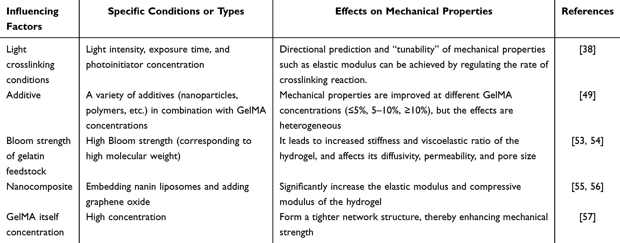 |
Table 1 Influencing Factors of GelMA Mechanics |
The mechanical properties of GelMA are typically weak, which limits its use in load-bearing bone applications.57 In addition, the Bloom strength of the gelatin raw material used in the preparation is equally crucial. Gelatin with higher Bloom strength can produce GelMA hydrogels with higher stiffness and a higher viscoelastic ratio, and can alter their diffusion properties and pore size.53,54 The toughness of GelMA can also be significantly enhanced by adding different concentrations of toughening agents or fillers such as graphene and nano-hydroxyapatite. In addition, altering the degree of crosslinking in GelMA has been shown to be an effective optimization strategy. By adjusting the photopolymerization conditions, researchers can control the crosslinking density of GelMA and thereby modify its mechanical properties. The higher the crosslinking density, the greater the scaffold’s stiffness and strength; however, it may also affect its biocompatibility and degradation rate.51 Therefore, optimizing the mechanical properties of GelMA scaffolds requires finding a balance between strength and biocompatibility.
Despite the versatility of GelMA, a persistent “stiffness-permeability paradox” remains a critical unsolved problem in scaffold design. While increasing the degree of methacryloyl substitution or crosslinking density enhances mechanical load-bearing capacity, it inevitably reduces hydrogel porosity and limits nutrient diffusion, often leading to necrotic cores in large-volume constructs. Current reinforcement strategies, such as adding inorganic fillers (eg, HAP, graphene), improve stiffness but usually introduce interface incompatibility or stress-shielding effects. Furthermore, most studies optimize these parameters in static environments, failing to account for the dynamic degradation rates that occur under physiological loading conditions. Achieving a balance in which degradation kinetics precisely match the rate of new bone formation remains a significant bioengineering challenge that has not yet been fully resolved.
Basic Properties of GelMA Hydrogel with Light-Curing 3D Printing Technology
Principles and Advantages of GelMA Light-Curing 3D Printing
3D printing is a manufacturing process that builds three-dimensional objects by adding materials layer by layer. The basic principle is to generate a three-dimensional model using computer-aided design (CAD) software, then slice the model into layers to guide the printer to deposit material layer by layer. Standard 3D printing techniques include SLA,58 Selective Laser Melting (SLM),59 Fused Deposition Modelling (FDM),60 and Selective Laser Sintering (SLS), among others. Each technology has distinct material requirements and printing methods. SLA uses photosensitive resin to build objects layer by layer via ultraviolet curing, whereas FDM builds objects layer by layer by heating extruded filaments.61 Light-curing 3D printing techniques, such as SLA and DLP, produce fine bone tissue scaffolds by initiating polymerization reactions with ultraviolet light and layer-by-layer curing of GelMA hydrogels. The advantage of light-curing 3D printing lies in its high degree of design flexibility and customization, enabling the fabrication of complex geometries to meet the demands of personalized medicine and bioengineering. One of the main advantages of light-curing 3D printing technology is its high print resolution, which can achieve micron- or even sub-micron precision. This high precision is crucial for manufacturing orthopedic scaffolds, particularly when fine porosity and complex geometries are required. Light-curing 3D printing can precisely simulate the ECM of natural bone tissue, thereby providing an ideal growth environment that promotes the adhesion, proliferation, and differentiation of osteoblasts.17,62
Comparison of the 3D Printing Process Compatibility
The light-curing properties of GelMA enable its compatibility with 3D printing technology, producing high-precision personalized scaffolds that simulate the microstructure of bone tissue. GelMA, a commonly used photocuring material, exhibits good biocompatibility and tunable degradability, thereby providing favourable support for bone tissue engineering. 3D printing supports a variety of photocurable biomaterials, such as GelMA, PEGDA, and their composites.63,64 These materials not only have excellent biocompatibility, but also mechanical properties, and degradation rates can be adjusted according to clinical needs. Compared with light-curing 3D printing technology, FDM mainly uses thermoplastics such as PLA and PCL, which lack biological activity and are not suitable for supporting cell growth;65–67 SLS technology, though capable of manufacturing metal or ceramic scaffolds, requires additional surface modification to be biocompatible;68,69 Inkjet printing is mainly used in soft tissue engineering and does not meet the mechanical requirements of orthopedic applications. Light-curing 3D printing technology enables the integration of biological factors, such as cells and growth factors, into scaffolds during printing, thereby producing multifunctional scaffolds that support bone tissue regeneration.26,70 In contrast, technologies like FDM and SLS have limited capabilities in this regard. FDM typically has lower resolution, is constrained by nozzle size, and cannot produce complex microscale structures; SLS technology is limited by powder particle size and lower print precision, making it difficult to achieve complex bone tissue scaffold structures (Table 2).
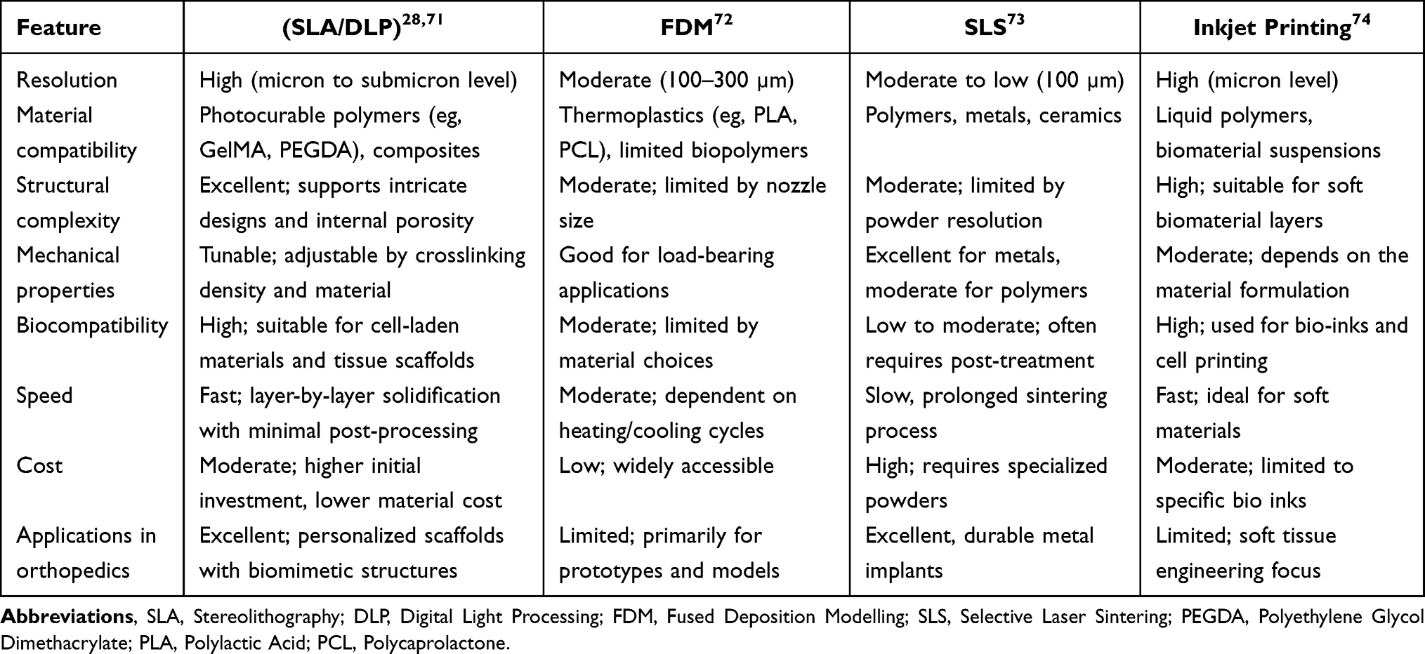 |
Table 2 Types of 3D Printing |
Advantages of GelMA Application in Bone Scaffolds
GelMA, as a novel biomaterial, is widely used in 3D printing technology due to its favourable biocompatibility and adjustable mechanical properties. The advantage of GelMA lies in its ability to cure rapidly via photopolymerization, forming a stable hydrogel structure, making it suitable for cell culture and tissue engineering.75 Moreover, the chemical structure of GelMA allows it to be combined with a variety of functional materials, which gives it significant application potential in areas such as bone tissue regeneration. When GelMA is combined with materials such as nano-hydroxyapatite (nHA), black phosphorus (BP), and nano-graphene oxide (rGO), it can enhance the scaffold’s mechanical properties and promote osteogenic differentiation and bone cell regeneration, thereby synergistically promoting bone tissue repair.76,77 In addition, GelMA can be combined with drug delivery systems to achieve local, sustained drug release, thereby facilitating the healing of bone defects. 3D printing technology using GelMA as a bioink not only improves printing accuracy but also supports cell growth and functional maintenance, thereby providing a suitable scaffold material for bone tissue regeneration.
Design and Optimization of GelMA Structures
Effects of Bone Scaffold Structures on Bone Regeneration
GelMA, as a biomaterial, offers significant advantages for bone defect regeneration, particularly for the design of highly adjustable bone scaffold structures. The structural design of bone scaffolds, including porosity, pore size, shape, and overall geometry, directly determines the effect of bone regeneration. GelMA exhibits excellent structural adaptability and can modulate porosity and pore size as needed to optimize the cell growth environment, thereby improving bone regeneration efficiency. Porosity is crucial for the migration, proliferation, and differentiation of bone cells. Proper porosity provides sufficient space to facilitate nutrient and oxygen diffusion and channels for waste removal, thereby accelerating bone regeneration.61 The porosity of GelMA can be precisely controlled by adjusting the print parameters, crosslinking degree, or material formulation. Studies have shown that GelMA bone scaffolds with moderate porosity can provide good support for the proliferation and differentiation of bone mesenchymal stem cells (BMSCs), thereby promoting bone tissue formation.75 GelMA bone scaffolds can be customized to meet the repair requirements of various bone defects. In addition to porosity, the size of the stent’s aperture is also an essential factor affecting bone regeneration. Proper pore size can effectively simulate the microstructure of bone tissue, providing better attachment surfaces and growth space for cells. By precisely controlling the pore size of the GelMA scaffold, a bone scaffold similar to the natural bone structure can be created to support the normal function of bone cells and promote the formation of new bone. The geometry and overall three-dimensional structure of bone scaffolds also significantly affect cell behaviour.64 3D printing technology enables the fabrication of complex geometries that simulate the microenvironment of natural bone. These structures not only enhance cell attachment and expansion but also improve the scaffold’s mechanical properties, providing sufficient support to withstand loads encountered during bone tissue repair. Studies have shown that GelMA scaffolds employ structures with stratified pores and reticular or porous designs that more effectively promote cell differentiation and mineralization and improve vascularization, thereby accelerating bone regeneration.36,78 Optimizing the structural design of GelMA scaffolds, particularly the adjustment of porosity, pore size, and geometry, can significantly improve their biocompatibility and mechanical properties. By adjusting the degree of GelMA cross-linking and incorporating bioactive molecules, the scaffold’s bioactivity (the ability to actively elicit pro-regenerative responses, including osteogenic support and angiogenic–osteogenic signalling modulation, rather than acting as a passive carrier) can be enhanced, thereby further improving the adhesion, proliferation, and differentiation of bone cells. The tunability of this structure makes GelMA an ideal bone scaffold material that can be customized to meet diverse clinical needs and the characteristics of bone defects, thereby providing more effective support and repair.
Optimization and Simulation of 3D Printing Design Parameters
In the design of GelMA 3D-printed scaffolds, optimizing design parameters is a key step toward improving scaffold performance. The design parameters include material concentration, print speed, layer thickness, and scaffold geometry, all of which affect the physical and biological properties of the final scaffold. Using computer simulation, design parameters can be systematically analyzed to predict their impact on the performance of 3D-printed personalized heart stents, thereby optimizing stent structure and improving therapeutic efficacy. Finite element analysis (FEA) is a widely used numerical simulation method that assesses the impact of design parameters, such as porosity, stent shape, and layer thickness, on mechanical properties by simulating stent behaviour under various loading conditions.79,80 FEA can analyze the stress distribution and deformation of honeycomb and mesh scaffolds under applied forces, ensuring that the scaffolds provide the necessary mechanical support without adversely affecting biocompatibility.81 The response surface method (RSM) is to explore the impact of multi-parameter combinations on stent performance by establishing a multivariable regression model, and to accelerate the optimization process while maintaining the balance between stent biocompatibility and mechanical performance by finding the optimal parameter combinations in the design space.82 Topological optimization methods can automatically adjust the material distribution and structural shape of scaffolds to maximize scaffold performance based on mechanical requirements, especially with significant advantages in the design of porous scaffolds.83 In recent years, machine learning and artificial intelligence technologies have also begun to be applied to the optimization of GelMA scaffolds, by analyzing a large amount of experimental data to predict the effects of different design parameters and automatically adjust the design scheme, further improving design efficiency and accuracy.84 In summary, these computer simulation and optimization methods not only improve the design accuracy of GelMA scaffolds, but also accelerate their clinical translation. For example, in the treatment of intervertebral disc degeneration, 3D cyclic-gradient GelMA hydrogel scaffolds, designed using computer-aided design, demonstrated good biocompatibility and degradability and showed significant therapeutic effects in experimental studies. In addition, numerical simulation methods have been widely applied to the study of intracranial flow-directed close-mesh scaffolds, thereby enhancing experimental efficiency by simulating mechanical properties and fluid dynamics, providing strong technical support for tissue engineering and regenerative medicine.
Applications
Bone tissue is a dynamic organ that is continually undergoing reconstruction. During this process, Osteoclasts (OCs) resorb aged bone tissue, whereas Osteoblasts (OBs) promote bone formation by synthesizing new bone matrix. They maintain bone metabolic balance by alternating between bone resorption and bone formation. The balance is not only dependent on the synergy between the two cell types. Still, it is also regulated by active factors, including RANKL and M-CSF, to ensure the steady progression of bone remodelling.85,86 OCs originate from hematopoietic stem cells and degrade bone by secreting acids and proteolytic enzymes, such as cathepsin K, which dissolve collagen and other gelatinous proteins during bone resorption. OBs are bone-forming cells that, under the guidance of transcription factors, are derived from BMSCs and differentiate into OBs. OBs secrete extracellular proteins, including osteocalcin, alkaline phosphatase, and type I collagen, and unmineralized bone-like secreted extracellular matrix, which then accumulates and mineralizes the calcium phosphate secreted by bone cells in the form of hydroxyapatite to produce bone tissue.38,87 But when the defect area exceeds the critical value for self-repair and reconstruction of bone tissue, the defect area will not heal on its own. Many factors influence bone defect repair and rebuilding, including gene expression regulation, cell migration, cell morphogenesis, and cell cycle progression. Effective coordination of the balance among osteoblasts, osteoclasts, and angiogenic cells within biological bone scaffolds is critical for promoting bone regeneration. GelMA, when combined with osteogenic agents or angiogenic factors, can enhance the osteogenic function of currently used composites. In addition, bone tissue, as a mineralized tissue, exhibits a specific elastic modulus, and the mechanical stiffness of GelMA is limited. By incorporating inorganic minerals with osteogenic-inducing functions into GelMA, the hydrogel’s elastic modulus can be increased to a certain extent. Therefore, it is vital to prepare mixed hydrogels from mixtures of two or more components to combine the unique advantages of individual elements within the composite hydrogel system.88,89
Research Progress of GelMA in Osteoarthritis (OA)
Osteoarthritis (OA) is characterized by progressive degeneration of articular cartilage, subchondral bone sclerosis, and osteophyte formation, which leads to pain and functional limitation. It is one of the high-burden diseases in an aging society.90,91 Existing treatment options, such as NSAIDs, intra-articular hyaluronic acid injection, and end-stage replacement, are mostly inclined towards analgesia and lubrication. They are challenging to reverse cartilage damage, with complications and life expectancy issues coexisting.92,93 GelMA, which combines ECM similarity, degradability, and tunability of mechanical and pore structures, can support and permeate the complex microenvironment of the joint, providing a material basis for the “anti-inflammatory + regenerative” combination treatment.
In terms of material design and loading strategies, GelMA is commonly used in combination with natural matrix dECM or MSC-ECM to enhance cell adhesion and an anti-inflammatory microenvironment. The former, when combined with microfracture surgery, can fill osteochondral defects, inhibit chondrocyte pyroptosis, and promote hyalecondroid regeneration. The latter supports cartilage repair by regulating macrophage metabolism through soluble factors, promoting M2 polarization, and alleviating synovitis.94 To address issues such as low systemic efficacy and significant side effects, GelMA was developed as a long-acting controlled-release platform, for example, by loading astragalus polysaccharides (APS) to activate Fgfr2 for proliferation and matrix synthesis and by loading curcumin to achieve “anti-inflammatory-chondrogenic differentiation” dual effects and increase local bioavailability.95 In cell therapy, GelMA can enhance cell retention and activity. Hypoxic pretreatment of CPCs, in combination with GelMA scaffolds, can activate the HIF-1α pathway, adapt to the joint’s hypoxic environment, and enhance repair.96 In addition, the three-dimensional microstructures constructed by GelMA can amplify the paracrine effects of MSC cells and promote cartilage regeneration.97 Signals such as MMP/ROS/acidity/mechanical stress can be “programmed” as release switches around the OA lesion. A microenvironment-responsive, physical-trigger GelMA response system can be constructed within the OA-specific pathological environment to trigger drug release during joint movement or under high MMP expression, thereby precisely targeting the lesion and reducing systemic exposure.98 He et al constructed ultrasensitive-piezoelectric microspheres with GelMA as the carrier and successfully achieved the dual effects of non-invasive analgesia and immunomodulation.99 Yu et al designed ROS-responsive, lubricated-surface injection microspheres to reduce oxidative damage and improve tribological performance in OA mice. In response to the observation that elevated intra-articular oxidative stress exacerbates chondrocyte damage, preventing chondrocyte apoptosis is an important step in the treatment of early OA.100 Yu et al developed strontium lanate SrR/GelMA composite hydrogels based on the Wnt/β-catenin signalling pathway in OA pathology. SrR/GelMA achieves early intervention by inhibiting Wnt/β-catenin and promoting chondrogenic differentiation of BMSCs.101 Drawing on the lubrication mechanism of synovial fluid, GelMA hydrogel microspheres with enhanced lubricity and controllable drug release were designed to significantly reduce the coefficient of friction between articular surfaces and mechanical damage. Meanwhile, the GelMA network within the microspheres can load anti-inflammatory drugs and enable local drug release via slow degradation, thereby alleviating inflammation.24,102 Overall, these regimens generally led to an increase in OARSI scores, upregulation of type II collagen/proteoglycans, improvements in cartilage thickness and microvessel density, and reductions in pain indicators in animal models.
At present, GelMA-based OA treatment research has developed in multiple directions and at multiple levels, including material modification (eg, composite dECM), the introduction of responsive groups, functional integration (eg, drug-controlled release, cell carriers), and mechanism exploration (eg, metabolic regulation, signalling pathway intervention), with significant progress. These studies not only provide diverse and innovative strategies for OA treatment but also deepen the understanding of material-cell-microenvironment interactions. However, there are still bottlenecks to be broken through in this field: first, some studies are still at the animal model stage, and there is a lack of human clinical trial data, which requires further verification of the safety and efficacy of the GelMA system in the human body; The complex physiological environment within the joints, such as frequent mechanical stress and immune cell infiltration, places higher demands on the long-term stability and functional persistence of GelMA; Third, the compatibility of GelMA degradation rate with cartilage repair cycle and the standardization process for large-scale production still need to be further optimized. In the future, promoting the clinical translation of GelMA-based treatment regimens through multidisciplinary intersections such as materials science, cell biology, and clinical medicine will become a core research direction, which is expected to bring new therapeutic hope to patients with OA (Table 3).
 |
Table 3 GelMA Studies for Osteoarthritis Treatment |
Steroid-Induced Femoral Head Necrosis
Hormone-induced femoral head necrosis (SONFH) is a condition in which bone tissue dies due to the disruption of the blood supply to the femoral head. As the condition progresses, the femoral head collapses, eventually leading to loss of hip joint function, often accompanied by joint pain and limited movement.103,104 Conventional conservative treatment has limited efficacy, and joint replacement is inevitable in the late stage. Tissue engineering repair offers new hope for patients with early-stage femoral head necrosis. The key to SONFH treatment lies in addressing three core issues: alleviating oxidative stress damage, restoring local blood supply, and promoting bone regeneration. Current research on GelMA focuses on these issues. Long-term hormone use leads to a substantial accumulation of reactive oxygen species (ROS) in the femoral head, which not only impairs mitochondrial function in BMSCs and inhibits their osteogenic differentiation but also activates pro-inflammatory macrophages (M1 type), intensifying the local inflammatory response and creating a vicious cycle. Conventional regimens lack targeted ROS-clearing mechanisms, leading to continuous deterioration of the bone repair microenvironment.
To address this issue, the Bai team constructed a composite scaffold of “metal-organic framework MOF-818 GelMA hydrogel + demineralized bone matrix (DBM)” to achieve the triple effect of “ROS clearance - immune regulation - bone vascular regeneration”.105 The study showed that when MOF-818 was modified with zinc, the activities of superoxide dismutase (SOD) and catalase were significantly enhanced, which helped to efficiently eliminate excessive reactive oxygen species (ROS) in the femoral head, thereby reducing mitochondrial damage in BMSCs and promoting the expression of osteogenic differency-related genes RUNX2 and OCN This result is consistent with studies on the mechanisms by which IGF1 and melatonin regulate osteogenic differentiation of BMSCs. Meanwhile, the composite scaffold promotes the conversion of M1-type macrophages to M2-type macrophages and significantly reduces the secretion of pro-inflammatory factors, such as IL-1β. DBM, as a natural bone matrix, provides bone morphogenetic protein (BMP) and binds to the mechanical support of GelMA to guide new bone growth along the scaffold, increasing trabecular bone density of the femoral head in animal experiments and reducing the risk of bone collapse. In SONFH, the hormone inhibits HIF-1α activity, thereby impairing angiogenic capacity. Deferoxamine (DFO) promotes angiogenesis by inhibiting the HIF-1α-degrading enzyme and thereby activating the HIF-1α pathway. However, DFO has a short half-life in the body, and direct injection is prone to “burst release”, and excessive local concentration can lead to BMSCs toxicity, limiting its clinical application. The sustained-release properties of GelMA hydrogel precisely address the “burst release problem” of DFO. By embedding DFO in GelMA hydrogel and adjusting the crosslinking degree of GelMA, the DFO release cycle is extended from 2 hours to 7 days, and the local concentration is maintained within the safe and effective range of 50–100μmol/L to avoid cytotoxicity. Sustained-release DFO continuously activates the HIF-1α pathway, increases VEGF expression, and the number of new blood vessels in the femoral head. At the same time, GelMA supported BMSC adhesion, and the two together promoted “vascular-bone regeneration” coupling. In the rabbit ONFH model, the area of osteonecrosis was reduced compared with the DFO-alone injection group.
GelMA hydrogel, as an integrated platform for SONFH treatment, can specifically address ROS-induced injury and insufficient blood supply by combining with MOF, DFO, nanoparticles, and other agents. It also features injectable and mechanically adjustable properties, making it suitable for minimally invasive surgical procedures. Significant results have been achieved in animal experiments, laying a foundation for clinical transformation. Clinical translation should focus on staging customized plans. In the future, it is expected to undergo clinical translation and serve as the core material for “head-preserving treatment” of early- and mid-stage SONFH, enabling most early- and mid-stage patients to avoid joint replacement and promoting progression from end-stage replacement to early, precise repair (Figure 2).
 |
Figure 2 Schematic illustration of the biomimetic multifunctional scaffold for the treatment of steroid-induced osteonecrosis of the femoral head. Reprinted from Bai L, Zhang X, Shen W et al Multifunctional Scaffold Comprising Metal-Organic Framework, Hydrogel, and Demineralized Bone Matrix for the Treatment of Steroid-Induced Femoral Head Necrosis. Small. Jan 2025;21(3): e2407758. doi:10.1002/smll.202407758.105 |
Osteoporotic Defect
Osteoporosis (OP) is a metabolic bone disease with a high global prevalence. The imbalance between bone resorption and bone formation leads to decreased bone density, microstructural damage, and an increased risk of fragility fractures. The prevalence is 30% to 50% in women and 10% to 20% in men among people over 50 years old, imposing a heavy burden on the social medical system.106,107 At present, treatment options for OP remain limited. Although drug therapy can slow bone loss, it cannot restore the damaged bone microarchitecture. In surgical management, conventional implants are prone to loosening and displacement because of low bone mass and reduced bone strength. Artificial bone exhibits insufficient biological activity and poor bone integration, and the risk of refracture after surgery is high. Traditional scaffold materials for tissue engineering repair have poor biocompatibility, are challenging to regulate the bone-formation microenvironment, and cannot be tailored to the patient’s bone-regeneration capacity.108 GelMA hydrogel, with its similar composition to natural bone matrix collagen, excellent biocompatibility, porous structure conducive to cell colonization and drug factor release, and adjustable mechanical properties, can specifically address many difficulties in OP treatment and has become one of the core materials in OP treatment research. The key to OP treatment lies in restoring the balance between bone formation and resorption, promoting repair of bone defects, and enhancing the efficiency of bone integration around implants. Current research on GelMA is centred on these goals, resulting in innovative, multi-dimensional treatment regimens.
Senescent OP (A-OP), the primary type of OP in the elderly population, is characterized by BMSCs’ senescence.109 With increasing age, the osteogenic differentiation capacity of BMSCs declines, and the secretion of inflammatory factors increases, further inhibiting bone formation. To address this challenge, the Fan team combined single-cell transcriptomics to precisely screen for key regulators of BMSCs’ senescence in A-OP and designed an A “GelMA hydrogel-modified titanium alloy implant” in which osteogenic gene expression in BMSCs was increased.109 In terms of enhancing implant osseointegration, the GelMA hydrogel forms A “bioactive interface” on the titanium alloy surface, providing a colonization scaffold for BMSCs and continuously regulating the bone formation microenvironment by sustained-release resveratrol, increasing the peri-implant osseointegration effect and reducing the risk of loosening in the A-OP animal model. The study employed single-cell transcriptomics in material design to precisely target pathological mechanisms and avoid the drawbacks of blind dosing in traditional regimens, offering new approaches to the precise treatment of A-OP (Figure 3).
 |
Figure 3 Schematic representation. (I) The dynamics of the skeletal stem cell and (II) its application to the design of a titanium implant for the treatment of senile osteoporosis. Reprinted from Fan W, Zheng T, Mao M et al A Study of Skeletal Stem Cell Dynamics and Its Potential Applications in the Design of a Titanium Implant for Senile Osteoporosis. Adv Sci (Weinh). Sep 2025;12(34): e06982. doi:10.1002/advs.202506982.109 |
OP patients exhibit insufficient synergy between bone and vascular regeneration during bone defect repair due to low bone mass and reduced vascular density, thereby limiting the efficacy of traditional repair materials.110 The He team combined SR-LDH with GelMA hydrogel to construct a synergistic system that activated osteoblasts via Sr2⁺ to promote osteogenic differentiation and angiogenesis. It sustainedly released Sr2⁺ through the porous structure of GelMA.111 In the OP animal model, the composite hydrogel significantly increased bone density and the number of new blood vessels, shortened the repair cycle, and its injectability meets the minimally invasive treatment requirements of elderly patients with OP (Figure 4).
 |
Figure 4 GelMA-QK/Sr-LDH@PDA hydrogel enhances vascularization in vitro. (A) Tube formation assay of Bend.3 cells on different hydrogels, with Matrigel as the control group. (B) Representative images of the migration assay of bEnd.3 cells at zero h and 36 h, and blank cell plate set as the control. (C) Quantification of annular structures in tube formation assay. (D) Quantification of annular branch points in tube formation assay. (E) Quantification of luminal diameter in the tube formation assay. (F) Quantification of bEnd.3 cell migration. (G and H) qRT-PCR results of vascularization-related marker genes. All groups are with the following designations: M for Matrigel, G for pure GelMA hydrogel, GL for GelMA/Sr-LDH@PDA composite hydrogel, GLQ for GelMA-QK/Sr-LDH@PDA composite hydrogel and C for control. Significant differences were indicated as *p < 0.05, **p < 0.01 and ***p < 0.001. Reprinted from He Y, Zeng F, Quan H et al Strontium-loaded multifunctional gelatin methacryloyl hydrogels for type-H vascularized bone regeneration under osteoporotic conditions. Mater Today Bio. Jun 2025;32: 101909. doi:10.1016/j.mtbio.2025.101909.111 |
Following fractures in OP patients, an abnormal local immune microenvironment leads to macrophage polarization toward the pro-inflammatory M1 phenotype, with the release of inflammatory factors, which inhibit osteoblast activity and intensify bone resorption.112 According to the study, this immune response pattern is particularly pronounced in patients with osteoporosis, resulting in significantly longer fracture-healing times than in healthy individuals, particularly among elderly patients. Conventional treatments focus on immobilization and calcium supplementation, neglecting immunomodulation and thereby impeding improvements in healing efficiency. The Zhou team designed an “IL-4 + bacterial exosome (BEVs) co-loaded GelMA hydrogel” to promote OP fracture repair through dual immunomodulation.113 IL-4 induces macrophage polarization to the anti-inflammatory M2 phenotype; BEVs carry the anti-inflammatory factor TGF-β to regulate the immune microenvironment; and their nanostructures enhance the stability of IL-4. GelMA hydrogel provides mechanical support and sustained-release IL-4 and BEVs, synergistically promoting osteoblast proliferation and angiogenesis (Figure 5). In OP animal fracture models, the regimen shortens fracture healing time. This study, for the first time, combines immunomodulation with bone regeneration, overcoming the limitations of traditional regimens and providing a new strategy for the efficient healing of OP fractures.
 |
Figure 5 Schematic representation of engineered probiotic EVs in a composite hydrogel for temporally controlled repair of OPF. Reprinted from Zhou G, Zhou Q, Li R et al Synthetically Engineered Bacterial Extracellular Vesicles and IL-4-Encapsulated Hydrogels Sequentially Promote Osteoporotic Fracture Repair. ACS Nano. Apr 29 2025;19(16):16,064–16083. doi:10.1021/acsnano.5c03106.113 |
With the increasing demand for precision in OP treatment, there is an urgent clinical need for smart stents that can respond to microenvironmental changes and precisely regulate bone formation. Some researchers have pointed out that nanomaterials can mimic the nanostructure of the natural bone matrix, enhance their affinity for bone tissue, and deliver growth factors precisely, thereby informing GelMA modification.114 Growth factors are key to bone regeneration, but their direct use poses risks. Combining them with GelMA to design sustained-release systems can enhance osteogenic effects. Future innovative scaffolds require multi-signal integration capabilities, and GelMA, owing to its high compatibility, is expected to serve as the core carrier. Currently, GelMA hydrogels have demonstrated remarkable efficacy in OP treatment research, with breakthroughs in implant modification, bone defect repair, and immune microenvironment regulation. The core advantage lies in its robust carrier properties, which enable the integration of multiple functional modules, achieve coordinated multi-mechanism regulation, and adapt to complex pathological environments. Future research could develop personalized GelMA systems based on pathological differences among OP subtypes.
Periodontitis and Alveolar Bone Regeneration
Periodontitis is a chronic inflammatory disease characterized by progressive alveolar bone defects, often accompanied by symptoms such as swollen gums and loose teeth. If the disease progresses, it will ultimately result in tooth loss.115 The core objective of current clinical treatment is to achieve the regeneration of dental supporting tissues, including alveolar bone, periodontal ligament, and cementum, as well as the formation of functional new attachments. However, traditional techniques such as bone grafting, guided tissue regeneration, and growth factor addition face limitations, including limited indications, poor adhesion of the binding epithelium, and easy collapse of the guiding membrane, which make them challenging to meet clinical needs.116 In contrast, the GelMA hydrogel offers significant advantages for the treatment of periodontitis. Its excellent biocompatibility can reduce immune rejection and create favourable conditions for tissue repair. With its customizable properties, it can precisely regulate mechanical properties and degradation rates to account for individual patient differences. Its robust functional adaptability supports the loading of growth factors, stem cells, and other active ingredients to target and promote periodontal tissue regeneration. The core advantage of GelMA hydrogel is its ability to be precisely adapted to the individual morphological characteristics of alveolar bone defects in patients. Through 3D printing, scaffolds with complex geometric structures can be fabricated to provide stable spatial support for periodontal tissue regeneration.
Zhu Y et al designed 3D bioprinted biomimetic periodontal modules with high structural integrity using GelMA/ECM as the cell-loading bioink.117 The module is not only mechanically stable, but also enables cell orientation guidance through high-precision terrain prompts, creating a favourable biochemical environment for the proliferation and differentiation of encapsulated cells. Meanwhile, the potent immunomodulatory activity of ECM reduces the release of pro-inflammatory factors TNF-α and IL-6 from M1-type macrophages, thereby alleviating local inflammation in the periodontal defect area and clearing environmental obstacles to bone regeneration.118,119 Periodontal Ligament Stem Cells (PDLSCs) are the primary seed cells for periodontal tissue regeneration and exhibit multi-directional differentiation potential.120,121 GelMA is an ideal carrier for the proliferation and differentiation of PDLSCs. Scaffolds prepared by bioprinting with 10% high-concentration GelMA hydrogel exhibit a higher compressive modulus and a lower swelling rate, although they slightly reduce the survival rate of PDLSCs. However, it can significantly enhance osteogenic differentiation ability by activating the eprinB2/EphB4 signalling pathway.117 Low-stiffness GelMA can optimize the microenvironment for cell survival and proliferation. The Wang team found that although low-stiffness GelMA hydrogels provide weaker mechanical support, they significantly increase PDLSC survival and are more conducive to cell proliferation, migration, and the expression of osteogenic genes RUNX2, COL1A1, and ALP.122
By constructing GelMA composite systems, the mechanical microenvironment can be precisely regulated, and the application of 3D bioprinting technology further enhances the precision with which the mechanical properties of GelMA are controlled. Yufei Ma et al prepared GelMA/PEGDA composite hydrogel microarrays by adjusting the ratio of GelMA to polyethylene glycol dimethacrylate (PEGDA).121 In vitro experiments confirmed that the composite system was most conducive to osteogenic differentiation of PDLSCs, and in vivo animal experiments also showed that its effect on new bone formation in rat alveolar bone defect models was significantly better than that of the single GelMA hydrogel control group.
Exosomes have immunomodulatory and tissue-regenerative activities and are emerging as functional biomarkers for periodontitis treatment; however, their short half-life and rapid clearance limit their clinical application.123,124 GelMA hydrogel can serve as an efficient carrier for exosomes, prolonging their retention time and biological activity within the periodontal lesion and thereby achieving a synergistic therapeutic effect. Immune-cell-derived exosomes regulate the periodontal inflammatory microenvironment. The Ya Cui team developed melatonin loaded m2-type macrophage-derived exosomes (Mel@M2-exos) and embedded them in injectable GelMA hydrogels M2-exos have the natural ability to target inflammatory sites and induce macrophages to polarize from pro-inflammatory M1 type to anti-inflammatory M2 type, reducing local inflammation; Meanwhile, melatonin released by exosomes alleviates excessive endoplasmic reticulum stress in PDLCs in an inflammatory environment, inhibits apoptosis, and restores their osteogenic, odontogenic, and odontogenic differentiation potential.125 Haiwei Zhuo’s team designed GelMA@MP196/Exos hydrogel, which induces neutrophil apoptosis, enhances macrophage phagocytosis, and effectively inhibits periodontal bone loss.126 Exosomes derived from dental pulp stem cells (DPSCs) can enhance alveolar bone regeneration efficiency. Yike Gao’s team used magnesium ions to activate DPSCs, thereby inducing the production of small extracellular vesicles (sEVs) enriched in miR-451a, which promote vascularized bone regeneration via the AKT/eNOS/NO signalling axis.127 The sustained release of sEVs was achieved through GelMA hydrogel, which significantly enhanced in vivo bone defect repair and effectively overcame the limited angiogenic ability of natural MSC-sEVs.
Infection is the primary driver of periodontitis progression and treatment failure, and the GelMA complex system can achieve synchronous regulation of antibacterial and bone-regenerative functions via slow degradation and controlled release. The Yuqi Ma team prepared nanoscale bismuth-doped GelMA electrospun fibre membranes that sustainably release bismuth nanoparticles (BiNPs), achieving a 100% killing rate against the key periodontitis pathogenic bacterium Porphyromonas gingivalis (P. gingivalis) and showing no significant cytotoxicity toward PDLSCs.128 By combining antioxidant and anti-inflammatory effects, it can improve the periodontal repair microenvironment and promote bone tissue repair and regeneration. The multifunctional injectable microspheres (GM@Fe-Cur/PDA/MH) developed by the Ziliang Xiu team encapsulate iron-curcurin nanoparticles (Fe-Cur NPs) in a GelMA matrix using microfluidic technology, with the surface modified by polydopamine (PDA) and loaded with minocycline hydrochloride (MH).129 The microspheres exhibit excellent retention capacity in the periodontal pocket, combining the photothermal effect of PDA with the antibacterial properties of MH to effectively kill pathogenic bacteria, induce macrophage polarization from pro-inflammatory M1 to anti-inflammatory M2, inhibit the release of inflammatory factors, and promote the expression of the osteogenic protein BMP-2. In the rat periodontitis model, the microsphere significantly inhibited inflammation and reduced alveolar bone resorption. In addition, the GelMA hydrogel complex loaded with RvD1 developed by the Zhe team can achieve sustained RvD1 release, significantly promote PDLC proliferation, reduce inflammatory cell infiltration, lower TNF-α and RANKL expression, and inhibit osteoclast generation.130 It also upregulates the expression of anti-inflammatory and tissue regenesis-related genes, effectively improving the periodontal repair microenvironment (Table 4).
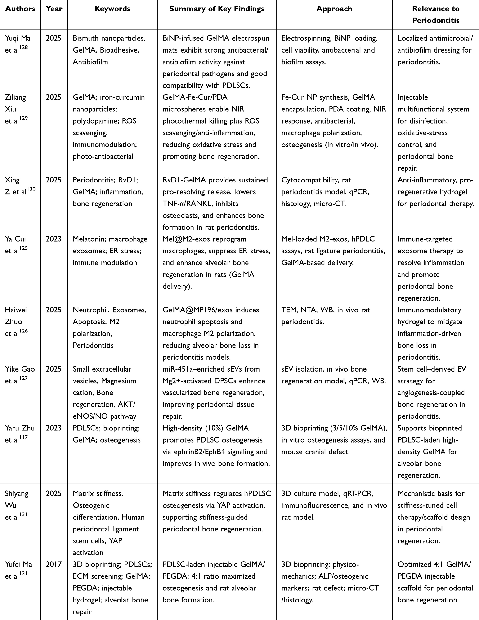 |
Table 4 GelMA Studies for Periodontitis Treatment |
With tunable mechanical properties, excellent biocompatibility, and multifunctional carrier characteristics, GelMA hydrogel offers key advantages in the “inflammation regulation-antibacterial treatment-tissue regeneration” process for the treatment of periodontitis. From regulating the mechanical strength of PDLSCs’ function to precise immune regulation via exosomes to loading multiple active ingredients as a drug-delivery platform, GelMA can achieve synergistic enhancement of therapeutic effects by combining with materials or technologies. In the future, as material modification technology continues to advance, the precision of 3D bioprinting and the mechanisms of stem cell and exosome regulation are expected to further enhance the role of GelMA hydrogels in personalized treatment of periodontitis, particularly for complex periodontal defect repair and clinical translational applications, where there remains substantial scope for exploration.
Applications of GelMA in Skeletal Organoids
Bone organoids leverage the self-organizing properties of stem cells to faithfully replicate the in vivo microenvironment and induce seed cells to form 3D organ microtissues that simulate bone tissue structure and physiological function.6,132 With ongoing research on bone organoids, a variety of bone organoid models, including trabeculae, callus, cartilage, and bone marrow, have been successfully constructed and play an essential role in the study of bone-defect-related diseases. Bone organoids are formed by the proliferation and differentiation of seed cells in vitro; seed cells include, but are not limited to, induced pluripotent stem cells, bone marrow mesenchymal stem cells, and periosteal cells.133–135 At present, researchers often induce the differentiation of seed cells into osteoblasts and osteoclasts and inoculate them into culture matrices to replicate bone formation and bone resorption units, thereby simulating the equilibrium state of bone remodelling at the organoid level. Seed cells can retain the donor’s genotype and exhibit high self-renewal and pluripotency, making them a powerful tool for studying and simulating bone defect-related diseases.136,137 The construction of bone organoids can be classified into scaffold- and non-scaffold-based methods depending on the quality of the medium. Scaffolds are organoid culture methods that use matrix gels, hydrogels, or other polymers to support cell growth and provide rich nutrient and cytokine environments for seed cells.138 Scaffolds are methods used to prevent cell adhesion and self-organization and to facilitate the formation of organoids. Standard methods include suspension culture, microarray, 3D printing, dynamic rotation culture, and microfluidic chips. Microfluidic chips enable continuous perfusion and spatiotemporal control of soluble cues, helping to overcome diffusion limitations and supporting more stable maturation of bone organoids. Given GelMA’s rapid photo-crosslinking and patternability, it is well-suited for forming perfusable microchannels and spatially defined niches within microfluidic devices. Studies have shown that GelMA can effectively support the growth and differentiation of bone organoid-associated cells and promote cell-matrix interactions.
GelMA can form porous networks through photocrosslinking, serving as the structural skeleton of bone organoids and providing space for BMSCs to attach, proliferate, and self-organize. Peng Luo et al found that the porous structure of a 10% concentration of GelMA hydrogel (HG-2) supported the complete extension of BMSCs to form intercellular connections and constructed HG-2/3D-BMSC organoids within 14 days.139 The elastic modulus of 10% GelMA precisely covered the optimal stiffness range for osteogenesis and chondrogenesis.117 It simultaneously promoted the differentiation of BMSCs into bone organoids, with differentiation toward osteoblasts and chondrocytes. Jian Wang et al used the porous structure of the GelMA/AlgMA/HAP composite scaffold, in which HAP released Ca2⁺/PO43− ions to promote BMSC mineralization. After 40 days of culture, the bone volume fraction of the organoids was significantly increased compared to pure GelMA, and the mechanical strength was close to that of loose bone.6 Successfully guided BMSCs to self-organize into trabecular bone structures. The maturation of bone organoids depends on the directed differentiation of BMSCs into osteogenesis or chondrogenesis. GelMA can guide cell fate through both mechanical property regulation and biochemical signal loading pathways to construct bone organoids.6 Guangyin Zhou et al constructed GelMA/DNA double network hydrogels (CGDE) with enhanced viscoelasticity through hydrogen bonding, increased storage modulus G’, increased stress relaxation rate, promoting BMSCs to self-organize into braided bone organoids, forming mineralized collagen networks after 21 days of culture, similar to the natural braided bone structure.23
GelMA has a unique advantage in loading and releasing biochemical signals. Because of its slow degradation, GelMA enables long-term, stable release of biochemical signals. This enables GelMA to provide a more persistent environment than traditional signal-release systems, thereby promoting long-term cell differentiation and tissue repair. In tissue engineering, GelMA, as a sustained-release carrier, can be loaded with cytokines, nanoparticles, or bioactive molecules to provide sustained biochemical signalling for skeletal organoids, demonstrating significant advantages for application.113 GelMA-loaded stem cell regulatory molecules induce cytoskeleton assembly through cell adhesion, activate the Yap/Tead4 mechanical conduction pathway, down-regulate Kat7, unlock the multi-directional differentiation potential of BMSCs, and enable the organoids to express osteogenic, chondrogenic, and other genes simultaneously. Promote the formation of bone organoids (Figure 6).139
 |
Figure 6 In vivo multidirectional differentiation ability of obtained grafts after osteogenic differentiation induction for 14 days. (A) H&E staining showing new tissue generation of different groups after 6 weeks. Scale bar =200μm. (B) Sirius red staining showing collagen fibre formation of different groups after 6 weeks. (C and E) Von Kossa staining and quantitative analysis results showing calcium salt deposition across different groups after 6 weeks; one-way analysis of variance, one-tailed. (D and F) Micro-CT scan reconstruction and quantitative analysis results providing the mineralization degrees of different groups after 6 weeks, one-way analysis of variance, one-tailed. Scale bar = 200 mm. All data are expressed as mean ± SD (n = 3; ***p < 0.001; ****p < 0.0001; ns means no significance). Reprinted from Luo P, Cheng Y, Luo Y et al Hydrogel enhanced organoid multidirectional differentiation via Yap/Tead4 mechanotransduction for accelerated tissue regeneration. ACS Appl Mater Interfaces. Jul 2 2025;17(26):37, 601–37616. doi:10.1021/acsami.5c06161.139 |
With ongoing advances in bone tissue engineering and regenerative medicine, GelMA has demonstrated distinct advantages for the construction of bone organoids. Its excellent biocompatibility and controllable mechanical properties enable GelMA to effectively promote the attachment, proliferation, and differentiation of bone cells, thereby simulating the complex structure and function of bone tissue. This property not only provides an ideal in vitro model for basic research but also lays a solid foundation for analyzing the mechanisms of bone diseases and developing new therapeutic strategies. When combined with bioprinting technology, GelMA is expected to enable greater precision and complexity in the fabrication of bone organoids to meet personalized medical needs. The introduction of innovative materials, such as responsive hydrogels or drug-delivery systems, will further enable GelMA to dynamically regulate the microenvironment, thereby enhancing the efficiency of bone tissue regeneration and therapeutic outcomes.
Clinical Translation Considerations and Challenges
Despite substantial progress in preclinical studies, clinical translation of GelMA-based nanocomposites for bone defect repair remains at an early stage. Most existing evidence is derived from in vitro experiments and small-animal models, whereas large-animal studies and well-designed clinical trials remain limited. This translational gap highlights the need for cautious interpretation of current outcomes and realistic assessment of clinical feasibility. Several key challenges must be addressed before GelMA-based systems can advance toward routine clinical use. First, although tunable, the mechanical strength of GelMA hydrogels remains insufficient for many load-bearing bone applications without reinforcement, raising concerns about long-term structural stability in vivo. Second, the photocrosslinking process, particularly when ultraviolet light is employed, may induce oxidative stress or DNA damage in encapsulated cells, necessitating optimization of crosslinking strategies, photoinitiator selection, and exposure parameters. From a translational perspective, issues related to large-scale manufacturing, batch-to-batch reproducibility, sterilization, and regulatory compliance under good manufacturing practice (GMP) standards also represent significant barriers. Furthermore, the degradation kinetics of GelMA scaffolds must be precisely matched with the rate of new bone formation to ensure sustained mechanical support while avoiding premature scaffold failure. Addressing these challenges through material optimization, advanced fabrication strategies, and rigorous in vivo validation could be critical for bridging the gap between experimental success and clinical application.
To bridge the gap between promising preclinical outcomes and clinical implementation, a stepwise and clinically oriented validation strategy is required. To strengthen translational readiness, future studies should move beyond proof-of-concept demonstrations and adopt clinically oriented validation pathways. In particular, a stepwise evidence hierarchy (in vitro → small-animal → large-animal) is needed, with careful selection of defect models that better approximate human biomechanics and healing timelines. From a practical standpoint, early clinical positioning may be more feasible in non-load-bearing or lower-load indications before expanding to high-load long-bone reconstruction, where mechanical demands and regulatory scrutiny are typically higher. In parallel, scalable and reproducible manufacturing must be prioritized, including defined quality attributes (eg, degree of methacrylation, rheological properties, crosslinking kinetics, and nanoparticle dispersion), sterilization compatibility, and batch-to-batch consistency under GMP-aligned workflows. Finally, translational studies should incorporate clinically meaningful endpoints—such as longitudinal imaging, quantitative biomechanical testing, safety/toxicity evaluation, and degradation–bone formation matching—to support a realistic pathway toward human application.
Conclusions and Future Perspectives
GelMA-based nanocomposites have emerged as a versatile and highly adaptable platform for bone defect regeneration, owing to their favourable biocompatibility, tunable mechanical properties, and the ability to integrate multifunctionally. Rather than serving as a single-purpose biomaterial, GelMA enables the coordinated regulation of cellular behaviour, immune responses, and angiogenic–osteogenic coupling, all of which are essential for effective bone repair.
Nevertheless, current advances remain predominantly at the preclinical stage, and significant challenges related to mechanical robustness, biosafety, degradation control, and translational scalability must be overcome. Importantly, the clinical potential of GelMA-based systems should be evaluated not only on the basis of biological performance but also with respect to regulatory feasibility, manufacturing reproducibility, and long-term in vivo safety.
Future research should prioritize large-animal validation, standardized fabrication protocols, and the development of clinically compatible crosslinking strategies. By integrating materials science, mechanobiology, immunology, and translational medicine, GelMA-based platforms may ultimately evolve from experimental constructs into clinically viable solutions for repairing complex bone defects.
Funding
This study was supported by Tianjin Education Commission Scientific Research Program (2024ZXZD001).
Disclosure
The authors report no conflicts of interest in this work.
References
1. Fan L, Chen S, Yang M, Liu Y, Liu J. Metallic Materials for Bone Repair. Adv Healthc Mater. 2024;13(3):e2302132. doi:10.1002/adhm.202302132
2. Li XL, Zhao YQ, Miao L, et al. Strategies for promoting neurovascularization in bone regeneration. Military Med Res. 2025;12(1):9. doi:10.1186/s40779-025-00596-1
3. Robin M, Mouloungui E, Castillo Dali G, et al. Mineralized collagen plywood contributes to bone autograft performance. Nature. 2024;636(8041):100–26. doi:10.1038/s41586-024-08208-z
4. Zhou S, Xiao C, Fan L, et al. Injectable ultrasound-powered bone-adhesive nanocomposite hydrogel for electrically accelerated irregular bone defect healing. J Nanobiotechnology. 2024;22(1):54. doi:10.1186/s12951-024-02320-y
5. Kurian AG, Singh RK, Patel KD, Lee JH, Kim HW. Multifunctional GelMA platforms with nanomaterials for advanced tissue therapeutics. Bioact Mater. 2022;8:267–295. doi:10.1016/j.bioactmat.2021.06.027
6. Wang J, Wu Y, Li G, et al. Engineering Large-Scale Self-Mineralizing Bone Organoids with Bone Matrix-Inspired Hydroxyapatite Hybrid Bioinks. Adv Mater. 2024;36(30):e2309875. doi:10.1002/adma.202309875
7. Huang L, Guo Z, Yang X, et al. Advancements in GelMA bioactive hydrogels: strategies for infection control and bone tissue regeneration. Theranostics. 2025;15(2):460–493. doi:10.7150/thno.103725
8. Meng J, Lu J, Jiang C, et al. Collagen hydrogel-driven pyroptosis suppression and combined microfracture technique delay osteoarthritis progression. Biomaterials. 2025;314:122817. doi:10.1016/j.biomaterials.2024.122817
9. Deng D, Li X, Zhang JJ, et al. Biotin-Avidin System-Based Delivery Enhances the Therapeutic Performance of MSC-Derived Exosomes. ACS Nano. 2023;17(9):8530–8550. doi:10.1021/acsnano.3c00839
10. Lei M, Liu H, Zhong Q, et al. Hydroxybutyl chitosan-based thermosensitive hydrogel enhanced osteoporotic bone regeneration through sustained release of alendronate and BMP-2. Carbohydr Polym. 2026;373:124609. doi:10.1016/j.carbpol.2025.124609
11. Tan B, Liu X, Chen S, et al. An injectable nano-hydroxyapatite-incorporated hydrogel with sustained release of Notoginsenoside R1 enhances bone regeneration by promoting angiogenesis through Notch1/Akt signaling. J Adv Res. 2025;2025:1. doi:10.1016/j.jare.2025.05.025
12. Zheng X, Zhang X, Wang Y, et al. Hypoxia-mimicking 3D bioglass-nanoclay scaffolds promote endogenous bone regeneration. Bioact Mater. 2021;6(10):3485–3495. doi:10.1016/j.bioactmat.2021.03.011
13. Ma B, Ma H, Gu B, et al. Injectable GelMA microsphere-integrated hyaluronic acid-polyphenol hydrogel system with multifunctional injury microenvironment modulation promotes peripheral nerve regeneration. Carbohydr Polym. 2025;368(Pt 2):124214. doi:10.1016/j.carbpol.2025.124214
14. Wang R, He X, Bai J, et al. Cerium Oxide Nanoparticles-Reinforced GelMA Hydrogel Loading Bone Marrow Stem Cells with Osteogenic and Inflammatory Regulatory Capacity for Bone Defect Repair. ACS Appl Mater Interfaces. 2024;16(49):67373–67384. doi:10.1021/acsami.4c15718
15. Alarcin E, Akguner ZP, Ozturk AB, et al. Biomimetic 3D bioprinted bilayer GelMA scaffolds for the delivery of BMP-2 and VEGF exogenous growth factors to promote vascularized bone regeneration in a calvarial defect model in vivo. Int J Biol Macromol. 2025;306(Pt 2):141440. doi:10.1016/j.ijbiomac.2025.141440
16. Beheshtizadeh N, Farzin A, Rezvantalab S, et al. 3D printing of complicated GelMA-coated Alginate/Tri-calcium silicate scaffold for accelerated bone regeneration. Int J Biol Macromol. 2023;229:636–653. doi:10.1016/j.ijbiomac.2022.12.267
17. Huang M, Zhang L, Cui J, et al. 3D printing of GelMA/nanohydroxyapatite/melanin nanoparticles composite hydrogel scaffolds for bone regeneration through immunomodulation. Int J Biol Macromol. 2025;306(Pt 2):141453. doi:10.1016/j.ijbiomac.2025.141453
18. You J, Li Y, Wang C, et al. Mild Thermotherapy-Assisted GelMA/HA/MPDA@Roxadustat 3D-Printed Scaffolds with Combined Angiogenesis-Osteogenesis Functions for Bone Regeneration. Adv Healthc Mater. 2024;13(22):e2400545. doi:10.1002/adhm.202400545
19. Jing X, Xu C, Su W, et al. Photosensitive and Conductive Hydrogel Induced Innerved Bone Regeneration for Infected Bone Defect Repair. Adv Healthc Mater. 2023;12(3):e2201349. doi:10.1002/adhm.202201349
20. Weng J, Chen Y, Zeng Y, et al. A novel hydrogel loaded with plant exosomes and stem cell exosomes as a new strategy for treating diabetic wounds. Mater Today Bio. 2025;32:101810. doi:10.1016/j.mtbio.2025.101810
21. Yue K, Trujillo-de Santiago G, Alvarez MM, Tamayol A, Annabi N, Khademhosseini A. Synthesis, properties, and biomedical applications of gelatin methacryloyl (GelMA) hydrogels. Biomaterials. 2015;73:254–271. doi:10.1016/j.biomaterials.2015.08.045
22. Lv B, Lu L, Hu L, et al. Recent advances in GelMA hydrogel transplantation for musculoskeletal disorders and related disease treatment. Theranostics. 2023;13(6):2015–2039. doi:10.7150/thno.80615
23. Zhu M, Zhang H, Zhou Q, et al. Dynamic GelMA/DNA Dual-Network Hydrogels Promote Woven Bone Organoid Formation and Enhance Bone Regeneration. Adv Mater. 2025;37(24):e2501254. doi:10.1002/adma.202501254
24. He X, He S, Xiang G, et al. Precise Lubrication and Protection of Cartilage Damage by Targeting Hydrogel Microsphere. Adv Mater. 2024;36(40):e2405943. doi:10.1002/adma.202405943
25. Li S, Sheng Y, You Y, et al. Study on VEGFA mRNA delivery via GelMA hydrogel-encapsulated extracellular vesicles for enhanced bone regeneration. Mater Today Bio. 2025;34:102144. doi:10.1016/j.mtbio.2025.102144
26. Li G, Gao F, Yang D, et al. ECM-mimicking composite hydrogel for accelerated vascularized bone regeneration. Bioact Mater. 2024;42:241–256. doi:10.1016/j.bioactmat.2024.08.035
27. Zhou X, Zou B, Chen Q, Yang G, Lai Q, Wang X. Construction of bilayer biomimetic periosteum based on SLA-3D printing for bone regeneration. Colloids Surf B Biointerfaces. 2025;246:114368. doi:10.1016/j.colsurfb.2024.114368
28. Qian Y, Gong J, Lu K, et al. DLP printed hDPSC-loaded GelMA microsphere regenerates dental pulp and repairs spinal cord. Biomaterials. 2023;299:122137. doi:10.1016/j.biomaterials.2023.122137
29. Nichol JW, Koshy ST, Bae H, Hwang CM, Yamanlar S, Khademhosseini A. Cell-laden microengineered gelatin methacrylate hydrogels. Biomaterials. 2010;31(21):5536–5544. doi:10.1016/j.biomaterials.2010.03.064
30. Yu H, Luo X, Li Y, et al. Advanced Hybrid Strategies of GelMA Composite Hydrogels in Bone Defect Repair. Polymers. 2024;16(21):3039. doi:10.3390/polym16213039
31. Zhu Y, Yu X, Liu H, et al. Strategies of functionalized GelMA-based bioinks for bone regeneration: recent advances and future perspectives. Bioact Mater. 2024;38:346–373. doi:10.1016/j.bioactmat.2024.04.032
32. Xu X, Zhang H, Li Y, et al. Chromatin remodeling and nucleoskeleton synergistically control osteogenic differentiation in different matrix stiffnesses. Mater Today Bio. 2023;20:100661. doi:10.1016/j.mtbio.2023.100661
33. Tan K, Yang Q, Han Y, et al. Elastic modulus of hydrogel regulates osteogenic differentiation via liquid-liquid phase separation of YAP. J Biomed Mater Res A. 2023;111(11):1781–1797. doi:10.1002/jbm.a.37590
34. Yang T, Fang Z, Zhang J, Zheng S. Physical cues in biomaterials modulate macrophage polarization for bone regeneration: a review. Front Bioeng Biotechnol. 2025;13:1640560. doi:10.3389/fbioe.2025.1640560
35. Zhuang Z, Zhang Y, Sun S, et al. Control of Matrix Stiffness Using Methacrylate-Gelatin Hydrogels for a Macrophage-Mediated Inflammatory Response. ACS Biomater Sci Eng. 2020;6(5):3091–3102. doi:10.1021/acsbiomaterials.0c00295
36. Ai Y, Dai F, Li W, et al. Photo-crosslinked bioactive BG/BMSCs@GelMA hydrogels for bone-defect repairs. Mater Today Bio. 2023;23:100882. doi:10.1016/j.mtbio.2023.100882
37. Clerkin S, Singh K, Davis JL, et al. Tuneable gelatin methacryloyl (GelMA) hydrogels for the directed specification of renal cell types for hiPSC-derived kidney organoid maturation. Biomaterials. 2025;322:123349. doi:10.1016/j.biomaterials.2025.123349
38. O’Connell CD, Zhang B, Onofrillo C, et al. Tailoring the mechanical properties of gelatin methacryloyl hydrogels through manipulation of the photocrosslinking conditions. Soft Matter. 2018;14(11):2142–2151. doi:10.1039/c7sm02187a
39. Pamplona R, González-Lana S, Romero P, Ochoa I, Martín-Rapún R, Sánchez-Somolinos C. Tuning of Mechanical Properties in Photopolymerizable Gelatin-Based Hydrogels for In Vitro Cell Culture Systems. ACS Appl Polymer Mater. 2023;5(2):1487–1498. doi:10.1021/acsapm.2c01980
40. Zhang H, Bré LP, Zhao T, Zheng Y, Newland B, Wang W. Mussel-inspired hyperbranched poly(amino ester) polymer as strong wet tissue adhesive. Biomaterials. 2014;35(2):711–719. doi:10.1016/j.biomaterials.2013.10.017
41. Miri AK, Hosseinabadi HG, Cecen B, Hassan S, Zhang YS. Permeability mapping of gelatin methacryloyl hydrogels. Acta Biomater. 2018;77:38–47. doi:10.1016/j.actbio.2018.07.006
42. Nguyen AH, McKinney J, Miller T, Bongiorno T, McDevitt TC. Gelatin methacrylate microspheres for controlled growth factor release. Acta Biomater. 2015;13:101–110. doi:10.1016/j.actbio.2014.11.028
43. Xu Q, Li J, Liu B, et al. Biodegradable copper-doped hollow mesoporous polydopamine nanoparticles for chemo/photothermal/chemodynamic synergistic therapy. J Drug Delivery Sci Technol. 2023;89:10515. doi:10.1016/j.jddst.2023.105015
44. Vigata M, Meinert C, Pahoff S, Bock N, Hutmacher DW. Gelatin Methacryloyl Hydrogels Control the Localized Delivery of Albumin-Bound Paclitaxel. Polymers. 2020;12(2):501. doi:10.3390/polym12020501
45. Sun M, Sun X, Wang Z, Guo S, Yu G, Yang H. Synthesis and Properties of Gelatin Methacryloyl (GelMA) Hydrogels and Their Recent Applications in Load-Bearing Tissue. Polymers. 2018;10(11):1290. doi:10.3390/polym10111290
46. Fairbanks BD, Schwartz MP, Bowman CN, Anseth KS. Photoinitiated polymerization of PEG-diacrylate with lithium phenyl-2,4,6-trimethylbenzoylphosphinate: polymerization rate and cytocompatibility. Biomaterials. 2009;30(35):6702–6707. doi:10.1016/j.biomaterials.2009.08.055
47. Kulkarni NS, Chauhan G, Goyal M, Sarvepalli S, Gupta V. Development of gelatin methacrylate (GelMa) hydrogels for versatile intracavitary applications. Biomater Sci. 2022;10(16):4492–4507. doi:10.1039/d2bm00022a
48. Noshadi I, Hong S, Sullivan KE, et al. In vitro and in vivo analysis of visible light crosslinkable gelatin methacryloyl (GelMA) hydrogels. Biomater Sci. 2017;5(10):2093–2105. doi:10.1039/c7bm00110j
49. Zhang Y, Sun M, Liu T, Hou M, Yang H. Effect of Different Additives on the Mechanical Properties of Gelatin Methacryloyl Hydrogel: a Meta-analysis. ACS Omega. 2021;6(13):9112–9128. doi:10.1021/acsomega.1c00244
50. Santana MDV, Magulas MBS, Brito GC, et al. Cryogenic 3D Printing of GelMA/Graphene Bioinks: improved Mechanical Strength and Structural Properties for Tissue Engineering. Int J Nanomed. 2024;19:10745–10765. doi:10.2147/IJN.S486868
51. Komosa ER, Lin WH, Ogle BM. Toward robust and reproducible pluripotent stem cell expansion in bioprinted GelMA constructs. Int J Bioprint. 2025;11(1):363–381. doi:10.36922/ijb.4633
52. Talaei A, O’Connell CD, Sayyar S, et al. Optimizing the composition of gelatin methacryloyl and hyaluronic acid methacryloyl hydrogels to maximize mechanical and transport properties using response surface methodology. J Biomed Mater Res B Appl Biomater. 2023;111(3):526–537. doi:10.1002/jbm.b.35169
53. Zambuto SG, Kolluru SS, Ferchichi E, et al. Evaluation of gelatin bloom strength on gelatin methacryloyl hydrogel properties. bioRxiv. 2023;2023:566924. doi:10.1101/2023.11.13.566924
54. Zambuto SG, Kolluru SS, Ferchichi E, et al. Evaluation of gelatin bloom strength on gelatin methacryloyl hydrogel properties. J Mech Behav Biomed Mater. 2024;154:106509. doi:10.1016/j.jmbbm.2024.106509
55. Elkhoury K, Sanchez-Gonzalez L, Lavrador P, et al. Gelatin Methacryloyl (GelMA) Nanocomposite Hydrogels Embedding Bioactive Naringin Liposomes. Polymers. 2020;12(12):2944. doi:10.3390/polym12122944
56. Gu Y, Miao F, Liu K, et al. Fabrication of gelatin methacryloyl/graphene oxide conductive hydrogel for bone repair. J biomater sci Poly ed. 2023;34(15):2076–2090. doi:10.1080/09205063.2023.2217063
57. Vigata M, O’Connell CD, Cometta S, Hutmacher DW, Meinert C, Bock N. Gelatin Methacryloyl Hydrogels for the Localized Delivery of Cefazolin. Polymers. 2021;13(22):3960. doi:10.3390/polym13223960
58. Kafle A, Luis E, Silwal R, Pan HM, Shrestha PL, Bastola AK. 3D/4D Printing of Polymers: fused Deposition Modelling (FDM), Selective Laser Sintering (SLS), and Stereolithography (SLA). Polymers. 2021;13(18):3101. doi:10.3390/polym13183101
59. Zhang L, Wang C, Zhang C, et al. High-Throughput Two-Photon 3D Printing Enabled by Holographic Multi-Foci High-Speed Scanning. Nano Lett. 2024;24(8):2671–2679. doi:10.1021/acs.nanolett.4c00505
60. Li M, Lei W, Yu W. FDM 3D Printing and Properties of WF/PBAT/PLA Composites. Molecules. 2024;29(21):5087. doi:10.3390/molecules29215087
61. Qin S, Wang W, Chen L, et al. 3D printed osteochondral lineage-specific biphasic scaffolds for functional repair of full-thickness articular cartilage defects in weight-bearing area. Biofabrication. 2025;17(3):9. doi:10.1088/1758-5090/ade8a9
62. Wei H, Chen W, Chen S, Zhang T, Xiao X. 3D printing of MOF-reinforced methacrylated gelatin scaffolds for bone regeneration. J biomater sci Poly ed. 2024;35(4):443–462. doi:10.1080/09205063.2023.2295057
63. Duan J, Cao Y, Shen Z, et al. 3D Bioprinted GelMA/PEGDA Hybrid Scaffold for Establishing an In Vitro Model of Melanoma. J Microbiol Biotechnol. 2022;32(4):531–540. doi:10.4014/jmb.2111.11003
64. Gao J, Li M, Cheng J, et al. 3D-Printed GelMA/PEGDA/F127DA Scaffolds for Bone Regeneration. J Funct Biomater. 2023;14(2):96. doi:10.3390/jfb14020096
65. Krobot Š, Melčová V, Menčík P, et al. Poly(3-hydroxybutyrate) (PHB) and Polycaprolactone (PCL) Based Blends for Tissue Engineering and Bone Medical Applications Processed by FDM 3D Printing. Polymers. 2023;15(10):2404. doi:10.3390/polym15102404
66. Paipa-Jabre-Cantu SI, Rodriguez-Salvador M, Castillo-Valdez PF. Revealing Three-Dimensional Printing Technology Advances for Oral Drug Delivery: application to Central-Nervous-System-Related Diseases. Pharmaceutics. 2025;17(4):445. doi:10.3390/pharmaceutics17040445
67. Sousa HC, Ruben RB, Viana JC. On the Fused Deposition Modelling of Personalised Bio-Scaffolds: materials, Design, and Manufacturing Aspects. Bioengineering. 2024;11(8):769. doi:10.3390/bioengineering11080769
68. Kauffmann P, Wolfer S, Gellhaus T, et al. Bone Regeneration in SLS-Manufactured Resorbable 3D-Scaffolds-An Experimental Pilot Study in Minipigs. Polymers. 2025;17(18):2498. doi:10.3390/polym17182498
69. Rodríguez-González R, Delgado LM, Pérez RA. Achievements in 3D printing of silica-based materials for bone tissue engineering. Colloids Surf B Biointerfaces. 2025;252:114640. doi:10.1016/j.colsurfb.2025.114640
70. Long J, Luo Y, Wang Y, et al. Nanosilicate-reinforced GelMA-PEGDA hydrogel promotes angiogenesis for bone regeneration. Int J Biol Macromol. 2024;277(Pt 2):133202. doi:10.1016/j.ijbiomac.2024.133202
71. Novotny J, Svobodova Z, Ilicova M, et al. Advantages of stereolithographic 3D printing in the fabrication of the Affiblot device for dot-blot assays. Mikrochimica Acta. 2024;191(8):442. doi:10.1007/s00604-024-06512-z
72. Bassand C, Benabed L, Charlon S, et al. 3D printed PLGA implants: APF DDM vs. FDM. J Controlled Rel. 2023;353:864–874. doi:10.1016/j.jconrel.2022.11.052
73. Wu Z, Shi C, Chen A, et al. Large-Scale, Abrasion-Resistant, and Solvent-Free Superhydrophobic Objects Fabricated by a Selective Laser Sintering 3D Printing Strategy. Adv Sci. 2023;10(9):e2207183. doi:10.1002/advs.202207183
74. Zaupa A, Terraza C, Abarzúa-Illanes PN, et al. A Psychrophilic GelMA: breaking Technical and Immunological Barriers for Multimaterial High-Resolution 3D Bioprinting. Biomacromolecules. 2023;24(1):150–165. doi:10.1021/acs.biomac.2c01019
75. Gao J, Ding X, Yu X, et al. Cell‐Free Bilayered Porous Scaffolds for Osteochondral Regeneration Fabricated by Continuous 3D‐Printing Using Nascent Physical Hydrogel as Ink. Adv Healthcare Mater. 2020;10(3):1404. doi:10.1002/adhm.202001404
76. Zhang X, Zhang H, Zhang Y, et al. 3D printed reduced graphene oxide-GelMA hybrid hydrogel scaffolds for potential neuralized bone regeneration. J Mater Chem B. 2023;11(6):1288–1301. doi:10.1039/d2tb01979e
77. Zhu Y, Shi Z, Pang Y, Zhou Y. Deferoxamine-loaded gelatin methacryloyl hydrogel endue 3D-printed PGCL-hydroxyapatite scaffold with angiogenesis, anti-oxidative and immunoregulatory capacities for facilitating bone healing. Int J Biol Macromol. 2025;295:139509. doi:10.1016/j.ijbiomac.2025.139509
78. Lee DN, Park JY, Seo YW, et al. Photo-crosslinked gelatin methacryloyl hydrogel strengthened with calcium phosphate-based nanoparticles for early healing of rabbit calvarial defects. J Periodontal Implant Sci. 2023;53(5):321–335. doi:10.5051/jpis.2203220161
79. Liu Z, Wang H, Yuan Z, et al. High-resolution 3D printing of angle-ply annulus fibrosus scaffolds for intervertebral disc regeneration. Biofabrication. 2022;15(1):71. doi:10.1088/1758-5090/aca71f
80. Schipani R, Scheurer S, Florentin R, Critchley SE, Kelly DJ. Reinforcing interpenetrating network hydrogels with 3D printed polymer networks to engineer cartilage mimetic composites. Biofabrication. 2020;12(3):035011. doi:10.1088/1758-5090/ab8708
81. Li H, Mao B, Zhong J, Li X, Sang H. Localized delivery of metformin via 3D printed GelMA-Nanoclay hydrogel scaffold for enhanced treatment of diabetic bone defects. J Orthop Translat. 2024;47:249–260. doi:10.1016/j.jot.2024.06.013
82. Li Z, Li S, Yang J, et al. 3D bioprinted gelatin/gellan gum-based scaffold with double-crosslinking network for vascularized bone regeneration. Carbohydr Polym. 2022;290:119469. doi:10.1016/j.carbpol.2022.119469
83. Zhang W, Wang N, Yang M, et al. Periosteum and development of the tissue-engineered periosteum for guided bone regeneration. J Orthop Translat. 2022;33:41–54. doi:10.1016/j.jot.2022.01.002
84. Deng B, Chen S, Lasaosa FL, et al. Predicting rheological properties of HAMA/GelMA hybrid hydrogels via machine learning. J Mech Behav Biomed Mater. 2025;168:107005. doi:10.1016/j.jmbbm.2025.107005
85. Jackett KN, Browne AT, Aber ER, Clements M, Kaplan RN. How the bone microenvironment shapes the pre-metastatic niche and metastasis. Nat Cancer. 2024;5(12):1800–1814. doi:10.1038/s43018-024-00854-6
86. Ye X, Huang X, Fu X, et al. Myeloid-like tumor hybrid cells in bone marrow promote progression of prostate cancer bone metastasis. J hematol oncol. 2023;16(1):46. doi:10.1186/s13045-023-01442-4
87. Wang Y, Jia J, Wang F, et al. Pre-metastatic niche: formation, characteristics and therapeutic implication. Signal Transduct Target Ther. 2024;9(1):236. doi:10.1038/s41392-024-01937-7
88. Dai J, Ni L, Jin C, et al. Esterase-responsive kartogenin composite hydrogel microspheres boost nucleus pulposus regeneration in intervertebral disc degeneration. Acta Biomater. 2025;198:131–150. doi:10.1016/j.actbio.2025.04.001
89. Jiao Y, Wang H, Zhao M, et al. Multifunctional nanocomposite hydrogel with dual-factor controlled release for stem cell recruitment, immunomodulation and bone remodeling. J Control Release. 2025;384:113893. doi:10.1016/j.jconrel.2025.113893
90. Courties A, Kouki I, Soliman N, Mathieu S, Sellam J. Osteoarthritis year in review 2024: epidemiology and therapy. Osteoarthritis Cartilage. 2024;32(11):1397–1404. doi:10.1016/j.joca.2024.07.014
91. Liu T, Wang J, Tong Y, et al. Integrating network pharmacology and animal experimental validation to investigate the action mechanism of oleanolic acid in obesity. J Transl Med. 2024;22(1):86. doi:10.1186/s12967-023-04840-x
92. Quicke JG, Conaghan PG, Corp N, Peat G. Osteoarthritis year in review 2021: epidemiology & therapy. Osteoarthritis Cartilage. 2022;30(2):196–206. doi:10.1016/j.joca.2021.10.003
93. Tran A, Gonzalez FM. Review of cooled radiofrequency ablation utilization for the treatment of symptomatic advanced knee arthritis and total knee arthroplasty. Skeletal Radiol. 2023;52(5):941–949. doi:10.1007/s00256-022-04058-w
94. Chen Z, Pang Q, Zhan J, et al. MSCs-derived ECM functionalized hydrogel regulates macrophage reprogramming for osteoarthritis treatment by improving mitochondrial function and energy metabolism. Mater Today Bio. 2024;29:101340. doi:10.1016/j.mtbio.2024.101340
95. Sun Q, Yin W, Ru X, Liu C, Song B, Qian Z. Dual role of injectable curcumin-loaded microgels for efficient repair of osteoarthritic cartilage injury. Front Bioeng Biotechnol. 2022;10:994816. doi:10.3389/fbioe.2022.994816
96. Feng K, Yu Y, Chen Z, et al. Injectable hypoxia-preconditioned cartilage progenitor cells-laden GelMA microspheres system for enhanced osteoarthritis treatment. Mater Today Bio. 2023;20:100637. doi:10.1016/j.mtbio.2023.100637
97. Li X, Li X, Yang J, et al. Living and Injectable Porous Hydrogel Microsphere with Paracrine Activity for Cartilage Regeneration. Small. 2023;19(17):e2207211. doi:10.1002/smll.202207211
98. Sun Q, Zhang Y, Hu B, et al. Development of a dual-responsive injectable GelMA/F127DA hydrogel for enhanced cartilage regeneration in osteoarthritis: harnessing MMP-triggered and mechanical stress-induced release of therapeutic agents. Int J Biol Macromol. 2025;304(Pt 1):140823. doi:10.1016/j.ijbiomac.2025.140823
99. Li X, Wang F, Zhang X, et al. Ultrasound-responsive piezoelectric analgesic microspheres alleviate osteoarthritis pain. J Control Release. 2025;385:114049. doi:10.1016/j.jconrel.2025.114049
100. Yu Z, Liu Y, Xiao R, et al. Microenvironment-Responsive Injectable Microspheres for Osteoarthritis Treatment via Oxidative Stress Inhibition and Sustained Lubrication. Adv Healthc Mater. 2025;14(32):e02637. doi:10.1002/adhm.202502637
101. Yu H, Liu Y, Yang X, et al. Strontium ranelate promotes chondrogenesis through inhibition of the Wnt/β-catenin pathway. Stem Cell Res Ther. 2021;12(1):296. doi:10.1186/s13287-021-02372-z
102. Han Y, Yang J, Zhao W, et al. Biomimetic injectable hydrogel microspheres with enhanced lubrication and controllable drug release for the treatment of osteoarthritis. Bioact Mater. 2021;6(10):3596–3607. doi:10.1016/j.bioactmat.2021.03.022
103. Xiang D, Zhou Y, Wang H, et al. Exosomal SPRY4 from adipogenic BMSCs impairs angiogenesis via the PTPRB/TIE2/PI3K axis in Steroid-induced osteonecrosis of the femoral head. Stem Cell Res Ther. 2025;16(1):375. doi:10.1186/s13287-025-04505-0
104. Zhang F, Wei L, Wang L, et al. Author Correction: FAR591 promotes the pathogenesis and progression of SONFH by regulating Fos expression to mediate the apoptosis of bone microvascular endothelial cells. Bone Res. 2024;12(1):30. doi:10.1038/s41413-024-00339-3
105. Bai L, Zhang X, Shen W, et al. Multifunctional Scaffold Comprising Metal–Organic Framework, Hydrogel, and Demineralized Bone Matrix for the Treatment of Steroid-Induced Femoral Head Necrosis. Small. 2025;21(3):e2407758. doi:10.1002/smll.202407758
106. Subarajan P, Arceo-Mendoza RM, Camacho PM. Postmenopausal Osteoporosis: a Review of Latest Guidelines. Endocrinol Metab Clinics North Am. 2024;53(4):497–512. doi:10.1016/j.ecl.2024.08.008
107. Yao Y, Cai X, Chen Y, Zhang M, Zheng C. Estrogen deficiency-mediated osteoimmunity in postmenopausal osteoporosis. Med Res Rev. 2025;45(2):561–575. doi:10.1002/med.22081
108. Ramchand SK, Leder BZ. Sequential Therapy for the Long-Term Treatment of Postmenopausal Osteoporosis. J Clin Endocrinol Metab. 2024;109(2):303–311. doi:10.1210/clinem/dgad496
109. Fan W, Zheng T, Mao M, et al. A Study of Skeletal Stem Cell Dynamics and Its Potential Applications in the Design of a Titanium Implant for Senile Osteoporosis. Adv Sci. 2025;12(34):e06982. doi:10.1002/advs.202506982
110. Lo HZ, Leow K, Hii R, Nerlekar N, Ebeling PR, Rodríguez AJ. Effect of osteoporosis medications on vascular and valvular calcification: a systematic review and meta-analysis. Osteoporosis Int. 2025;36(5):779–799. doi:10.1007/s00198-025-07468-3
111. He Y, Zeng F, Quan H, et al. Strontium-loaded multifunctional gelatin methacryloyl hydrogels for type-H vascularized bone regeneration under osteoporotic conditions. Mater Today Bio. 2025;32:101909. doi:10.1016/j.mtbio.2025.101909
112. Zhang YW, Cao MM, Li YJ, et al. The Preventive Effects of Probiotic Prevotella histicola on the Bone Loss of Mice with Ovariectomy-Mediated Osteoporosis. Microorganisms. 2023;11(4):950. doi:10.3390/microorganisms11040950
113. Zhou G, Zhou Q, Li R, et al. Synthetically Engineered Bacterial Extracellular Vesicles and IL-4-Encapsulated Hydrogels Sequentially Promote Osteoporotic Fracture Repair. ACS Nano. 2025;19(16):16064–16083. doi:10.1021/acsnano.5c03106
114. Xiao L, Lin J, Chen R, et al. Sustained Release of Melatonin from GelMA Liposomes Reduced Osteoblast Apoptosis and Improved Implant Osseointegration in Osteoporosis. Oxid Med Cell Longev. 2020;2020:6797154. doi:10.1155/2020/6797154
115. Walther KA, Gröger S, Vogler JAH, Wöstmann B, Meyle J. Inflammation indices in association with periodontitis and cancer. Periodontology. 2024;96(1):281–315. doi:10.1111/prd.12612
116. Liu Y, Li T, Sun M, et al. ZIF-8 modified multifunctional injectable photopolymerizable GelMA hydrogel for the treatment of periodontitis. Acta Biomater. 2022;146:37–48. doi:10.1016/j.actbio.2022.03.046
117. Zhu Y, Wang W, Chen Q, et al. Bioprinted PDLSCs with high-concentration GelMA hydrogels exhibit enhanced osteogenic differentiation in vitro and promote bone regeneration in vivo. Clin Oral Investig. 2023;27(9):5153–5170. doi:10.1007/s00784-023-05135-7
118. Zhang L, Nie F, Zhao J, et al. PGRN is involved in macrophage M2 polarization regulation through TNFR2 in periodontitis. J Transl Med. 2024;22(1):407. doi:10.1186/s12967-024-05214-7
119. Liu W, Song A, Wu Y, et al. Enhanced immunomodulation and periodontal regeneration efficacy of subgingivally delivered progranulin-loaded hydrogel as an adjunct to non-surgical treatment for Class II furcation involvement in dogs. J Clin Periodontol. 2024;51(6):774–786. doi:10.1111/jcpe.13955
120. Ma Y, Ji Y, Huang G, Ling K, Zhang X, Xu F. Bioprinting 3D cell-laden hydrogel microarray for screening human periodontal ligament stem cell response to extracellular matrix. Biofabrication. 2015;7(4):044105. doi:10.1088/1758-5090/7/4/044105
121. Ma Y, Ji Y, Zhong T, et al. Bioprinting-Based PDLSC-ECM Screening for in Vivo Repair of Alveolar Bone Defect Using Cell-Laden, Injectable and Photocrosslinkable Hydrogels. ACS Biomater Sci Eng. 2017;3(12):3534–3545. doi:10.1021/acsbiomaterials.7b00601
122. Wang W, Zhu Y, Li J, et al. Bioprinting EphrinB2-Modified Dental Pulp Stem Cells with Enhanced Osteogenic Capacity for Alveolar Bone Engineering. Tissue Eng Part A. 2023;29(7–8):244–255. doi:10.1089/ten.TEA.2022.0180
123. Lin H, Chen H, Zhao X, et al. Advances of exosomes in periodontitis treatment. J Transl Med. 2022;20(1):279. doi:10.1186/s12967-022-03487-4
124. Xie Q, Gu J, Sun Y, et al. Therapeutic Potential of Ginger Exosome-Like Nanoparticles for Alleviating Periodontitis-Induced Tissue Damage. Int J Nanomed. 2024;19:11941–11956. doi:10.2147/ijn.S483091
125. Cui Y, Hong S, Xia Y, et al. Melatonin Engineering M2 Macrophage-Derived Exosomes Mediate Endoplasmic Reticulum Stress and Immune Reprogramming for Periodontitis Therapy. Adv Sci. 2023;10(27):e2302029. doi:10.1002/advs.202302029
126. Zhuo H, Zhang S, Wang H, Deng J, Zhang X. Gelatin methacryloyl @MP196/exos hydrogel induced neutrophil apoptosis and macrophage M2 polarization to inhibit periodontal bone loss. Colloids Surf B Biointerfaces. 2025;248:114466. doi:10.1016/j.colsurfb.2024.114466
127. Gao Y, Li X, Ding Y, et al. MiR-451a-Enriched Small Extracellular Vesicles Derived from Mg(2+)-Activated DPSCs Induce Vascularized Bone Regeneration through the AKT/eNOS/NO Axis. ACS Appl Mater Interfaces. 2025;17(21):31345–31356. doi:10.1021/acsami.5c02551
128. Ma Y, Ghosh S, Kong X, et al. Nanoscale bismuth infused bioadhesive gelatin methacryloyl electrospun mats demonstrate excellent antibiofilm activity and biocompatibility. Int J Biol Macromol. 2025;312:144103. doi:10.1016/j.ijbiomac.2025.144103
129. Xiu Z, Zhu Y, Li X, et al. A multifunctional injectable microsphere with enhanced near-infrared photo-antibacterial, ROS scavenging, and anti-inflammatory properties for periodontitis treatment. Theranostics. 2025;15(9):3750–3780. doi:10.7150/thno.107793
130. Xing Z, Liu J, Cai J, et al. The Application of Resolvin D1-Loaded Gelatin Methacrylate in a Rat Periodontitis Model. Pharmaceutics. 2024;17(1):16. doi:10.3390/pharmaceutics17010016
131. Wu S, Chai Z, Yang Y, et al. Effect of Matrix Stiffness on the Osteogenic Differentiation of Human Periodontal Ligament Stem Cells in a Three-Dimensional Culture Hydrogel: a Preliminary Study. ACS Biomater Sci Eng. 2025;11(9):5616–5626. doi:10.1021/acsbiomaterials.5c01151
132. Bai L, Zhou D, Li G, Liu J, Chen X, Su J. Engineering bone/cartilage organoids: strategy, progress, and application. Bone Res. 2024;12(1):66. doi:10.1038/s41413-024-00376-y
133. Frenz-Wiessner S, Fairley SD, Buser M, et al. Generation of complex bone marrow organoids from human induced pluripotent stem cells. Nature Methods. 2024;21(5):868–881. doi:10.1038/s41592-024-02172-2
134. Wang J, Zhou D, Li R, et al. Protocol for engineering bone organoids from mesenchymal stem cells. Bioact Mater. 2025;45:388–400. doi:10.1016/j.bioactmat.2024.11.017
135. Chen R, Dong H, Raval D, et al. Sfrp4 is required to maintain Ctsk-lineage periosteal stem cell niche function. Proc Natl Acad Sci USA. 2023;120(46):e2312677120. doi:10.1073/pnas.2312677120
136. Huang Y, Cao D, Zhang M, et al. Exploring the impact of PDGFD in osteosarcoma metastasis through single-cell sequencing analysis. Cellular Oncol. 2024;47(5):1715–1733. doi:10.1007/s13402-024-00949-3
137. Sánchez-de-Diego C, Yada RC, Sethakorn N, et al. Engineering the bone metastatic prostate cancer niche through a microphysiological system to report patient-specific treatment response. Commun Biol. 2025;8(1):961. doi:10.1038/s42003-025-08384-2
138. Burdis R, Chariyev-Prinz F, Browe DC, et al. Spatial patterning of phenotypically distinct microtissues to engineer osteochondral grafts for biological joint resurfacing. Biomaterials. 2022;289:121750. doi:10.1016/j.biomaterials.2022.121750
139. Luo P, Cheng Y, Luo Y, et al. Hydrogel Enhanced Organoid Multidirectional Differentiation via Yap/Tead4 Mechanotransduction for Accelerated Tissue Regeneration. ACS Appl Mater Interfaces. 2025;17(26):37601–37616. doi:10.1021/acsami.5c06161
 © 2026 The Author(s). This work is published and licensed by Dove Medical Press Limited. The
full terms of this license are available at https://www.dovepress.com/terms
and incorporate the Creative Commons Attribution
- Non Commercial (unported, 4.0) License.
By accessing the work you hereby accept the Terms. Non-commercial uses of the work are permitted
without any further permission from Dove Medical Press Limited, provided the work is properly
attributed. For permission for commercial use of this work, please see paragraphs 4.2 and 5 of our Terms.
© 2026 The Author(s). This work is published and licensed by Dove Medical Press Limited. The
full terms of this license are available at https://www.dovepress.com/terms
and incorporate the Creative Commons Attribution
- Non Commercial (unported, 4.0) License.
By accessing the work you hereby accept the Terms. Non-commercial uses of the work are permitted
without any further permission from Dove Medical Press Limited, provided the work is properly
attributed. For permission for commercial use of this work, please see paragraphs 4.2 and 5 of our Terms.
Recommended articles
Cell Sheet Technology as an Engineering-Based Approach to Bone Regeneration
You Q, Lu M, Li Z, Zhou Y, Tu C
International Journal of Nanomedicine 2022, 17:6491-6511
Published Date: 20 December 2022
Anti-Infection Efficacy, Osteogenesis Potential, and Biocompatibility of 3D Printed PLGA/Nano-Hydroxyapatite Porous Scaffolds Grafted with Vancomycin/DOPA/rhBMP-2 in Infected Rabbit Bone Defects
Ke re mu ALM, Abulikemu M, Liang Z, Abulikemu A, Tuxun A
International Journal of Nanomedicine 2025, 20:6399-6421
Published Date: 21 May 2025
Enhancing Bone Repair with β-TCP-Based Composite Scaffolds: A Review of Design Strategies and Biological Mechanisms
Ni X, Feng J, Liang M, Zhou F, Xia Y, Dong Z, Xue Q, Li Z, Pu F, Xia P
Orthopedic Research and Reviews 2025, 17:313-340
Published Date: 14 July 2025
Plant Exosome-Loaded Intelligent Hydrogels for Osteoporotic Bone Regeneration: Mechanisms and Applications
Li L, Ye C, Wu ZQ, Wu R
International Journal of Nanomedicine 2025, 20:15863-15881
Published Date: 29 December 2025