
Back to Journals » Clinical Interventions in Aging » Volume 21
Free Triiodothyronine as a Biomarker for Ventricular Arrhythmia Following Myocardial Infarction: A Multicenter Prospective Cohort Study
Authors Deng L, Gao Y, Pan L, Wang Q  , Deng Y, Wang J , Huang J, Cai D , Zhang Y, Zhu W, Bao X, You H, Cai L, Chen Q , Wei J, Wang RX, Sun L
Received 22 August 2025
Accepted for publication 16 January 2026
Published 27 January 2026 Volume 2026:21 562233
DOI https://doi.org/10.2147/CIA.S562233
Checked for plagiarism Yes
Review by Single anonymous peer review
Peer reviewer comments 2
Editor who approved publication: Dr Zhi-Ying Wu
Li Deng,1,2,* Yunlai Gao,1,* Lu Pan,1,* Qingjie Wang,2 Ye Deng,2 Jingyi Wang,2 Jianya Huang,2 Dabei Cai,2 Yang Zhang,3 Wenwu Zhu,4 Xiaojing Bao,1 Huayan You,1 Lei Cai,1 Qianwen Chen,1 Jun Wei,3 Ru-Xing Wang,1 Ling Sun1
1Department of Cardiology, The Affiliated Wuxi People’s Hospital of Nanjing Medical University, Wuxi Medical Center, Nanjing Medical University, Wuxi People’s Hospital, Wuxi, Jiangsu, 214001, People’s Republic of China; 2Department of Cardiology, The Third Affiliated Hospital of Nanjing Medical University, Changzhou, Jiangsu, 213000, People’s Republic of China; 3Department of Cardiovascular Surgery, The Affiliated Hospital of Xuzhou Medical University, Xuzhou, 221000, People’s Republic of China; 4Department of Cardiology, Xuzhou Central Hospital, Xuzhou Clinical School of Nanjing Medical University, Xuzhou Institute of Cardiovascular Disease, Xuzhou, Jiangsu, 221006, People’s Republic of China
*These authors contributed equally to this work
Correspondence: Ru-Xing Wang, Department of Cardiology, The Affiliated Wuxi People’s Hospital of Nanjing Medical University, Wuxi Medical Center, Nanjing Medical University, Wuxi People’s Hospital, 299# Qingyang Road, Wuxi, Jiangsu, 214001, People’s Republic of China, Email [email protected] Ling Sun, Department of Cardiology, The Affiliated Wuxi People’s Hospital of Nanjing Medical University, Wuxi Medical Center, Nanjing Medical University, Wuxi People’s Hospital, 299# Qingyang Road, Wuxi, Jiangsu, 214001, People’s Republic of China, Email [email protected]
Background: The relationship between plasma free triiodothyronine (FT3) levels and the risk of ventricular arrhythmias (VA) in patients with acute myocardial infarction (AMI) remains unclear.
Objective: This study aimed to investigate whether the level of FT3 influences VA in patients with AMI.
Methods: A multicenter prospective study was conducted to collect patients diagnosed with AMI from three centers between January 2018 and December 2021. Patients were categorized into VA and Non-VA groups. FT3 levels were compared between groups, and multivariate logistic regression analyses were performed to evaluate the relationship.
Results: A total of 3277 consecutive patients (mean age: 64.6 years) were included, with 123 (3.9%) developing VA during hospitalization. The VA group had significantly lower FT3 levels compared to the Non-VA group ([3.74 ± 0.77 pmol/L] vs [4.13 ± 0.81 pmol/L], P < 0.001). Multivariate analysis identified FT3 level as an independent predictor of VA risk (adjusted odds ratio [OR]: 0.761; 95% confidence interval [CI]: 0.591– 0.980; P = 0.035). A dose-dependent association was observed, with progressively lower VA risks across increasing FT3 quartiles (Ptrend = 0.007). Each 1 standard deviation decrease in FT3 levels was associated with a 19.9% increased VA risk (OR = 0.801; 95% CI: 0.651– 0.984; P = 0.035).
Conclusion: This study confirmed a significant association between plasma FT3 levels and the risk of ventricular arrhythmias during hospitalization in patients with myocardial infarction. Low FT3 levels are associated with an increased risk of VA in patients with AMI.
Keywords: free triiodothyronine, acute myocardial infarction, ventricular arrhythmia, biomarker, prospective cohort study, diagnosis and prediction
Introduction
Global Burden of Disease studies and World Health Organization data underscore the persistent public health burden of acute myocardial infarction (AMI), with over 10 million new cases annually and mortality rates remaining stubbornly high.1–3 Ventricular arrhythmias (VA), one of the most life-threatening complications of AMI, account for a substantial proportion of sudden cardiac deaths (SCD) during hospitalization.4–6 Previous studies have demonstrated alarmingly high incidence rates of SCD, often precipitated by unrecognized structural or functional abnormalities in the cardiovascular system, such as subclinical ischemia, genetic ion channel defects, or inflammatory cascades.7,8 These findings reveal potential undiscovered mechanisms underlying arrhythmogenic triggers, highlighting the critical need for advanced biomarkers to enable early detection and risk stratification in asymptomatic high-risk populations.9–12 Although the Global Registry of Acute Coronary Events (GRACE) scores and molecular biomarkers (such as high-sensitivity troponin, microRNA) can effectively predict overall mortality,13 few of these indicators take into account metabolic or endocrine regulatory factors, and some of the indicators are also difficult to obtain. This highlights the urgent need for measurable biological markers that can rapidly detect high-risk AMI, so as to achieve rapid identification and targeted in-depth monitoring.
Thyroid hormones exert multifaceted effects on cardiovascular homeostasis through nuclear receptor-mediated gene regulation in cardiomyocytes.14 Free triiodothyronine (FT3), the biologically active thyroid hormone, plays a critical role in myocardial energy metabolism, calcium handling, and gap junction function; these processes are of vital importance for maintaining normal cardiac electrophysiology.15–18 Emerging evidence indicates that “low T3 syndrome” (characterized by reduced total triiodothyronine with normal thyroid-stimulating hormone [TSH]) occurs in 30–50% of patients with acute cardiovascular events, potentially exacerbating myocardial remodeling and oxidative stress through adaptive metabolic reprogramming.19–21
Although thyroid dysfunction has been linked to adverse cardiovascular outcomes, the association between thyroid hormone levels and VA risk in AMI patients remains poorly understood. In particular, whether baseline plasma FT3 concentration—an inexpensive, widely available, and rapidly obtainable laboratory parameter—can serve as an independent predictor of ventricular arrhythmias in the acute phase of myocardial infarction has not been systematically investigated in large cohorts. This study aims to investigate the dose-response relationship between baseline FT3 levels and VA occurrence during hospitalization in a large cohort of AMI patients, with the goal of identifying novel biomarkers for optimizing arrhythmia risk management.
Materials and Methods
Ethics Statements
This study was approved by the Ethics Committee of The Third Affiliated Hospital of Nanjing Medical University (approval number: 2018-YK005-01) and Xuzhou Central Hospital (approval number: XZXY-LK-20211021-037). The study is in accordance with the principles of the Declaration of Helsinki as revised in 2013. This trial is registered with the Chinese Clinical Trial Registry (ChiCTR1800014583, Registration date: 22 January 2018). All the enrolled patients from the Third Affiliated Hospital of Nanjing Medical University and Xuzhou Central Hospital had signed informed consent.
Study Participants
This multicenter prospective cohort study included AMI patients admitted between January 2018 and December 2021 from three clinical centers: Xuzhou Central Hospital and two hospital areas of Nanjing Medical University Third Affiliated Hospital (Chengzhong branch and Yanghu branch). The two hospital areas are geographically separated by 16 km. The study workflow is presented in Figure 1. Inclusion criteria: (1) Inpatient diagnosis of AMI, based on the diagnostic criteria of the European Society of Cardiology/American College of Cardiology (ESC/ACC) for AMI.22 (2) Patients with a history of percutaneous coronary intervention (PCI) during hospitalization. (3) Older than 18 years. Exclusion criteria: (1) Patients with heart failure, autoimmune diseases, recent infections, pregnancy, severe liver or kidney dysfunction, myocarditis, or a history of previous heart surgery. (2) Patients with thyroid disease or taking medications known to interfere with thyroid function. (3) Incomplete or missing medical records. A total of 3456 AMI patients were initially enrolled and 3277 patients were ultimately included in the subsequent analysis after applying inclusion and exclusion criteria (Figure 1).
 |
Figure 1 Flowchart for patient selection. Patient selection, exclusion criteria and setting of study endpoints. |
Data Collections
All data were recorded from Electronic Medical Record System. Clinical data during hospitalization were collected, including demographics (gender, age), baseline vital signs (systolic blood pressure, diastolic blood pressure, heart rate), comorbidities (hypertension, diabetes mellitus), body mass index (BMI), smoking, alcohol consumption, laboratory indices (White blood cell count (WBC), neutrophil percentage, free triiodothyronine (FT3), free thyroxine (FT4), thyroid stimulating hormone (TSH), surgery-related indices.
Blood Collection
After admission, the patient had their blood drawn for thyroid hormone testing. The collected blood sample was immediately sent to the laboratory and subjected to low-temperature centrifugation for plasma separation. The thyroid hormone levels were measured using enhanced chemiluminescence. The reference ranges for adult thyroid function are as follows: FT3: 3.1 to 6.8 pmol/L; FT4: 12 to 22 pmol/L; TSH: 0.27 to 4.2 μIU/mL. In the three research centers, plasma FT3 levels were tested by three testing instruments from the same manufacturer (Roche Diagnostics of Switzerland) (Centers 1 and 2 used the Roche Cobas 8000 E602; Center 3 used the Roche Cobas 8000-1). These three instruments belong to the Roche Cobas 8000 product series and have the same detection principle (enhanced chemiluminescence) and core reaction system, which lays the foundation for the consistency of the test results. All tests were calibrated using the same international standards, and each center used the reference ranges provided by the same manufacturer. And monthly, each center sends quality control samples to the other two centers for mutual testing for cross-calibration (the coefficient of variation between the two determinations is less than 8%), which significantly reduces the risk of systematic bias in this design of the study.
Endpoints
The study endpoint was the incidence of VA during hospitalization. The incidence of ventricular arrhythmia (VA) during hospitalization, defined as sustained ventricular tachycardia (sustained VT, ≥30 seconds or requiring urgent intervention), ventricular flutter or ventricular fibrillation (VF).23 Patients admitted to the cardiac care unit (CCU) underwent continuous bedside electrocardiographic monitoring with real-time waveform transmission. Patients in the general wards are all equipped with continuous mobile cardiac telemetry devices (MCT) or long-term patch-type electrocardiogram recorders. The data from these devices will be automatically uploaded to a centralized platform at least once a day, allowing for real-time review of parameters related to arrhythmias. For suspected arrhythmic events not confirmed by 12-lead ECG at the exact time of detection, cross-validation was mandatory using contemporaneous clinical records (nursing notes, patient symptoms, and ambulatory blood pressure recordings) to exclude clinically irrelevant artifacts or false-positive signals. All suspected endpoint events were adjudicated by three senior cardiovascular specialists who were blinded to treatment allocation. Events were confirmed based on majority vote.
Statistical Analysis
Measurement data conforming to normal distribution were expressed as mean ± standard deviation ( ± s) using t-test; count data were expressed as cases (rate) using χ2 test and Fisher’s exact probability method. The continuous relationship between VA risk and FT3 levels was determined using a restricted cubic spline regression model. Univariate and multifactorial logistic regression analyses were used to assess the effect of FT3 level on the risk of VA during hospitalization in patients with AMI. The results were presented as odds ratio (OR) and 95% confidence interval. P < 0.05 was considered as statistically significant difference. Statistical analysis was carried out using SPSS25.0 and R software (version4.1.2). Graphs were created using R software and GraphPad Prism (version9.5.0).
Results
Among the 3277 enrolled patients, 123 (3.9%) AMI patients developed VA during hospitalization. The distribution of VA subtypes was as follows: 35 cases (28.5%) had isolated VT, 26 cases (21.1%) had isolated VF, and 62 cases (50.4%) had both VT and VF. As shown in Table 1, compared to the non-VA group, the VA group exhibited older age, higher proportion of females, lower body mass index (BMI), lower smoking rates, and higher prevalence of hypertension, diabetes, Killip class >2, and hypokalemia (serum potassium <3.5 mmol/L). Concurrently, the VA group demonstrated lower hemoglobin (Hb) levels, higher serum creatinine (SCr) levels, and lower incidence of atrial fibrillation (P < 0.05) (Table 1). Notably, FT3 levels were significantly lower in the VA group compared to the Non-VA group ([3.74 ± 0.77 pmol/L] vs [4.13 ± 0.81 pmol/L], P < 0.001), while no significant differences were observed in thyroid-stimulating hormone (TSH) or free thyroxine (FT4) levels between the two groups (Figure 2). Results from Table 2 revealed that the VA group had higher utilization of clopidogrel but lower use of ticagrelor and angiotensin-converting enzyme inhibitors/angiotensin receptor blockers (ACEI/ARB) (P<0.05), with no statistically significant difference in culprit vessel distribution between groups (P>0.05) (Table 2). Baseline characteristics for the three centers are provided in Supplementary Table 1. To control for confounding factors, univariate and multivariate regression analyses were performed on variables with baseline P<0.05 (Figure 3). Model 1was adjusted for sex and age alone. Model 2 further incorporated all variables with P<0.05 from Table 1, demonstrating an association between FT3 levels and VA risk. Model 3 additionally included medication-related variables with P<0.05. In the fully adjusted model (Model 3), lower FT3 levels, non-use of ACEI/ARB, hypertension, Killip class ≥2, and hypokalemia were independently associated with increased odds of VA (all P<0.05). Multivariate regression analysis demonstrated that each unit decrease in FT3 levels was independently associated with higher odds of VA (OR: 0.761, 95% CI: 0.591–0.980, P=0.035).
 |
Table 1 Baseline Characteristics |
 |
Table 2 Medications Treatments and Angiographic Characteristics of Enrolled VA Patients and Participants |
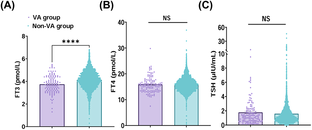 |
Figure 2 Comparison of thyroid-related hormone levels between VA and Non-VA groups. (A) Violin plots reflect the distribution and probability density of FT3 between the two groups. Purple represents the VA group and cyan represents the Non-VA group. (B) Reflects the distribution and probability density of FT4 between the two groups. (C) Reflects the distribution and probability density of TSH between the two groups. The center box of each violin plot represents the interquartile range (IQR), and the horizontal line inside the box represents the median. ****P < 0.0001; NS indicates that there is no significant difference between the VA and Non-VA groups (P > 0.05). |
 |
Figure 3 Univariate and multivariate logistic regression analyses of population-wide risk of VA development. The horizontal coordinates represent Odds Ratios (OR), with <1 indicating a decrease in odds and >1 indicating an increase in odds. |
Subsequently, we categorized the patients into four groups based on FT3 quartiles. As shown in Table 3, FT3 levels were significantly associated with the risk of ventricular arrhythmias (VA) (comparison of Q4 and Q1, unadjusted OR=0.237, 95% CI: 0.131–0.429, Ptrend <0.001; comparison of Q4 and Q1, in Model 3 OR = 0.432, 95% CI: 0.224–0.832, Ptrend =0.007). After multivariate adjustment, a decrease of 1 standard deviation in FT3 was associated with a 19.9% increased risk of VA (OR = 0.801, 95% CI: 0.651–0.984, P = 0.035) (Table 3). This result was consistent with the findings from the restricted cubic spline regression analysis (Figure 4A). Furthermore, in subgroup analyses by gender and age, we observed a progressive decrease in the OR with increasing FT3 levels. This trend appeared more pronounced in elderly and female subgroups, although these analyses were limited by small event numbers and should be interpreted with caution (Figure 4B–E). In the correlation analysis, FT3 was significantly positively correlated with body mass index (BMI) (r = 0.16, P < 0.001), DBP (r = 0.12, P < 0.001), Hb (r = 0.36, P < 0.001), and FT4 (r = 0.24, P < 0.001). It was significantly negatively correlated with age (r = −0.34, P < 0.001), heart rate (r = −0.093, P < 0.001), neutrophil percentage (r = −0.20, P < 0.001), and creatinine (r = −0.19, P < 0.001) (Figure 5A and B).
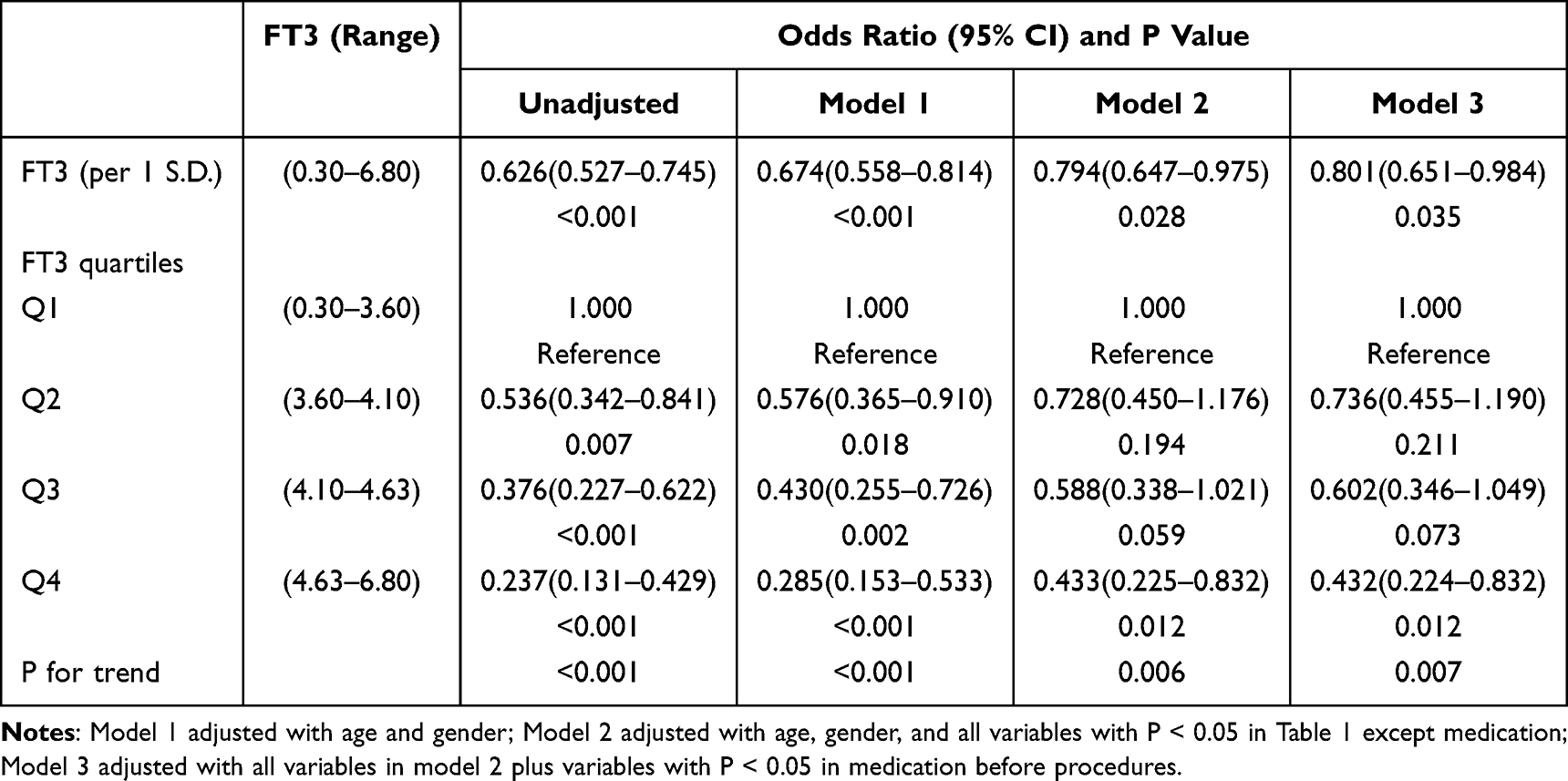 |
Table 3 FT3 Levels and Risk of VA in the Whole Population |
 |
Figure 4 Relationship between FT3 levels and risk of VA in restricted cubic spline models in different groups. Odd ratios are indicated by the red line, and 95% CI is indicated by the shaded area derived from the three-node restricted cubic spline regression. The horizontal reference line shown as a dashed line indicates an odd ratio of 1.0, and the vertical reference line is the corresponding FT3 at this point in time. (A) Relationship between FT3 level and risk of VA incidence in the entire study cohort. (B) Relationship between FT3 level and the risk of VA in the male cohort. (C) Relationship between FT3 level and the risk of VA in the female cohort. (D) The relationship between FT3 levels and the risk of VA in the cohort under the age of 60. (E) The relationship between FT3 levels and the risk of VA in the cohort aged 60 and above. |
 |
Figure 5 Correlations between different variables in patients with AMI. The diagonal lines indicate the distribution of each variable; the bottom of the diagonal line shows a binary scatterplot with a fitted present; the top of the diagonal line shows the correlation coefficients and significance levels, *** P < 0.001, ** P < 0.01. (A) Correlation analysis between clinical variables and FT3. (B) Correlation analysis between laboratory variables and FT3. Abbreviations: BMI, body mass index; SBP, systolic blood pressure; DBP, diastolic blood pressure; HR, heart rate; FT3, free triiodothyronine; FT4, free thyroxine; Neu, Neutrophil percentage; Hgb, hemoglobin; Cr, serum creatinine; UA, Uric acid. |
Discussion
Our study confirmed a VA incidence rate of 3.9% in AMI patients. In the Global Utilization of Streptokinase and t-PA for Occluded Coronary Arteries I (GUSTO-I) trial, the occurrence rate of ventricular tachycardia/ventricular fibrillation (VT/VF) in ST-elevation myocardial infarction (STEMI) patients was 10.2%, which was associated with early reperfusion therapy.24 The STEMI management guidelines in 2017 also indicate that ventricular fibrillation incidence ranges between 6–10% in STEMI patients, while atrial fibrillation prevalence may vary between 10–21%.25 The inconsistent phenomenon observed in our study regarding the incidence of VA can be attributed to the following factors: (1) The severity of the underlying conditions of the patients varies. Age and gender differences have an impact on the risk of VA occurrence; (2) The potential antiarrhythmic effects of medications such as ACEI/ARBs and dapagliflozin used in patient management.
This investigation revealed a significant association between low free triiodothyronine (FT3) levels and VA risk after AMI. While the thyroid primarily secretes thyroxine (T4), most circulating T3 derives from peripheral T4 conversion via deiodinases. Elevated FT4 levels may promote cardiovascular disease (CVD) development, which subsequently reduces deiodinase activity, establishing a positive correlation between FT4 and CVD but an inverse relationship with FT3. Notably, FT3 has been identified as an independent predictor of all-cause and cardiovascular mortality in diabetic populations.26,27
Emerging evidence suggests that hypothyroidism increases cardiovascular risk, particularly arrhythmia susceptibility.28–30 Ion channel dysfunction represents a critical mechanism underlying arrhythmogenesis.31,32 Clinical case reports have documented hypothyroidism-induced Brugada-like electrocardiographic abnormalities.33 Furthermore, population studies by Gussekloo et al demonstrated positive correlations between FT4 concentrations and all-cause/cardiovascular mortality.34 In heart failure patients, Kannan et al identified FT4 levels as predictive of composite endpoints including mortality and cardiac device implantation. Notably, early-stage hyperthyroidism and altered thyroid set-point regulation may also contribute to ventricular arrhythmia risk.35 While thyroid hormone replacement therapy has shown efficacy in reducing ischemic heart disease in hypothyroid patients,36 existing research predominantly focuses on global thyroid status or TSH levels, with limited exploration of FT3’s specific role in VA.
Our analysis further demonstrated that AMI patients in the lowest FT3 quartile exhibited significantly elevated VA risk. Each unit decrease in FT3 level was associated with a 19.9% increase in VA risk, with a numerically stronger association observed in elderly and female patients, although the number of events in these subgroups was limited, precluding definitive conclusions. However, this observation is only an assumption and needs to be verified in a larger cohort in the future. The pathophysiological basis for this association involves multiple mechanisms.
Our research also found that the neutrophil percentage was strongly correlated with the level of FT3. The inflammatory response plays a key role in the process of AMI. The massive release of inflammatory factors may interfere with the metabolic process of thyroid hormones. Previous studies have shown that long-term administration of interleukin-6 can significantly inhibit plasma FT3.37,38 Concurrently, the inflammatory state also affects the electrophysiological properties of cardiomyocytes, thereby increasing the risk of ventricular arrhythmias.39,40
FT3 directly modulates cardiac ion channel function through genomic and non-genomic pathways. Experimental evidence indicates FT3-mediated regulation of sarcoplasmic reticulum Ca2⁺-ATPase and phospholamban expression,41 along with rapid modulation of sodium, potassium, and calcium channel kinetics.42–44 Hypothyroxinemia reduces potassium channel expression, prolonging action potential duration and effective refractory period while enhancing repolarization heterogeneity. This is a key mechanism in recurrent arrhythmias.45–47 Notably, Barbara et al demonstrated gender-specific impairment of myocardial electrical coupling protein connexin-43 (Cx43) expression in hypothyroidism, particularly exacerbating asynchronous electrical activity and the susceptibility to torsade de pointes ventricular tachycardia in female models.48
FT3 plays a key regulatory role in mitochondrial bioenergetic metabolism by optimizing the tricarboxylic acid cycle and enhancing oxidative phosphorylation.41,49,50 Under ischemic conditions, low FT3 levels impair ATP production, compromising Na⁺/K⁺-ATPase function and calcium homeostasis. This metabolic crisis potentiates after depolarizations and calcium overload, creating a pro-arrhythmic milieu. The precise interplay between thyroid hormone status and post-ischemic ionic remodeling warrants further investigation.
These findings suggest that plasma FT3 may serve as a useful prognostic biomarker for identifying AMI patients at higher risk of in-hospital ventricular arrhythmias. Patients with low FT3 levels represent a higher-risk subgroup who may benefit from closer electrocardiographic monitoring during the acute phase. Further prospective studies are needed to confirm these findings and to explore whether FT3-guided risk stratification can improve clinical outcomes in patients with AMI. This study has several significant advantages, thereby enhancing the reliability and general applicability of the research results. First, this study adopted a large-scale, multi-center, and prospective design, involving three clinical centers and 3277 consecutive patients with acute myocardial infarction. Compared with previous single-center studies, it significantly reduced selection bias and improved external validity. Second, we applied advanced statistical methods, including restricted cubic spline curves to describe the dose-response relationship, multiple imputation for missing data, and pearson analysis. All of these enhanced the reliability of the research results.
Limitations
Despite these strengths, several limitations merit consideration. First of all, although this was a multicenter study, all hospitals were within a single province in China, which may limit generalizability. Future multicenter studies in different provinces with larger sample sizes are required to validate these results. Secondly, our study focused solely on in-hospital outcomes, leaving the long-term prognostic implications of FT3 levels following hospital discharge unexplored. This represents an important direction for subsequent research endeavors. Thirdly, although we have adjusted for key clinical and laboratory confounding factors in the multivariate analysis, unmeasured variables such as unmeasured inflammatory markers or unknown drugs that affect thyroid metabolism may still have residual confounding factors. Future studies should include continuous measurements of these inflammatory and drug factors to minimize such biases.
Conclusions
This study confirmed a significant association between plasma FT3 levels and the risk of ventricular arrhythmias during hospitalization in patients with myocardial infarction. Low FT3 levels are associated with an increased risk of VA in patients with AMI.
Abbreviations
AMI, acute myocardial infarction; BMI, body mass index; DBP, diastolic blood pressure; FT3, free triiodothyronine; FT4, free thyroxine; NSTEMI, Non-ST-elevation myocardial infarction; STEMI, ST-elevation myocardial infarction; SBP, systolic blood pressure; VA, ventricular arrhythmias; WBC, white blood cell.
Data Sharing Statement
The datasets used and analyzed during the current study are available from the corresponding authors upon reasonable request 12 months after publication, and will remain accessible for 5 years. Individual deidentified participant data will not be shared to ensure participant confidentiality.
Funding
This study was supported by grants from National Natural Science Foundation of China (Grant No. 82270328), Natural Science Foundation of Jiangsu Province (BK20221229), Key Project of Jiangsu Provincial Health Commission (K2024014), Construction Project of High Level Hospital of Jiangsu Province (GSPJS202404), Changzhou High Level Medical Talents Training Project (2022CZBJ054), Young Talents Program of Wuxi Medical Center to Nanjing Medical University (WMCQ202403), General Projects of Wuxi Medical Center to Nanjing Medical University (WMCG202408), Clinical Research Project of Wuxi Medical Center to Nanjing Medical University (WMCC202505) and Changzhou Talent Program (CQ20240067).
Disclosure
The authors declare no competing interests in this work.
References
1. Roth GA, Mensah GA, Johnson CO. et al. Global Burden of Cardiovascular Diseases and Risk Factors, 1990–2019: update From the GBD 2019 Study. J Am Coll Cardiol. 2020;76(25):2982–13. doi:10.1016/j.jacc.2020.11.010
2. Timmis A, Aboyans V, Vardas P, et al. European Society of Cardiology: the 2023 Atlas of Cardiovascular Disease Statistics. Eur Heart J. 2024;45(38):4019–4062. doi:10.1093/eurheartj/ehae466
3. Liu S, Li Y, Zeng X, et al. Burden of Cardiovascular Diseases in China, 1990–2016: findings From the 2016 Global Burden of Disease Study. JAMA Cardiol. 2019;4(4):342–352. doi:10.1001/jamacardio.2019.0295
4. Anderson JL, Morrow DA. Acute Myocardial Infarction. N Engl J Med. 2017;376(21):2053–2064. doi:10.1056/NEJMra1606915
5. Zipes DP, Wellens HJ. Sudden cardiac death. Circulation. 1998;98(21):2334–2351. doi:10.1161/01.cir.98.21.2334
6. Gheeraert PJ, De Buyzere ML, Taeymans YM, et al. Risk factors for primary ventricular fibrillation during acute myocardial infarction: a systematic review and meta-analysis. Eur Heart J. 2006;27(21):2499–2510. doi:10.1093/eurheartj/ehl218
7. Kanani J, Modi K. A Rare Case of Sudden Death due to Rupture of Saccular Descending Thoracic Aortic Aneurysm with Dissection. Heart Views. 2024;25(4):270–274. doi:10.4103/heartviews.heartviews_100_24
8. Kanani J, Sheikh MI. Ruptured dissecting aorta: an uncommon cause of sudden death—An autopsy study. Cirugía Cardiovascular. 2025. doi:10.1016/j.circv.2024.11.009
9. Martin SS, Aday AW, Allen NB, et al. Heart Disease and Stroke Statistics: a Report of US and Global Data From the American Heart Association. Circulation. 2025;151(8):e41–e660. doi:10.1161/cir.0000000000001303
10. Crea F. Focus on the European Society of Cardiology: guidelines on hypertension, Atlas of cardiovascular disease statistics, and Consensus statement on obesity. Eur Heart J. 2024;45(38):3897–3900. doi:10.1093/eurheartj/ehae664
11. Marijon E, Narayanan K, Smith K, et al. The Lancet Commission to reduce the global burden of sudden cardiac death: a call for multidisciplinary action. Lancet. 2023;402(10405):883–936. doi:10.1016/s0140-6736(23)00875-9
12. Zhang M, Tong Z, Wang N, et al. Novel Protein-Based Biomarkers of Out-of-hospital Sudden Cardiac Death After Myocardial Infarction. Circ Arrhythm Electrophysiol. 2025;18(4):e013217. doi:10.1161/circep.124.013217
13. Fox KA, Fitzgerald G, Puymirat E, et al. Should patients with acute coronary disease be stratified for management according to their risk? Derivation, external validation and outcomes using the updated GRACE risk score. BMJ Open. 2014;4(2):e004425. doi:10.1136/bmjopen-2013-004425
14. Öksüz SB T, Şahin M. Thyroid and cardiovascular diseases. Turk J Med Sci. 2024;54(7):1420–1427. doi:10.55730/1300-0144.5927
15. Olanrewaju OA, Asghar R, Makwana S, et al. Thyroid and Its Ripple Effect: impact on Cardiac Structure, Function, and Outcomes. Cureus. 2024;16(1):e51574. doi:10.7759/cureus.51574
16. Spartalis M, Spartalis E, Paschou SA, Iliopoulos DC, Siasos G. The Thyroid-cardiac Axis: thyroid Function, Cardiac Rhythmology, and Sudden Cardiac Death. Endocr Metab Immune Disord Drug Targets. 2024;24(10):1127–1130. doi:10.2174/0118715303243951231024112208
17. Casis O, Echeazarra L, Sáenz-Díez B, Gallego M. Deciphering the roles of triiodothyronine (T3) and thyroid-stimulating hormone (TSH) on cardiac electrical remodeling in clinical and experimental hypothyroidism. J Physiol Biochem. 2024;80(1):1–9. doi:10.1007/s13105-023-01000-z
18. Alonso H, Fernández-Ruocco J, Gallego M, et al. Thyroid stimulating hormone directly modulates cardiac electrical activity. J Mol Cell Cardiol. 2015;89(Pt B):280–286. doi:10.1016/j.yjmcc.2015.10.019
19. Passino C, Pingitore A, Landi P, et al. Prognostic value of combined measurement of brain natriuretic peptide and triiodothyronine in heart failure. J Card Fail. 2009;15(1):35–40. doi:10.1016/j.cardfail.2008.08.008
20. Cerillo AG, Storti S, Kallushi E, et al. The low triiodothyronine syndrome: a strong predictor of low cardiac output and death in patients undergoing coronary artery bypass grafting. Ann Thorac Surg. 2014;97(6):2089–2095. doi:10.1016/j.athoracsur.2014.01.049
21. Zhao Y, Wang W, Zhang K, Tang YD. Association Between Low T3 Syndrome and Poor Prognosis in Adult Patients With Acute Myocarditis. Front Endocrinol. 2021;12:571765. doi:10.3389/fendo.2021.571765
22. Alpert JS, Thygesen K, Antman E, Bassand JP. Myocardial infarction redefined--a consensus document of The Joint European Society of Cardiology/American College of Cardiology Committee for the redefinition of myocardial infarction. J Am Coll Cardiol. 2000;36(3):959–969. doi:10.1016/s0735-1097(00)00804-4
23. Al-Khatib SM, Stevenson WG, Ackerman MJ, et al. AHA/ACC/HRS Guideline for Management of Patients With Ventricular Arrhythmias and the Prevention of Sudden Cardiac Death: a Report of the American College of Cardiology/American Heart Association Task Force on Clinical Practice Guidelines and the Heart Rhythm Society. J Am Coll Cardiol. 2018;72(14):e91–e220. doi:10.1016/j.jacc.2017.10.054
24. Newby KH, Thompson T, Stebbins A, et al. Sustained ventricular arrhythmias in patients receiving thrombolytic therapy: incidence and outcomes. The GUSTO Investigators. Circulation. 1998;98(23):2567–2573. doi:10.1161/01.cir.98.23.2567
25. Ibánez B, James S, Agewall S, et al. ESC Guidelines for the management of acute myocardial infarction in patients presenting with ST-segment elevation. Rev Esp Cardiol. 2017;70(12):1082. doi:10.1016/j.rec.2017.11.010
26. Zhu P, Lao G, Chen C, et al. TSH levels within the normal range and risk of cardiovascular and all-cause mortality among individuals with diabetes. Cardiovasc Diabetol. 2022;21(1):254. doi:10.1186/s12933-022-01698-z
27. de Vries TI, de Valk HW, van der Graaf Y, et al. Normal-range thyroid-stimulating hormone levels and cardiovascular events and mortality in type 2 diabetes. Diabet Res Clin Pract. 2019;157:107880. doi:10.1016/j.diabres.2019.107880
28. Fazio S, Palmieri EA, Lombardi G, Biondi B. Effects of thyroid hormone on the cardiovascular system. Recent Prog Horm Res. 2004;59:31–50. doi:10.1210/rp.59.1.31
29. Brenner R, Bilz S, Busch S, et al. Arrhythmias in thyroid dysfunction. Herzschrittmacherther Elektrophysiol. 2024;35(3):183–192. doi:10.1007/s00399-024-01030-0
30. Stamatouli A, Bedoya P, Yavuz S. Hypothyroidism: cardiovascular Endpoints of Thyroid Hormone Replacement. Front Endocrinol. 2019;10:888. doi:10.3389/fendo.2019.00888
31. Keating MT, Sanguinetti MC. Molecular and cellular mechanisms of cardiac arrhythmias. Cell. 2001;104(4):569–580. doi:10.1016/s0092-8674(01)00243-4
32. Splawski I, Shen J, Timothy KW, et al. Spectrum of mutations in long-QT syndrome genes. KVLQT1, HERG, SCN5A, KCNE1, and KCNE2. Circulation. 2000;102(10):1178–1185. doi:10.1161/01.cir.102.10.1178
33. Bioletto F, Cuboni D, Varaldo E, et al. Severe hypothyroidism as a trigger for Brugada-type ECG abnormalities: a case report and literature review. Arch Endocrinol Metab. 2024;68:e230027. doi:10.20945/2359-4292-2023-0027
34. Gussekloo J, van Exel E, de Craen AJ, et al. Thyroid status, disability and cognitive function, and survival in old age. JAMA. 2004;292(21):2591–2599. doi:10.1001/jama.292.21.2591
35. Müller P, Dietrich JW, Lin T, et al. Usefulness of Serum Free Thyroxine Concentration to Predict Ventricular Arrhythmia Risk in Euthyroid Patients With Structural Heart Disease. Am J Cardiol. 2020;125(8):1162–1169. doi:10.1016/j.amjcard.2020.01.019
36. Keating Jr FR, Parkin TW, Selby JB, Dickinson LS. Treatment of heart disease associated with myxedema. Prog Cardiovasc Dis. 1961;3:364–381. doi:10.1016/s0033-0620(61)90004-4
37. Deng MC, Erren M, Lütgen A, et al. Interleukin-6 correlates with hemodynamic impairment during dobutamine administration in chronic heart failure. Int J Cardiol. 1996;57(2):129–134. doi:10.1016/s0167-5273(96)02805-7
38. Zoccali C, Tripepi G, Cutrupi S, Pizzini P, Mallamaci F. Low triiodothyronine: a new facet of inflammation in end-stage renal disease. J Am Soc Nephrol. 2005;16(9):2789–2795. doi:10.1681/asn.2005040356
39. Cooper Jr LT. Myocarditis. N Engl J Med. 2009;360(15):1526–1538. doi:10.1056/NEJMra0800028
40. Hulsmans M, Sam F, Nahrendorf M. Monocyte and macrophage contributions to cardiac remodeling. J Mol Cell Cardiol. 2016;93:149–155. doi:10.1016/j.yjmcc.2015.11.015
41. Klein I, Ojamaa K. Thyroid hormone and the cardiovascular system. N Engl J Med. 2001;344(7):501–509. doi:10.1056/nejm200102153440707
42. Klein I. Thyroid hormone and the cardiovascular system. Am J Med. 1990;88(6):631–637. doi:10.1016/0002-9343(90)90531-h
43. Dillmann WH. Biochemical basis of thyroid hormone action in the heart. Am J Med. 1990;88(6):626–630. doi:10.1016/0002-9343(90)90530-q
44. Polikar R, Burger AG, Scherrer U, Nicod P. The thyroid and the heart. Circulation. 1993;87(5):1435–1441. doi:10.1161/01.cir.87.5.1435
45. Klein I, Danzi S. Thyroid disease and the heart. Circulation. 2007;116(15):1725–1735. doi:10.1161/circulationaha.106.678326
46. Antzelevitch C, Burashnikov A. Overview of Basic Mechanisms of Cardiac Arrhythmia. Card Electrophysiol Clin. 2011;3(1):23–45. doi:10.1016/j.ccep.2010.10.012
47. Tse G. Mechanisms of cardiac arrhythmias. J Arrhythm. 2016;32(2):75–81. doi:10.1016/j.joa.2015.11.003
48. Bačová BS, Vinczenzová C, Žurmanová J, et al. Altered thyroid status affects myocardial expression of connexin-43 and susceptibility of rat heart to malignant arrhythmias that can be partially normalized by red palm oil intake. Histochem Cell Biol. 2017;147(1):63–73. doi:10.1007/s00418-016-1488-6
49. Leitch SP, Brown HF. Effect of raised extracellular calcium on characteristics of the Guinea-pig ventricular action potential. J Mol Cell Cardiol. 1996;28(3):541–551. doi:10.1006/jmcc.1996.0050
50. Dillmann WH. Cellular action of thyroid hormone on the heart. Thyroid. 2002;12(6):447–452. doi:10.1089/105072502760143809
 © 2026 The Author(s). This work is published and licensed by Dove Medical Press Limited. The
full terms of this license are available at https://www.dovepress.com/terms
and incorporate the Creative Commons Attribution
- Non Commercial (unported, 4.0) License.
By accessing the work you hereby accept the Terms. Non-commercial uses of the work are permitted
without any further permission from Dove Medical Press Limited, provided the work is properly
attributed. For permission for commercial use of this work, please see paragraphs 4.2 and 5 of our Terms.
© 2026 The Author(s). This work is published and licensed by Dove Medical Press Limited. The
full terms of this license are available at https://www.dovepress.com/terms
and incorporate the Creative Commons Attribution
- Non Commercial (unported, 4.0) License.
By accessing the work you hereby accept the Terms. Non-commercial uses of the work are permitted
without any further permission from Dove Medical Press Limited, provided the work is properly
attributed. For permission for commercial use of this work, please see paragraphs 4.2 and 5 of our Terms.
Recommended articles
Identification of Immuno-Inflammation-Related Biomarkers for Acute Myocardial Infarction Based on Bioinformatics
You H, Dong M
Journal of Inflammation Research 2023, 16:3283-3302
Published Date: 7 August 2023
Identification of Oxidative Stress-Related Biomarkers in Acute Myocardial Infarction

Sun Y, Wang M, Tan X, Zhang H, Yang S
International Journal of General Medicine 2023, 16:4805-4818
Published Date: 26 October 2023