
Back to Journals » Breast Cancer: Targets and Therapy » Volume 18

Expression Profiling of SWI/SNF Complex Subunits in Triple-Negative Breast Cancer Using Tissue Microarray and Immunohistochemistry
Authors Wang S, Gao C, Wang Q, Xu J 
Received 9 March 2026
Accepted for publication 25 June 2026
Published 18 July 2026 Volume 2026:18 606605
DOI https://doi.org/10.2147/BCTT.S606605
Checked for plagiarism Yes
Review by Single anonymous peer review
Peer reviewer comments 3
Editor who approved publication: Professor Harikrishna Nakshatri
Shang Wang,1 Chen Gao,2 Qi Wang,2 Jing Xu2
1Department of Pathology, School of Basic Medicine, Qingdao University, Qingdao, People’s Republic of China; 2Department of Pathology, Qingdao Central Hospital, University of Health and Rehabilitation Sciences (Qingdao Central Medical Group), Qingdao, People’s Republic of China
Correspondence: Jing Xu, Department of Pathology, Qingdao Central Hospital, University of Health and Rehabilitation Sciences (Qingdao Central Medical Group), Num. 127, Siliunan Road, Qingdao, Shandong, 266042, People’s Republic of China, Tel +86-0532-84851283, Email [email protected]
Introduction: The SWI/SNF (SWItch/Sucrose Non - Fermentable) complex is a multi - subunit, ATPase - dependent chromatin - remodeling complex involved in regulating key cellular processes. Tumors with SWI/SNF loss tend to be poorly differentiated and aggressive. Triple - negative breast cancer (TNBC) typically has a high histological grade and a poor prognosis. Given the limited reports on the SWI/SNF complex in breast cancer, we focused on its role in TNBC.
Methods: Using tissue microarray and immunohistochemistry (IHC), we evaluated the expression of four important subunits of the SWI/SNF complex (SMARCB1, SMARCA2, ARID1A, and SMARCA4) in 104 TNBC tissues, which included 96 cases of conventional TNBC and 8 cases of low - grade TNBC. Additionally, we evaluated the endogenous expression of the four subunits in five breast cancer cell lines by real - time quantitative PCR (RT - qPCR). These cell lines consisted of two TNBC cell lines (MDA - MB - 231, MDA - MB - 468) and three non - TNBC cell lines (MCF - 7, T47D, and MDA - MB - 453).
Results: The IHC results indicated that the deletion rate of SMARCA2 was the highest in TNBC compared with other subunits (91/104, 87.5%) (p < 0.0001), and SMARCA4 had the highest retention rate (98/104, 94.2%). Whether low - grade TNBC was excluded or not, there was no significant difference between the deletion rate and various clinicopathological parameters. Consistently, we found that SMARCA2 exhibited the lowest expression level across all five cell lines (p < 0.01). SMARCA4 showed the highest expression in all cell lines except MDA - MB - 453, in which ARID1A expression was the highest.
Conclusion: These results indicate that TNBC is characterized by a high proportion of SMARCA2 deletion and positive expression of SMARCA4, which are significant molecular features. The roles and potential mechanisms of these two factors in TNBC require further investigation.
Keywords: triple-negative breast cancer, SWI-SNF complex, SMARCA2, SMARCA4, chromatin remodelling, immunohistochemistry
Introduction
Conventional triple - negative breast cancer (TNBC) is a highly aggressive breast cancer subtype, characterized by poor differentiation and a poor prognosis. TNBC accounts for approximately 15% to 20% of all breast carcinomas and more frequently affects pre - menopausal women under 40 years of age. More than 50% of patients experience a relapse within the first 3 to 5 years after diagnosis.1–3 There is an increasing need to explore novel therapeutic targets and develop more effective treatments to improve the prognosis of TNBC patients.
A retrospective multicentric study found that the deletion of components of the SWI/SNF (SWItch/Sucrose Non - Fermentable) complex was one of the significant molecular alterations in treatment - refractory or treatment - resistant invasive breast carcinoma.4 Chromatin remodeling is one of the key mechanisms for the dynamic regulation of gene expression. It is facilitated by various protein/protein complexes, including the multi - subunit SWI/SNF complex. This complex is a crucial regulator of nucleosome positioning. It utilizes the energy from ATP hydrolysis to reposition nucleosomes, thus regulating key cellular processes such as transcription and DNA repair.5–7 The SWI/SNF complex subunits can be classified into three functionally distinct categories: an ATPase - dependent catalytic subunit (SMARCA2/BRM or SMARCA4/BRG1), a targeting subunit thought to confer functional specificity to the complex (ARID1A, ARID1B, or PBRM1), and the remaining core and variant subunit.8 These proteins are essential for binding either to DNA or to other proteins.
Among the numerous subunits of the SWI/SNF complex, mutations occurred most commonly in SMARCA4, as well as in subunits believed to confer functional specificity (ARID1A, ARID1B, and PBRM1).8 Concurrent loss of SMARCA2 and SMARCA4 expression results in synthetic lethality regarding cancer cell survival and proliferation.9 Compared to the primary tumor, ARID1A is one of the few genes that are more frequently mutated in metastatic breast cancer.10 SMARCB1 (also known as INI1, SNF5, and BAF47) is a highly conserved core subunit of the human SWI/SNF complex and is essential for the complex’s ATP - dependent chromatin - remodeling activity. Homozygous inactivation of SMARCB1 is commonly observed in nearly all rhabdomyosarcoma tumors, yet its expression status in TNBC remains uncharacterized.11 Given the above findings, we selected four important subunits of the SWI/SNF complex: two catalytic subunits, SMARCA2 and SMARCA4; one targeting subunit, ARID1A; and one core subunit, SMARCB1, as the research subjects. We aimed to detect their protein expression in TNBC tissues and their endogenous expression in five breast cancer cell lines. Our hypotheses were as follows: (1) The SWI/SNF complex subunits are differentially expressed in TNBC; (2) The expression of the SWI/SNF complex subunits correlates with clinicopathological characteristics, such as tumor grade, lymph node involvement, Ki - 67 proliferation index, and other indicators.
Materials and Methods
Tissue Specimens
A total of 104 paraffin - embedded TNBC tissue specimens were selected from Qingdao Central Hospital, University of Health and Rehabilitation Sciences, between 2017 and 2024. Inclusion criteria: Patients had no prior history of radiotherapy or chemotherapy. Cases had available results of IHC and/or dual - color silver in situ hybridization or fluorescence in situ hybridization. Tumor diameter was ≥ 1.5 cm. Exclusion criteria: Biopsy specimens obtained via needle aspiration. Cases with incomplete clinical data.
According to the classification in Felipe C Geyer’s research, conventional TNBC shows high - grade morphology and aggressive biological behavior. Low - grade TNBC, which can be broadly classified as salivary - gland - like tumors of the breast and includes histological entities based on specific genetic alterations regardless of the anatomical site of origin, has an indolent behavior.12
As shown in Figure 1A, there were 96 cases of conventional TNBC. These included 80 cases of high - grade invasive carcinoma, no special type (NST) (including 1 case of microglandular adenosis - related carcinoma (MGAC), and the associated carcinoma was NST), 1 case of invasive pleomorphic lobular carcinoma (P - ILC), 1 case of invasive papillary carcinoma (IPC) (with micropapillary carcinoma components), 8 cases of metaplastic carcinoma (MPC) (including 2 cases of high - grade spindle cell metaplastic carcinoma (MSCC), 5 cases of matrix - producing carcinoma, and 1 case with heterologous mesenchymal differentiation), 5 cases of high - grade carcinoma with apocrine differentiation (C - AD), and 1 case of lymphoepithelioma - like carcinoma (LELC). Correspondingly, 8 cases of low - grade TNBC were collected, including 4 cases of low - grade C - AD, 1 case of adenoid cystic carcinoma (AdCC), 2 cases of malignant adenomyoepithelioma (M - AME) (the malignant components were myoepithelial carcinoma), and 1 case of mucinous cystadenocarcinoma (MCAC). The clinicopathological data were also collected. It should be noted that the sample size of low - grade TNBC was small, and this imbalance might introduce statistical bias.
 |
Figure 1 (A) It presented the composition of 104 cases of triple-negative breast cancer, including 96 cases of traditional triple-negative breast cancer and 8 cases of low-grade triple-negative breast cancer. It also showed the histological types of 8 cases of metaplastic carcinomas. (B) Deletion expression of SWI/SNF complex in 104 cases of TNBC with different histological types. The subunits are shown in columns and cases in rows. The deletion and positive IHC results were marked with 0 and 1 points respectively. Groups of histological subtypes are depicted in phenotype bar. (C) Endogenous expression of SWI/SNF subunits (SMARCB1, SMARCA2, ARID1A, and SMARCA4) in breast cancer cell lines. The type of error bars is SD. * indicates a significant difference compared to group SMARCA4 (*, p < 0.05), # indicates a significant difference compared to group ARID1A (#, p < 0.05). |
Tissue Microarray Construction
The tissue microarrays were constructed to be of sufficient size to ensure reliable visualization of internal controls (normal breast tissue). Representative cores, each with a 5 - mm diameter, were arranged in a standardized 3×4 grid pattern from each paraffin - embedded TNBC specimen.
Immunohistochemistry(IHC)
IHC staining was carried out on the Roche VENTANA platform. Paraffin - embedded tissues were cut into 4 - µm slides and stained with a set of IHC markers: SMARCB1 (nuclear staining), SMARCA2 (nuclear and cytoplasmic staining), ARID1A (nuclear staining), and SMARCA4 (nuclear staining). The sources and clones of the antibodies are listed in Table 1.
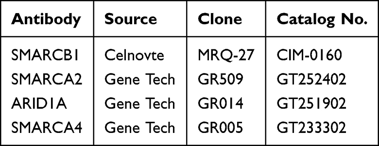 |
Table 1 Sources and Clone of the Antibodies |
After baking at 60°C for 30 minutes, the sections were automatically deparaffinized and hydrated. The VENTANA ULTRA CC1 solution was incubated at 95°C for 64 minutes for antigen retrieval. The primary antibody was incubated at 37°C for 32 minutes, followed by the secondary antibody at 37°C for 8 minutes. After DAB staining and hematoxylin counter - staining for bluing, the sections underwent dehydration, clearing, and mounting with neutral gum.
IHC expression was evaluated using the Allred scoring system. All scoring was independently conducted by two pathologists. Expression deletion was defined as a complete loss or a significant reduction in nuclear expression in tumor cells compared to strong nuclear expression in background stromal cells or non - tumor epithelial cells. Each tissue microarray was accompanied by a positive control (normal breast tissue) to guarantee the accuracy of the experimental results.
Cell Culture
The five human breast cancer cell lines—MDA - MB - 231, MDA - MB - 468, MCF - 7, T47D, and MDA - MB - 453—were all obtained from the Shanghai Institutes for Biological Sciences. During the experiments, all cell lines were used within passages 5 to 20. The cells were cultured in high - glucose DMEM (Sparkjade, Shandong, China) supplemented with 10% fetal bovine serum or in RPMI 1640 medium (BasalMedia, Shanghai, China). All cells were kept at 37°C in an environment with 5% CO2.
Real-Time Quantitative PCR (RT-qPCR)
Total RNA was extracted using Trizol reagent (Vazyme, R0016), and the A260/A280 ratio was measured by a spectrophotometer (Merinton, SMA - 4000). A total of 1 µg of purified RNA was reverse - transcribed to cDNA with the Evo M - MLV RT Premix for qPCR (Accurate Biology, AG11706) under the following conditions: 37 °C for 15 min, 85 °C for 5 s, and 4 °C for hold. The resulting cDNA was then subjected to RT - qPCR analysis.
RT - qPCR was performed using the SYBR Green qPCR Mix (Vazyme, Q311). The RT - qPCR program steps were as follows: 95 °C for 30s, followed by 40 cycles of 95 °C for 10s and 60 °C for 30s, and finally 95 °C for 15s, 60 °C for 60s, and 95 °C for 15s. The 2−ΔΔCt method was used to calculate the relative mRNA amounts of target genes (SMARCB1, SMARCA2, ARID1A, and SMARCA4) relative to the endogenous GAPDH control. The sequences of RT - qPCR primers are listed in Table 2.
 |
Table 2 The Primer Sequences Used for RT-qPCR Analysis |
Statistical Analysis
Data were analyzed using GraphPad Prism software. The chi - square test and Fisher’s exact tests were used to analyze the expression of SWI/SNF subunits in TNBC tissues. The chi - square test with Yates’ correction and Fisher’s exact tests were applied to assess the association between the expression of SWI/SNF subunits and the clinicopathological features of TNBC. One - way analysis of variance was performed to compare the endogenous expression of SWI/SNF subunits in the five breast cancer cell lines. Statistical significance was considered at p < 0.05.
Results
Expression of SWI/SNF Subunits (SMARCB1, SMARCA2, ARID1A, and SMARCA4) in TNBC Tissues (Figure 1B and Table 3)
Among 104 TNBC cases, IHC results indicated that the deletion rates of SMARCB1, SMARCA2, ARID1A, and SMARCA4 were 7.7% (8/104), 87.5% (91/104), 9.6% (10/104), and 5.8% (6/104), respectively. Whether low - grade TNBC was excluded or not, the loss rate of SMARCA2 was the highest in TNBC compared with other SWI/SNF subunits (p < 0.0001). Furthermore, we observed nuclear - cytoplasmic co - localization of SMARCA2. On the other hand, SMARCA4 expression was positive in the majority of cases (98/104, 94.2%), showing the highest expression rate among the four subunits in TNBC.
 |
Table 3 Expression of SWI/SNF Family Members in TNBC Tissues |
Regarding the histological subtypes of the 104 cases of TNBC, the deletion rate of the four subunits in NST (Figure 2A) was similar to that in the total TNBC cases. For P - ILC (Figure 2B), only SMARCA4 was positive. In 8 cases of MPC (Figure 2C and D), SMARCA2 was deleted in all cases, and SMARCA4 was lost in 2 cases, with a much higher SMARCA4 deletion rate than other subtypes of TNBC (P < 0.05). While in 9 cases of C - AD (Figure 2E), SMARCA4 and ARID1A were both positive, accompanied by SMARCB1 deletion in 1 case and SMARCA2 deletion in 8 cases. Next, AdCC (Figure 3A), M - AME (Figure 3B), MCAC (Figure 3C), LELC (Figure 3D), and MGAC (Figure 3E) were analyzed: in 2 cases of M - AME, SMARCA2 was deleted in both, SMARCA4 was deleted in 1 case, while SMARCB1 and ARID1A were both positive. In the cases of AdCC, MCAC, and MGAC, there was only SMARCA2 deletion. In the LELC case, only SMARCA4 was positive. In the IPC case, all four subunits were positive.
 |
Figure 2 H and E and IHC results of representative cases-1. i represents H and E staining; ii–v indicate IHC staining for SMARCB1, SMARCA2, ARID1A and SMARCA4 respectively. (A) NST with grade III case: IHC results showed deletion of SMARCB1, SMARCA2, ARID1A expression and SMARCA4. (B) P-ILC case: discohesive cells arranged in a cord-like pattern. IHC results showed deletion expression of SMARCB1, SMARCA2, and ARID1A, and positive expression of SMARCA4. (C) MPC with matrix producing case: IHC results showed deletion of SMARCA2, and positive expression of SMARCB1, ARID1A, and SMARCA4. (D) High-grade MSCC case: IHC results showed deletion expression of SMARCA2, ARID1A, and SMARCA4, and with positive expression of SMARCB1. (E) High-grade C-AD case: with eosinophilic cytoplasm. IHC results showed deletion expression of SMARCA2 and SMARCB1, and with positive expression of ARID1A and SMARCA4. All panels, original magnification ×400. |
 |
Figure 3 H and E and IHC staining results of representative cases-2. i represents H and E staining; ii–v indicate IHC staining for SMARCB1, SMARCA2, ARID1A and SMARCA4 respectively. (A) AdCC case: Basaloid cell nests are observed within a hyalinized stroma, with centrally located true glandular lumina exhibiting pink discharge. IHC results showed deletion expression of SMARCA2, with positive expression of SMARCB1, ARID1A, and SMARCA4. (B) M-AME case: Tumor cell nests show biphasic differentiation into both epithelial and myoepithelial components. IHC results showed deletion expression of SMARCA2, with positive expression of SMARCB1, ARID1A, and SMARCA4. (C) MCAC case: Cystically dilated lumina are observed, containing complex glands with intra- and extracellular mucin, along with the loss of the surrounding basement membrane. IHC results showed deletion expression of SMARCA2, with positive expression of SMARCB1, ARID1A, and SMARCA4. (D) LELC case: Tumor cells exhibit vesicular nuclei, with stromal infiltration by lymphocytes. IHC results showed deletion expression of SMARCA2, SMARCB1 and ARID1A, and positive expression of SMARCA4. (E) MGAC case: At the tumor periphery, areas of microglandular adenosis are noted, with coarse eosinophilic granules in tumor cytoplasm, and the associated carcinoma is NST. IHC results showed deletion expression of SMARCA2, and positive expression of SMARCB1, ARID1A, and SMARCA4. All panels, original magnification ×400. |
Co - Deletion of SWI/SNF Subunits (SMARCB1, SMARCA2, ARID1A, and SMARCA4) Expression in TNBC
Among the 104 cases of TNBC, 91 cases showed the deletion of SMARCA2 expression. Among these, 8 cases had the simultaneous deletion of SMARCB1 expression (including 5 cases of NST, 1 case of P - ILC, 1 case of C - AD, and 1 case of LELC); 10 cases had a simultaneous deletion of ARID1A expression (including 7 cases of NST, 1 case of P - ILC, 1 case of MSCC, and 1 case of LELC); and 6 cases had a simultaneous deletion of SMARCA4 expression (including 3 cases of NST, 1 case of MPC, 1 case of MSCC, and 1 case of M - AME). Additionally, there were 3 cases with simultaneous deletion of SMARCB1, SMARCA2, and ARID1A expression (1 case of NST, 1 case of P - ILC, and 1 case of LELC); 1 case with simultaneous deletion of SMARCA2, ARID1A, and SMARCA4 expression (MSCC); and 1 case with simultaneous deletion of all four subunits expression (NST). Furthermore, there were 13 cases with positive expression of all four subunits (including 11 cases of NST, 1 case of IPC, and 1 case of C - AD). Among the 91 cases with subunit deletions, all had deletion of SMARCA2 alone or in combination with the other three subunits, and there were no cases with deletion of any of the other three subunits alone.
Correlation Between SWI/SNF Subunits Deletion (SMARCB1, SMARCA2, ARID1A, and SMARCA4) and Various Clinicopathological Parameters of TNBC
We evaluated the correlation between the deletion rates of SWI/SNF subunits (SMARCB1, SMARCA2, ARID1A, and SMARCA4) and various clinicopathological parameters of TNBC. Whether low - grade TNBC was excluded or not, the deletion rates of the four subunits showed no significant difference with various clinicopathological parameters, including age, histological grade, grade of TILs, tumor size, lymph node (LN) metastasis, and Ki - 67 percentage (Table 4).
 |
Table 4 Association Between SWI/SNF Subunits Expression and Clinicopathological Features in 104 Cases of TNBC |
Endogenous Expression of SWI/SNF Subunits (SMARCB1, SMARCA2, ARID1A, and SMARCA4) in Breast Cancer Cell Lines (Figure 1C)
We compared the endogenous expression of four subunits between TNBC cell lines (MDA - MB - 231, MDA - MB - 468) and non - TNBC cell lines (MCF - 7, T47D, and MDA - MB - 453). RT - qPCR results confirmed that, as shown in Figure 1C, compared with ARID1A and SMARCA4, both SMARCB1 and SMARCA2 exhibited low expression across all breast cancer cell lines, including the TNBC cell lines. There was no significant difference between SMARCB1 and SMARCA2 expression, but SMARCA2 showed the lowest expression level. The highest expression of SMARCA4 was observed in all cell lines except the MDA - MB - 453 cell line, which had the highest expression of ARID1A.
Discussion
The catalytic function of the SWI/SNF complex depends on one of its two mutually exclusive ATP - dependent helicases, SMARCA2 or SMARCA4, which share substantial protein sequence homology.13 Given that TNBC is more invasive and has a poorer prognosis compared to other types of breast cancer, we focused on the expression of SWI/SNF subunits in TNBC. The SMARCA2 and SMARCA4 ATPases may promote breast cancer cell proliferation by regulating cell - cycle progression, through independent mechanisms and with at least partially non - overlapping roles.14 SMARCA2 has been identified as a potent synthetic - lethal target in SMARCA4 - deficient cancers, and vice versa. SMARCA4 plays a complex role, acting as either a tumor suppressor or a tumor promoter, depending on the context. It has been regarded as both a prognostic marker and a potential therapeutic target in breast cancer. Low SMARCA4 expression is associated with a more favorable prognosis,15 while in TNBC, SMARCA4 promotes cancer progression by upregulating lipid synthesis.16
In our study, we found that the SMARCA2 expression deletion rate (87.5%) in TNBC was the highest among other SWI/SNF subunits (SMARCB1, ARID1A, and SMARCA4). Additionally, SMARCA2 showed the lowest endogenous expression level across the breast - cancer cell lines (MDA - MB - 231, MDA - MB - 468, MCF - 7, T47D, and MDA - MB - 453). This suggests that it may play a unique biological role in TNBC. As one of the ATPase subunits in the SWI/SNF complex, the loss of SMARCA2 may severely compromise chromatin - remodeling functions, resulting in widespread dysregulation of gene expression and genomic instability.14 As another ATPase subunit mutually exclusive with SMARCA2, previous studies have shown that the loss of expression of both SMARCA2 and SMARCA4 is synthetically lethal to the survival and development of cancer cells. SMARCA2 has been identified as an effective synthetic - lethal target for SMARCA4 - deficient cancers.9,17 Our results indicated that the expression deletion rate of SMARCA4 was the lowest among the four subunits, with positive expression in most TNBC samples. Moreover, our RT - qPCR results in cell lines showed significantly higher expression of SMARCA4 compared to the other subunits in TNBC cell lines, suggesting a potentially crucial function of SMARCA4 in TNBC. Nevertheless, we did not delve further into the effects of the concurrent deletion of SMARCA4 and SMARCA2 on the progression of TNBC; this will be the primary focus of our future research.
Interestingly, SMARCA4 exerts different functions under varying conditions. SMARCA4 genetic alterations (mutations or deletions) are mainly observed in small - cell lung cancer and ovarian small - cell carcinoma.18 In TNBC, SMARCA4 plays dual roles in tumor progression, acting as either a promoter or a suppressor. Studies have shown that SMARCA4 can directly bind to gene promoters, increasing DNA accessibility and thereby activating the transcription of Rho GTPase - activating protein 29 (ARHGAP29). This up - regulation of ARHGAP29 expression modulates RHOA signaling, ultimately inhibiting the growth and metastasis of breast cancer.19 In addition, SMARCA4 interacts with other proteins to form complexes and jointly regulates the proliferation of TNBC cells. When combined with SOX4, SMARCA4 directly activates the transcription of hexokinase 2 (HK2), enhancing glycolysis and promoting TNBC cell proliferation. At the same time, it modulates the expression of TGFBR2, which initiates the activation of the PI3K - AKT pathway and thus influences TNBC progression.20
Our findings suggest that SMARCA4 may play an important role in TNBC, and SMARCA2, as the mutually - exclusive ATPase subunit, is mostly deleted. However, in 8 MPC cases, SMARCA4 was deleted in 2 cases, and the deletion rate was much higher than that of other TNBC subtypes. MPC is a rare subtype of breast cancer, defined as mammary carcinoma with squamous or mesenchymal differentiation, which may include spindle - cell, chondroid, osseous, or rhabdomyoid differentiation patterns.21 MPC is characterized by a larger tumor size, less lymph - node metastasis, a higher histology grade, and a worse prognosis.22 However, the limited number of MPC cases included in this study may affect the reliability of our conclusions. In future work, we will increase the sample size of MPC and other special subtypes of breast cancer. Additionally, we will conduct targeted studies by integrating multi-omics analysis and cellular functional experiments.
One study found that SMARCB1 was homozygously inactivated in nearly all cases of rhabdoid tumors, a rare type of pediatric malignancy.11 SMARCB1 knockout mice were also predisposed to developing similar tumors,23 further demonstrating that SMARCB1 is a genuine tumor - suppressor gene. Since then, additional studies have identified mutations in other SWI/SNF subunits (ARID1A, PBRM1, SMARCA4) in various tumors, including ovarian clear - cell carcinoma, clear - cell renal cell carcinoma, and non - small - cell lung cancer.24–26
In breast cancer research, mutations in ARID1A, ARID1B, and PBRM1 have been reported,8,27,28 suggesting a tumor - suppressor role for the SWI/SNF complex. Notably, ARID1A is one of the few genes found to be more frequently mutated in metastatic breast cancer than in primary tumors. Mutations in ARID1A have also been implicated in mediating resistance to endocrine therapy in metastatic ER - positive breast cancer.29 Previous studies have confirmed that ARID1A functions as a tumor suppressor within breast cancer.30
In contrast, the loss rates of SMARCB1 and ARID1A were 7.7% and 9.6%, respectively. These lower loss rates suggest that their roles in TNBC may differ from that of SMARCA2, or that their loss may contribute less to the carcinogenicity of TNBC. Perhaps they exert more limited effects in certain TNBC subtypes or specific contexts. Our research was in line with previous studies. For instance, the deletion of SMARCB1 is very common in certain soft - tissue tumors, such as malignant rhabdoid tumors,11 but is relatively rare in breast cancer. ARID1A shows a high mutation rate in several cancer types, such as clear - cell ovarian carcinoma and endometrial cancer,24,26 yet has a relatively low deletion rate in TNBC.
In this study, we found that the deletion of any SWI/SNF subunit was always accompanied by the deletion of SMARCA2, and isolated deletion of the other three subunits was not detected. Previous studies on multiple cancer types have shown that mutations in different SWI/SNF subunits are not mutually exclusive, and the frequency of co - mutations is approximately what would be expected randomly. This suggests that these subunits have non - redundant functions, and their combined abnormalities may gradually impair the complex. Thus, we speculate that the deletion of SMARCA2 may be a key initiating event that drives the dysfunction of the SWI/SNF complex in TNBC, and the concomitant deletion of other subunits could potentially exacerbate this functional impairment.
Through RT - qPCR analysis comparing four SWI/SNF subunits between TNBC cell lines (MDA - MB - 231, MDA - MB - 468) and non - TNBC lines (MCF - 7, T47D, MDA - MB - 453), we identified the characteristic high proportion of SMARCA2 deletion and SMARCA4 positive expression. The low mRNA expression of SMARCA2 in TNBC cell lines is consistent with the high deletion rate of SMARCA2 protein observed by IHC, further strengthening the reliability of this study. This conserved expression profile suggests a possible role of SWI/SNF complexes in breast cancer pathogenesis. Notably, ARID1A is aberrantly expressed in MDA - MB - 453 cells. As a core subunit of the SWI/SNF complex, ARID1A mediates cell - specific chromatin remodeling, which may reflect the requirements for subtype - specific chromatin remodeling.
One study analyzed a series of 6026 primary and metastatic breast cancers using targeted capture sequencing. Alterations in SMARC core subunits (SMARCA4, SMARCB1, and SMARCA2) were identified in less than 1% of all breast cancers. These included 27 primary and 30 recurrent/metastatic tumors, and 12% (7/57) had histologic patterns in the form of rhabdoid, composite rhabdoid, sarcomatoid, or anaplastic features. In this study, 30 cases of IHC detection showed that SMARC proteins (SMARCB1, SMARCA2, and SMARCA4) were preserved in nearly all tumors analyzed (26/30, 87%).31 Our study found a high deletion rate of SMARCA2 in TNBC. Although our study did not assess SMARCA2 mutations, the minimal mutation rates (<1%) reported in prior genomic studies strongly suggest that non - mutational mechanisms predominantly drive SMARCA2 deletion in TNBC. These mechanisms may include post - transcriptional regulation, protein stability control, and epigenetic silencing. Epigenetic regulation plays a vital role in the progression of TNBC. In follow - up studies, we will further investigate the post - translational modification functions of SMARC proteins in TNBC.
Consistent with Ulicna et al’s report that core SWI/SNF subunits (eg, SMARCB1, ARID1A) are predominantly localized in the nucleus, our IHC results confirmed the strict nuclear localization of these subunits in TNBC.32 This conservation supports their canonical functions in chromatin remodeling. We observed nuclear - cytoplasmic co - localization of SMARCA2, in line with Ulicna et al’s biochemical fractionation data demonstrating the cytoplasmic presence of SMARCA2. This dual - localization pattern may reflect its bifunctional roles: participating in chromatin remodeling within the nucleus while potentially regulating translation (eg, via mRNA or ribosome binding) in the cytoplasm. As reported by Ulicna et al, cytoplasmic SMARCA4 predominantly functions in translational regulation. Mutations in SMARCA4 can reduce overall translation activity and render cells sensitive to inhibitors targeting specific translational pathways. In the nucleus, by contrast, SMARCA4 mainly mediates ATP - dependent chromatin remodeling, which involves transcriptional regulation, DNA damage repair, cell - cycle control, and other biological processes. In breast cancer (51% of cases showed nuclear - cytoplasmic co - expression), our TNBC cohort exhibited exclusively nuclear SMARCA4. This discrepancy may occur because SMARCA4 in TNBC may preferentially exert oncogenic functions within the nucleus, or low - abundance cytoplasmic SMARCA4 was undetectable by our IHC protocol.
The significantly high deletion rate of SMARCA2 and high expression of SMARCA4 in TNBC provide new directions for clinical diagnosis and treatment, as their expression status may serve as a potential biomarker for predicting patient prognosis. For instance, developing inhibitors against other members of the SWI/SNF complex, such as SMARCA4, or targeting cell - survival pathways that depend on SMARCA2 loss through synthetic - lethal strategies may offer new treatment options for TNBC patients. Since deletion of SMARCA4 appears to increase the radiosensitivity of tumor cells, inhibitors of this ATPase may serve as adjuvant agents for radiotherapy. Additionally, although the expression - loss rates of SMARCB1, ARID1A, and SMARCA4 are relatively low, their potential biological roles and impacts in specific patient populations still warrant further investigation.
Our study had certain limitations. First, the sample size of this study was relatively small; future research should expand the sample size to further validate the conclusions and their correlation with various clinicopathological parameters. Second, IHC has a degree of subjectivity, so future studies should incorporate molecular - biology techniques, such as gene sequencing and CRISPR/Cas9 gene - knockout experiments, to further explore the specific effects of SMARCA2 loss on the biological behavior of TNBC cells. Third, while the NST cases constituted the predominant category, the limited sample sizes of other subtypes precluded meaningful statistical analysis. Future multi - center collaborations assembling larger cohorts of rare subtypes (eg, MPC, C - AD, and M - AME) are needed to validate these preliminary observations. Furthermore, regarding the reliability of the results, this study has several major limitations: the absence of in - vitro and in - vivo functional validation, the use of a single reference gene for RT - qPCR normalization, and the lack of inter - observer agreement assessment for IHC scoring.
In summary, this study reveals the expression status of key subunits of the SWI/SNF complex in TNBC. Specifically, it shows the high deletion rate of SMARCA2 and the high retention rate of SMARCA4, suggesting a potential association with TNBC pathogenesis. This finding provides new insights into the diagnosis and treatment of TNBC.
Ethics Approval and Consent to Participate
Our study complies with the Declaration of Helsinki. The cases in this research were from Qingdao Central Hospital, University of Health and Rehabilitation Sciences. This study (KY202415701) was reviewed and approved by the Medical Ethics Committee of Qingdao Central Medical Group on October 17, 2024.
Author Contributions
All authors made a significant contribution to the work reported, whether that is in the conception, study design, execution, acquisition of data, analysis and interpretation, or in all these areas; took part in drafting, revising or critically reviewing the article; gave final approval of the version to be published; have agreed on the journal to which the article has been submitted; and agree to be accountable for all aspects of the work. Dr. Jing Xu took part in drafting and revising the article, and gave final approval of the version to be published. Shang Wang is responsible for the acquisition of data, analysis, interpretation and manuscript draft. Chen Gao took part in make tissue arrays. Qi Wang took part in performing immunohistochemistry.
Funding
This work was funded by the Qingdao Medical and Health Research Guidance Project (Grant No. 2023 - WJZD202), the National Human Genetic Resources Sharing Service Platform (Grant No. PT - 2024 - 0303), and the Qingdao Medical and Health Excellent Discipline Leader Program (Grant No. Qingwei Science and Education Character (2019) No. 6).
Disclosure
The authors report no conflicts of interest in this work.
References
1. Haffty BG, Yang Q, Reiss M, et al. Locoregional relapse and distant metastasis in conservatively managed triple negative early-stage breast cancer. J Clin Oncol. 2006;24(36):5652–13. doi:10.1200/JCO.2006.06.5664
2. Li Y, Zhang H, Merkher Y, et al. Recent advances in therapeutic strategies for triple-negative breast cancer. J Hematol Oncol. 2022;15(1):121. doi:10.1186/s13045-022-01341-0
3. Wu SY, Wang H, Shao ZM, Jiang YZ. Triple-negative breast cancer: new treatment strategies in the era of precision medicine. Sci China Life Sci. 2021;64(3):372–388. doi:10.1007/s11427-020-1714-8
4. Pose Lapausa P, Soria Comes T, Calabria I, Maestu Maiques I. Molecular characterization, via next-generation sequencing, of refractory or resistant invasive breast carcinoma. Cureus. 2021;13(11):e19528. doi:10.7759/cureus.19528
5. Wilson BG, Roberts CW. SWI/SNF nucleosome remodellers and cancer. Nat Rev Cancer. 2011;11(7):481–492. doi:10.1038/nrc3068
6. Kwon H, Imbalzano AN, Khavari PA, Kingston RE, Green MR. Nucleosome disruption and enhancement of activator binding by a human SW1/SNF complex. Nature. 1994;370(6489):477–481. doi:10.1038/370477a0
7. Kadoch C, Crabtree GR. Mammalian SWI/SNF chromatin remodeling complexes and cancer: mechanistic insights gained from human genomics. Sci Adv. 2015;1(5):e1500447. doi:10.1126/sciadv.1500447
8. Shain AH, Pollack JR. The spectrum of SWI/SNF mutations, ubiquitous in human cancers. PLoS One. 2013;8(1):e55119. doi:10.1371/journal.pone.0055119
9. Hoffman GR, Rahal R, Buxton F, et al. Functional epigenetics approach identifies BRM/SMARCA2 as a critical synthetic lethal target in BRG1-deficient cancers. Proc Natl Acad Sci U S A. 2014;111(8):3128–3133. doi:10.1073/pnas.1316793111
10. Yates LR, Knappskog S, Wedge D, et al. Genomic evolution of breast cancer metastasis and relapse. Cancer Cell. 2017;32(2):169–184e7. doi:10.1016/j.ccell.2017.07.005
11. Versteege I, Sevenet N, Lange J, et al. Truncating mutations of hSNF5/INI1 in aggressive paediatric cancer. Nature. 1998;394(6689):203–206. doi:10.1038/28212
12. Geyer FC, Berman SH, Marchio C, et al. Genetic analysis of microglandular adenosis and acinic cell carcinomas of the breast provides evidence for the existence of a low-grade triple-negative breast neoplasia family. Mod Pathol. 2017;30(1):69–84. doi:10.1038/modpathol.2016.161
13. Mashtalir N, D’Avino AR, Michel BC, et al. Modular organization and assembly of SWI/SNF family chromatin remodeling complexes. Cell. 2018;175(5):1272–1288e20. doi:10.1016/j.cell.2018.09.032
14. Li K, Wang B, Hu H. Research progress of SWI/SNF complex in breast cancer. Epigenetics Chromatin. 2024;17(1):4. doi:10.1186/s13072-024-00531-z
15. Bai J, Mei P, Zhang C, et al. BRG1 is a prognostic marker and potential therapeutic target in human breast cancer. PLoS One. 2013;8(3):e59772. doi:10.1371/journal.pone.0059772
16. Wu Q, Madany P, Dobson JR, et al. The BRG1 chromatin remodeling enzyme links cancer cell metabolism and proliferation. Oncotarget. 2016;7(25):38270–38281. doi:10.18632/oncotarget.9505
17. Cantley J, Ye X, Rousseau E, et al. Selective PROTAC-mediated degradation of SMARCA2 is efficacious in SMARCA4 mutant cancers. Nat Commun. 2022;13(1):6814. doi:10.1038/s41467-022-34562-5
18. Schoenfeld AJ, Bandlamudi C, Lavery JA, et al. The genomic landscape of SMARCA4 alterations and associations with outcomes in patients with lung cancer. Clin Cancer Res. 2020;26(21):5701–5708. doi:10.1158/1078-0432.CCR-20-1825
19. Sun Z, Li Z, Wei Y, Xu L, Hang X, Kang Y. SMARCA4 inhibits breast cancer progression and metastasis through RHOA suppression. Cancer Res. 2025;85(10):1803–1818. doi:10.1158/0008-5472.CAN-24-2801
20. Mehta GA, Angus SP, Khella CA, et al. SOX4 and SMARCA4 cooperatively regulate PI3k signaling through transcriptional activation of TGFBR2. NPJ Breast Cancer. 2021;7(1):40. doi:10.1038/s41523-021-00248-2
21. Narayan P, Kostrzewa CE, Zhang Z, et al. Metaplastic carcinoma of the breast: matched cohort analysis of recurrence and survival. Breast Cancer Res Treat. 2023;199(2):355–361. doi:10.1007/s10549-023-06923-1
22. Tray N, Taff J, Adams S. Therapeutic landscape of metaplastic breast cancer. Cancer Treat Rev. 2019;79:101888. doi:10.1016/j.ctrv.2019.08.004
23. Roberts CW, Galusha SA, McMenamin ME, Fletcher CD, Orkin SH. Haploinsufficiency of Snf5 (integrase interactor 1) predisposes to malignant rhabdoid tumors in mice. Proc Natl Acad Sci U S A. 2000;97(25):13796–13800. doi:10.1073/pnas.250492697
24. Wiegand KC, Shah SP, Al-Agha OM, et al. ARID1A mutations in endometriosis-associated ovarian carcinomas. N Engl J Med. 2010;363(16):1532–1543. doi:10.1056/NEJMoa1008433
25. Jones S, Wang TL, Shih Ie M, et al. Frequent mutations of chromatin remodeling gene ARID1A in ovarian clear cell carcinoma. Science. 2010;330(6001):228–231. doi:10.1126/science.1196333
26. Matias-Guiu X, Stewart CJR. Endometriosis-associated ovarian neoplasia. Pathology. 2018;50(2):190–204. doi:10.1016/j.pathol.2017.10.006
27. Xia W, Nagase S, Montia AG, et al. BAF180 is a critical regulator of p21 induction and a tumor suppressor mutated in breast cancer. Cancer Res. 2008;68(6):1667–1674. doi:10.1158/0008-5472.CAN-07-5276
28. Mamo A, Cavallone L, Tuzmen S, et al. An integrated genomic approach identifies ARID1A as a candidate tumor-suppressor gene in breast cancer. Oncogene. 2012;31(16):2090–2100. doi:10.1038/onc.2011.386
29. Xu G, Chhangawala S, Cocco E, et al. ARID1A determines luminal identity and therapeutic response in estrogen-receptor-positive breast cancer. Nat Genet. 2020;52(2):198–207. doi:10.1038/s41588-019-0554-0
30. Guo X, Zhang Y, Mayakonda A, et al. ARID1A and CEBPalpha cooperatively inhibit UCA1 transcription in breast cancer. Oncogene. 2018;37(45):5939–5951. doi:10.1038/s41388-018-0371-4
31. Schwartz CJ, Pareja F, da Silva EM, et al. Histologic and genomic features of breast cancers with alterations affecting the SWI/SNF (SMARC) genes. Mod Pathol. 2021;34(10):1850–1859. doi:10.1038/s41379-021-00837-3
32. Ulicna L, Kimmey SC, Weber CM, et al. The interaction of SWI/SNF with the ribosome regulates translation and confers sensitivity to translation pathway inhibitors in cancers with complex perturbations. Cancer Res. 2022;82(16):2829–2837. doi:10.1158/0008-5472.CAN-21-1360
 © 2026 The Author(s). This work is published and licensed by Dove Medical Press Limited. The
full terms of this license are available at https://www.dovepress.com/terms
and incorporate the Creative Commons Attribution
- Non Commercial (unported, 4.0) License.
By accessing the work you hereby accept the Terms. Non-commercial uses of the work are permitted
without any further permission from Dove Medical Press Limited, provided the work is properly
attributed. For permission for commercial use of this work, please see paragraphs 4.2 and 5 of our Terms.
© 2026 The Author(s). This work is published and licensed by Dove Medical Press Limited. The
full terms of this license are available at https://www.dovepress.com/terms
and incorporate the Creative Commons Attribution
- Non Commercial (unported, 4.0) License.
By accessing the work you hereby accept the Terms. Non-commercial uses of the work are permitted
without any further permission from Dove Medical Press Limited, provided the work is properly
attributed. For permission for commercial use of this work, please see paragraphs 4.2 and 5 of our Terms.
Recommended articles
SMARCA2 Deficiency While Preserving SMARCA4 in Lung Adenocarcinoma Combined with Abnormal β-Catenin Expression
Dai X, Feng X, Li J, Peng F
Cancer Management and Research 2025, 17:1961-1970
Published Date: 12 September 2025