Back to Journals » International Journal of Nanomedicine » Volume 21
Emerging Salivary Exosome Nanodiagnostics for Early Oral Squamous Cell Carcinoma: Platforms, Biomarkers, and Future Perspectives
Authors Zhao LX ![]() , Wang ET, Jia WH, Wang MH
, Wang ET, Jia WH, Wang MH ![]() , Pan Z, Dong L, Wen HQ, Xu WH
, Pan Z, Dong L, Wen HQ, Xu WH ![]() , Xu R
, Xu R ![]()
Received 28 October 2025
Accepted for publication 8 January 2026
Published 24 January 2026 Volume 2026:21 577306
DOI https://doi.org/10.2147/IJN.S577306
Checked for plagiarism Yes
Review by Single anonymous peer review
Peer reviewer comments 3
Editor who approved publication: Professor Dong Wang
Lai-Xi Zhao,1,* En-Tang Wang,2,* Wen-Hao Jia,1 Mo-Han Wang,5 Zhao Pan,4 Liang Dong,4 Hui-Qin Wen,3 Wen-Hua Xu,1 Rui Xu1
1College & Hospital of Stomatology, Anhui Medical University, Anhui Province Key Laboratory of Oral Diseases Research, Hefei, 230032, People’s Republic of China; 2Stomatology Hospital, School of Stomatology, Zhejiang University School of Medicine, Zhejiang Provincial Clinical Research Center for Oral Diseases, Key Laboratory of Oral Biomedical Research of Zhejiang Province, Cancer Center of Zhejiang University, Engineering Research Center of Oral Biomaterials and Devices of Zhejiang Province, Hangzhou, 310000, People’s Republic of China; 3Department of Blood Transfusion, The First Affiliated Hospital of Anhui Medical University, Hefei, 230022, People’s Republic of China; 4Zhejiang Cancer Hospital, Hangzhou Institute of Medicine (HIM), Chinese Academy of Sciences, Hangzhou, Zhejiang, 310022, People’s Republic of China; 5Department of Oral and Maxillofacial Surgery, Ninth People’s Hospital, College of Stomatology, Shanghai Jiao Tong University School of Medicine, Shanghai, 200011, People’s Republic of China
*These authors contributed equally to this work
Correspondence: Rui Xu, Email [email protected] Wen-Hua Xu, Email [email protected]
Abstract: Early oral squamous cell carcinoma (OSCC) is frequently missed because conventional imaging and biopsy are poorly suited to detect subtle, preclinical molecular changes. Emerging salivary exosome nanodiagnostics aims to convert an anatomically proximal, information-rich nanoscale biospecimen into quantitative and repeatable readouts for screening and longitudinal monitoring. Clinical translation, however, is constrained by four bottlenecks: (i) pre-analytical variability in saliva collection, processing, and storage; (ii) yield–purity trade-offs during vesicle enrichment; (iii) low-abundance tumor-derived signals masked by abundant background vesicles; and (iv) biomarker heterogeneity that limits robustness across cohorts. Here, we integrate the landscape of salivary exosomal biomarkers (nucleic acids, proteins, and other cargo) and synthesize nanotechnology-enabled solutions that directly address these barriers, including microfluidic and affinity-based enrichment, nucleic-acid amplification circuits (eg, RCA, HCR, CHA) for signal gain, and optical/electrochemical transduction (eg, SPR, SERS) compatible with microliter-scale samples. Beyond listing platforms, we propose a unifying framework: co-design capture and readout to preserve biological specificity, match biomarker modality to detection physics, and prioritize multi-marker panels with transparent analytical benchmarks. Finally, we outline a translational roadmap spanning standardization, reproducibility, prospective multicenter validation, manufacturability, and regulatory readiness to accelerate clinically actionable early OSCC detection.
Keywords: oral squamous cell carcinoma, salivary exosomes, exosomal biomarkers, liquid biopsy, nanodiagnostics, nanoplatform
Introduction
Oral squamous cell carcinoma (OSCC) is the most common subtype of head and neck squamous cell carcinoma (HNSCC), primarily affecting the mucosa of the tongue, buccal cavity, gingiva, palate, and floor of the mouth.1 According to recent global statistics, approximately 389,485 new cases of lip and oral cavity cancer were diagnosed, and188230related deaths occurred in 2022.2 Despite significant advances in therapeutic interventions, the survival rate and quality of life for OSCC patients remain suboptimal.3,4 This can largely be attributed to the high rates of recurrence and distant metastasis associated with the disease.5,6 Furthermore, the 5-year survival rate for patients with advanced OSCC is typically less than 20%, in stark contrast to the 80% survival rate for those diagnosed at an early stage.7 Consequently, early detection and accurate diagnosis of OSCC are essential for enabling personalized, targeted, and timely treatment strategies that can significantly improve patient outcomes.
The diagnosis of OSCC is commonly based on general clinical examination,8 radiological imaging,9 and surgical biopsy,10 owing to the tumor’s typically superficial location. However, these diagnostic methods primarily identify clinically apparent symptoms, which often leads to a diagnosis at later stages, thereby resulting in a poorer prognosis. Specifically, these techniques struggle to detect subtle biomolecular changes in early-stage tumor tissues, leading to the missed opportunity for early diagnosis and intervention.11,12
Liquid biopsy has emerged as a novel, accurate, and minimally invasive strategy for cancer detection, enabling both early diagnosis and longitudinal monitoring by analyzing tumor-associated biomolecules in body fluids. These analytes include tumor-derived proteins, circulating tumor DNA, circulating tumor cells, and extracellular vesicles (EVs), particularly exosomes.13 While blood remains the most widely utilized medium in liquid biopsy studies,14 other biofluids—such as urine, cerebrospinal fluid, and saliva—are also gaining attention for their diagnostic potential. Saliva, in particular, has been employed in the diagnosis of a range of inflammatory, metabolic, and immune-related conditions.15,16 More recently, its relevance in cancer diagnostics—especially for oral malignancies—has come under increasing focus.17 Notably, for OSCC, saliva may offer diagnostic performance comparable to, or even superior to, blood-based assays.18,19 Due to the close anatomical proximity of OSCC lesions to the oral cavity, tumor-derived components—including exfoliated cells, nucleic acids, cytokines, and EVs—can be directly secreted into saliva.20 Furthermore, saliva collection is entirely non-invasive, painless, and does not induce irritation or damage to tumor tissue, making it an ideal sampling method for repeated assessments and population-level screening.21 Compared to blood, saliva is easier to collect, more economical to store, and does not require anticoagulants or specialized preservation agents, further enhancing its feasibility in clinical and remote settings. However, the diagnostic performance of whole saliva is often hindered by the presence of enzymatic activity and heterogeneous contaminants, which can degrade nucleic acids and proteins, thereby limiting sensitivity and specificity. To circumvent these issues, growing attention has been directed toward salivary exosomes—a more stable and biochemically enriched subpopulation of EVs—as a promising source of OSCC-specific biomarkers.21
Exosomes are nanosized, membrane-bound vesicles (typically 40–160 nm in diameter) secreted by most cell types.22 They function as critical mediators of intercellular communication by transporting nucleic acids, proteins, lipids, and metabolites that reflect the physiological or pathological state of their cells of origin.23–25 Owing to their stability in biological fluids and their capacity to encapsulate disease-relevant cargo, exosomes have emerged as attractive candidates for non-invasive diagnostics and real-time disease monitoring. In recent years, saliva-derived exosomes have been extensively investigated for their utility in OSCC detection. These vesicles carry tumor-specific molecular signatures, and alterations in their concentration or content may serve as early indicators of disease onset and progression.26,27 For example, Nakamichi et al reported increased levels of exosomal marker protein Alix in the saliva of OSCC patients compared to healthy individuals.28 Winck et al employed label-free liquid chromatography-tandem mass spectrometry (LC-MS/MS) proteomics to demonstrate differential expression patterns in salivary EVs associated with immune responses in OSCC patients.29 Similarly, Langevin et al identified a diagnostic model based on the salivary exosomal miRNAs miR-10b and miR-486-5p, which were significantly altered in OSCC patients.30 Hofmann et al further profiled 119 overlapping miRNAs in plasma- and saliva-derived exosomes from HNSCC patients, some of which were significantly associated with disease-free survival.31 Collectively, these findings underscore the potential of salivary exosomes as a minimally invasive liquid biopsy platform for OSCC. Their molecular profiling can provide real-time insights into tumor biology, aid in early disease detection, and support personalized therapeutic decision-making.
In this review, we provide a comprehensive overview of the current advancements in the application of salivary exosomes for OSCC. We begin by describing the biogenesis and physicochemical properties of exosomes, followed by an evaluation of saliva collection protocols and isolation techniques tailored for diagnostic use. We then discuss recent progress in exosome detection and characterization technologies, encompassing both conventional analytical methods and emerging biosensor-based platforms. Special emphasis is placed on the molecular constituents of salivary exosomes—including nucleic acids, proteins, and lipids—and their potential utility as diagnostic and prognostic biomarkers in OSCC. Finally, we examine the clinical relevance, practical challenges, and future perspectives of salivary exosome-based liquid biopsy for early cancer detection and disease monitoring. We aim to poll and discuss the updated research evidence and perspectives on the collection, separation, and detection technologies of salivary exosomes, and to introduce several components in salivary exosomes with potential to serve as diagnostic biomarkers for OSCC. This work would serve as a timely and comprehensive resource for researchers and clinicians pursuing non-invasive, exosome-driven strategies for the early diagnosis and personalized management of OSCC.
Biogenesis and Sources of Salivary Exosomes
Exosomes Biogenesis
Salivary exosomes hold great promise as diagnostic markers for oral squamous cell carcinoma, as they contain a wealth of specific biomarkers from the source cells. This characteristic is closely related to their biological formation mechanism. The biogenesis of exosomes is a dynamic and highly regulated process that remains difficult to observe directly (Figure 1). Exosomes can be formed either through direct outward budding from the plasma membrane (PM) or via a more complex endocytic pathway involving inward budding,32 and they can also be released by intracellular plasma membrane-connected compartments (IPMC).33 In the endocytosis pathway, the PM undergoes invagination to form early endosomes. These early endosomes can either recycle back to the PM—referred to as “recycling endosomes”—or progress through maturation by continuous inward budding, giving rise to numerous intraluminal vesicles (ILVs). Endosomes containing numerous ILVs are termed multivesicular bodies (MVBs).34 While the majority of MVBs fuse with lysosomes, resulting in degradation of their contents, a subset of MVBs enriched with specific molecules—such as the tetraspanin CD63, lysosomal-associated membrane proteins Lysosome-associated membrane glycoprotein 1 (LAMP1) and Lysosome-associated membrane glycoprotein 2 (LAMP2), or the major histocompatibility complex II (MHC-II)—can instead fuse with the PM.35 This fusion event facilitates the release of ILVs into the extracellular space, at which point they are defined as exosomes.
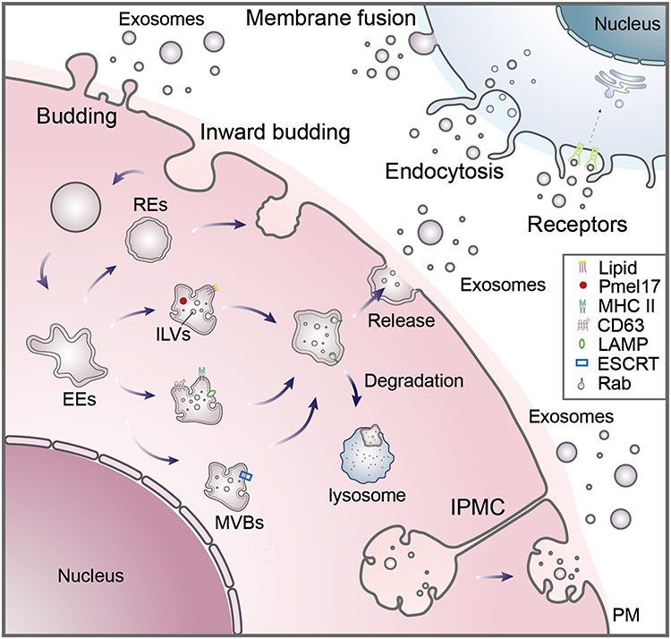 |
Figure 1 Biogenesis of Exosomes. Exosomes are small extracellular vesicles generated through multiple cellular pathways. They can originate from: (1) direct outward budding of the plasma membrane (PM); (2) inward budding of the PM, followed by maturation through the endocytic pathway; and (3) intracellular compartments (IPMCs) associated with the PM. In the canonical endocytic route, the plasma membrane invaginates to form early endosomes (EEs), which either recycle back to the PM or mature by inward budding to generate numerous intraluminal vesicles (ILVs), resulting in multivesicular bodies (MVBs). While most MVBs fuse with lysosomes for degradation, a subset—enriched in proteins such as CD63, lysosome-associated membrane glycoprotein 1/2 (LAMP1/2), and major histocompatibility complex II (MHC II)—fuses with the PM to release ILVs into the extracellular milieu as exosomes. Exosome formation is tightly regulated by the endosomal-sorting complex required for transport (ESCRT) machinery, although ESCRT-independent mechanisms also exist. For instance, CD63 can mediate protein sorting into ILVs independently of ESCRT, and the melanosome-specific protein Pre-melanosomal protein (Pmel17), along with certain lipids, can contribute to ILV biogenesis in an ESCRT-independent manner. The Rab GTPase family orchestrates the transport, docking, and fusion of MVBs with the PM. Once secreted, exosomes interact with recipient cells through surface receptor binding, direct membrane fusion, or endocytic uptake, thereby modulating downstream intracellular signaling pathways. |
The biogenesis of exosomes is orchestrated by multiple protein complexes that regulate distinct steps of ILVs formation and MVBs maturation. Central to this process is the endosomal-sorting complex required for transport (ESCRT), which is involved in sorting cargo into ILVs, membrane remodeling, vesicle budding, and scission.22,36 Additionally, the Rab family of small GTPases regulates MVBs size, intracellular trafficking, and docking at the PM to mediate exosome release.37–40 The syndecan–syntenin–ALIX complex also contributes to ILVs formation by facilitating membrane curvature and cargo recruitment.41 The ESCRT machinery consists of four core subcomplexes—ESCRT-0, -I, -II, and -III—comprising more than 30 proteins, along with several accessory factors such as ALG-2-interacting protein X (ALIX), Vacuolar Protein Sorting 4 (VPS4), and vesicle trafficking 1 (VTA1). These complexes assemble sequentially on the endosomal membrane to drive vesicle budding.42 ESCRT-0 initiates the process by recognizing and clustering ubiquitinated transmembrane proteins; ESCRT-I and -II promote membrane invagination, while ESCRT-III mediates final vesicle scission, releasing ILVs into the MVBs lumen.43 Although ESCRT-dependent pathways are primarily responsible for sorting ubiquitinated cargo into exosomes,44 protein ubiquitination is not an absolute requirement, and alternative mechanisms exist. Exosome biogenesis can also proceed via ESCRT-independent pathways involving specific lipids and tetraspanins. For instance, enzymes such as neutral sphingomyelinase and phospholipase D2 promote ceramide-dependent inward budding of the MVBs membrane.45,46 Tetraspanins, including CD63, facilitate the selective incorporation of proteins—such as melanoma-associated antigens and ceramides—into ILVs independent of the ESCRT machinery.47 Other molecules, such as heat shock cognate protein 70 (HSC70), aid in cargo recruitment (eg, transferrin receptors),48 while pre-melanosomal protein (Pmel17), a melanosome-specific protein with a specialized lumenal domain, can also drive ILVs formation in a lipid-dependent, ESCRT-independent manner.49 Importantly, ESCRT-dependent and ESCRT-independent pathways are not mutually exclusive; both mechanisms may coexist within a single cell or even within distinct MVBs subpopulations.32 Beyond protein-mediated mechanisms, exosome biogenesis is also influenced by the cell of origin, intracellular mechanical forces, growth factors, and various physicochemical conditions.39 Upon release into the extracellular environment, exosomes travel via the circulatory system to distant target tissues, where they interact with recipient cells through surface receptor binding.50 They subsequently initiate downstream signaling cascades or transfer their cargo through membrane fusion, endocytosis, macropinocytosis, or phagocytosis, thereby modulating recipient cell behavior and function.32
Sources of Salivary Exosomes
Saliva is a readily accessible biofluid, secreted by various salivary glands located throughout the oral cavity. Once released into the oral environment, it becomes a complex mixture that includes not only glandular secretions but also serum exudates from the gingival sulcus, and secretions from the nasal and pharyngeal mucosa.51 Saliva is composed predominantly of water, in addition to a diverse array of proteins, enzymes, food debris, shed cells, and microorganisms. It plays a vital role in several physiological processes, including digestion, lubrication, antimicrobial defense, tissue repair, pH buffering, and immune modulation.52 There is a dynamic exchange of substances between the blood and saliva. Biomolecules from the circulatory system can enter saliva through passive ultrafiltration, exudation, or active transport.53 It is estimated that 20–30% of the human salivary proteome overlaps with that of plasma, leading to saliva being referred to as a “plasma ultrafiltrate” or the “mirror of the body”.54 In addition to systemic circulation, salivary components also originate from gingival crevicular fluid and resident oral microbiota.55
Salivary exosomes originate from multiple cellular sources (Figure 2). Exosomes produced by cells located spatially close to the oral cavity can directly enter the saliva. The exosomes produced by the distal cells of the body can transport their cargo through the vascular system to the salivary glands, which are then secreted along with the saliva into the oral cavity.56 Tumors in superficial locations of the oral cavity, such as the tongue, buccal mucosa, and gingiva, come into direct contact with saliva. Exosomes derived from these tumor tissues maintain high and detectable concentrations in saliva.28,57 In addition, various exosomes from distal tumors are also present in saliva. Studies in animals have demonstrated that exosomes from pancreatic cancer cells or exosome-like microcarriers from lung cancer cells reach the oral cavity through circulation, thereby delivering tumor-associated biomarkers into saliva.58,59 Almost all normal cells commonly found in the oral cavity, including epithelial cells and keratinocytes, are capable of producing exosomes.60,61 Salivary gland cells also produce exosomes and enter the oral cavity with saliva via the salivary gland ducts.62 Exosomes may also be produced by the large number of bacteria, and fungi present in the oral cavity.63 While this complexity provides a rich reservoir of physiological and pathological information, it also presents significant analytical challenges, particularly in distinguishing tumor-derived exosomes from background vesicle populations.
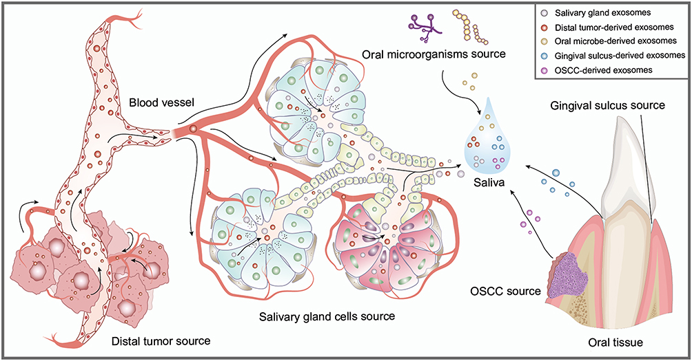 |
Figure 2 Sources of exosomes in the salivary environment. Exosomes in saliva can be directly released into the oral cavity by oral squamous cell carcinoma (OSCC) cells and other epithelial cells of the oral mucosa. Exosomes derived from distal tumor tissues may enter the systemic circulation and be transported to the salivary glands, where they are subsequently secreted into saliva via salivary ducts. The gingival crevicular fluid (GCF), a serous exudate originating from the gingival sulcus, is also enriched with exosomes. Additionally, saliva contains abundant extracellular vesicles of microbial origin, including those secreted by commensal and pathogenic bacteria and fungi. Exosomes are also actively secreted by salivary gland epithelial cells and released into saliva through the glandular ductal system. |
Salivary Exosome Components and Potential OSCC Biomarkers
The components of exosomes are sorted from the contents of the origin cell, partially reflecting the characteristics of the origin cell. Exosomes are mainly composed of components such as proteins, nucleic acids, lipids, sugars, and metabolites (Figure 3). Exosomes mediate intercellular communication in different physiological and pathological environments by delivering these components.64 Salivary exosomes also harbor unique biomarkers derived from OSCC cells. Among the most widely used techniques for proteomic analysis of exosomes are Western blotting (WB) and flow cytometry, both of which are effective for detecting surface protein markers. When combined with immunocapture strategies, the sensitivity and specificity of these methods can be significantly enhanced.65,66 For exosomal nucleic acid analysis, commonly employed approaches include polymerase chain reaction (PCR), quantitative reverse transcription PCR (qRT-PCR), and various next-generation sequencing technologies.67 Through continued research efforts, the molecular composition of salivary exosomes has been progressively elucidated, thereby driving the development of novel diagnostic strategies for the early detection of OSCC (Table 1).
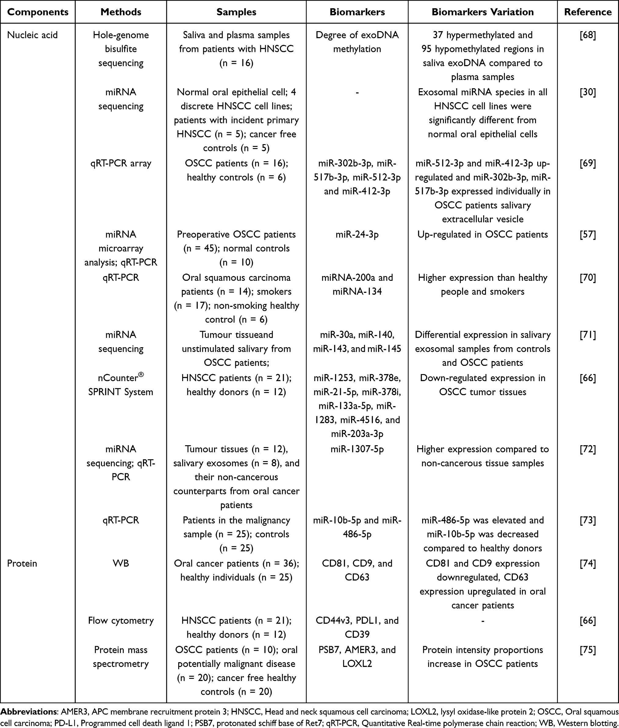 |
Table 1 Potential OSCC Biomarkers in Salivary Exosomes |
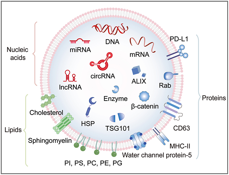 |
Figure 3 Molecular composition of salivary exosomes. The ability of exosomes to modulate the immune response is attributed to the presence of molecules such as programmed cell death ligand 1 (PD-L1) and major histocompatibility complex II (MHC-II) on their surface. Proteins associated with the biogenesis of exosomes include tumor susceptibility gene 101 (TSG101), ALG-2-interacting protein X (ALIX), CD63, and Rab proteins. Proteins involved in cell signaling pathways, such as β-catenin, are also present in exosomes. In addition, exosomes contain heat shock proteins (HSPs) and metabolic enzymes. Notably, aquaporin-5 in salivary exosomes highlights the difference between exosomes secreted by salivary glands and those from other sources. Exosomal lipids mainly include cholesterol, sphingomyelin, phosphatidylcholine (PC), phosphatidylserine (PS), phosphatidylethanolamine (PE), phosphatidylglycerol (PG), phosphatidylinositol (PI), and phosphatidic acid (PA). The major nucleic acid components in salivary exosomes are DNA, RNA, microRNAs (miRNA), circular RNAs (circRNAs), and long non-coding RNAs (lncRNA). |
Nucleic Acid
Exosomes are rich in nucleic acids, including genomic DNA and various RNAs. RNAs are divided into mRNA and non-coding RNA.76,77 Nucleic acids in exosomes reach target cells to enable signaling and ultimately influence their behavior.78 Since tumorigenesis and progression are often associated with abnormal changes in genetic material, the study of exosomal nucleic acids is a focal point for cancer diagnosis using EVs. EVs secreted by tumor cells are enriched with information about these abnormal nucleic acids and can reflect tumor cell status. Researchers can identify potential OSCC diagnostic biomarkers by investigating the association between altered salivary exosome gene expression patterns and OSCC status.
DNA
Back in 2016, F. A. San Lucas et al attempted to analyze the nucleic acid composition of shed exosomes in biofluid samples from patients with pancreaticobiliary cancers. They found that tumor DNA information, including mutations in Notch Receptor 1 (NOTCH1) and BRCA2 DNA repair associated gene (BRCA2), were robustly expressed in shed exosomes. The results suggest that liquid biopsies of shed exosomes may provide a comprehensive analytical assessment of tumors. This approach excludes the necessity of direct tumor sampling for visceral cancers and demonstrates the potential of exosomal DNA in the field of cancer diagnostics.79 Wang et al analyzed DNA in circulating exosomes from pheochromocytoma (PCC) and paraganglioma (PGL) patients and mice. They found that double-stranded DNA (dsDNA) fragments were present in the circulating exosomes of the patients. These fragments were highly consistent with the genomes of the paired tumors. Demonstrated that exosomal dsDNA can be used as a noninvasive biomarker for tumor diagnosis.80 We only retrieved that Mouadh Barbirou et al directly investigated the diagnostic performance of salivary exosomal DNA. They compared their potential as biomarkers for HNSCC by collecting cell-free DNA (cfDNA) and exosomal DNA (exoDNA) from blood and saliva samples of HNSCC patients. In their study, they conducted whole-genome bisulfite sequencing of paired cfDNA and exoDNA in saliva and plasma. There were more methylation differences in saliva exoDNA compared to plasma, demonstrating the potential of salivary exoDNA as a biomarker for HNSCC.68
Overall, current research on salivary exosomal DNA is sparse, and further studies are needed to explore its diagnostic potential in OSCC.
mRNA
Several studies have shown that tumor-derived exosomal mRNA is important for intercellular communication. Specifically, exosomal mRNAs are closely associated with tumor growth, angiogenesis, cell proliferation, immune escape, drug resistance, metastasis, and other behaviors.81 Several studies have investigated the diagnostic potential of salivary mRNA. Li et al used microarray analysis to compare the differences in gene expression in saliva supernatants from OSCC patients and controls. They found that the expression levels of 1679 genes in the saliva supernatants of OSCC patients were significantly different from those of controls. Seven cancer-related mRNA biomarkers (including transcripts of IL8, IL1B, DUSP1, HA3, OAZ1, S100P, and SAT) were upregulated at least 3.5-fold in the salivary supernatants of OSCC patients. ROC curves and classification modeling showed that the combination of these biomarkers had high sensitivity and specificity for the differentiation of OSCC patients from controls.82 The study by Gleber-Netto et al included a large number of clinical samples to investigate the diagnostic potential of multiple salivary transcriptome biomarkers, proteome biomarkers, and risk factor combinations for OSCC. They found that the level of the transcriptome marker DUSP1 in the saliva of OSCC patients was significantly lower than that of healthy controls and the potentially malignant oral disorders group. A multivariate model combining salivary markers and risk factors associated with oral cancer showed good diagnostic efficacy in differentiating OSCC from controls. The results of this study provide clues for the early non-invasive diagnosis of OSCC.83
We have not found studies exploring the potential of salivary exosomal mRNA in OSCC diagnosis. Since mRNA can be stabilized in salivary exosomes, it is feasible to use salivary exosomal mRNA as an entry point for the study of non-invasive methods of disease diagnosis.84
Non-Coding RNA (ncRNA)
The ncRNAs are a class of RNA molecules that do not code for proteins, which are mainly divided into microRNAs (miRNAs), transfer RNAs (tRNAs), small interfering RNAs (siRNAs), piwi-interacting RNA (piRNA), circular RNAs (circRNAs), and long non-coding RNAs (lncRNAs).85,86 In OSCC, ncRNAs can be involved in regulating behaviors such as cell proliferation, metastasis, and invasion.87 The study of exosomal ncRNAs will contribute to early cancer diagnosis and the discovery of new targets for effective cancer therapy.
MiRNAs are undoubtedly the stars of tumor diagnostics compared to other ncRNAs. Several of studies have shown that exosomes derived from tumor cells contain a large number of miRNAs, which are considered to be promising diagnostic biomarkers for a variety of cancer types.32 Depending on the cell type from which they originate, these miRNAs can either promote or inhibit cancer growth.81 MiRNAs are among the most abundant small RNAs in human saliva, and this feature is more pronounced in salivary exosomes, attributed to the exosome membrane’s protective effect on miRNAs.88,89 Abnormal expression of exosomal miRNAs may be an excellent diagnostic biomarker in OSCC diagnostic studies. Scott Langevin et al cultured four HNSCC cell lines and normal oral epithelial cells, collected supernatant exosomes, and sequenced miRNAs. All four HNSCC cell lines had highly overlapping exosomal miRNA profiles, and all differed significantly from normal oral epithelial cells. In their work, miRNA demonstrated relatively low sensitivity but high specificity as a non-invasive salivary biomarker for HNSCC, highlighting its strong potential for diagnosis of tumors.30 Chiara Gai et al collected unstimulated saliva from OSCC patients and normal control subjects, obtained exosomes by centrifugation, and finally assessed the expression levels of miRNAs by qRT-PCR. They found a differential expression of miRNAs in salivary exosomes of OSCC patients compared to normal controls. MiR-302b-3p and miR-517b-3p were expressed only in salivary exosomes of OSCC patients, whereas miR-512-3p and miR-412-3p were upregulated. They also discussed the potential of saliva as a diagnostic biofluid, noting that saliva collection technology offers superior cost-effectiveness and operational feasibility compared to blood samples.69 He et al used miRNA microarray technology to obtain differentially expressed miRNAs from salivary exosomes of healthy controls and OSCC patients. The qRT-PCR validation revealed a significant increase of miR-24-3p in the salivary exosomes of pre-surgical OSCC patients compared to controls. ROC analysis showed miR-24-3p to have extremely high diagnostic accuracy for OSCC ([AUC] = 0.738, P = 0.02), suggesting that salivary exosomal miR-24-3p has the potential as a novel diagnostic biomarker for OSCC.57 Amina Fouad Farag et al utilised qRT-PCR to analyse the changes in microRNA expression in saliva from healthy individuals, smokers, and patients with OSCC. They found that OSCC patients had significantly higher expression of miRNA-200a and miRNA-134 than the other two groups.70 Shanaya Patel et al constructed a combinatorial miRNA model consisting of salivary exosomes miR-30a, miR-140, miR-143, and miR-145 for oral cancer diagnosis. ROC analysis showed that these miRNAs had high sensitivity and specificity for OSCC, and their diagnostic results were consistent with the characteristics of the tissue samples. Subsequent miRNA-mRNA regulatory network analyses also revealed that these miRNAs correlate with oral cancer behaviors such as disease progression, recurrence, and chemotherapy resistance. This multiple miRNA signature has higher efficacy in early detection and is clinically relevant to the disease progression and overall survival rate of OSCC patients, which is of great significance for the research on the early diagnosis of cancer.71 To establish a diagnostic tool for HNSCC in synergy with plasma exosomes, Linda Hofmann et al analyzed the characterization of saliva-derived exosomes from patients with HNSCC and healthy individuals. In their miRNA profiling results, eight miRNAs had significantly lower expression ratios in the HNSCC group compared to the healthy individuals group, with miR-203 a-3p showing the largest fold change and miR-133a-5p showing the highest significance.66 Subsequent study performed a comprehensive and exploratory comparison of exosomal miRNA profiles from plasma and salivary in HNSCC patients. Their proposed two tumor-specific exosomal miRNA panels from plasma and saliva showed great potential in diagnosing HNSCC patients, Union for International Cancer Control (UICC) staging, and human papillomavirus (HPV) status. These results advance the diagnostic applications of salivary exosomal miRNAs.31 In a study conducted by Aditi Patel et al, saliva, tumor tissues, and adjacent non-cancerous tissues were collected from patients with oral cancer and subjected to miRNA sequencing analysis. The results revealed that the expression level of miR-1307-5p was significantly elevated in both salivary exosome samples and cancerous tissues compared to non-cancerous controls. This enrichment has been hypothesized to serve as a potential indicator of poor prognosis in affected patients.72 Similarly, Cosmin Ioan Faur et al employed qRT-PCR to assess the differential expression of salivary exosomal miR-10b-5p and miR-486-5p in patients diagnosed with oral cavity and oropharyngeal squamous cell carcinoma (OPC), in comparison to healthy volunteers. Their findings demonstrated that miR-486-5p was upregulated in the patient group, whereas miR-10b-5p was downregulated, suggesting their potential utility as diagnostic biomarkers for oral and oropharyngeal malignancies.73 qPCR-based detection kits hold potential for transformation into cost-controlled, high-throughput, and relatively straightforward tumor screening or clinical testing programs.
The role of ncRNAs such as lncRNAs, circRNAs, tRNAs, siRNAs, and piRNAs in the progression of OSCC has been extensively investigated. The lncRNA TIRY can facilitate oral cancer proliferation, invasion, and metastasis by promoting epithelial-mesenchymal transition.90,91 CircRNA induces tumor immune escape in OSCC92. These ncRNAs can be used as biomarkers for cancer diagnosis and prognosis, as their expression is associated with various clinical variables in cancer, and investigating them will contribute to the discovery of new diagnostic and therapeutic strategies for OSCC.93–95 However, studies on ncRNAs other than miRNAs in the field of salivary exosome biopsy have not been conducted.
Protein
Research on the exosomal proteome facilitates the search for unique disease biomarkers. Until 2024, the Vesiclepedia database contains 566 911protein entries. Some of the common proteins are HSP and cytoskeleton proteins. Many conserved proteins are shared between different EVs, and many more are different because of the way EVs are synthesized and the way proteins are sorted in the cell of origin, reflecting specific characteristics of the cell of origin.96 Changes in these proteins can be informative for tumor diagnosis. The ability of exosomes to regulate the immune response is due to the presence of molecules such as PD-L1 and MHC-II on the surface. According to electron microscopy, salivary exosomes are also abundant in tetraspanins.84 Tetraspanins are widely involved in biological functions such as protein transport and signaling. For example, CD63 is involved in ESCRT-independent exosome biogenesis.47 The proteins associated with the ESCRT-dependent pathway in salivary exosomes are Hrs, flotillin, tumor susceptibility gene 101 (TSG101), and ALIX. Rab proteins, membrane transport proteins, and annexins are responsible for the regulation of membrane binding during exosome biogenesis and remain in the exosome after its formation. Several proteins that play important roles in cell signaling pathways, such as β-catenin, Wnt5B, or Notch ligand Delta-like 4, have also been identified in exosomes.53 In addition, exosomes contain heat shock proteins, cytoskeletal proteins (actin and myosin), integrins, and metabolic enzymes. Finally, water channel protein-5 and others in salivary exosomes highlight the difference between exosomes secreted by the salivary glands and exosomes from other sources.97 Numerous studies have shown that variations in the protein fractions of salivary exosomes are closely associated with OSCC status. To investigate the differences in exosomes in the oral fluid of oral cancer patients and healthy individuals, Ayelet Zlotogorski-Hurvitz et al conducted a clinical study. They obtained exosomes by ultracentrifugation and then analyzed these exosome samples for morphological and molecular differences using AFM, NTA, ELISA, and WB. The results showed that exosomes in the oral fluid of oral cancer patients not only had increased particle concentrations and particle sizes but also had changes in protein expression levels on their surfaces. CD81 and CD9 expression were reduced, while CD63 expression was higher.74 Fatima Qadir et al suggested that exosomes derived from cancer cells can express surrogate oncogenic markers such as the membrane protein CEP55. That exosomal CEP55 in saliva or blood can be used as a cancer biomarker for the non-invasive diagnosis and prognosis of HNSCC.98 Linda Hofmann et al found that exosomes isolated from the saliva of HNSCC patients contained high levels of CD44v3, PD-L1, and CD39. Changes in the expression of these proteins partly reflect the immunosuppressive microenvironment induced by HNSCC and are expected to be diagnostic biomarkers. Their research not only explores the molecular composition of salivary exosomes but also delves into their immunosuppressive functions, integrating both aspects. By combining the most discriminative surface protein panel with a streamlined miRNA panel and pairing them with efficient magnetic bead capture technology, a “multiparametric liquid biopsy tool” can be developed for head and neck cancer auxiliary diagnosis, prognosis assessment, and prediction of immunotherapy efficacy.66 Natalie Bozyk et al also focused on the effect of OSCC on salivary exosome protein fractions, quantifying salivary exosome protein content from OSCC patients, oral potentially malignant disease (OPMD) patients, and healthy controls using mass spectrometry. They found that a screened panel of three proteins highly expressed in OSCC salivary exosomes, protonated schiff base of Ret7 (PSB7), APC membrane recruitment protein 3 (AMER3), and lysyl oxidase-like protein 2 (LOXL2), could serve as highly sensitive biomarkers for differentiation between healthy controls and patients with OSCC. This study prospectively enrolled patients with OPMD and successfully developed a proteomics detection panel capable of distinguishing healthy individuals from OPMD patients. This achievement directly aligns with core clinical needs for early detection and risk stratification, demonstrating significant translational medical value.75
Lipids
Similar to the cell membrane, the surface of exosomes is composed of lipid bilayers. Compared to the plasma membrane of the origin cell, the membrane of exosomes is more robust and resistant to degradation, which may be attributed to their higher cholesterol, desaturated lipids, and sphingomyelin content.99,100 Therefore, researchers often describe lipids on the surface of exosomes as “natural barriers” or “transporters” for proteins and nucleic acids. Exosomal lipids mainly include cholesterol, sphingomyelin, phosphatidylcholine (PC), phosphatidylserine (PS), phosphatidylethanolamine (PE), phosphatidylglycerol (PG), phosphatidylinositol (PI) and phosphatidic acid (PA), with cholesterol being the most abundant.101 In exosomes from different sources, the abundance of these lipids varies. We did not retrieve studies on lipid components in salivary exosomes and tumor diagnosis. Research in this area would be beneficial to deepen the understanding of exosome formation mechanisms and promote early diagnosis of tumors.
Technologies for Saliva Sampling and Salivary Exosome Characterization and Detection
Saliva Sample Collection
To obtain sufficient diagnostic information from saliva for OSCC, standardized and appropriate saliva collection methods are essential (Figure 4). To minimize contamination and variability, current protocols generally recommend collecting saliva samples from fasting individuals in the morning. Participants should avoid alcohol consumption for at least 24 hours prior to collection to prevent mucosal irritation, and refrain from eating, drinking, smoking, and performing oral hygiene procedures for at least 8 hours before sampling.51,102 It is advised that subjects stand during collection, as salivary flow rates have been reported to be higher in the standing position compared to sitting or lying down.103 Saliva can be either whole saliva or glandular saliva. Additionally, based on whether external stimulation is applied during the collection process, samples can be classified as stimulated or unstimulated saliva.104
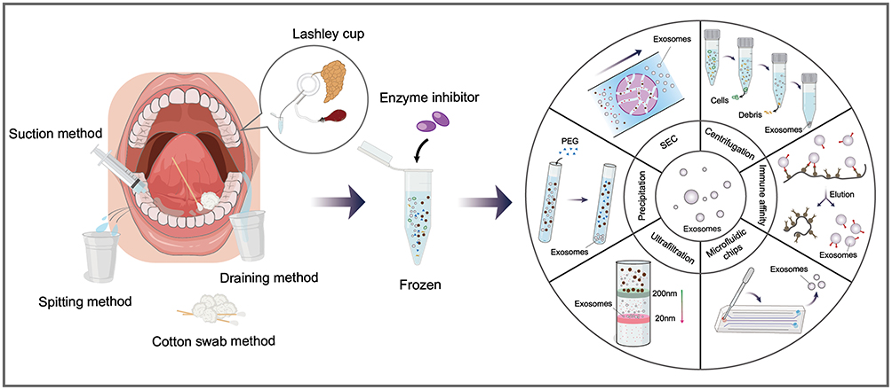 |
Figure 4 Saliva collection techniques and salivary exosome isolation methods. Whole saliva is collected by four main methods: the spitting method, the draining method, the suction method, and the cotton swab method. Salivary gland saliva is collected by a cannula, Lashley cup, or modified Carlson Crittenden device. Saliva sample preservation requires the addition of enzyme inhibitors and transfer under frozen conditions. Common exosome isolation methods include centrifugation, ultrafiltration, sedimentation, size exclusion chromatography (SEC), and immunoaffinity capture. In addition to these, there are emerging technologies such as microfluidic chips. |
Whole saliva, also referred to as mixed saliva, is the most commonly collected form. Four principal techniques are used for whole saliva collection: Spitting method, which is simple, suitable for all age groups, and associated with relatively low inter-individual variability.102 Draining method, which involves allowing saliva to drip passively from the lower lip into a container, thereby minimizing irritation and preserving the native state of the sample. Suction method, which utilizes pipettes or syringes to aspirate saliva directly from the oral cavity floor.105 Cotton swab method, in which a cotton swab is placed in the oral cavity until saturated and then transferred to a collection container.51 In contrast, glandular saliva collection requires specialized devices such as cannulas, the Lashley cup, or the modified Carlson–Crittenden device to isolate saliva from specific salivary glands.104,106 In recent years, companies such as DNA Genotek, QIAGEN, and Malvern Medical Developments have developed customized alternative kits or devices for saliva collection. These kits often include preservatives or employ specially designed containers and materials to ensure sample stability and prevent contamination. They are widely used in clinical and molecular diagnostic settings, including for the detection of viral diseases such as mumps and rubella.107,108 If not for immediate use, saliva samples need to be frozen as soon as possible after initial centrifugation. Saliva samples can be stored at either −20 °C or −80 °C, and repeated freezing and thawing should be avoided.109,110 In addition, protease inhibitors should be added to minimize degradation of protein content during storage and analysis.
Unstimulated whole saliva is generally considered to have greater diagnostic potential than stimulated glandular saliva in the context of OSCC diagnosis. Unlike glandular saliva, whole saliva is in direct contact with OSCC lesions and is therefore more likely to contain exosomes derived from OSCC. Although stimulated saliva yields a larger volume over the same collection period, it is often more dilute, resulting in lower exosome concentrations and reduced sensitivity for diagnostic applications.103 Further clinical studies are warranted to validate the diagnostic value of different saliva types and collection methodologies in OSCC. Optimizing sampling strategies is crucial, as poor-quality or improperly collected samples can compromise the accuracy of diagnostic results. As research advances, the development of more user-friendly, cost-effective, and high-sensitivity saliva collection technologies is expected to further enhance the clinical utility of salivary diagnostics for OSCC.
Isolation of Salivary Exosomes
Exosome isolation is one of the key steps in exosome research. There are a range of well-developed techniques for exosome extraction from saliva and other samples. Common exosome isolation methods include centrifugation, ultrafiltration, precipitation, size exclusion chromatography, and immunoaffinity capture. In addition, innovative technologies such as microfluidic chips are also emerging (Figure 4).
The centrifugation method is the most common isolation method and is currently the gold standard for exosome isolation.111 By generating centrifugal force through the rotation of rotors, the centrifugation method allows different particles in a liquid to settle at various speeds. The centrifugation methods commonly applied to exosomes include differential ultracentrifugation and density gradient centrifugation. The former gradually removes cell debris, apoptotic vesicles, and other components from the sample by adjusting the centrifugal force.112 The latter utilizes sucrose, nycodenz, and iodoexanol media to form density gradients, leading to an orderly distribution of particles with different densities in different density compartments of the media.113 The centrifugation method is suitable for large-volume samples but also suffers from drawbacks including time-consuming and low purity.114 Moreover, the centrifugation process could disrupt the morphology and biological activity of exosomes.115
The ultrafiltration method isolates EVs by forcing the sample through a membrane with specific pore sizes, which can be adjusted to screen for different sizes of EVs. The ultrafiltration method is easily operated and does not require expensive equipment, but the membrane life and separation efficiency are often significantly reduced due to clogging of the membrane pores.116 Moreover, the size consistency and purity of ultrafiltered vesicles are inferior, requiring additional processing to separate exosomes from contaminating proteins.117
The precipitation method utilizes materials such as the hydrophilic polymer polyethylene glycol (PEG) to trap water molecules in the sample, continually decreasing the solubility of the exosome to facilitate its settling. The precipitation method is convenient to employ, with high exosome yield but low purity and high cost, and there are already many mature kits on the market utilizing the precipitation method to achieve exosome isolation.100,118
The size exclusion chromatography (SEC) method separates based on particle size. When the sample flows through SEC fillers (such as SEC columns) with specific pores, the fillers adsorb proteins and lipids with smaller sizes. In comparison, larger-sized exosomes flow faster and are preferentially eluted into the sample collection tubes at specific stages. The advantages of the SEC method are high product purity and minimal influence on exosome characteristics. However, the implementation of the SEC method requires special equipment and is hardly applied to large-volume samples.114,119
The immunoaffinity capture method utilizes ligands or indicators with specific immunoaffinity to recognize protein markers present on exosomes, such as transmembrane proteins and heat shock proteins (HSP). The immunoaffinity capture method allows simultaneous exosome isolation, quantification, and subpopulation identification, which has great potential for disease diagnosis, especially liquid biopsy.114,120,121
Microfluidics technology has continued to develop in recent years, leading to the development of microfluidic chips capable of rapid, automated isolation of exosomes.122 Microfluidic chips can be equipped with microfilters, nano-arrays, and nanowires while utilizing acoustic nanofiltration, dielectrophoretic segregation, or tangential flow filtration to achieve efficient and high-purity exosome isolation.123 Microfluidic chips enable high throughput in situ exosome isolation and analysis, and as it continues to develop, the cost will decrease.
These technologies perform differently when applied to different types of samples and combining them may help to maximize the benefits and avoid the drawbacks.124 For example, the samples after centrifugation or ultrafiltration followed by size exclusion chromatography can provide better exosome samples for proteomics and functional studies.125 Conjugation of specific antibodies on microfluidic chips enables efficient exosome isolation.126
In recent years, exosome isolation technologies have been under constant improvement and innovation, with some lower-cost, more efficient, and more personalized technologies emerging. For example, Suchi Gupta et al simplified the centrifugation step by introducing a sucrose cushion into the centrifugation process. Because of the simplification of the centrifugation step, the time and cost spent as well as the destruction of exosomes are reduced.127 Gu et al have developed an acoustic fluid centrifugation technology that combines acoustic wave actuation and spin of a fluidic droplet to achieve rapid concentration and size-based separation of nanoparticles. Researchers have developed a variety of latex and magnetic beads encapsulating specific receptors that target exosomal membrane markers such as CD63 and Alix. The beads adsorbed exosomes were then placed in special buffers to release the target exosomes.128 Novel magnetic beads coated with polycationic polymers can also utilize the negatively charged phosphatidylserine on the exosome surface to isolate exosomes in large quantities from sample.129 Feng et al coated an amphiphile-dendrimer supramolecular probe (ADSP) on nitrocellulose membranes for exosome capture. The ADSP is composed of highly branched globular dendrimers functionalized with amphotericin B (AMB) molecules. The AMB molecule on ADSP can interact with EVs and promote EVs segregation. They also combined ADSP with an automated printing workstation to enable high throughput capture and analysis of EVs. In addition, this workstation can also be used for the exploration of glycosylation modification on the surface of EVs, which provides a new tool for the functional research of EVs.130 Recently, Zhang et al developed a multivalent cholesterol-modified paranemic crossover DNA construct by DNA nanotechnology. As an effective synthetic nano glue, this structure promotes the rapid merging of nanoscale vesicles into micrometer-sized clusters, followed by low-speed centrifugation to enable rapid enrichment of EVs within minutes. This method effectively avoids the destruction of the structure of EVs by complex ultracentrifugation and provides an efficient and reliable tool for EVs research.131 In conclusion, the exosome isolation method continues to be optimized and innovated and will certainly provide a more robust foundation for the research and application of exosomes in the future.
The isolation of exosomes from saliva poses unique technical challenges compared to other biological fluids such as blood. Saliva collection methods—such as spitting, draining, or suction—typically yield limited sample volumes, rendering volume-dependent techniques like differential ultracentrifugation less practical. As a result, isolation protocols for salivary exosomes must prioritize high recovery efficiency and purity from minimal input volumes. In this context, precipitation-based commercial kits and immunomagnetic bead capture methods have shown favorable compatibility with saliva samples, as they require smaller volumes while maintaining effective isolation performance.132,133 However, the heterogeneous composition of saliva introduces additional complications. Salivary exosomes are often contaminated with food debris, digestive enzymes, and microbial components, all of which can interfere with downstream analysis and compromise diagnostic accuracy. To address this, enzyme inhibitors are commonly added during sample processing, and preliminary purification steps—such as low-speed centrifugation or filtration—are employed to remove large particulate matter. These pre-treatment steps, however, may also risk compromising the structural integrity or biological activity of exosomes. Moreover, the inherently higher viscosity of saliva compared to other biofluids necessitates dilution with suitable buffers prior to isolation, which can introduce variability in exosome yield and concentration.113 Currently, there is no standardized, universally accepted protocol for the isolation of salivary exosomes. Future efforts should focus on the development and validation of reproducible workflows tailored to the physicochemical properties of saliva. Importantly, such protocols must also account for their impact on the fidelity of downstream molecular profiling and the clinical applicability of exosome-based diagnostic assays.
Salivary Exosomes Characterization and Observation
The characterization of salivary exosomes is essential for assessing sample quality, optimizing diagnostic workflows, and enabling the identification of robust and reproducible biomarkers. Accurate characterization provides critical information on exosome size distribution, concentration, morphology, and surface marker expression, thereby enhancing the reliability of subsequent molecular analyses. A range of analytical techniques—such as nanoparticle tracking analysis (NTA), dynamic light scattering (DLS), tunable resistive pulse sensing (TRPS), and various electron microscopy methods—have been widely employed in salivary exosome research. These tools not only facilitate quality control of isolated exosomes but also support the validation of their diagnostic potential in the context of OSCC and other pathologies.
Saliva Exosome Characterization
Extensive physicochemical heterogeneity exists between exosomes. Characterization of exosomes is essential for quality assessment of clinical saliva samples, disease diagnosis and diagnostic markers studies (Table 2).
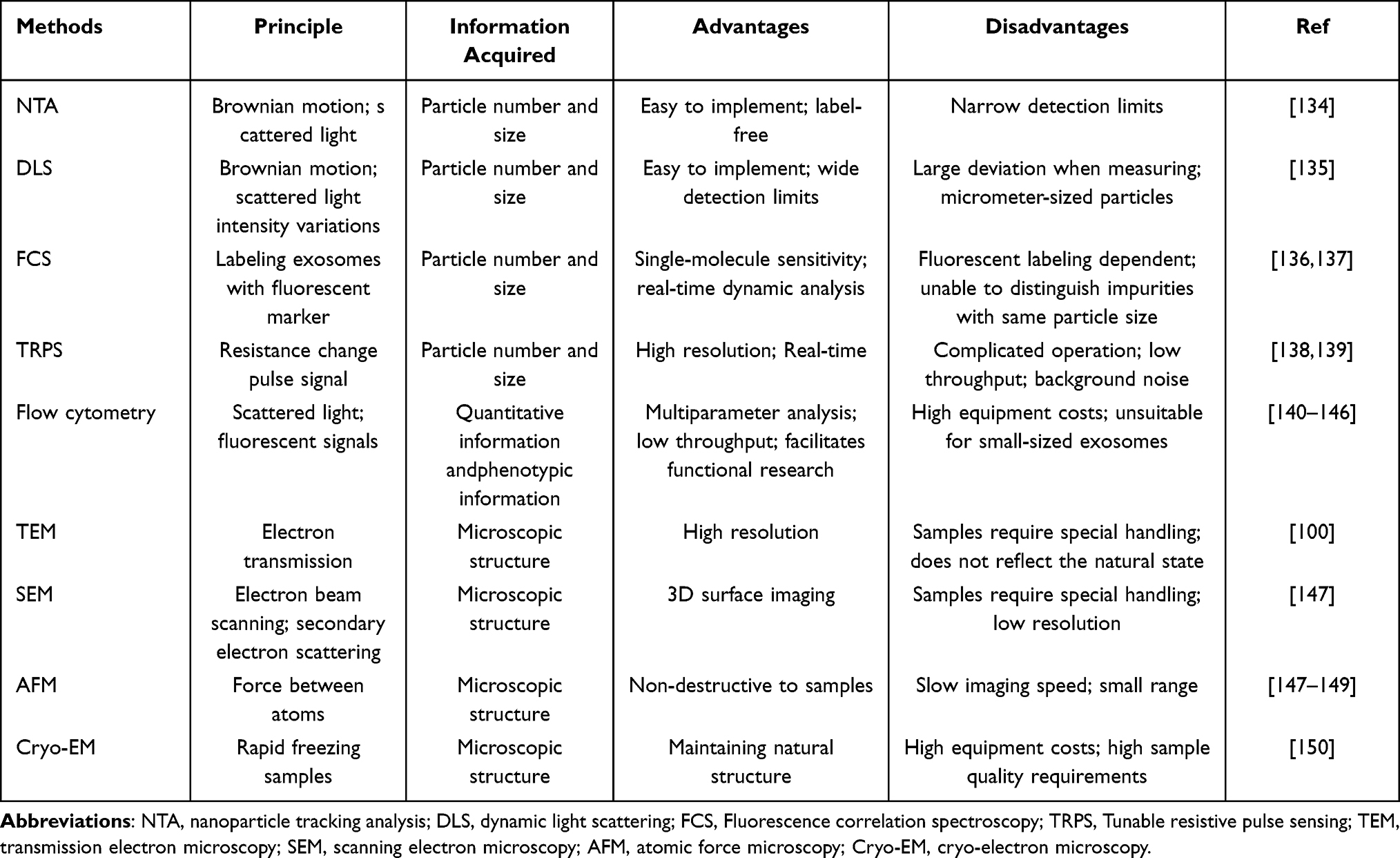 |
Table 2 Characterization Technologies for Exosomes |
As the preferred technology for characterization of exosomes, NTA utilizes the Brownian motion of exosomes in solution. The NTA captures the variation of scattered light from exosomes in the field of view under laser irradiation by the camera and then calculates the sample concentration and particle size.151 The principle and operation of NTA are simple and suitable for analyzing multiple sets of samples. However, the concentration of vesicles is often underestimated due to limitations of the instrumental detection of particle size.134
The principle of DLS is similar to the NTA. Particle size and concentration are inferred by using a fast photon detector to monitor the temporal intensity changes in the scattered light produced by the laser irradiation of particles.147 The DLS offers a broader detection range, however, its measurements are susceptible to the physicochemical properties of the particles. In samples containing a small number of micrometer-sized exosomes, this sensitivity may lead to significant deviations in particle size estimation.135
Fluorescence correlation spectroscopy (FCS) has also been commonly used for exosome characterization. It uses the fluctuation of fluorescent signals generated by fluorescently labeled molecules as they move through a small volume of sample to analyze the characteristics of the labeled molecules. In addition, it can also be used to characterize proteins on the surface of exosomes.136 The primary advantage of FCS over DLS is its ability to detect individual fluorescently labeled molecules. This feature makes it possible to have a detection limit below 50 nm while being less prone to erroneous results when detecting larger particles.137
TRPS technology is also capable of determining exosome particle concentration and size.138 TRPS measures the transient change in electrical resistance produced by individual nanoscale particles as they pass through a tunably sized pore embedded in an elastic membrane and determines the number and size of particles passing through the pore by monitoring and analyzing the change in electrical current. TRPS features high-resolution, real-time analysis. For exosome identification, TRPS suffers from certain important drawbacks, such as the possibility of membrane clogging with repeated use, followed by a decrease in assay stability. At the same time, the background noise of the instrument system could obscure the signals of tiny vesicles, resulting in a decrease in sensitivity.139
Flow cytometry is also frequently applied to exosome characterization.140,141 However, the scattering intensity of exosomes typically drops below the detection limit of most flow cytometers.142 To detect exosomes by normal flow cytometry, it is necessary to first increase the surface area of the exosome sample by binding it with fluorescein-conjugated antibodies or silica beads.143,144 Another serious obstacle is the clustering of small exosomes, where conventional flow cytometers have difficulty in resolving the scattered signals generated by multiple exosomes that are clustered and only record them as a single object.145 In recent years, with the development of nanoscale flow cytometry (nFCM), the analysis of vesicles has become more precise, which will expand the detection limit of exosomes.146
In addition to the above common exosome characterization technology, microfluidics and single particle interferometric reflectance imaging sensor (SP-IRIS) are also rapidly developing, and their application in exosome characterization is also worth expecting.123,152
Salivary Exosome Observation
Exosomes are nanoscale membrane vesicle structures, for direct observation of their natural morphological structure and assessment of the purity of the sample, the primary tool is the microscope. The most common microscopes used for observing exosomes include transmission electron microscopy (TEM), scanning electron microscopy (SEM), and atomic force microscopy (AFM).100,148 TEM images exosomes on carriers such as carbon film and copper mesh in a vacuum environment, and these exosome samples require fixation, dehydration, or staining beforehand. The images obtained by TEM could not fully reflect the natural state of exosomes in the vivo but rather appeared as “cup-like” or “teat-like” structures. The SEM utilizes an electron beam to scan over the surface of a conductive sample and detects the secondary electrons emitted for imaging.147 AFM images exosomes with a scanning probe that interacts with molecules on their surface. Compared to electron microscopes, AFM presents the advantage of being able to make measurements directly in aqueous solution, and the disadvantage of being prone to deformation or rupture of exosomes.149 The cryo-electron microscopy (Cryo-EM) has also been used for exosome structure observation to avoid the shortcomings of the above three commonly used microscopes. Imaging of samples on vitreous ice insulated by liquid nitrogen avoids damage to exosomes by fixation, dehydration, or electron beam irradiation.150 Overall, microscopes have low throughput and cannot be used for accurate counting of exosomes, but there is no substitute for their role in morphological observation and purity assessment.
Salivary Exosomes Detection
The primary goal of exosome detection is to selectively identify specific extracellular vesicles or their molecular constituents within complex biological samples, thereby facilitating early and accurate tumor diagnosis.147 In recent years, a range of advanced detection technologies has been developed, including aptamer-based recognition systems, DNA-based biosensors, nanomaterial-assisted capture platforms, and plasmonic sensing strategies. These methodologies have demonstrated remarkable sensitivity and specificity, particularly in detecting low-abundance exosomes within heterogeneous biological matrices. Their application to salivary exosome analysis is especially promising, given the limited sample volumes and the need for high diagnostic precision in OSCC. By selectively targeting tumor-associated biomarkers enriched within salivary exosomes, these platforms enable precise isolation and molecular profiling from minimal input volumes. The resulting biomolecular recognition events are subsequently converted into quantifiable diagnostic signals through thermal, optical, electrical, or chemical transduction mechanisms, thereby supporting the development of robust liquid biopsy tools for OSCC detection (Table 3).
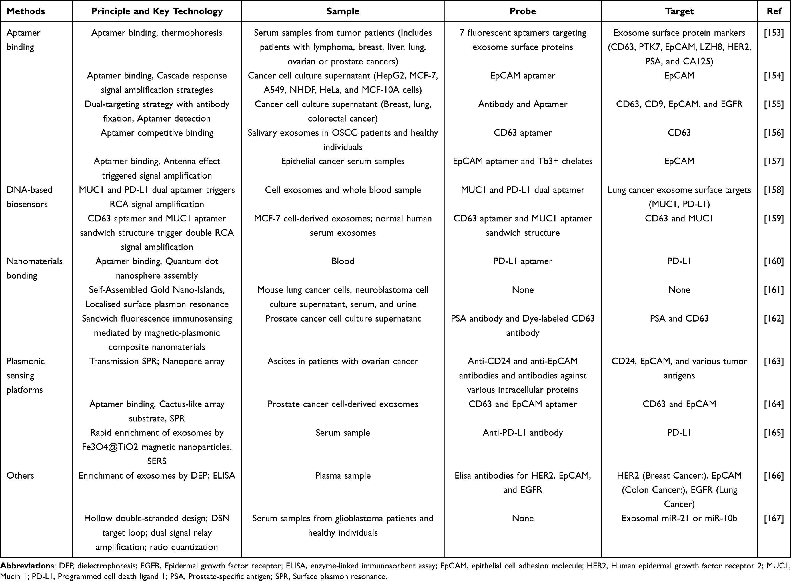 |
Table 3 Salivary Exosome Detection Technologies |
Aptamer Binding
The most commonly used of these technologies is aptamer binding. The aptamer is a small synthetic oligonucleotide sequence or a short polypeptide that binds to the corresponding ligand with high affinity and specificity.168 As a novel, efficient, and rapid identification research platform in biomedicine, it possesses a great deal of research support and huge application potential in exosome detection and tumor diagnosis. The aptamer could be chemically synthesized and exhibit higher thermal/chemical stability, higher tissue penetration, higher product homogeneity, higher immunogenicity, and lower toxicity than traditional antibodies.169 Researchers have developed a variety of fluorescent or electrochemical sensors utilizing aptamer binding technologies that exhibit excellent sensitivity and specificity in detecting exosomes. Liu et al developed an aptamer and thermophoretic-based method to enrich and detect tumor-derived exosomes. The method utilizes seven fluorescent aptamers targeting specific surface protein markers to label tumor-derived exosomes. The surface fluorescence intensity of exosomes from different tumors correlates with the expression level of their surface proteins. Combined with thermophoretic enrichment and linear discriminant analysis, the method demonstrated excellent performance in clinical validation in 102 patients with various types of cancers ranging from stage I to IV.153 Kuang et al designed an aptamer sensor for cancer exosomes that express epithelial cell adhesion molecule (EpCAM) proteins specifically. The EpCAM aptamer formed a hemin/G-quadruplex DNAzyme with Hemin, which catalyzes the tetramethylbenzidine-hydrogen peroxide (TMB-H2O2) system to produce a colorimetric signal. When exosomes expressing EpCAM proteins are added to this system, the DNAzyme structure is destroyed and stops catalyzing the coloration reaction, resulting in a decrease in absorbance. The detection limit of this method is 3.94×10⁵ particles per milliliter, and it shows favorable specificity and high sensitivity to the exosomes with abnormal EpCAM protein expression produced by some cancer cells.154 Cuong Viet Pham et al developed a method to detect EVs using aptamers and fluorescence polarization. This method first immobilizes EVs with a high-specificity antibody and then uses fluorescence polarization guided by a tiny aptamer to detect EVs in the sample. The two-pronged approach effectively removes impurities from the sample, and the specificity of the exosome assay increases significantly.155 Li et al successfully developed a label-free aptamer sensor for discriminating salivary exosomes from OSCC patients and healthy individuals. The sensor utilizes silica microspheres as the substrate, loaded carbon dots as the luminophore, and CD63 aptamer as the specific identification unit. Exosome binding to the CD63 aptamer will induce the release of carbon dots, allowing an increase in the intensity of the fluorescence assay. The difference in fluorescence intensity can be utilized to identify exosomes from OSCC patients and healthy individuals.156 Zeng et al designed a spontaneously foldable stem-and-loop structure called Antenna-Hairpin-Tb by integrating computer-assisted design. They hybridized this structure with an EpCAM-specific aptamer to construct a duplex aptasensor. When this sensor binds to EpCAM, the Antenna-Hairpin-Tb is dehybridized from the sensor and folded, triggering an antenna effect that significantly enhances the time-resolved luminescence of chelated Tb3+. In subsequent diagnostic experiments, the sensor was able to discriminate epithelial cancer serum samples from non-epithelial cancer serum samples in a short time with 100% sensitivity, specificity, precision, and accuracy.157 The superior performance of aptamer technology, compared to traditional antibody-based detection methods, stems from its exceptional capabilities. An increasing number of studies have recognized the potential of aptamers in oral diseases.170 In the detection of clinical saliva samples, aptamers exhibit high resistance to enzymatic reactions present. They can also precisely bind to targets via base complementary pairing or spatial conformation recognition, thus avoiding non-specific binding to proteins, lipids, and other abundant substances in saliva. Overall, the challenge in translating tumor marker aptamers to clinical applications lies in the time-consuming, technically demanding, and costly early design process. However, they offer advantages such as the potential for large-scale, low-cost production through chemical synthesis, with biological preparation costs significantly lower than those of antibodies. Consequently, the final diagnostic kit costs are expected to be kept at a relatively low level.
DNA-Based Biosensors
DNA-based biosensors are a type of sensors that utilize DNA signal amplification technology, and they increase detection sensitivity by increasing the strength of the DNA detectable signal. DNA signal amplification technology provides an excellent research tool for diagnosis, genetic assays, and biomedical fields. It involves technologies such as catalytic hairpin assembly (CHA), hybrid chain reaction (HCR), and rolling-cycle amplification (RCA).171 He et al developed portable sensors for analyzing tumor-derived exosomes without isolation and labeling. The sensor targets typical biomarkers on exosomes and regulates the amount of parallel RCA reaction product through the affinity of the aptamer to the target, which in turn affects the fluorescence intensity of quantum dots (QDs) and methylene blue (MB). Finally, a simple hand-held fluorometer can be used to complete the detection. In subsequent experiments, the sensor could analyze clinical samples quickly and sensitively, and the results were highly consistent with pathology reports.158 Zhao et al constructed a dual aptamer sandwich-type electrochemical aptamer sensor by a four-way-junction (4-WJ) triggered double rolling circle amplification (RCA)-assisted MB/G-quadruplex strategy. Exosomes are first captured by the CD63 aptamer on the sensor, and then a sandwich structure is formed utilizing a Mucin 1 (MUC1) protein-specific aptamer. Subsequently, the 3’ end of the mucin-1 aptamer facilitated 4-WJ formation with the assistance of the probe. Finally, the two DNA templates bound to the 4-WJ terminus trigger the RCA reaction, while the products of RCA capture a large number of MB indicators and amplify the electrochemical signals.159 These studies have illustrated the potential applications of DNA sensors in blood samples. When applied to saliva samples, which may contain lower concentrations of biomarkers, such as miRNA, the performance of DNA sensors remains impressive. Their signal amplification function can enhance minute binding signals to detectable levels. Specially designed DNA sequences, such as those incorporating stem-loop structures, can also improve their resistance to interference from impurities in saliva, preventing the non-specific activation of signals.
Nanomaterials Bonding
Common nanomaterials include quantum dots, gold nanoparticles, graphene, and MnO2 nanosheets.172 These small-sized nanomaterials exhibit unique optical, electrical, and electromagnetic properties. Nanomaterials bound to exosomes absorb and emit light at specific wavelengths, allowing their detection with spectroscopic technology. To accurately quantify the tumor exosome programmed cell death ligand 1 (PD-L1) protein, Zhang et al developed a signal amplification method based on aptamer binding and DNA scaffold hybridization-triggered assembly of quantum dot nanospheres for signal amplification. The method enables two-color phenotyping of exosomes to accurately screen for cancer and assess PD-L1 expression levels through machine learning.160 Thakur et al report a localized surface plasmon resonance (LSPR) biosensor scheme based on self-assembled gold nano-islands (SAM-AuNIs). The LSPR interferometer is capable of capturing differences in biophysical interactions, distinguishing target exosomes from other background vesicles. Their design provides a convenient way to fabricate mass-produced and low-cost biosensor substrates.161 Focusing on the excellent properties of multifunctional carbon nanomaterials, Lee et al developed a magneto-fluorescence immunosensor strategy using silver nanoparticles and magnetic iron oxide nanoparticles modified graphene. The anti-prostate specific antigen is immobilized on the platform, and when the antigen captures exosomes from prostate cancer cells, an applied magnetic field can separate them from the sample. The introduction of a Dye-tetraspanin antibody induces the formation of a “magnetic capture-fluorescence detection” sandwich structure, and the fluorescence intensity of the dye changes according to the number of exosomes in the sample. The platform can be used for rapid isolation and direct detection of exosomes.162 When handling saliva samples, the high specific surface area of nanomaterials can enhance the sensor’s capture efficiency for salivary exosomes. Additionally, their unique physicochemical properties, such as plasma effects and electrochemical activity, can amplify the detection signal, thereby addressing the issue of low concentrations of salivary exosomes.
Plasmonic Sensing Platforms
In the field of exosome detection, plasma sensors have made significant advances. These sensors stimulate surface-plasmon resonance (SPR) through the interaction of light with nanostructures, enabling rapid, label-free, highly sensitive, and real-time detection of exosomes.163,173 Common plasmonic sensing platforms can be categorized into SPR sensors, surface-enhanced Raman spectroscopy (SERS) sensors, and plasmonic-enhanced fluorescence (PEF) sensors. These plasma sensing platforms are also easily translatable. By modifying the sensor chip with aptamers, non-specific binding of other components in saliva could be reduced, enabling precise detection in complex saliva samples. Combining their features with AI deep learning can obtain diagnostic models enabling rapid diagnosis of multiple cancer types.174 The development of plasma sensing platforms is heading towards miniaturization, portability, integration, and intelligence, which is more favorable for exosome detection and cancer diagnosis. Hyungsoon Im et al reported a novel portable SPR chip based on SPR for simultaneous detection of proteins in intact exosomes and lysates. The chip includes arrays of nanopores in a metal membrane, each containing an aptamer for the target exosome. When bound to a target exosomal protein, the chip will produce a spectral shift or intensity change proportional to the protein level. In subsequent experiments, they successfully detected CD24- and EpCAM-positive exosomes from ovarian cancer cells.163 Jia et al designed and fabricated an integrated microfluidic chip equipped with a cactus-like array substrate (CAS) based on SERS to enable the detection of prostate lymph node cancer exosomes. The CAS structure was formed by the self-assembly of bilayer polystyrene microspheres onto a polyethylene terephthalate film. The CAS, labeled with CD63 aptamer, and the SERS nanoprobes, labeled with EpCAM aptamer, were then combined into a “sandwich” structure and applied to the microfluidic chip. The results showed that the detection limit of this microarray for exosomes in cancer cells was up to 1 particle/μL.164 Recently, Pang et al developed an integrated detection method based on Fe3O4@TiO2 magnetic nanoparticles with SERS technology to achieve direct capture and quantitative analysis of serum exosomal PD-L1. In the method, Fe3O4@TiO2 is first used to enrich exosomes through the binding of TiO2 to the hydrophilic phosphate head of the exosome phospholipid. The Au@Ag@MBA SERS label modified by an anti-PD-L1 antibody was then introduced to capture exosomes. Quantification of PD-L1 enables individualized exosomal PD-L1 dynamic monitoring and diagnosis of non-small cell lung cancer.165 Plasmonic sensing platforms remain far from routine clinical application. It is a “platform-based” or “instrument-based” technology, not a “reagent-based” one. Its core value is embedded in specific nanomaterials and SERS detection platforms. To achieve broader clinical adoption, its principles must be integrated into more mature, low-cost, high-throughput, and portable clinical testing platforms.
Other Exosome Detection Technologies
The recent application of some emerging technologies in the field of exosome detection is also remarkable. For example, Minsu Park et al have developed an integrated platform of dielectrophoresis (DEP) and enzyme linked immunosorbent assay (ELISA) technologies for the high-purity isolation and ultra-high sensitivity detection of small EVs in plasma. The platform analyzes three biomarkers, PD-L1, EpCAM, and epidermal growth factor receptor, with a colorimetric method. In subsequent diagnostic experiments, its diagnostic accuracy for a variety of tumors was greater than 90%.166 Niu et al innovatively developed intelligent molecular cleavage and dual-signal relay amplification-based ratiometric, which provides a breakthrough solution for early diagnosis and dynamic monitoring of glioblastoma. The hollow double-stranded design of this technology effectively blocks non-specific cleavage by duplex-specific nuclease (DSN). The cascade reaction will be triggered to achieve double signal amplification when the target miRNA is introduced. This technology is capable of tracking dynamic changes in exosomal RNA and accurately capturing the pattern of miRNA temporal fluctuations, which has great potential for diagnosis and monitoring of various diseases.167
In summary, these exosome detection technologies are currently undergoing rapid development and are beginning to make their appearance in the field of early tumor diagnosis. They can also provide important inspiration for the new type liquid biopsy strategies aimed at screening tumors such as OSCC.
Potential and Challenges of Salivary Exosomes in the Early Diagnosis of OSCC
Salivary exosomes have emerged as a compelling candidate for non-invasive diagnostics in OSCC, offering a combination of anatomical accessibility, biomolecular richness, and compatibility with emerging detection technologies.175 Their ability to reflect tumor-specific molecular alterations and their stability in the salivary microenvironment endow them with distinct advantages over conventional diagnostic modalities. Nevertheless, clinical translation remains limited due to persistent technical and biological challenges that must be addressed through coordinated research efforts (Figure 5).
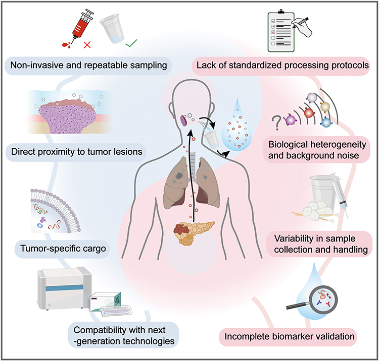 |
Figure 5 The promise and obstacles of salivary exosomes in the early diagnosis of OSCC. As carriers of tumor-specific biomarkers, salivary exosomes are in direct anatomical proximity to OSCC lesions, making them an ideal medium for the early detection and molecular investigation of oral tumors. Their non-invasive origin confers significant clinical advantages—saliva collection is simple, painless, and repeatable, thereby minimizing patient discomfort and enhancing the feasibility of exosome-based diagnostics in routine and large-scale screenings. Moreover, exosomes have become a focal point in biomedical research, and the continuous refinement of analytical tools and protocols has further strengthened their applicability in cancer diagnostics. However, several obstacles hinder the clinical translation of salivary exosome-based diagnostics. The variability in saliva collection techniques, sampling tools, and the lack of standardized analytical workflows limit reproducibility and personalization, while also increasing cost and operational complexity. Additionally, the inherent heterogeneity of salivary exosomes—originating from diverse cellular and microbial sources—combined with high biological background noise in clinical samples, poses substantial challenges in accurately isolating and characterizing tumor-derived vesicles. |
Diagnostic Potential of Salivary Exosomes
Direct Proximity to Tumor Lesions
Unlike other biofluids, saliva maintains direct physical contact with primary OSCC sites—particularly in the tongue, buccal mucosa, and gingiva—facilitating the direct secretion of tumor-derived exosomes into the oral cavity.21 This anatomical advantage enhances the concentration and specificity of tumor-related exosomes in saliva, bypassing systemic dilution and offering a window into localized tumor biology.
Tumor-Specific Molecular Cargo
Exosomal cargos—including miRNAs, proteins, DNA fragments, and metabolites—are dynamically altered during tumorigenesis. Multiple studies have identified promising OSCC-associated markers in salivary exosomes: elevated exosomal Alix expression, immune-related proteomic changes, miRNA signatures such as miR-10b, miR-486-5p, and overlapping miRNA panels found in both saliva and plasma.31,73 These findings underscore the potential of salivary exosomes as molecular fingerprints for OSCC.
Non-Invasive and Repeatable Sampling
Saliva collection is simple, painless, and well tolerated, allowing repeated sampling for longitudinal monitoring of disease progression or therapeutic response. Unlike tissue biopsies, which are invasive and limited in frequency, salivary diagnostics are patient-friendly and highly suitable for community-based screening programs and post-treatment surveillance.19,176
Compatibility with Next-Generation Technologies
Recent advances in biosensing, such as microfluidic platforms, digital PCR, nanoparticle-enhanced assays, and machine learning-based analysis, have significantly improved the sensitivity, specificity, and scalability of exosome detection.177,178 These innovations make it feasible to profile exosomal biomarkers in small saliva volumes with high throughput and clinical relevance.
Key Barriers to Clinical Translation
Despite these advantages, several challenges currently impede the widespread adoption of salivary exosomes in routine clinical diagnostics:
Biological Heterogeneity and Background Noise
Saliva contains exosomes secreted not only by tumor cells, but also by salivary glands, epithelial cells, immune cells, and oral microbiota (including bacteria and fungi) (Figure 2). This diverse origin complicates the identification and isolation of tumor-specific exosomes, especially in early-stage OSCC, where the signal-to-noise ratio is low. Establishing standardized procedures for the collection and processing of saliva samples will help to overcome this obstacle. Furthermore, practical strategies that can improve diagnostic specificity remain to be developed. First, continue identifying more ideal multi-omics markers. Ideal targets should be highly expressed on tumor-derived exosomes while being lowly or non-expressed on normal cell-derived exosomes. Technologically, multi-antibody or aptamer-modified nanoprobes/chips should be developed. Immobilizing 2–3 antibodies or aptamers on capture platforms to selectively capture exosomal subsets expressing multiple corresponding targets simultaneously would facilitate enhanced specificity. In clinical practice, by utilizing the expression levels, ratios, and combination patterns of multiple biomarkers as input features, machine learning can train composite models capable of distinguishing tumors from non-tumors. The performance of such models typically far surpasses that of any single indicator. This layered, combinatorial strategy holds promise for progressively overcoming the bottleneck of salivary exosome specificity, enabling the differentiation of unique exosomes and facilitating the early diagnosis of OSCC.
Lack of Standardized Isolation and Processing Protocols
Existing isolation techniques—such as ultracentrifugation, polymer-based precipitation, and size-exclusion chromatography—often suffer from limitations in specificity, yield, and scalability.124 Inter-laboratory reproducibility remains poor, hindering regulatory approval and large-scale clinical implementation.
Variability in Sample Collection and Handling
Saliva composition is highly susceptible to circadian rhythms, hydration status, dietary intake, and collection protocols (eg, stimulated vs unstimulated saliva).52 Although unstimulated whole saliva is generally preferred for OSCC diagnosis due to its closer proximity to tumor sites, no universally accepted standard for sampling currently exists.
Incomplete Biomarker Validation
While numerous candidate exosomal biomarkers have been proposed, few have been validated in large-scale, multicenter, or longitudinal cohorts. The lack of external validation limits the robustness, reproducibility, and regulatory acceptance of these markers for clinical application.
Future Directions and Strategic Priorities
To realize the clinical promise of salivary exosomes in the early diagnosis of OSCC, a multi-tiered roadmap is essential—encompassing technical optimization, biomarker discovery, clinical validation, and translational infrastructure development. The following areas should be considered high-priority targets for future research and development:
Standardization of Pre-Analytical and Analytical Procedures
Uniformity in saliva collection, storage, and processing is crucial for ensuring reproducibility across studies and clinical settings. Future efforts should establish international consensus guidelines that account for variables such as:
Type of saliva (stimulated vs unstimulated; whole vs glandular);
Collection time and subject preparation (eg, fasting, oral hygiene, medication use);
Use of preservatives, enzyme inhibitors, and storage conditions (eg, freeze-thaw cycles).
Moreover, exosome isolation workflows must be standardized in terms of methodology (eg, centrifugation parameters, filtration cutoff, affinity reagents) to reduce batch effects and enable cross-laboratory comparability. Based on the research evidence we have collected, a simple framework for sample selection, collection, storage, and pretreatment for OSCC testing can be proposed to ensure sample quality. First, the saliva sample collected for OSCC testing should be as close as possible to the body’s baseline state, specifically non-stimulated whole saliva. Before collection, subjects should avoid alcohol consumption for 24 hours, refrain from eating, drinking, or smoking for 8 hours, and suspend oral hygiene practices. Collection should be performed in the early morning with subjects standing in a fasted state. Saliva samples should be properly stored in storage containers and transported at a low temperature (4°C) to the isolation and testing platform. Enzyme inhibitors and preservatives should be applied during transport to ensure sample stability. Before exosome detection, impurities in saliva (such as cell debris and food residues) must be removed using low-speed centrifugation and filtration methods. Commercial kits based on precipitation methods and immunomagnetic bead capture are particularly suitable for salivary exosome isolation, as they require minimal sample volume while maintaining effective performance. High-quality exosomes ensure diagnostic accuracy for OSCC. As research evidence advances, this framework can be progressively refined to accommodate diverse research and clinical scenarios.
Development of High-Throughput, Point-of-Care Detection Platforms
To transition salivary exosome diagnostics from bench to bedside, rapid and scalable detection platforms are indispensable. Promising directions include:
Microfluidic-integrated biosensors capable of simultaneously capturing, enriching, and analyzing exosomes within minutes;
Electrochemical and plasmonic sensors for label-free, real-time detection of specific exosomal biomarkers;
AI-assisted signal interpretation and diagnostic classification, leveraging machine learning to integrate complex biomarker panels into actionable clinical outputs;
Portable diagnostic devices for use in community clinics or remote settings, supporting decentralized screening programs.
Discovery and Validation of Robust Multi-Marker Panels
The clinical utility of salivary exosomes will be contingent on the identification of reproducible and OSCC-specific biomarker panels. Future biomarker discovery should:
Integrate multi-omic strategies (miRNA, lncRNA, protein, lipidomic, methylation patterns) to improve sensitivity and specificity;
Apply single-vesicle profiling techniques to resolve heterogeneity and identify subpopulations enriched in tumor-related cargo;
Include longitudinal sample tracking in patients undergoing treatment to assess temporal biomarker dynamics for monitoring recurrence or therapeutic efficacy;
Validate findings in large, multi-center, prospective cohorts that reflect real-world clinical diversity and heterogeneity.
Integration Into Risk Stratification and Clinical Decision-Making Models
To move beyond proof-of-concept, salivary exosome assays must be incorporated into clinical workflows that enhance personalized care. This includes:
Combining salivary exosome data with patient risk factors, such as tobacco use, HPV status, or family history, to create integrative risk prediction tools;
Evaluating the utility of salivary exosomes as complementary tools to imaging or histopathology, especially in indeterminate or early-stage lesions;
Developing predictive algorithms for treatment response (eg, immunotherapy, chemoradiotherapy), using exosomal PD-L1, MHC molecules, or immunomodulatory miRNAs as dynamic indicators.
Understanding Functional Roles and Biological Mechanisms
Beyond diagnostics, elucidating how salivary exosomes actively participate in OSCC pathophysiology can provide additional clinical insights. Future studies should:
Investigate how tumor-derived exosomes contribute to immune evasion, angiogenesis, epithelial–mesenchymal transition (EMT), and local invasion;
Explore interactions between tumor and stromal exosomes, including those from fibroblasts, immune cells, or microbiota;
Assess how exosomal content changes over the course of tumor evolution, potentially serving as a window into tumor heterogeneity and resistance mechanisms.
Regulatory Pathways and Commercial Translation
To ensure smooth clinical implementation, regulatory and industry-oriented pathways must also be considered. Priorities include:
Establishing analytical and clinical performance benchmarks for diagnostic kits;
Promoting industry-academic collaboration for the development of CE-IVD/FDA-cleared exosome-based saliva diagnostic tools;
Engaging with regulatory bodies early to define acceptable endpoints and validation frameworks for salivary exosome assays.
With sustained interdisciplinary collaboration and technological innovation, salivary exosomes hold the potential to revolutionize OSCC screening and early detection, facilitating more timely intervention and ultimately improving patient survival.
Conclusion
With nearly 390 000 new cases diagnosed globally each year and persistently low survival rates, OSCC represents a significant global health burden.2,7 While advanced-stage OSCC can often be identified through clinical examination and confirmed via surgical biopsy, early-stage lesions are typically asymptomatic and subtle in appearance, resulting in frequent misdiagnosis or delayed detection.179 This highlights an urgent need for reliable, non-invasive diagnostic strategies capable of identifying OSCC at an early stage. Liquid biopsy has emerged as a powerful approach for the early detection of malignancies by capturing molecular alterations that are not readily observable through traditional diagnostic methods. Among various liquid biopsy sources, salivary exosomes have recently attracted increasing attention due to their anatomical proximity to oral lesions, ease of collection, and potential to harbor tumor-derived molecular signatures. In this review, we systematically summarize the current knowledge surrounding the collection, isolation, characterization, and detection of salivary exosomes, with particular emphasis on their molecular components relevant to OSCC. Furthermore, we critically discuss both the advantages and the challenges associated with using salivary exosomes as a diagnostic target in the context of liquid biopsy. Overall, the intrinsic properties of salivary exosomes—combined with recent technological advances in exosome enrichment, profiling, and biosensing—position them as promising candidates for non-invasive, early OSCC detection. However, further research is warranted to identify robust biomarkers, standardize sampling and processing protocols, and validate their clinical utility through large-scale, prospective studies. Salivary exosome-based diagnostics may not only enhance early tumor detection but could also transform the broader landscape of precision oncology.
Abbreviations
4-WJ, Four-way-junction; ADSP, Amphiphile-dendrimer supramolecular probe; AFM, Atomic force microscopy; ALIX, ALG-2-interacting protein X; AMB, Amphotericin B; AMER3, APC membrane recruitment protein 3; BRCA2, BRCA2 DNA repair associated gene; CAS, Cactus-like array substrate; cfDNA, Cell-free DNA; CHA, Catalytic hairpin assembly; circRNAs, Circular RNAs; Cryo-EM, Cryo-electron microscopy; DEP, Dielectrophoresis; DLS, Dynamic light scattering; DSN, Duplex-specific nuclease; dsDNA, Double-stranded DNA; EGFR, Epidermal growth factor receptor; ELISA, Enzyme linked immunosorbent assay; EMT, Epithelial–mesenchymal transition; EpCAM, Epithelial cell adhesion molecule; ESCRT, Endosomal-sorting complex required for transport; EVs, Extracellular vesicles; exoDNA, Exosomal DNA; FCS, Fluorescence correlation spectroscopy; HCR, Hybrid chain reaction; HNSCC, Head and neck squamous cell carcinoma; HSC70, Heat shock cognate protein 70; HSP, Heat shock proteins; HPV, Human papillomavirus; ILVs, Intraluminal vesicles; IPMC, Intracellular plasma membrane-connected compartments; LAMP1, Lysosome-associated membrane glycoprotein 1; LAMP2, Lysosome-associated membrane glycoprotein 2; LC-MS/MS, Liquid chromatography-tandem mass spectrometry; lncRNAs, Long non-coding RNAs; LOXL2, Lysyl oxidase-like protein 2; LSPR, Localized surface plasmon resonance; MB, Methylene blue; MHC-II, Major histocompatibility complex II; MUC1, Mucin 1; miRNAs, MicroRNAs; MVBs, Multivesicular bodies; ncRNA, Non-coding RNA; nFCM, Nanoscale flow cytometry; NOTCH1, Notch Receptor 1; NTA, Nanoparticle tracking analysis; OPC, Oropharyngeal squamous cell carcinoma; OPMD, Oral potentially malignant disease; OSCC, Oral squamous cell carcinoma; PA, Phosphatidic acid; PC, Phosphatidylcholine; PCC, Pheochromocytoma; PCR, Polymerase chain reaction; PD-L1, Programmed cell death ligand 1; PE, Phosphatidylethanolamine; PEF, Plasmonic-enhanced fluorescence; PEG, Polymer polyethylene glycol; PG, Phosphatidylglycerol; PGL, Paraganglioma; PI, Phosphatidylinositol; piRNA, Piwi-interacting RNA; PM, Plasma membrane; Pmel17, Pre-melanosomal protein; PS, Phosphatidylserine; PSB7, Protonated schiff base of Ret7; QDs, Quantum dots; qRT-PCR, Quantitative reverse transcription PCR; RCA, Rolling-cycle amplification; SAM-AuNIs, Self-assembled gold nano-islands; SEC, Size exclusion chromatography; SEM, Scanning electron microscopy; SERS, Surface-enhanced Raman spectroscopy; siRNAs, Small interfering RNAs; SP-IRIS, Single particle interferometric reflectance imaging sensor; SPR, Surface plasmon resonance; TEM, Transmission electron microscopy; TMB-H2O2, Tetramethylbenzidine-hydrogen peroxide; tRNAs, Transfer RNAs; TRPS, Tunable resistive pulse sensing; UICC, Union for International Cancer Control; VPS4, Vacuolar Protein Sorting 4; VTA1, vesicle trafficking 1; WB, Western blotting.
Author Contributions
All authors made a significant contribution to the work reported, whether that is in the conception, study design, execution, acquisition of data, analysis and interpretation, or in all these areas; took part in drafting, revising or critically reviewing the article; gave final approval of the version to be published; have agreed on the journal to which the article has been submitted; and agree to be accountable for all aspects of the work.
Funding
The Basic and Clinical Cooperative Research and Promotion Program of Anhui Medical University (2023xkjT046), the Research Fund of Anhui Institute of Translational Medicine (2023zhyx-C90), the Disciplinary Construction Project in School of Dentistry, Anhui Medical University (2023xkfyhz01), the National Natural Science Foundation of China (22475192), and the Research and Development Project of Stomatology Hospital Zhejiang University School of Medicine (No. RD2022YYYB01).
Disclosure
The authors report no conflicts of interest in this work.
References
1. Carnielli CM, Macedo CCS, De Rossi T, et al. Combining discovery and targeted proteomics reveals a prognostic signature in oral cancer. Nat Commun. 2018;9(1):3598. doi:10.1038/s41467-018-05696-2
2. Bray F, Laversanne M, Sung H, et al. Global cancer statistics 2022: GLOBOCAN estimates of incidence and mortality worldwide for 36 cancers in 185 countries. CA Cancer J Clin. 2024;74(3):229–30. doi:10.3322/caac.21834
3. Omar E. Current concepts and future of noninvasive procedures for diagnosing oral squamous cell carcinoma--a systematic review. Head Face Med. 2015;11:6. doi:10.1186/s13005-015-0063-z
4. Visacri MB, Ferrari GB, Dias P, et al. Quality of life of patients with squamous cell carcinoma of the head and neck receiving high-dose cisplatin chemotherapy and radiotherapy. South Med J. 2015;108(6):343–349. doi:10.14423/smj.0000000000000298
5. da Silva SD, Hier M, Mlynarek A, Kowalski LP, Alaoui-Jamali MA. Recurrent oral cancer: current and emerging therapeutic approaches. Front Pharmacol. 2012;3:149. doi:10.3389/fphar.2012.00149
6. Bugshan A, Farooq I. Oral squamous cell carcinoma: metastasis, potentially associated malignant disorders, etiology and recent advancements in diagnosis. F1000Res. 2020;9:229. doi:10.12688/f1000research.22941.1
7. van der Waal I. Are we able to reduce the mortality and morbidity of oral cancer; some considerations. Med Oral Patol Oral Cir Bucal. 2013;18(1):e33–7. doi:10.4317/medoral.18486
8. Walsh T, Warnakulasuriya S, Lingen MW, et al. Clinical assessment for the detection of oral cavity cancer and potentially malignant disorders in apparently healthy adults. Cochrane Database Syst Rev. 2021;12(12):Cd010173. doi:10.1002/14651858.CD010173.pub3
9. Flygare L, Erdogan ST, Söderkvist K. PET/MR versus PET/CT for locoregional staging of oropharyngeal squamous cell cancer. Acta Radiol. 2023;64(5):1865–1872. doi:10.1177/02841851221140668
10. Walsh T, Macey R, Kerr AR, Lingen MW, Ogden GR, Warnakulasuriya S. Diagnostic tests for oral cancer and potentially malignant disorders in patients presenting with clinically evident lesions. Cochrane Database Syst Rev. 2021;7(7):Cd010276. doi:10.1002/14651858.CD010276.pub3
11. Foser S, Maiese K, Digumarthy SR, Puig-Butille JA, Rebhan C. Looking to the future of early detection in cancer: liquid biopsies, imaging, and artificial intelligence. Clin Chem. 2024;70(1):27–32. doi:10.1093/clinchem/hvad196
12. Martins I, Ribeiro IP, Jorge J, et al. Liquid biopsies: applications for cancer diagnosis and monitoring. Genes. 2021;12(3):349. doi:10.3390/genes12030349
13. Heitzer E, Haque IS, Roberts CES, Speicher MR. Current and future perspectives of liquid biopsies in genomics-driven oncology. Nat Rev Genet. 2019;20(2):71–88. doi:10.1038/s41576-018-0071-5
14. Wu P, He X, Fan J, et al. Electrochemical cytosensors for non-invasive liquid biopsy: detection procedures and technologies for circulating tumor cells. Biosens Bioelectron. 2024;267:116818. doi:10.1016/j.bios.2024.116818
15. Zhang CZ, Cheng XQ, Li JY, et al. Saliva in the diagnosis of diseases. Int J Oral Sci. 2016;8(3):133–137. doi:10.1038/ijos.2016.38
16. Giannobile WV, Wong DT. Salivary diagnostics: oral health and beyond! J Dent Res. 2011;90(10):1153–1154. doi:10.1177/0022034511420436
17. Kinane DF, Gabert J, Xynopoulos G, Guzeldemir-Akcakanat E. Strategic approaches in oral squamous cell carcinoma diagnostics using liquid biopsy. Periodontol 2000. 2024;96(1):316–328. doi:10.1111/prd.12567
18. Chiamulera MMA, Zancan CB, Remor AP, Cordeiro MF, Gleber-Netto FO, Baptistella AR. Salivary cytokines as biomarkers of oral cancer: a systematic review and meta-analysis. BMC Cancer. 2021;21(1):205. doi:10.1186/s12885-021-07932-3
19. Nonaka T, Wong DTW. Saliva Diagnostics. Annu Rev Anal Chem. 2022;15(1):107–121. doi:10.1146/annurev-anchem-061020-123959
20. Kaczor-Urbanowicz KE, Wei F, Rao SL, et al. Clinical validity of saliva and novel technology for cancer detection. Biochim Biophys Acta Rev Cancer. 2019;1872(1):49–59. doi:10.1016/j.bbcan.2019.05.007
21. Ferrari E, Pezzi ME, Cassi D, Pertinhez TA, Spisni A, Meleti M. Salivary cytokines as biomarkers for oral squamous cell carcinoma: a systematic review. Int J Mol Sci. 2021;22(13):6795. doi:10.3390/ijms22136795
22. Kalluri R, LeBleu VS. The biology, function, and biomedical applications of exosomes. Science. 2020;367(6478):eaau6977. doi:10.1126/science.aau6977
23. Cheng L, Hill AF. Therapeutically harnessing extracellular vesicles. Nat Rev Drug Discov. 2022;21(5):379–399. doi:10.1038/s41573-022-00410-w
24. Liu J, Ren L, Li S, et al. The biology, function, and applications of exosomes in cancer. Acta Pharm Sin B. 2021;11(9):2783–2797. doi:10.1016/j.apsb.2021.01.001
25. Zhou B, Xu K, Zheng X, et al. Application of exosomes as liquid biopsy in clinical diagnosis. Signal Transduct Target Ther. 2020;5(1):144. doi:10.1038/s41392-020-00258-9
26. Ghosh AK, Nath A, Elangovan E, Banerjee A, Ramalingam K, Sethuraman S. Exploring salivary biomarkers for tumor diagnosis: a narrative review. Cureus. 2024;16(7):e65725. doi:10.7759/cureus.65725
27. Sharma S, Gillespie BM, Palanisamy V, Gimzewski JK. Quantitative nanostructural and single-molecule force spectroscopy biomolecular analysis of human-saliva-derived exosomes. Langmuir. 2011;27(23):14394–14400. doi:10.1021/la2038763
28. Nakamichi E, Sakakura H, Mii S, et al. Detection of serum/salivary exosomal Alix in patients with oral squamous cell carcinoma. Oral Dis. 2021;27(3):439–447. doi:10.1111/odi.13565
29. Winck FV, Prado Ribeiro AC, Ramos Domingues R, et al. Insights into immune responses in oral cancer through proteomic analysis of saliva and salivary extracellular vesicles. Sci Rep. 2015;5:16305. doi:10.1038/srep16305
30. Langevin S, Kuhnell D, Parry T, et al. Comprehensive microRNA-sequencing of exosomes derived from head and neck carcinoma cells in vitro reveals common secretion profiles and potential utility as salivary biomarkers. Oncotarget. 2017;8(47):82459–82474. doi:10.18632/oncotarget.19614
31. Hofmann L, Abou Kors T, Ezić J, et al. Comparison of plasma- and saliva-derived exosomal miRNA profiles reveals diagnostic potential in head and neck cancer. Front Cell Dev Biol. 2022;10:971596. doi:10.3389/fcell.2022.971596
32. Zhou X, Xie F, Wang L, et al. The function and clinical application of extracellular vesicles in innate immune regulation. Cell Mol Immunol. 2020;17(4):323–334. doi:10.1038/s41423-020-0391-1
33. Pegtel DM, Gould SJ. Exosomes. Annu Rev Biochem. 2019;88:487–514. doi:10.1146/annurev-biochem-013118-111902
34. Kalluri R. The biology and function of extracellular vesicles in immune response and immunity. Immunity. 2024;57(8):1752–1768. doi:10.1016/j.immuni.2024.07.009
35. Colombo M, Raposo G, Théry C. Biogenesis, secretion, and intercellular interactions of exosomes and other extracellular vesicles. Annu Rev Cell Dev Biol. 2014;30:255–289. doi:10.1146/annurev-cellbio-101512-122326
36. Villarroya-Beltri C, Baixauli F, Gutiérrez-Vázquez C, Sánchez-Madrid F, Mittelbrunn M. Sorting it out: regulation of exosome loading. Semin Cancer Biol. 2014;28:3–13. doi:10.1016/j.semcancer.2014.04.009
37. Hessvik NP, Llorente A. Current knowledge on exosome biogenesis and release. Cell Mol Life Sci. 2018;75(2):193–208. doi:10.1007/s00018-017-2595-9
38. Kowal J, Tkach M, Théry C. Biogenesis and secretion of exosomes. Curr Opin Cell Biol. 2014;29:116–125. doi:10.1016/j.ceb.2014.05.004
39. Gurunathan S, Kang MH, Kim JH. A comprehensive review on factors influences biogenesis, functions, therapeutic and clinical implications of exosomes. Int J Nanomedicine. 2021;16:1281–1312. doi:10.2147/ijn.S291956
40. Mashouri L, Yousefi H, Aref AR, Ahadi AM, Molaei F, Alahari SK. Exosomes: composition, biogenesis, and mechanisms in cancer metastasis and drug resistance. Mol Cancer. 2019;18(1):75. doi:10.1186/s12943-019-0991-5
41. Baietti MF, Zhang Z, Mortier E, et al. Syndecan-syntenin-ALIX regulates the biogenesis of exosomes. Nat Cell Biol. 2012;14(7):677–685. doi:10.1038/ncb2502
42. Gill DJ, Teo H, Sun J, et al. Structural insight into the ESCRT-I/-II link and its role in MVB trafficking. EMBO j. 2007;26(2):600–612. doi:10.1038/sj.emboj.7601501
43. Hanson PI, Cashikar A. Multivesicular body morphogenesis. Annu Rev Cell Dev Biol. 2012;28:337–362. doi:10.1146/annurev-cellbio-092910-154152
44. Katzmann DJ, Babst M, Emr SD. Ubiquitin-dependent sorting into the multivesicular body pathway requires the function of a conserved endosomal protein sorting complex, ESCRT-I. Cell. 2001;106(2):145–155. doi:10.1016/s0092-8674(01)00434-2
45. Trajkovic K, Hsu C, Chiantia S, et al. Ceramide triggers budding of exosome vesicles into multivesicular endosomes. Science. 2008;319(5867):1244–1247. doi:10.1126/science.1153124
46. Ghossoub R, Lembo F, Rubio A, et al. Syntenin-ALIX exosome biogenesis and budding into multivesicular bodies are controlled by ARF6 and PLD2. Nat Commun. 2014;5:3477. doi:10.1038/ncomms4477
47. van Niel G, Charrin S, Simoes S, et al. The tetraspanin CD63 regulates ESCRT-independent and -dependent endosomal sorting during melanogenesis. Dev Cell. 2011;21(4):708–721. doi:10.1016/j.devcel.2011.08.019
48. Géminard C, De Gassart A, Blanc L, Vidal M. Degradation of AP2 during reticulocyte maturation enhances binding of hsc70 and Alix to a common site on TFR for sorting into exosomes. Traffic. 2004;5(3):181–193. doi:10.1111/j.1600-0854.2004.0167.x
49. Babst M. MVB vesicle formation: ESCRT-dependent, ESCRT-independent and everything in between. Curr Opin Cell Biol. 2011;23(4):452–457. doi:10.1016/j.ceb.2011.04.008
50. Liu YJ, Wang C. A review of the regulatory mechanisms of extracellular vesicles-mediated intercellular communication. Cell Commun Signal. 2023;21(1):77. doi:10.1186/s12964-023-01103-6
51. Krahel A, Hernik A, Dmitrzak-Weglarz M, Paszynska E. Saliva as diagnostic material and current methods of collection from oral cavity. Clin Lab. 2022;68(10). doi:10.7754/Clin.Lab.2022.211224
52. Carpenter GH. The secretion, components, and properties of saliva. Annu Rev Food Sci Technol. 2013;4:267–276. doi:10.1146/annurev-food-030212-182700
53. Cheshmi B, Cheshomi H. Salivary exosomes: properties, medical applications, and isolation methods. Mol Biol Rep. 2020;47(8):6295–6307. doi:10.1007/s11033-020-05659-1
54. Bandhakavi S, Stone MD, Onsongo G, Van Riper SK, Griffin TJ. A dynamic range compression and three-dimensional peptide fractionation analysis platform expands proteome coverage and the diagnostic potential of whole saliva. J Proteome Res. 2009;8(12):5590–5600. doi:10.1021/pr900675w
55. Zhang Y, Sun J, Lin CC, Abemayor E, Wang MB, Wong DT. The emerging landscape of salivary diagnostics. Periodontol 2000. 2016;70(1):38–52. doi:10.1111/prd.12099
56. Nonaka T, Wong DTW. Saliva-exosomics in cancer: molecular characterization of cancer-derived exosomes in saliva. Enzymes. 2017;42:125–151. doi:10.1016/bs.enz.2017.08.002
57. He L, Ping F, Fan Z, et al. Salivary exosomal miR-24-3p serves as a potential detective biomarker for oral squamous cell carcinoma screening. Biomed Pharmacother. 2020;121:109553. doi:10.1016/j.biopha.2019.109553
58. Yang J, Wei F, Schafer C, Wong DT. Detection of tumor cell-specific mRNA and protein in exosome-like microvesicles from blood and saliva. PLoS One. 2014;9(11):e110641. doi:10.1371/journal.pone.0110641
59. Lau C, Kim Y, Chia D, et al. Role of pancreatic cancer-derived exosomes in salivary biomarker development. J Biol Chem. 2013;288(37):26888–26897. doi:10.1074/jbc.M113.452458
60. Wang R, Shi Y, Lv Y, Xie C, Hu Y. The novel insights of epithelial-derived exosomes in various fibrotic diseases. Biomed Pharmacother. 2024;174:116591. doi:10.1016/j.biopha.2024.116591
61. Sjoqvist S, Kasai Y, Shimura D, et al. Oral keratinocyte-derived exosomes regulate proliferation of fibroblasts and epithelial cells. Biochem Biophys Res Commun. 2019;514(3):706–712. doi:10.1016/j.bbrc.2019.04.202
62. Gonzalez-Begne M, Lu B, Han X, et al. Proteomic analysis of human parotid gland exosomes by multidimensional protein identification technology (MudPIT). J Proteome Res. 2009;8(3):1304–1314. doi:10.1021/pr800658c
63. Liu C, Udawatte NS, Liaw A, et al. Microbial DNA profiles of bacterial extracellular vesicles from 3D salivary polymicrobial biofilms - A pilot study. Adv Healthc Mater. 2025;14(6):e2403300. doi:10.1002/adhm.202403300
64. Isola AL, Chen S. Exosomes: the messengers of health and disease. Curr Neuropharmacol. 2017;15(1):157–165. doi:10.2174/1570159x14666160825160421
65. Faur CI, Rotaru H, Osan C, et al. Salivary exosomal microRNAs as biomarkers for head and neck cancer detection-a literature review. Maxillofac Plast Reconstr Surg. 2021;43(1):19. doi:10.1186/s40902-021-00303-9
66. Hofmann L, Medyany V, Ezić J, et al. Cargo and functional profile of saliva-derived exosomes reveal biomarkers specific for head and neck cancer. Front Med. 2022;9:904295. doi:10.3389/fmed.2022.904295
67. Miceli RT, Chen TY, Nose Y, et al. Extracellular vesicles, RNA sequencing, and bioinformatic analyses: challenges, solutions, and recommendations. J Extracell Vesicles. 2024;13(12):e70005. doi:10.1002/jev2.70005
68. Barbirou M, Miller A, Baek D, et al. Abstract 2293: comparisons of saliva versus plasma cfDNA and exosomal DNA as potential biomarkers for head and neck cancer. Cancer Research. 2024;84(6_Supplement):2293. doi:10.1158/1538-7445.AM2024-2293
69. Gai C, Camussi F, Broccoletti R, et al. Salivary extracellular vesicle-associated miRNAs as potential biomarkers in oral squamous cell carcinoma. BMC Cancer. 2018;18(1):439. doi:10.1186/s12885-018-4364-z
70. Farag A, Sabry D, Hassabou N, Alaa El-din Y. MicroRNA-134/MicroRNA-200a derived salivary exosomes are novel diagnostic biomarkers of oral squamous cell carcinoma. Egypt Dent J. 2021;67:367–377. doi:10.21608/edj.2020.47990.1317
71. Patel S, Patel A, Mandlik D, Patel K, Swamy K, Tanavde V. A novel 4 miRNA signature from salivary exosomes regulates multiple signalling pathways and predicts tumour progression in oral squamous cell carcinoma. 2022.
72. Patel A, Patel S, Patel P, Mandlik D, Patel K, Tanavde V. Salivary exosomal miRNA-1307-5p predicts disease aggressiveness and poor prognosis in oral squamous cell carcinoma patients. Int J Mol Sci. 2022;23(18):10639. doi:10.3390/ijms231810639
73. Faur CI, Roman RC, Jurj A, et al. Salivary exosomal MicroRNA-486-5p and MicroRNA-10b-5p in oral and oropharyngeal squamous cell carcinoma. Medicina. 2022;58(10):1478. doi:10.3390/medicina58101478
74. Zlotogorski-Hurvitz A, Dayan D, Chaushu G, Salo T, Vered M. Morphological and molecular features of oral fluid-derived exosomes: oral cancer patients versus healthy individuals. J Cancer Res Clin Oncol. 2016;142(1):101–110. doi:10.1007/s00432-015-2005-3
75. Bozyk N, Tang KD, Zhang X, et al. Salivary exosomes as biomarkers for early diagnosis of oral squamous cell carcinoma. Oral Oncol Rep. 2023;6:100017. doi:10.1016/j.oor.2023.100017
76. Jeppesen DK, Fenix AM, Franklin JL, et al. Reassessment of exosome composition. Cell. 2019;177(2):428–445.e18. doi:10.1016/j.cell.2019.02.029
77. Sato-Kuwabara Y, Melo SA, Soares FA, Calin GA. The fusion of two worlds: non-coding RNAs and extracellular vesicles--diagnostic and therapeutic implications (Review). Int J Oncol. 2015;46(1):17–27. doi:10.3892/ijo.2014.2712
78. Lee Y, El Andaloussi S, Wood MJ. Exosomes and microvesicles: extracellular vesicles for genetic information transfer and gene therapy. Hum Mol Genet. 2012;21(R1):R125–34. doi:10.1093/hmg/dds317
79. San Lucas FA, Allenson K, Bernard V, et al. Minimally invasive genomic and transcriptomic profiling of visceral cancers by next-generation sequencing of circulating exosomes. Ann Oncol. 2016;27(4):635–641. doi:10.1093/annonc/mdv604
80. Wang L, Li Y, Guan X, Zhao J, Shen L, Liu J. Exosomal double-stranded DNA as a biomarker for the diagnosis and preoperative assessment of pheochromocytoma and paraganglioma. Mol Cancer. 2018;17(1):128. doi:10.1186/s12943-018-0876-z
81. Bano A, Vats R, Yadav P, Bhardwaj R. Exosomics in oral cancer diagnosis, prognosis, and therapeutics - An emergent and imperative non-invasive natural nanoparticle-based approach. Crit Rev Oncol Hematol. 2022;178:103799. doi:10.1016/j.critrevonc.2022.103799
82. Li Y, St John MA, Zhou X, et al. Salivary transcriptome diagnostics for oral cancer detection. Clin Cancer Res. 2004;10(24):8442–8450. doi:10.1158/1078-0432.Ccr-04-1167
83. Gleber-Netto FO, Yakob M, Li F, et al. Salivary biomarkers for detection of oral squamous cell carcinoma in a taiwanese population. Clin Cancer Res. 2016;22(13):3340–3347. doi:10.1158/1078-0432.Ccr-15-1761
84. Palanisamy V, Sharma S, Deshpande A, Zhou H, Gimzewski J, Wong DT. Nanostructural and transcriptomic analyses of human saliva derived exosomes. PLoS One. 2010;5(1):e8577. doi:10.1371/journal.pone.0008577
85. Balakittnen J, Weeramange CE, Wallace DF, et al. Noncoding RNAs in oral cancer. Wiley Interdiscip Rev RNA. 2023;14(3):e1754. doi:10.1002/wrna.1754
86. Umapathy VR, Natarajan PM, Swamikannu B. Molecular and therapeutic roles of non-coding RNAs in oral cancer-a review. Molecules. 2024;29(10):2402. doi:10.3390/molecules29102402
87. Huang F, Xin C, Lei K, Bai H, Li J, Chen Q. Noncoding RNAs in oral premalignant disorders and oral squamous cell carcinoma. Cell Oncol. 2020;43(5):763–777. doi:10.1007/s13402-020-00521-9
88. Michael A, Bajracharya SD, Yuen PS, et al. Exosomes from human saliva as a source of microRNA biomarkers. Oral Dis. 2010;16(1):34–38. doi:10.1111/j.1601-0825.2009.01604.x
89. Bahn JH, Zhang Q, Li F, et al. The landscape of microRNA, Piwi-interacting RNA, and circular RNA in human saliva. Clin Chem. 2015;61(1):221–230. doi:10.1373/clinchem.2014.230433
90. Jin N, Jin N, Bu W, et al. Long non-coding RNA TIRY promotes tumor metastasis by enhancing epithelial-to-mesenchymal transition in oral cancer. Exp Biol Med. 2020;245(7):585–596. doi:10.1177/1535370220903673
91. Lu N, Jiang Q, Xu T, et al. LncOCMRL1 promotes oral squamous cell carcinoma growth and metastasis via the RRM2/EMT pathway. J Exp Clin Cancer Res. 2024;43(1):267. doi:10.1186/s13046-024-03190-w
92. Chen Y, Li Z, Liang J, et al. CircRNA has_circ_0069313 induced OSCC immunity escape by miR-325-3p-Foxp3 axes in both OSCC cells and Treg cells. Aging. 2022;14(10):4376–4389. doi:10.18632/aging.204068
93. Ng KW, Anderson C, Marshall EA, et al. Piwi-interacting RNAs in cancer: emerging functions and clinical utility. Molecular Cancer. 2016;15(1):5. doi:10.1186/s12943-016-0491-9
94. Hu Y, Xu M, Liu M, Peng H. Comparison of saliva and blood derived cell free RNAs for detecting oral squamous cell carcinoma. Sci Rep. 2025;15(1):4645. doi:10.1038/s41598-025-88472-9
95. Zhao SY, Wang J, Ouyang SB, Huang ZK, Liao L. Salivary circular RNAs Hsa_Circ_0001874 and Hsa_Circ_0001971 as novel biomarkers for the diagnosis of oral squamous cell carcinoma. Cell Physiol Biochem. 2018;47(6):2511–2521. doi:10.1159/000491624
96. Chitti SV, Gummadi S, Kang T, et al. Vesiclepedia 2024: an extracellular vesicles and extracellular particles repository. Nucleic Acids Res. 2024;52(D1):D1694–d1698. doi:10.1093/nar/gkad1007
97. Ishikawa Y, Pieczonka TD, Bragiel AM. Membrane transporters in salivary exosomes and microvesicles as biomarkers of systemic or oral disease. Journal of Oral Biosciences. 2014;56(4):110–114. doi:10.1016/j.job.2014.06.001
98. Qadir F, Aziz MA, Sari CP, et al. Transcriptome reprogramming by cancer exosomes: identification of novel molecular targets in matrix and immune modulation. Mol Cancer. 2018;17(1):97. doi:10.1186/s12943-018-0846-5
99. Record M, Carayon K, Poirot M, Silvente-Poirot S. Exosomes as new vesicular lipid transporters involved in cell-cell communication and various pathophysiologies. Biochim Biophys Acta. 2014;1841(1):108–120. doi:10.1016/j.bbalip.2013.10.004
100. Miron RJ, Zhang Y. Understanding exosomes: part 1-Characterization, quantification and isolation techniques. Periodontol 2000. 2023;94(1):231–256. doi:10.1111/prd.12520
101. Skotland T, Hessvik NP, Sandvig K, Llorente A. Exosomal lipid composition and the role of ether lipids and phosphoinositides in exosome biology. J Lipid Res. 2019;60(1):9–18. doi:10.1194/jlr.R084343
102. Reseco L, Molina-Crespo A, Atienza M, Gonzalez E, Falcon-Perez JM, Cantero JL. Characterization of extracellular vesicles from human saliva: effects of age and isolation techniques. Cells. 2024;13(1):95. doi:10.3390/cells13010095
103. Navazesh M. Methods for collecting saliva. Ann N Y Acad Sci. 1993;694:72–77. doi:10.1111/j.1749-6632.1993.tb18343.x
104. Priya YS, Prathibha M. Methods of collection of saliva-A Review. Int J Oral Health Dent. 2017;3:149–153.
105. Bhattarai K Raj, Kim H, Chae H. Compliance with Saliva Collection Protocol in Healthy Volunteers: Strategies for Managing Risk and Errors. Int J Med Sci. 2018;15(8):823–831. doi:10.7150/ijms.25146
106. Yoshizawa JM, Schafer CA, Schafer JJ, Farrell JJ, Paster BJ, Wong DT. Salivary biomarkers: toward future clinical and diagnostic utilities. Clin Microbiol Rev. 2013;26(4):781–791. doi:10.1128/cmr.00021-13
107. Urbizu A, Arnaldo L, Beyer K. Obtaining miRNA from saliva-comparison of sampling and purification methods. Int J Mol Sci. 2023;24(3):2386. doi:10.3390/ijms24032386
108. Vyse AJ, Cohen BJ, Ramsay ME. A comparison of oral fluid collection devices for use in the surveillance of virus diseases in children. Public Health. 2001;115(3):201–207. doi:10.1038/sj/ph/1900751
109. Hansen AM, Garde AH, Persson R. Sources of biological and methodological variation in salivary cortisol and their impact on measurement among healthy adults: a review. Scand J Clin Lab Invest. 2008;68(6):448–458. doi:10.1080/00365510701819127
110. Granger DA, Fortunato CK, Beltzer EK, Virag M, Bright MA, Out D. Focus on methodology: salivary bioscience and research on adolescence: an integrated perspective. J Adolesc. 2012;35(4):1081–1095. doi:10.1016/j.adolescence.2012.01.005
111. Yang XX, Sun C, Wang L, Guo XL. New insight into isolation, identification techniques and medical applications of exosomes. J Control Release. 2019;308:119–129. doi:10.1016/j.jconrel.2019.07.021
112. Jeppesen DK, Hvam ML, Primdahl-Bengtson B, et al. Comparative analysis of discrete exosome fractions obtained by differential centrifugation. J Extracell Vesicles. 2014;3:25011. doi:10.3402/jev.v3.25011
113. Martins TS, Vaz M, Henriques AG. A review on comparative studies addressing exosome isolation methods from body fluids. Anal Bioanal Chem. 2023;415(7):1239–1263. doi:10.1007/s00216-022-04174-5
114. Trevisan França de Lima L, Müller Bark J, Rasheduzzaman M, Weeramange CE, Punyadeera C. Isolation and characterization of salivary exosomes for cancer biomarker discovery. Methods Mol Biol. 2022;2504:101–112. doi:10.1007/978-1-0716-2341-1_8
115. Yu LL, Zhu J, Liu JX, et al. A comparison of traditional and novel methods for the separation of exosomes from human samples. Biomed Res Int. 2018;2018:3634563. doi:10.1155/2018/3634563
116. Li P, Kaslan M, Lee SH, Yao J, Gao Z. Progress in Exosome Isolation Techniques. Theranostics. 2017;7(3):789–804. doi:10.7150/thno.18133
117. Kimiz-Gebologlu I, Oncel SS. Exosomes: large-scale production, isolation, drug loading efficiency, and biodistribution and uptake. J Control Release. 2022;347:533–543. doi:10.1016/j.jconrel.2022.05.027
118. Zlotogorski-Hurvitz A, Dayan D, Chaushu G, et al. Human saliva-derived exosomes: comparing methods of isolation. J Histochem Cytochem. 2015;63(3):181–189. doi:10.1369/0022155414564219
119. Konoshenko MY, Lekchnov EA, Vlassov AV, Laktionov PP. Isolation of extracellular vesicles: general methodologies and latest trends. Biomed Res Int. 2018;2018:8545347. doi:10.1155/2018/8545347
120. Mondal SK, Whiteside TL. Immunoaffinity-based isolation of melanoma cell-derived and T cell-derived exosomes from plasma of melanoma patients. Methods Mol Biol. 2021;2265:305–321. doi:10.1007/978-1-0716-1205-7_23
121. Cao Y, Qin Y, Cheng Q, Zhong J, Han B, Li Y. Bifunctional nanomaterial enabled high-specific isolation of urinary exosomes for cervical cancer metabolomics analysis and biomarker discovery. Talanta. 2025;285:127280. doi:10.1016/j.talanta.2024.127280
122. Wang C, Zhang Y, Wang J, et al. Single-cell isolation chip integrated with multicolor barcode array for high-throughput single-cell exosome profiling in tissue samples. Adv Mater. 2024;37:e2411259. doi:10.1002/adma.202411259
123. Contreras-Naranjo JC, Wu HJ, Ugaz VM. Microfluidics for exosome isolation and analysis: enabling liquid biopsy for personalized medicine. Lab Chip. 2017;17(21):3558–3577. doi:10.1039/c7lc00592j
124. Lai JJ, Chau ZL, Chen SY, et al. Exosome processing and characterization approaches for research and technology development. Adv Sci. 2022;9(15):e2103222. doi:10.1002/advs.202103222
125. Benedikter BJ, Bouwman FG, Vajen T, et al. Ultrafiltration combined with size exclusion chromatography efficiently isolates extracellular vesicles from cell culture media for compositional and functional studies. Sci Rep. 2017;7(1):15297. doi:10.1038/s41598-017-15717-7
126. Chen C, Skog J, Hsu CH, et al. Microfluidic isolation and transcriptome analysis of serum microvesicles. Lab Chip. 2010;10(4):505–511. doi:10.1039/b916199f
127. Gupta S, Rawat S, Arora V, et al. An improvised one-step sucrose cushion ultracentrifugation method for exosome isolation from culture supernatants of mesenchymal stem cells. Stem Cell Res Ther. 2018;9(1):180. doi:10.1186/s13287-018-0923-0
128. Jiawei S, Zhi C, Kewei T, Xiaoping L. Magnetic bead-based adsorption strategy for exosome isolation. Front Bioeng Biotechnol. 2022;10:942077. doi:10.3389/fbioe.2022.942077
129. Kim H, Shin S. ExoCAS-2: rapid and pure isolation of exosomes by anionic exchange using magnetic beads. Biomedicines. 2021;9(1):28. doi:10.3390/biomedicines9010028
130. Feng X, Shen A, Zhang W, et al. High-throughput capture and in situ protein analysis of extracellular vesicles by chemical probe-based array. Nat Protoc. 2025;20(4):1057–1081. doi:10.1038/s41596-024-01082-z
131. Zhang L, Yang C, Li J, et al. Efficient and rapid enrichment of extracellular vesicles using DNA nanotechnology-enabled synthetic nano-glue. Anal Chem. 2025;97(3):1557–1564. doi:10.1021/acs.analchem.4c03842
132. Yao C, Xin H, Liu S, et al. Change and significance of connexin 43 in saliva extracellular vesicles from oral lichen planus patients. Arch Oral Biol. 2025;173:106217. doi:10.1016/j.archoralbio.2025.106217
133. Liu C, Seneviratne CJ, Palma C, et al. Immunoaffinity-enriched salivary small extracellular vesicles in periodontitis. Extracell Vesicles Circ Nucl Acids. 2023;4(4):698–712. doi:10.20517/evcna.2023.48
134. Filipe V, Hawe A, Jiskoot W. Critical evaluation of Nanoparticle Tracking Analysis (NTA) by nanosight for the measurement of nanoparticles and protein aggregates. Pharm Res. 2010;27(5):796–810. doi:10.1007/s11095-010-0073-2
135. Tian X, Nejadnik MR, Baunsgaard D, Henriksen A, Rischel C, Jiskoot W. A comprehensive evaluation of nanoparticle tracking analysis (Nanosight) for characterization of proteinaceous submicron particles. J Pharm Sci. 2016;105(11):3366–3375. doi:10.1016/j.xphs.2016.08.009
136. Fu X, Song Y, Masud A, Nuti K, DeRouchey JE, Richards CI. High-throughput fluorescence correlation spectroscopy enables analysis of surface components of cell-derived vesicles. Anal Bioanal Chem. 2020;412(11):2589–2597. doi:10.1007/s00216-020-02485-z
137. Szatanek R, Baj-Krzyworzeka M, Zimoch J, Lekka M, Siedlar M, Baran J. The methods of choice for extracellular vesicles (EVs) characterization. Int J Mol Sci. 2017;18(6):1153. doi:10.3390/ijms18061153
138. Maas SL, De Vrij J, Broekman ML. Quantification and size-profiling of extracellular vesicles using tunable resistive pulse sensing. J Vis Exp. 2014;(92):e51623. doi:10.3791/51623
139. Anderson W, Lane R, Korbie D, Trau M. Observations of tunable resistive pulse sensing for exosome analysis: improving system sensitivity and stability. Langmuir. 2015;31(23):6577–6587. doi:10.1021/acs.langmuir.5b01402
140. Liu H, Tian Y, Xue C, Niu Q, Chen C, Yan X. Analysis of extracellular vesicle DNA at the single-vesicle level by nano-flow cytometry. J Extracell Vesicles. 2022;11(4):e12206. doi:10.1002/jev2.12206
141. Choi D, Montermini L, Jeong H, Sharma S, Meehan B, Rak J. Mapping subpopulations of cancer cell-derived extracellular vesicles and particles by nano-flow cytometry. ACS Nano. 2019;13(9):10499–10511. doi:10.1021/acsnano.9b04480
142. Coumans FAW, Brisson AR, Buzas EI, et al. Methodological guidelines to study extracellular vesicles. Circ Res. 2017;120(10):1632–1648. doi:10.1161/circresaha.117.309417
143. Libregts S, Arkesteijn GJA, Németh A, Nolte-’t Hoen ENM, Wauben MHM. Flow cytometric analysis of extracellular vesicle subsets in plasma: impact of swarm by particles of non-interest. J Thromb Haemost. 2018;16(7):1423–1436. doi:10.1111/jth.14154
144. Pospichalova V, Svoboda J, Dave Z, et al. Simplified protocol for flow cytometry analysis of fluorescently labeled exosomes and microvesicles using dedicated flow cytometer. J Extracell Vesicles. 2015;4:25530. doi:10.3402/jev.v4.25530
145. General S, Théry C. The fifth international meeting of ISEV, ISEV2016, Rotterdam, The Netherlands, 4 - 7 May, 2016. J Extracell Vesicles. 2016;5:31552. doi:10.3402/jev.v5.31552
146. Tian Y, Gong M, Hu Y, et al. Quality and efficiency assessment of six extracellular vesicle isolation methods by nano-flow cytometry. J Extracell Vesicles. 2020;9(1):1697028. doi:10.1080/20013078.2019.1697028
147. Rupert DLM, Claudio V, Lässer C, Bally M. Methods for the physical characterization and quantification of extracellular vesicles in biological samples. Biochim Biophys Acta Gen Subj. 2017;1861(1 Pt A):3164–3179. doi:10.1016/j.bbagen.2016.07.028
148. Sharma S, Rasool HI, Palanisamy V, et al. Structural-mechanical characterization of nanoparticle exosomes in human saliva, using correlative AFM, FESEM, and force spectroscopy. ACS Nano. 2010;4(4):1921–1926. doi:10.1021/nn901824n
149. Parisse P, Rago I, Ulloa Severino L, et al. Atomic force microscopy analysis of extracellular vesicles. Eur Biophys J. 2017;46(8):813–820. doi:10.1007/s00249-017-1252-4
150. Yuana Y, Koning RI, Kuil ME, et al. Cryo-electron microscopy of extracellular vesicles in fresh plasma. J Extracell Vesicles. 2013:2. doi:10.3402/jev.v2i0.21494
151. Soo CY, Song Y, Zheng Y, et al. Nanoparticle tracking analysis monitors microvesicle and exosome secretion from immune cells. Immunology. 2012;136(2):192–197. doi:10.1111/j.1365-2567.2012.03569.x
152. Daaboul GG, Gagni P, Benussi L, et al. Digital detection of exosomes by interferometric imaging. Sci Rep. 2016;6:37246. doi:10.1038/srep37246
153. Liu C, Zhao J, Tian F, et al. Low-cost thermophoretic profiling of extracellular-vesicle surface proteins for the early detection and classification of cancers. Nat Biomed Eng. 2019;3(3):183–193. doi:10.1038/s41551-018-0343-6
154. Kuang J, Fu Z, Sun X, et al. A colorimetric aptasensor based on a hemin/EpCAM aptamer DNAzyme for sensitive exosome detection. Analyst. 2022;147(22):5054–5061. doi:10.1039/d2an01410f
155. Pham CV, Chowdhury R, Patel S, et al. An aptamer-guided fluorescence polarisation platform for extracellular vesicle liquid biopsy. J Extracell Vesicles. 2024;13(9):e12502. doi:10.1002/jev2.12502
156. Li LS, Chen XH, Pei Y, et al. A label-free fluorescence aptasensor for salivary exosomes based on a nano-micro dual-scale signal amplification strategy. Talanta. 2025;287:127638. doi:10.1016/j.talanta.2025.127638
157. Zeng Y, Yu Y, Zhu N, et al. An ultrasensitive duplex aptasensor featuring the target-triggered lanthanide luminescence antenna effect for wash-free detection of epithelial cancerous exosomes. ACS Sens. 2025;10(2):932–943. doi:10.1021/acssensors.4c02709
158. He Y, Zeng X, Xiong Y, Shen C, Huang K, Chen P. Portable aptasensor based on parallel rolling circle amplification for tumor-derived exosomes liquid biopsy. Adv Sci. 2024;11(32):e2403371. doi:10.1002/advs.202403371
159. Zhao Z, Yang S, Tang X, et al. DNA four-way junction-driven dual-rolling circle amplification sandwich-type aptasensor for ultra-sensitive and specific detection of tumor-derived exosomes. Biosens Bioelectron. 2024;246:115841. doi:10.1016/j.bios.2023.115841
160. Zhang YP, Chen HJ, Hu Y, et al. Accurate cancer screening and prediction of PD-L1-guided immunotherapy efficacy using quantum dot nanosphere self-assembly and machine learning. Nano Lett. 2024;24(5):1816–1824. doi:10.1021/acs.nanolett.3c05060
161. Thakur A, Qiu G, Ng SP, et al. Direct detection of two different tumor-derived extracellular vesicles by SAM-AuNIs LSPR biosensor. Biosens Bioelectron. 2017;94:400–407. doi:10.1016/j.bios.2017.03.036
162. Lee J, Lee JH, Mondal J, et al. Magnetofluoro-immunosensing platform based on binary nanoparticle-decorated graphene for detection of cancer cell-derived exosomes. Int J Mol Sci. 2022;23(17):9619. doi:10.3390/ijms23179619
163. Im H, Shao H, Park YI, et al. Label-free detection and molecular profiling of exosomes with a nano-plasmonic sensor. Nat Biotechnol. 2014;32(5):490–495. doi:10.1038/nbt.2886
164. Jia H, Meng W, Gao R, et al. Integrated SERS-microfluidic sensor based on nano-micro hierarchical cactus-like array substrates for the early diagnosis of prostate cancer. Biosensors. 2024;14(12):579. doi:10.3390/bios14120579
165. Pang Y, Shi J, Yang X, Wang C, Sun Z, Xiao R. Personalized detection of circling exosomal PD-L1 based on Fe(3)O(4)@TiO(2) isolation and SERS immunoassay. Biosens Bioelectron. 2020;148:111800. doi:10.1016/j.bios.2019.111800
166. Park M, Lee CH, Noh H, et al. High-precision extracellular-vesicle isolation-analysis integrated platform for rapid cancer diagnosis directly from blood plasma. Biosens Bioelectron. 2025;267:116863. doi:10.1016/j.bios.2024.116863
167. Niu X, Guo Y, Li N, et al. Intelligent molecular cleavage and dual-signal relay amplification ratiometric strategy for high-sensitivity analysis and dynamic monitoring of exosomal RNA in glioma. Biosens Bioelectron. 2025;274:117205. doi:10.1016/j.bios.2025.117205
168. Althomali RH, Hamoud Alshahrani S, Qasim Almajidi Y, et al. Current trends in nanomaterials-based electrochemiluminescence aptasensors for the determination of antibiotic residues in foodstuffs: a comprehensive review. Crit Rev Anal Chem. 2024;54(8):3252–3268. doi:10.1080/10408347.2023.2238059
169. Ali MH, Elsherbiny ME, Emara M. Updates on aptamer research. Int J Mol Sci. 2019;20(10):2511. doi:10.3390/ijms20102511
170. Yang Y, Yang Z, Liu H, Zhou Y. Aptamers in dentistry: diagnosis, therapeutics, and future perspectives. Biomater Sci. 2025;13(6):1368–1378. doi:10.1039/d4bm01233j
171. Mo L, He W, Li Z, et al. Recent progress in the development of DNA-based biosensors integrated with hybridization chain reaction or catalytic hairpin assembly. Front Chem. 2023;11:1134863. doi:10.3389/fchem.2023.1134863
172. Li D, Wu Y, Gan C, Yuan R, Xiang Y. Bio-cleavable nanoprobes for target-triggered catalytic hairpin assembly amplification detection of microRNAs in live cancer cells. Nanoscale. 2018;10(37):17623–17628. doi:10.1039/c8nr05229h
173. Lin X, Zhu J, Shen J, Zhang Y, Zhu J. Advances in exosome plasmonic sensing: device integration strategies and AI-aided diagnosis. Biosens Bioelectron. 2024;266:116718. doi:10.1016/j.bios.2024.116718
174. Shin H, Choi BH, Shim O, et al. Single test-based diagnosis of multiple cancer types using Exosome-SERS-AI for early stage cancers. Nat Commun. 2023;14(1):1644. doi:10.1038/s41467-023-37403-1
175. Nair S, Tang KD, Kenny L, Punyadeera C. Salivary exosomes as potential biomarkers in cancer. Oral Oncol. 2018;84:31–40. doi:10.1016/j.oraloncology.2018.07.001
176. Setti G, Pezzi ME, Viani MV, et al. Salivary MicroRNA for diagnosis of cancer and systemic diseases: a systematic review. Int J Mol Sci. 2020;21(3):907. doi:10.3390/ijms21030907
177. Xiong H, Huang Z, Yang Z, et al. Recent progress in detection and profiling of cancer cell-derived exosomes. Small. 2021;17(35):e2007971. doi:10.1002/smll.202007971
178. Wang J, Ma P, Kim DH, Liu BF, Demirci U. Towards microfluidic-based exosome isolation and detection for tumor therapy. Nano Today. 2021;37:101066. doi:10.1016/j.nantod.2020.101066
179. Brinkmann O, Kastratovic DA, Dimitrijevic MV, et al. Oral squamous cell carcinoma detection by salivary biomarkers in a serbian population. Oral Oncol. 2011;47(1):51–55. doi:10.1016/j.oraloncology.2010.10.009
 © 2026 The Author(s). This work is published and licensed by Dove Medical Press Limited. The
full terms of this license are available at https://www.dovepress.com/terms
and incorporate the Creative Commons Attribution
- Non Commercial (unported, 4.0) License.
By accessing the work you hereby accept the Terms. Non-commercial uses of the work are permitted
without any further permission from Dove Medical Press Limited, provided the work is properly
attributed. For permission for commercial use of this work, please see paragraphs 4.2 and 5 of our Terms.
© 2026 The Author(s). This work is published and licensed by Dove Medical Press Limited. The
full terms of this license are available at https://www.dovepress.com/terms
and incorporate the Creative Commons Attribution
- Non Commercial (unported, 4.0) License.
By accessing the work you hereby accept the Terms. Non-commercial uses of the work are permitted
without any further permission from Dove Medical Press Limited, provided the work is properly
attributed. For permission for commercial use of this work, please see paragraphs 4.2 and 5 of our Terms.
Recommended articles
Photodynamic Therapy for Oral Squamous Cell Carcinoma: Current Status, Challenges, and Prospects

Zhang W, Chen S, Bai Z, Gan M, Chen M, Zhang Y, Liu S, Liu D
International Journal of Nanomedicine 2024, 19:10699-10710
Published Date: 22 October 2024
Immunohistochemical Markers in the Early Identification of Oral Squamous Cell Carcinoma: A Systematic Review
Alshammari AF, Madfa AA, Anazi BA, Mohammed GE, Alkurdi KA
Clinical, Cosmetic and Investigational Dentistry 2025, 17:363-373
Published Date: 12 August 2025
Enhancing Early Detection of Pancreatic Cancer in Genetically Predisposed Individuals: Integrating Advanced Imaging Modalities with Emerging Biomarkers and Liquid Biopsy
Abdel-Razeq R, Mansour A, Barbar M, Abu Shanap M, Sharaf B, Tamimi F, Mansour R, Muhanna A, Al-Othman Y, Hammad H, Shakhatreh M, Mahafdah S, Bani Hani H, Abdel-Razeq H
Biologics: Targets and Therapy 2025, 19:511-523
Published Date: 30 August 2025
Non-Invasive Techniques for Early Detection of Oral Squamous Cell Carcinoma: A Narrative Review
Yang J, Yuan Z, Mei S, Liu H, Xiang Q, Song E
Cancer Management and Research 2026, 18:569352
Published Date: 4 February 2026