
Back to Journals » Journal of Pain Research » Volume 19
Effect of Repeated Pulsed Radiofrequency for Trigeminal Herpes Zoster Neuralgia: A Retrospective Study
Authors Wang B  , Xia JR , Li JY , Du ZJ, Yu JM
Received 18 August 2025
Accepted for publication 6 December 2025
Published 9 January 2026 Volume 2026:19 557560
DOI https://doi.org/10.2147/JPR.S557560
Checked for plagiarism Yes
Review by Single anonymous peer review
Peer reviewer comments 3
Editor who approved publication: Dr Rune Häckert Christensen
Bo Wang, Ju-Rong Xia, Jin-Yu Li, Zhong-Ju Du, Jian-Ming Yu
Department of Anesthesiology and Pain Medicine, Hangzhou Third People’s Hospital, Hangzhou, Zhejiang, People’s Republic of China
Correspondence: Jian-Ming Yu, Department of Anesthesiology and Pain Medicine, Hangzhou Third People’s Hospital, No. 38 West Lake Road, Hangzhou, Zhejiang, 310009, People’s Republic of China, Tel +86 15068180821, Email [email protected]
Background: The quality of life for patients suffering from trigeminal herpes zoster neuralgia (TG-HZN) is significantly compromised due to the persistent pain associated with this condition. Pulsed radiofrequency (PRF) has emerged as an effective treatment option. Currently, most patients opt for a single PRF treatment; however, some individuals report inadequate analgesic effects, short durations of pain relief, and overall instability in efficacy. Repeated PRF may enhance therapeutic benefits and improve patient satisfaction.
Objective: This retrospective study aimed to evaluate the efficacy of repeated PRF compared to a single PRF in patients with TG-HZN.
Patients and Methods: We conducted a retrospective analysis of data from 107 TG-HZN patients who underwent CT-guided selective extracranial PRF neurolysis targeting the mandibular branch within the foramen ovale during a one-week hospitalization period. Participants were divided into two groups: single PRF (once PRF, Group S-PRF) and repeated PRF (twice PRF, Group R-PRF). To assess outcomes pre- and post-treatment, we utilized the numeric rating scale (NRS), Pittsburgh Sleep Quality Index (PSQI), and measured the incidence of clinically significant trigeminal postherpetic neuralgia (TG-PHN).
Results: The NRS and PSQI scores of the group R-PRF were significantly lower than those of the group S-PRF at the 1st, 4th, 8th, 12th, and 24th weeks post-treatment (P < 0.05). Additionally, the incidence of clinically meaningful TG-PHN in the group R-PRF was notably lower than that in the group S-PRF at the 12th and 24th weeks post-treatment (P < 0.05).
Conclusion: Repeated pulsed radiofrequency treatments demonstrated substantial efficacy in managing trigeminal herpes zoster neuralgia while also exhibiting superior effectiveness in decreasing the incidence of clinically significant trigeminal postherpetic neuralgia.
Keywords: pulsed radiofrequency, trigeminal, herpes zoster neuralgia, repeated, retrospective
Introduction
Trigeminal herpes zoster is caused by infection of the trigeminal nerve by the varicella zoster virus,1 accounting for approximately 16.3% of herpes zoster cases.2 Neuralgia is among the most common and severe symptoms associated with this condition.3,4 Trigeminal herpes zoster neuralgia (TG-HZN) presents a diverse range of pain characteristics, including needle-like and electric shock-like sensations, which may be continuous or paroxysmal in nature within the skin innervated by the affected ganglia.5 As a form of refractory neuropathic pain, TG-HZN has a significant likelihood of progressing to trigeminal postherpetic neuralgia (TG-PHN,6 the chronic neuralgia resulting from TG-HZN, persisting for more than 3 months). The pathogenesis of PHN is complex and unclear, involves intricate alterations in pain signaling pathways, these changes lead to an increased pain response due to nociceptor sensitization, heightened sensitivity from local inflammatory mediators, enhanced excitability of pain pathways, and reduced inhibitory control.7,8 Consequently, treatment outcomes are often unsatisfactory, severely impacting patients’ quality of life.9,10 Therefore, Therefore, advancing the treatment window for effectively managing TG-HZN is crucial in reducing the occurrence of TG-PHN. Single pulsed radiofrequency (S-PRF, once PRF treatment within hospitalization) has emerged as a therapeutic modality for managing TG-HZN,11,12 however, a subset of patients does not experience satisfactory relief from this singular intervention.13 Notably, PRF does not damage surrounding tissues and allows reproducible application techniques, repeated pulsed radiofrequency (R-PRF, twice PRF treatments within hospitalization) has been effectively and safely utilized in managing neuralgia-like lumbosacral radicular pain patients.14 Nevertheless, it remains unclear whether R-PRF can significantly enhance long-term treatment outcomes for TG-HZN patients. This study aims to retrospectively evaluate the clinical efficacy of repeated PRF in treating patients with TG-HZN.
Patients and Methods
Study Patients
This study received approval from the Ethics Committee of Hangzhou Third People’s Hospital (2025KA137) and registered with the Chinese Clinical Trial Registry (ChiCTR2500103682) conducted in accordance with the Declaration of Helsinki. The requirement for written consent from patients was waived, as we ensured that all patient information and treatment records remained anonymous to the researchers involved. We collected clinical data from the hospital database and analyzed the records of TG-HZN patients who underwent PRF in the pain department between February 2021 and September 2024.
Inclusion criteria: (1) Participants must have a confirmed diagnosis of herpes zoster affecting the mandibular branch of the trigeminal nerve, characterized by round to bean-sized papules arranged in a band-like pattern within the area supplied by this nerve, which subsequently evolve rapidly into blisters;15 (2) Individuals aged over 18 years; (3) Duration of TG-HZN less than one month; (4) Administration of pregabalin for at least one week, commencing at a bedtime dose of 75 mg and escalated by 75 mg per day up to a maximum daily dosage of 300 mg; (5) Numeric Rating Scale (NRS) score exceeding 5 on a scale ranging from 0 to 10.
Exclusion criteria: (1) Inability to accurately describe the nature of pain to investigators due to factors such as language barriers or neuropsychiatric disorders; (2) Presence of preexisting chronic neuralgia unrelated to the current condition; (3) Contraindications for puncture procedures, including coagulopathy, infection at the site of puncture, or presence of tumors; (4) Patients who have been lost to follow-up or cannot be contacted for subsequent assessments.
Clinical Procedure
CT-guided selective extracranial radiofrequency neurolysis of the mandibular branch within the foramen ovale was conducted as per the methodology described by Huang et al.16 Patients were positioned on the CT treatment bed, and continuous monitoring of blood pressure, heart rate, pulse oxygen saturation, and electrocardiogram was conducted. A 5 mm thick semi-coronal scan facilitated visualization of the foramen ovale (FO), allowing for precise simulation of the needle trajectory using CT software. The skin entry point was meticulously selected to ensure a safe pathway free from bony obstruction; measurements were taken regarding both the needle entry angle and the depth from the skin entry point to the opening of the FO. Following the predetermined parameters of angle, path, and depth, a PRF trocar (22 gauge, 10 cm electrode with a 10 mm active tip, PMF-22-100-10, Baylis Medical Inc., Montreal, QC, Canada) was advanced under intermittent CT guidance until it reached the desired location (Figure 1). The RFE4 radiofrequency generator (Beijing Neo Science Co., Ltd., Beijing, China) was connected. A sensory test at 50 Hz with an amplitude of 0.2–0.5 mA was performed to elicit paresthesia in areas corresponding to mandibular branch distribution. Additionally, an exercise test at 2 Hz with an amplitude of 0.2–0.5 mA induced rhythmic quivering of the mandible, this confirmed that the needle tip was correctly positioned. Subsequent settings utilized on the pain treatment generator included: an initial output voltage set at 50 V that was gradually increased to maximum tolerable levels without causing discomfort in conscious patients, pulse temperature maintained at 42°C; pulse duration fixed at 20 ms; pulse rate established at 2 Hz; and total pulse time set for 600 seconds. After treatment, patients applied icing to the puncture site for 30 mins. Patients were categorized into two groups based on PRF times within one week of hospitalization: Group S-PRF underwent once PRF treatment, while Group R-PRF underwent twice PRF treatments.
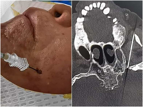 |
Figure 1 Image of puncture and CT shows the needle to the target position. |
Outcomes
Baseline: Researchers collected demographic and disease-related information from patients, including age, gender, duration of illness, affected side, comorbidities, pregabalin, as well as pre-treatment scores on the Numeric Rating Scales (NRS) and the Pittsburgh Sleep Quality Index (PSQI). The NRS is a scale ranging from 0 to 10; where 0 indicates no pain, 1–3 points indicate mild pain, 4–6 points signify moderate pain, and 7–10 points denote severe pain. Additionally, the PSQI was employed to assess the subjects’ sleep quality over the past month. The total score for all factors in the PSQI can range from 0 to 21. A PSQI score between 0 to 5 reflects good sleep quality; a total score of 6 to 10 suggests fair sleep quality; a score between 11 and 15 indicates that sleep quality is average; while a total score ranging from 16 to 21 signifies poor sleep quality. This data was gathered through an electronic medical record system.
The investigators systematically collected the outcomes included the NRS and PSQI scores at 1st, 4th, 8th, 12th, and 24th weeks afterwards. Additionally, assessed the incidence of clinically meaningful TG-PHN at 12th, and 24th weeks following treatment. The study also recorded side effects after treatment (eg, nerve damage, hematoma formation, and local infections) in both groups. A portion of the data was obtained from medical records, while further information was gathered during follow-up clinic visits and telephone interviews.
Statistical Analysis
SPSS 20.0 software (SPSS, Illinois, United States) was used to data analysis, and the GraphPad Prism 5.0 software was used to plotting the charts. Enumeration data were presented as counts and rates were compared using the χ2 test, the expected frequencies of the contingency table were calculated to ensure that all cells had an expected frequency greater than 5, thereby satisfying the conditions for applying the chi-square test. Measurement data were presented as mean ± standard deviation and compared using the t-test, and to assess variable data at different time points, a repeated measures analysis of variance (RM-ANOVA) was conducted. All quantitative data were evaluated for normality with the Shapiro–Wilk test and assessed for homogeneity of variance using Levene’s test. The results indicated that all datasets followed a normal distribution (all P > 0.05) and met the assumption of homogeneity of variance (all P > 0.05). P < 0.05 was considered statistically significant.
Results
A total of 114 patients were enrolled in this study. Among them, 4 patients were lost to follow-up and 3 patients withdrew (Figure 2). Ultimately, 107 patients were included in the analysis.
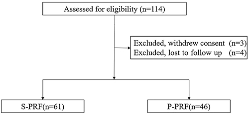 |
Figure 2 Flow diagram of the study patients. Abbreviations: S-PRF, single pulsed radiofrequency; R-PRF, repeated pulsed radiofrequency. |
The baseline characteristics of patients, including age, gender, duration of illness, affected side, comorbidities, pregabalin, pre-treatment scores of the NRS and PSQI, exhibited no significant differences between the two groups (P > 0.05, Table 1).
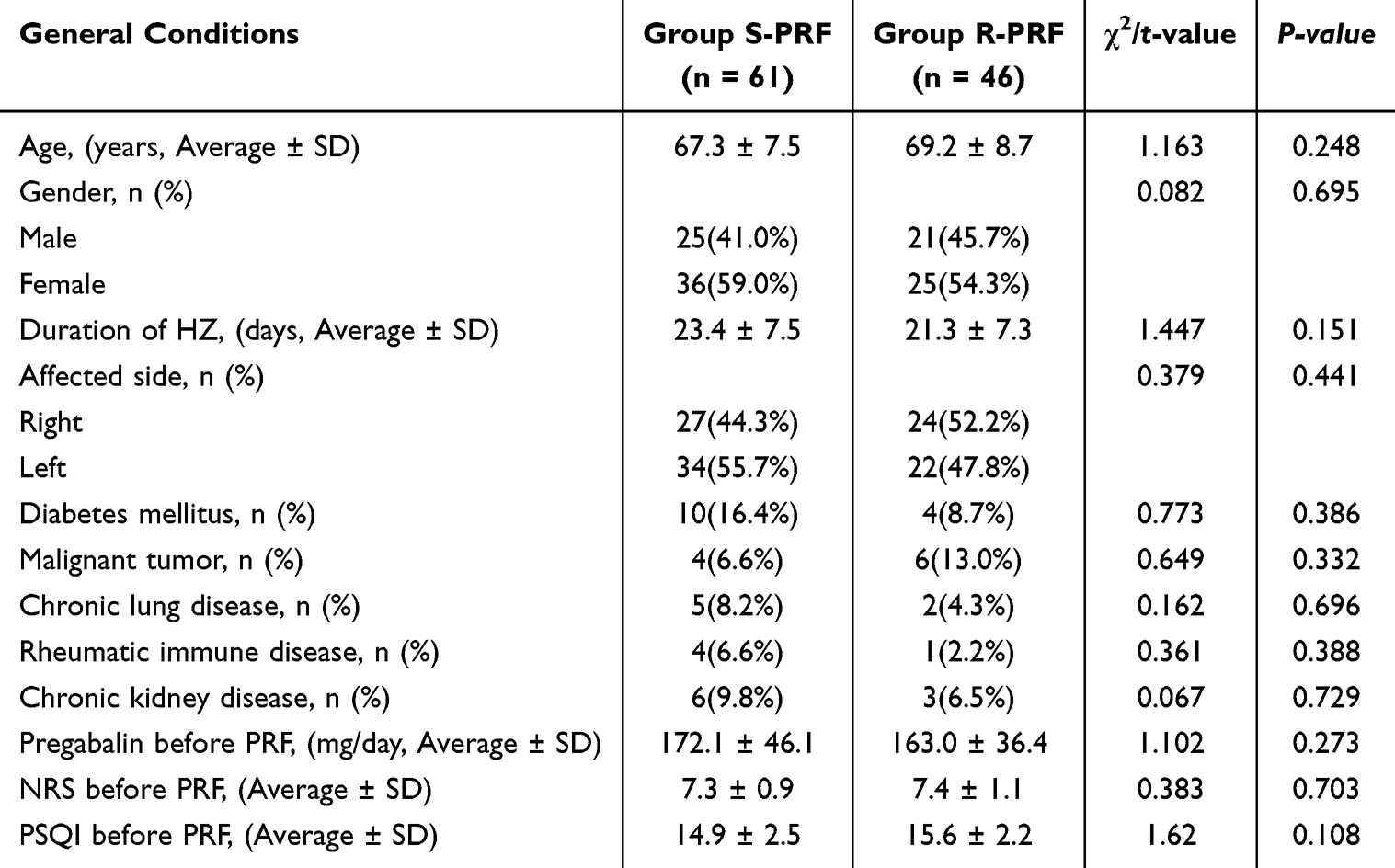 |
Table 1 Basic Characteristics of the Participants |
The NRS and PSQI scores of the group R-PRF were significantly lower than those of the group S-PRF at the 1st, 4th, 8th, 12th, and 24th weeks post-treatment (P < 0.05) (Figures 3 and 4). Additionally, the incidence of clinically meaningful TG-PHN in the group R-PRF was notably lower than that in the group S-PRF at the 12th and 24th weeks post-treatment (P < 0.05) (Table 2). No side effects associated with the treatment were reported in either group.
 |
Table 2 The Incidence of Clinically Meaningful PHN in the 12-Week and 24-Week Post-Treatment |
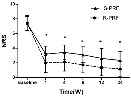 |
Figure 3 The NRS of the patients in the two groups. *P < 0.05 for Group R-PRF vs Group S-PRF. Abbreviations: NRS, numeric rating scale; S-PRF, single pulsed radiofrequency; R-PRF, repeated pulsed radiofrequency. |
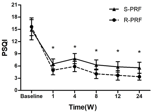 |
Figure 4 The PSQI of the patients in the two groups. *P < 0.05 for Group R-PRF vs Group S-PRF. Abbreviations: PSQI, Pittsburgh Sleep Quality Index; S-PRF, single pulsed radiofrequency; R-PRF, repeated pulsed radiofrequency. |
Discussion
In this study, we innovatively applied R-PRF technology for the treatment of TG-HZN. Our findings indicate that the group R-PRF exhibited lower NRS scores, experienced greater pain relief, and demonstrated superior sleep quality compared to the group S-PRF for TG-HZN. Notably, the incidence of clinically meaningful TG-PHN in the group R-PRF was significantly lower than that in the group S-PRF at the 12th and 24th weeks post-treatment.
Our research revealed a significant improvement in NRS and sleep quality across both groups when compared to pre-treatment levels, thereby reinforcing the effectiveness and precision of PRF therapy for HZN. PRF is a minimally invasive technique that delivers pulsed current to the targeted nerve.17 It reversibly interrupts nerve impulse transmission within small or unmyelinated fibers in the peripheral nervous system.18 The high-frequency alternating current produced by PRF interacts with target tissues to induce plasticity-related modifications that affect the processing of pain signals along central pathways. This process upregulates c-fos expression (the expression product of immediate early gene known as a molecular marker of neural activity19) and modulates levels of substance P and endorphins within the central nervous system. Consequently, it exerts an inhibitory effect on pain while simultaneously promoting both the expression and secretion of endogenous opioid peptides, thus contributing to the long-term analgesic effects associated with PRF treatment.20
In this study, the NRS and PSQI scores in the R-PRF group were significantly lower than that of the S-PRF group. Most patients in the group R-PRF experienced mild pain, while a majority of patients in the group S-PRF continued to report moderate pain, indicating a significant difference in sleep quality. For patients undergoing treatment, the group R-PRF achieved clinical outcomes that aligned with expected therapeutic effects. Zhang et al21 conducted research comparing single-PRF and repeated-PRF for the acute herpes zoster neuropathy patients with neck, thoracic, and lumbar ganglion, their findings revealed that the NRS in the R-PRF treatment were significantly reduced than that of the S-PRF at 4, 8, and12 weeks post-treatment, these results corroborate our study’s findings. The repeated stimulation of targeted nerves via R-PRF led to prolonged durations of stimulation, which resulted in cumulative effects. This approach facilitated rapid repair of damaged nerves, regulated neurological function, enhanced immune cell activity, improved neuroplasticity, and reduced central sensitization.22,23 Consequently, these factors contributed to sustained clinical efficacy over time.
In this study, the incidence rate of clinically meaningful TG-PHN in the group S-PRF at 12 weeks post-treatment was found to be 26.2%. According to Song et al,24 S-PRF demonstrated an efficacy rate of 74.7% at three months after treatment for acute/subacute zoster-related trigeminal neuralgia patients. Additionally, Jia25 conducted a retrospective analysis on the efficacy and safety of CT-guided PRF in patients with acute zoster-related trigeminal neuralgia, reporting an effective rate of 62.4% at three months post-treatment. The results from these two studies were consistent with those observed in our research, suggesting that the S-PRF may experience a ceiling effect, the duration of action for S-PRF is relatively short, leading to limited intensity of effect and consequently preventing the achievement of higher therapeutic outcomes. The incidence of clinically meaningful TG-PHN in the group R-PRF at the 12th and 24th weeks after treatment was recorded as 8.7% and 4.3%, respectively, significantly lower than that observed in the group S-PRF (26.2% and 18%). In a retrospective study by Wen et al,26 it was shown that R-PRF resulted in significantly lower VAS scores across all follow-up time points while achieving a superior overall efficacy rate compared to S-PRF (86.7% vs 60.6%) for patients suffering from subacute postherpetic neuralgia affecting thoracic and abdominal dermatomes. These findings not only support previous conclusions but also indicate enhanced efficacy over earlier studies, which may be attributed to factors such as patient characteristics involving acute herpetic neuralgia and variations in pulsed radiofrequency duration.
The timing of treatment is crucial in clinical practice, particularly for patients suffering from acute herpes zoster neuralgia, where nerve repair is the primary objective. Numerous studies have demonstrated that therapeutic effects during the acute phase are significantly superior to those observed in the chronic phase.27,28 Factors such as dorsal horn atrophy, degeneration of fibers within sensory ganglia, loss of cell axons and myelin, as well as fibrosis may contribute to diminished efficacy of PRF in patients with postherpetic neuralgia.29 Therefore, it is imperative that PRF interventions be implemented early in the treatment strategy for patients experiencing moderate-to-severe pain associated with HZN.
In this study, we collected the side effects caused by PRF (eg, nerve damage, hematoma formation, and local infections) in the two groups. An accurate preoperative evaluation of patients was conducted to exclude surgical contraindications. Intraoperative CT real-time guidance during puncture ensured comprehensive visualization and precise operation throughout the entire procedure. Additionally, utilizing a PRF temperature of 42°C was found not to damage nerves or surrounding tissues. Ultimately, no side effects related to the treatment were reported in either group within our study, thereby confirming the safety profile of R-PRF treatment.
The current study presents several limitations that should be addressed in future research. First, patients were recruited from a single center, resulting in a relatively small sample size. Expanding the sample size and collaborating with additional research centers may prove beneficial. Second, this study was not randomized and utilized a retrospective design; therefore, prospective randomized controlled trials are needed to provide more robust evidence. Third, this study only evaluated the efficacy of twice PRF treatments; it remains unclear whether an increased frequency of PRF stimulation would yield better outcomes.
Conclusion
R-PRF was a more effective treatment in alleviating the trigeminal herpes zoster neuralgia, and superior effectiveness in reducing the incidence of clinically significant trigeminal postherpetic neuralgia than S-PRF.
Data Sharing Statement
Data used to support the findings of this study are available from the corresponding author upon request.
Ethical Approval
The Ethics Committee of Hangzhou Third People’s Hospital (2025KA137) and Chinese Clinical Trial Registry (ChiCTR2500103682) approved this research.
Informed Consent
Informed consent was obtained from all individual participants included in the study.
Acknowledgments
We would like to thank the editorial board for their review and criticism in improving this paper.
Author Contributions
All authors made a significant contribution to the work reported, whether that is in the conception, study design, execution, acquisition of data, analysis and interpretation, or in all these areas; took part in drafting, revising or critically reviewing the article; gave final approval of the version to be published; have agreed on the journal to which the article has been submitted; and agree to be accountable for all aspects of the work.
Funding
This research was supported by the Hangzhou Joint Fund of the Zhejiang Provincial Natural Science Foundation of China under Grant No. LHZY24H090001 and the Zhejiang Province Traditional Chinese Medicine Science and Technology Project under Grant No. 2025ZL465. The sponsors had no role in the design or conduct of this research.
Disclosure
The authors declare that they have no competing interests in this research.
References
1. Saguil A, Kane S, Mercado M, et al. Herpes zoster and postherpetic neuralgia: prevention and management. Am Family Phys. 2017;96(10):656–8.
2. Tsau PW, Liao MF, Hsu JL, et al. Clinical presentations and outcome studies of cranial nerve involvement in herpes zoster infection: a retrospective single-center analysis. J Clin Med. 2020;9(4):946. doi:10.3390/jcm9040946
3. Patil A, Goldust M, Wollina U. Herpes zoster: a review of clinical manifestations and management. Viruses. 2022;14(2):192. doi:10.3390/v14020192
4. Jie-Lim DZ, Tey HL, Alferez-Salada BM, et al. Herpes zoster and post-herpetic neuralgia-diagnosis, treatment, and vaccination strategies. Pathogens. 2024;13(7). doi:10.3390/pathogens13070596
5. Niederer RL, Meyer JJ, Liu K, et al. Herpes zoster ophthalmicus clinical presentation and risk factors for loss of vision. Am J Ophthalmol. 2021;226(06):83–89. doi:10.1016/j.ajo.2021.02.002
6. Johnson RW, Rice AS. Clinical practice. Postherpetic neuralgia. New Engl J Med. 2014;371(16):1526–1533. doi:10.1056/NEJMcp1403062
7. Niemeyer CS, Harlander-Locke M, Bubak AN, et al. Trigeminal postherpetic neuralgia: from pathophysiology to treatment. Current Headache Rep. 2024;28(4):295–306. doi:10.1007/s11916-023-01209-z
8. DaSilva AF, DosSantos MF. The role of sensory fiber demography in trigeminal and postherpetic neuralgias. J Dent Res. 2012;91(1):17–24. doi:10.1177/0022034511411300
9. Liu DY, Chen JS, Lin CY, et al. Subcutaneous peripheral nerve stimulation for treatment of acute/subacute herpes zoster-related trigeminal neuralgia: a retrospective research. Clin J Pain. 2021;37(12):867–871. doi:10.1097/AJP.0000000000000981
10. Adriaansen EJM, Jacobs JG, Vernooij LM, et al. Herpes zoster and post herpetic neuralgia. Pain Pract. 2024;11(04). doi:10.1111/papr.13423
11. Wang CH, Dou Z, Yan MW, et al. Efficacy and safety of pulsed radiofrequency in herpes zoster related trigeminal neuralgia: a systematic review and Meta-Analysis. J Pain Res. 2023;16:341–355. doi:10.2147/JPR.S396209
12. Liu DY, Chen JS, Fang ZZ, et al. Pulsed radiofrequency of the trigeminal ganglion for treating postherpetic neuralgia of the ophthalmic branch. Pain Res Manag. 2021;30:6638392. doi:10.1155/2021/6638392
13. Chen L, Li J, Liu H, et al. Interventions for zoster associated pain: a retrospective study based on the clinical database. Front Neurol. 2022;13:1056171. doi:10.3389/fneur.2022.1056171
14. Nagda JV, Davis CW, Bajwa ZH, et al. Retrospective review of the efficacy and safety of repeated pulsed and continuous radiofrequency lesioning of the dorsal root ganglion/segmental nerve for lumbar radicular pain. Pain Physician. 2011;14(4):371–376. doi:10.36076/ppj.2011/14/371
15. Cohen JI. Clinical practice: herpes zoster. New Engl J Med. 2013;369(3):255–263. doi:10.1056/NEJMcp1302674
16. Huang B, Xie KY, Chen YJ, et al. Bipolar radiofrequency ablation of mandibular branch for refractory V3 trigeminal neuralgia. J Pain Res. 2019;12:1465–1474. doi:10.2147/JPR.S197967
17. Ojango C, Raguso M, Fiori R, et al. Pulse-dose radiofrequency treatment in pain management-initial experience. Skeletal Radiol. 2018;47(5):609–618. doi:10.1007/s00256-017-2854-8
18. Tun K, Cemil B, Gurcay AG, et al. Ultrastructural evaluation of pulsed radiofrequency and conventional radiofrequency lesions in rat sciatic nerve. Surg Neurol. 2009;72(5):496–500. doi:10.1016/j.surneu.2008.11.016
19. Chung LY. A brief introduction to the transduction of neural activity into fos signaldevelopment & reproduction. Develop Reproduct. 2015;19(2):61–67. doi:10.12717/DR.2015.19.2
20. Kim HJ, Ahn HS, Lee JY, et al. Effects of applying nerve blocks to prevent postherpetic neuralgia in patients with acute herpes zoster: a systematic review and meta-analysis. Korean J Pain. 2017;30(1):3–17. doi:10.3344/kjp.2017.30.1.3
21. Zhang EM, Fei Y, Xu LS, et al. Effect of repeated high-voltage long-duration pulsed radiofrequency on herpetic neuralgia. Pain Physician. 2022;25(7):E1047–E1055.
22. Irwin MR, Opp MR. Sleep health: reciprocal regulation of sleep and innate immunity. Neuropsychopharmacology. 2017;42(1):129–155. doi:10.1038/npp.2016.148
23. Vallejo R, Tilley DM, Williams J, et al. Pulsed radiofrequency modulates pain regulatory gene expression along the nociceptive pathway. Pain Physician. 2013;16(5):E601–E613. doi:10.36076/ppj.2013/16/e601
24. Song YH, Yu ZH, Guan JJ, et al. Efficacy of gasserian ganglion high-voltage, long-duration pulsed radiofrequency combined with block on acute/subacute zoster-related trigeminal neuralgia. Pain Res Manag. 2024;1992483. doi:10.1155/2024/1992483
25. Jia YT, Shen Y, Meng L, et al. Efficacy, safety, and predictors of response to pulsed radiofrequency therapy for acute zoster-related trigeminal neuralgia patients: a multicenter retrospective study. Pain Physician. 2022;25(4):E523–E530.
26. Wen HC, Wang Y, Cheng H, et al. Outcomes of twice repeated high-voltage long-duration pulsed radiofrequency treatment in subacute postherpetic neuralgia: a retrospective single-center analysis. J Pain Res. 2024;17:2043–2050. doi:10.2147/JPR.S465251
27. Kim KH, Jo DH, Kim ED. Pulsed radiofrequency to the dorsal root ganglion in acute herpes zoster and postherpetic neuralgia. Pain Physician. 2017;20(3):E411–418. [PMID: 28339440]. doi:10.36076/ppj.2017.E418
28. Wang H, Zhang DD, Wang SY, et al. Comparison of the efficacy of pulsed radiofrequency in treating acute herpetic neuralgia and postherpetic neuralgia in the thoracic segment. Front Neurol. 2024;15(1425796). doi:10.3389/fneur.2024.1425796
29. Bennett GJ, Watson CP. Herpes zoster and postherpetic neuralgia: past, present and future. Pain Res Manag. 2009;14(4):275–282. doi:10.1155/2009/380384
 © 2026 The Author(s). This work is published and licensed by Dove Medical Press Limited. The
full terms of this license are available at https://www.dovepress.com/terms
and incorporate the Creative Commons Attribution
- Non Commercial (unported, 4.0) License.
By accessing the work you hereby accept the Terms. Non-commercial uses of the work are permitted
without any further permission from Dove Medical Press Limited, provided the work is properly
attributed. For permission for commercial use of this work, please see paragraphs 4.2 and 5 of our Terms.
© 2026 The Author(s). This work is published and licensed by Dove Medical Press Limited. The
full terms of this license are available at https://www.dovepress.com/terms
and incorporate the Creative Commons Attribution
- Non Commercial (unported, 4.0) License.
By accessing the work you hereby accept the Terms. Non-commercial uses of the work are permitted
without any further permission from Dove Medical Press Limited, provided the work is properly
attributed. For permission for commercial use of this work, please see paragraphs 4.2 and 5 of our Terms.
Recommended articles
Outcomes of Single versus Repeated High-Voltage Pulsed Radiofrequency for Thoracic Herpes Zoster Neuralgia in Patients with Diabetes Mellitus: A Retrospective Study
Shen Q, Zhang H, Yu J, Wang B
Journal of Pain Research 2026, 19:618445
Published Date: 29 June 2026