")
Back to Journals » Veterinary Medicine: Research and Reports » Volume 12
Effect of a Topical Formulation on Infective Viral Load in Lambs Naturally Infected with Orf Virus
Authors Lacasta D , Reina R, Ruiz de Arcaute M, Ferrer LM, Benito AA, Tejedor MT , Echeverria I, Ruiz H, Martinez Cardenas S, Windsor PA
Received 12 February 2021
Accepted for publication 12 April 2021
Published 9 June 2021 Volume 2021:12 Pages 149—158
DOI https://doi.org/10.2147/VMRR.S306355
Checked for plagiarism Yes
Review by Single anonymous peer review
Peer reviewer comments 3
Editor who approved publication: Professor Young Lyoo
Delia Lacasta,1 Ramses Reina,2 Marta Ruiz de Arcaute,1 Luis Miguel Ferrer,1 Alfredo Angel Benito,3 Maria Teresa Tejedor,4 Irache Echeverria,2 Hector Ruiz,1 Silvia Martinez Cardenas,1 Peter Andrew Windsor5
1Animal Pathology Department, Instituto Agroalimentario de Aragón-IA2 (Universidad de Zaragoza-CITA), Veterinary Faculty of Zaragoza, Zaragoza, 50013, Spain; 2Instituto de Agrobiotecnología (CSIC-Gobierno de Navarra), Mutilva, 31192, Navarra, Spain; 3EXOPOL S.L, San Mateo de Gállego, Zaragoza, Spain; 4Anatomy, Embryology and Animal Genetics Department, CIBER CV (Universidad de Zaragoza-IIS), Veterinary Faculty of Zaragoza, Zaragoza, 50013, Spain; 5Sydney School of Veterinary Science, The University of Sydney, Camden, NSW, 2570, Australia
Correspondence: Delia Lacasta
Facultad de Veterinaria de Zaragoza, C/Miguel Servet 177, Zaragoza, 50013, Spain
Tel +34 609676727
Email [email protected]
Introduction: Orf is a highly contagious eruptive viral disease of the skin and mucosa of sheep and goats. Although vaccination with live or attenuated orf virus is the preferred option for disease control, the vaccine is unavailable in many countries. Treatment of orf lesions involves standard hygiene and in numerous cases, management of presumptive secondary infections with antibiotics, increasing risks of antimicrobial resistance (AMR). The wound dressing formulation Tri-Solfen® containing two local anaesthetics (lignocaine and bupivacaine), adrenaline and an antiseptic (cetrimide) in a gel formulation was developed for pain relief in sheep undergoing surgical husbandry procedures in Australia. Recently, TS therapy was found to reduce suffering and enhance recovery in cattle and buffalo with oral and skin lesions due to foot-and-mouth disease (FMD) virus infection. It was noted that TS has a low pH and is potentially viricidal, potentially aiding disease control.
Methods: One-month-old lambs (n=14), naturally infected with orf, were recruited from a farm during a natural outbreak of the disease. The animals were selected at the early stages of the infection and randomly divided into two cohorts: Group A (n=11) treated with the topical wound gel formulation (TS); and Group B (n=3) an untreated control group. Swabs were obtained before treatment (T0) and on days one (T1), 3 (T2) and 5 (T3) post-treatment, then submitted to direct DNA extraction with real-time PCR quantification, plus incubation with primary tissue cultures from ovine skin fibroblasts (OSF) and T-immortalized goat embryonic fibroblasts (TIGEF).
Results: Although no significant differences were found in the clinical progression of the lesions and PCR quantification (p=0.722) between these small cohorts, there was a significant difference (p< 0.05) in reduction in infective viral load between the groups when assessed in OSF cell cultures between T0 and T3.
Conclusion: These preliminary findings suggest that treatment of early stage lesions with this TS may reduce the infective viral load present in orf lesions.
Keywords: sheep, contagious ecthyma, wound formulation, local therapy
Introduction
Contagious ecthyma, also known as orf, contagious pustular dermatitis, sore mouth or scabby mouth, is a highly contagious eruptive skin condition of sheep, goats and other ruminants, although zoonotic transmission also occurs. Orf disease affects mainly young animals in the first year of their life, with severe outbreaks generally associated with intensive sheep husbandry or transport. Orf virus belongs to the genus Parapoxvirus, family Poxviridae, sub-family Chordopoxvirinae.1 It is a pathogen with worldwide distribution, causing significant financial losses in livestock production. Transmission of orf is from direct or indirect contact with the orf virus from pustules of infected animals containing-virus or live vaccines, causing painful lesions in sheep and goats, and potentially people, especially farmers and veterinarians. Orf is very resistant in the environment, particularly in dry atmospheres, with the virus shown to be infective for up to 17 years.2 Clinical presentation in lambs or kids includes the formation of vesicles, papules, pustules with a yellowish creamy and/or proliferative appearance, then scabs that finally become dry and shed, usually with no scar remaining.1 Although the most common lesions can be found in the oral cavity, these can be extended to the skin of the face, ears, mammary gland, feet, flanks, scrotum and perianal area.3
Disease diagnosis is usually based on clinical presentation. However, laboratory testing and confirmation may be critical as some of the differential diagnoses are notifiable diseases, especially foot-and-mouth disease (FMD), bluetongue (BT), sheep pox and peste des petits ruminants (PPR). Laboratory diagnosis can be achieved through electron microscopy, PCR based on the amplification of B2L4 or VIR5 genomic regions, and/or, isolation of the virus in cell culture using a variety of primary and continuous cell lines.
Vaccination is the preferred option to control the disease. Live virus and attenuated vaccines usually provide adequate protection against orf virus, albeit not life-long, with their annual administration considered the best strategy to control the disease in flocks and herds.6 However, orf vaccine is currently unavailable in most European countries, and where available, existing live-attenuated or scab-based vaccines may revert to virulence and are not fully protective. This could be due to genetic deletions present in attenuated vaccines, that may significantly reduce their immunogenicity and efficacy, and genetic heterogeneity of field strains.7–9
The treatment of this disorder involves standard hygiene practices and in numerous cases, management of presumptive secondary infections, usually with antibiotic treatments.2 As reducing antibiotic use in livestock has become a priority for the management of antimicrobial resistance (AMR) risk, there is a need for treatment protocols that provide more effective topical treatment of orf cases. Recent studies have confirmed the efficacy of the wound therapy formulation Tri-Solfen® (TS) (Animal Ethics Pty Ltd, Australia) for reducing pain and hastening healing of skin and mucosal lesions in sheep and cattle. The dressing formulation contains two local anaesthetics (lignocaine and bupivacaine), adrenaline and an antiseptic (cetrimide) in a gel formulation that creates a barrier effect, numbing the pain of lesions, rapidly reducing their infectivity, and hastening healing, potentially reducing the weight loss in affected individuals. The TS product was developed for pain relief in sheep undergoing surgical husbandry procedures in Australia. It is also registered for use in cattle in Australia and New Zealand for husbandry procedures and more recently in Laos as a therapy for reducing suffering and enhancing recovery of lesions FMD.10–12 Registration of TS in Europe is pending, although it has been shown to be efficacious in the provision of pain relief and more rapid healing of husbandry lesions in sheep in Australia and Spain.10,13,14 As TS has a pH of 2.7, it has a potential viricidal impact that may reduce transmission risks, with the formulation potentially aiding healing and avoiding the need for other treatments, including antibiotics.11,12 This paper describes a preliminary investigation of orf therapy with TS, examining the potential antiviral roles and healing properties of the pain-relief formulation in naturally infected lambs with orf, through viral genome real-time PCR quantification and tissue culture in ovine primary cells.
Materials and Methods
All procedures were conducted under Project Licence PI 40/19 approved by the Ethics Committee for Animal Experiments from the University of Zaragoza. The care and use of animals were performed according to the Spanish Policy for Animal Protection RD53/2013, meeting the European Union Directive 2010/63 on the protection of animals used for experimental and other scientific purposes.
Animals
One-month-old lambs (n=14) with similar weights and naturally infected with orf were recruited from a farm where a natural outbreak of orf disease was occurring. The 1800 Lacaune sheep farm was located in a small village of the Pyrenees mountains. Due to high prolificacy (2.2), some lambs were routinely artificially fed milk by an artificial lactation machine. These lambs were kept in a separate pen with proper cleaning and disinfection conditions, although the farmers reported the lambs frequently had recurrent orf infections. The lambs selected for the experiment were in the early stages of orf infection when lesions were initially diagnosed. The lambs were co-located in a separate pen and randomly divided into two cohorts, with Group A (n=11) consisting of animals with orf lesions treated with TS and Group B (n=3), an orf-infected control group remaining untreated. All lambs were subjected to the same management, with artificial feeding conducted until weaning at 45 days of age. From trial commencement, the lambs also had ad libitum access to compound feedstuff and straw. No other treatments were administered throughout the duration of the study.
Sampling
Sterile cotton swabs were inserted into the lesions prior to the application of treatments (T0) to confirm the presence of orf virus. This was followed by a similar sampling of the lesions on days 1 (T1), 3 (T2) and 5 (T3) post-treatment of all the lesions observed on the lambs.
Samples of 3mL of whole blood were also collected from the jugular vein through a vacutainer system into EDTA tubes to perform haematology in all the animals prior to (T0) and ten days (T4) following treatment, respectively.
Treatment Application
Following confirmation of the presence of orf virus in the lesions, a single spray of 1.5mL of TS was applied liberally with a spray gun to all orf lesions in Group A lambs, with the Group B lambs remaining untreated.
Clinical Progression
Clinical examination of all lambs was performed by observers blinded to treatment. This occurred daily for 11 days to determine the clinical progression of the lesions, with data collected on the changes occurring in the lesions. In addition, photographic records of the lesions were taken at three angles, including the two lateral profiles and an anterior (frontal) image with the mouth open to observe and record lesions occurring inside the mouth. Images were also taken with a ruler to provide size comparisons. The number of lesions and the morphological appearance of these lesions were chronologically recorded from each animal, described as papules, pustules, proliferations, crusts, or erosions.
Haematology
Haematology was performed using an automatic haematological counter IDEXX ProcyteDx (IDEXX laboratories, Westbrook, ME, USA). Measured parameters were leucocytes (K/mL), erythrocytes (M/mL), haemoglobin (g/dL), haematocrit (%), platelets (K/mL), MCV (Mean Corpuscular Volume; fL), MCH (mean corpuscular haemoglobin; pg), MCHC (mean corpuscular haemoglobin concentration; g/dL) and reticulocytes (K/mL). White blood cells were assessed by counts of neutrophils (K/mL-1), lymphocytes (K/mL), monocytes (K/mL), basophils (K/mL) and eosinophils (K/mL).
Real-Time PCR
Sterile swab samples taken from the lesions at T0, before treatment, and T1, T2 and T3 were submitted to direct DNA extraction and further real-time PCR quantification. Nucleic acid extraction was performed using the commercial MagMAX™ Pathogen RNA/DNA kit (Thermo Fisher Scientific) and the automated magnetic particle processor KingFisher Flex System (Thermo Fisher Scientific), following the manufacturer’s instructions. Extracted DNA samples were stored at −80°C until end of sampling in order to evaluate all of them in a single run of real-time PCR quantification. Orf virus detection was performed using the commercial qPCR kit EXOone Contagious Ecthyma (Exopol, Spain) which targets the BL2 gene that encodes a major envelope viral antigen. The kit contains a quantified synthetic positive control, and also an endogenous control to avoid false-negative results. Amplification was performed on a FAST 7500 cycler (Applied Biosystems), and a cut-off value for positive samples was established at cycle quantification (Cq) values lower than 38.
Virus Culture
Sterile swabs collected at T0, T1, T2 and T3 were submitted to incubation with primary tissue cultures from ovine skin fibroblasts (OSF) and T-immortalized goat embryonic fibroblasts (TIGEF). Briefly, swabs were immersed in 2mL of tissue culture medium, DMEM supplemented with 1% glutamine, 2% foetal bovine serum and 2% antibiotics (Sigma Aldrich) and then added to cells. Cells were incubated at 37°C, 5% CO2 atmosphere for 5 days. DNA extraction was performed in cells using E.Z.N.A Blood DNA Mini Kit (Omega bio-tek). Orf virus detection and viral load quantification were conducted by real-time quantitative PCR in an Agilent AriaMx machine using commercial PCR EXOone Contagious Ecthyma (Exopol, Spain).
Statistical Analysis of Results
Statistical analyses were performed using IBM SPSS statistics version 26 (2019) software (IBM, Armonk, NY, USA). Shapiro–Wilk test was applied to quantitative variables for assessing normal distribution. Where normal distribution was assessed, two ways mixed ANOVA were applied when considering repeated measures (within-subject factor: repeated measure; between-subject factor: group). Where quantitative variables were not normally distributed, non-parametric tests were applied (Mann–Whitney´s U-test for independent samples and Wilcoxon test for paired samples). Viral load data were analysed by T-Student’s test for unrelated samples. Percentages were compared by Pearson chi-square test and Fisher´s exact test. A difference was considered statistically significant when p≤ 0.050.
Results
At sampling performed before treatment (T0), all lambs tested positive to orf virus on real-time PCR, with Cq values ranging from 23.1 to 35.9 (threshold value <38).
Clinical Progression
Lesion progression was daily analysed by clinical examination for eleven days following treatment in addition to a review of lesion progression from images using three different angles. Firstly, the progression of the lesion surface was calculated with the formula of the area of the circle (πr2), adding the left, right and anterior surfaces of each type of lesions (papules, pustules, proliferations, crusts and erosions) (Table 1). No differences were found in any type of lesions throughout the study between treated and untreated lambs.
 |
Table 1 Mean and Standard Deviation (SD) for the Total Surface of Lesions (cm2), per Day, Group and Type of Lesion. Group A (n=11) Consisting of Animals with Orf Lesions Treated with TS and Group B (n=3), an Orf-Infected Control Group Remaining Untreated |
In addition, the total number of each type of lesion per lamb was analysed, with no differences noted between the groups (Table 2). However, as expected, the total number of papules (initial orf lesion) decreased throughout the study, whereas there was no difference in the number of pustules and proliferations, with the number of crusts increasing as days progressed. Finally, there were no differences in the number of lambs presenting each type of lesion at each time point of the study.
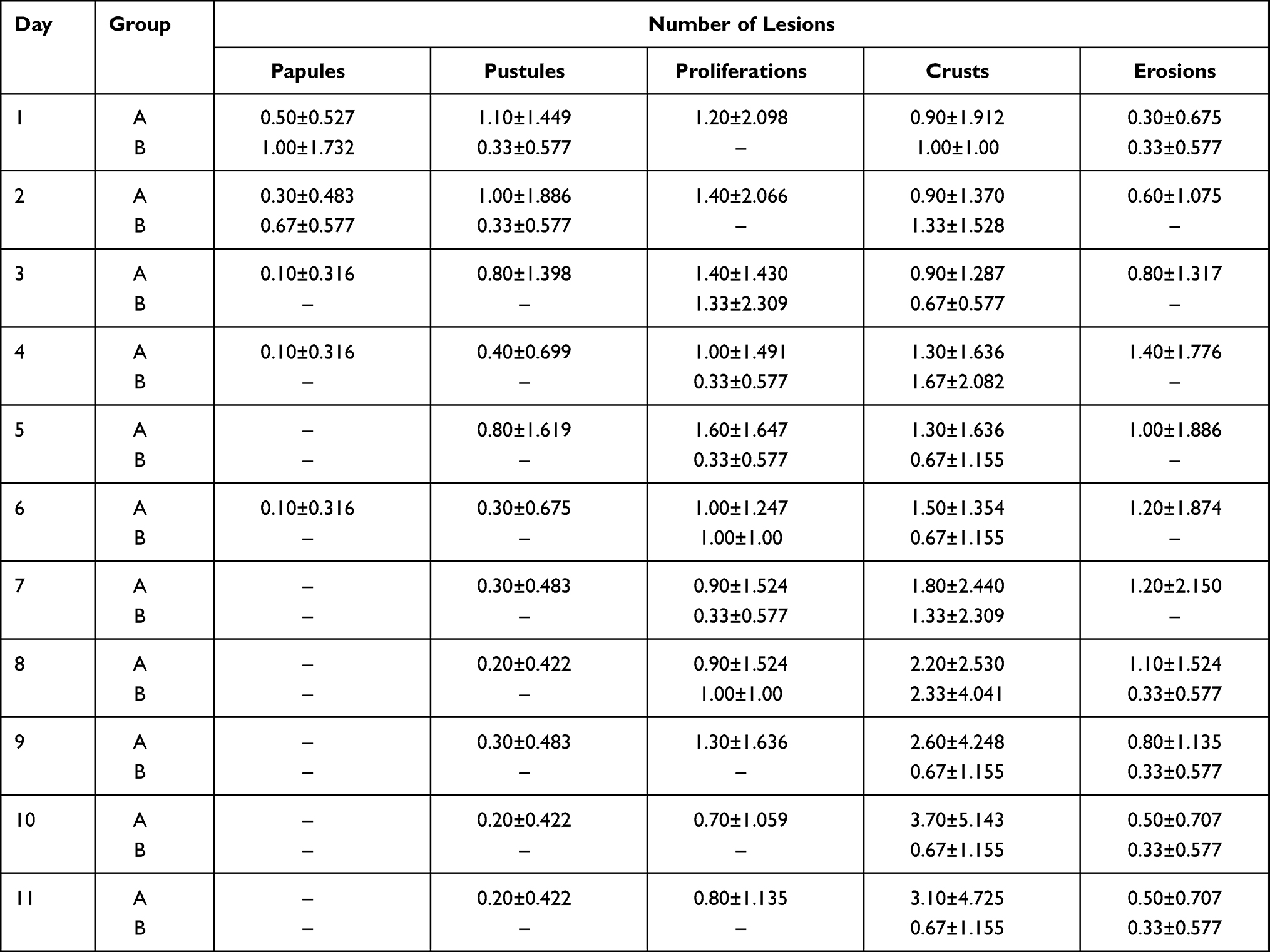 |
Table 2 Mean and Standard Deviation (SD) for the Total Number of Lesions per Day, Group and Type of Lesion. Group A (n=11) Consisting of Animals with Orf Lesions Treated with TS and Group B (n=3), an Orf-Infected Control Group Remaining Untreated |
The number and percentage of lambs with lesion per day, group and type of lesion are displayed (Table 3). No differences were found in any type of lesions throughout the study between treated and untreated lambs.
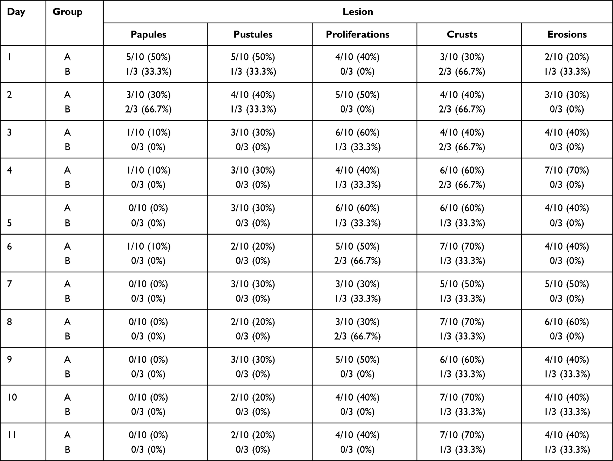 |
Table 3 Number and Percentage of Lambs That Presented the Different Type of Orf Lesions Each Day. Group A (n=11) Consisting of Animals with Orf Lesions Treated with TS and Group B (n=3), an Orf-Infected Control Group Remaining Untreated |
Haematology
All blood parameters analysed remained within normal ranges throughout the entire study, with the means and standard deviation of the haematological parameters analysed displayed (Table 4). A slight neutrophilia was observed on the eleventh day of study from the lambs of group A, and no significant differences were found between groups (p>0.050). Although a significant increase was observed in erythrocytes, total leukocytes, neutrophils and monocytes on day 11 in comparison with day 1, the MCV and MCH had significantly decreased on the 11th day.
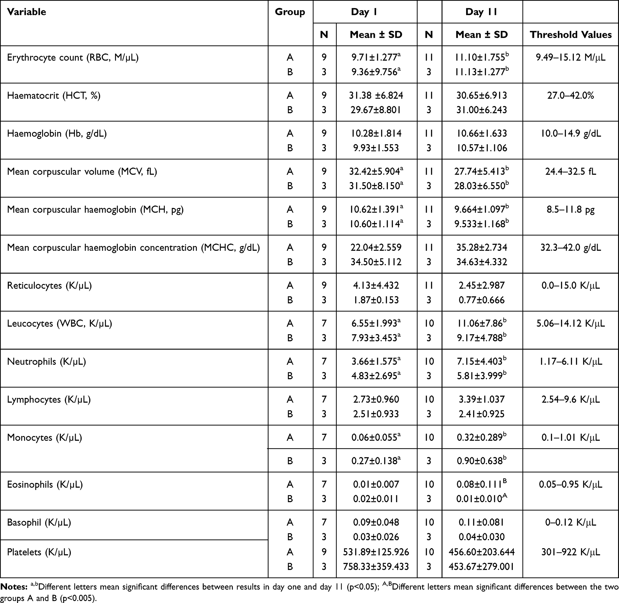 |
Table 4 Means and Standard Deviation (SD) of the Haematological Parameters. Group A (n=11) Consisting of Animals with Orf Lesions Treated with TS and Group B (n=3), an Orf-Infected Control Group Remaining Untreated |
Real-Time PCR
All animals tested positive in all samples, except one lamb that tested negative in T2 but returned positive in T3. For the statistical study, the Cq values obtained at T0 and T3 were analysed, and no significant differences were observed between treated and untreated animals (p=0.722). The mean Cq was 29.63 in T0, 28.52 in T1, 29.62 in T2 and 29.17 in T3 (Table 5).
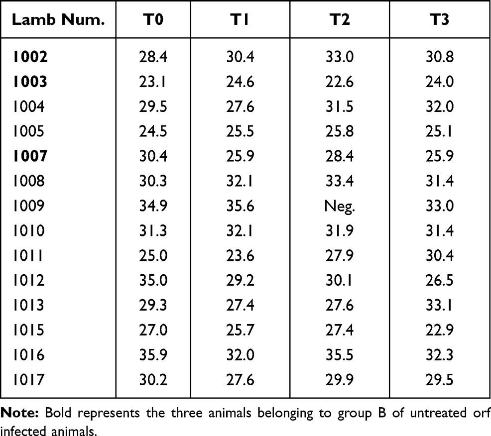 |
Table 5 Real-Time PCR Cq Values of Each Analysed Lamb in T0, Immediately Before Treatment, and on Days 1 (T1), 3 (T2) and 5 (T3) Post-Treatment |
Virus Culture
Cotton swabs submitted to incubation with primary tissue cultures from ovine skin fibroblasts (OSF) and T-immortalized goat embryonic fibroblasts (TIGEF) showed a viral load reduction in T0 and T3 between treated and untreated animals in both groups throughout the experiment (Figures 1 and 2). However, statistical differences were only found in the viral load difference obtained in the case of OSF cells (p<0.05). TIGEF showed no difference in control animals comparing T0 and T3 despite treated animals displaying lower viral loads at the end of the experiment (p<0.05).
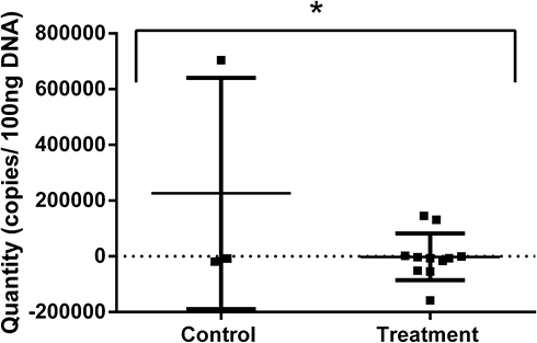 |
Figure 1 Orf viral load in ovine skin fibroblasts (OSF) incubated with swab samples from naturally infected lambs obtained from control and treated groups. Data shown are the differences in viral load from T0 to T3 in control and treated animals (*p< 0.05). |
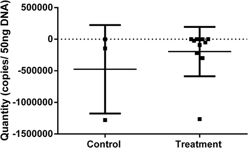 |
Figure 2 Orf viral load in T-immortalized goat fibroblasts (TIGEF) incubated with samples from naturally infected lambs obtained from control and treated groups. Data shown are the differences in viral load from T0 to T3 in control and treated animals (p> 0.05). |
Discussion
In recent years, the novel topical anaesthetic wound formulation TS developed for reduced pain and enhanced wound healing in surgical husbandry procedures in livestock, has been suggested as potentially appropriate for the management of other disorders in a variety of species.10, Most recently, TS was found to be efficacious for the treatment of foot-and-mouth disease (FMD) in cattle and buffalo in Laos and cattle in Cameroon.11,12 In these studies, clinical observations and lesion measurements confirmed that improved welfare and healing occurred following treatment of FMD lesions, with treated cattle and buffalo achieving both superior appetite and lesion healing scores with a more rapid reduction in dimensions of lesions than other groups.11,12 In Laos, it was noted that as the presentation of affected animals for treatment and reporting of outbreaks improved, the use of TS had potential to improve disease surveillance, reduce the socioeconomics impacts of outbreaks, and reduce risks of AMR.12
These studies also speculated that TS might have a viricidal effect against FMD virus if applied prior to or at the time of lesion rupture, potentially limiting virus transmission during FMD outbreaks.12 As TS has a pH of 2.7–2.9, if applied early in the course of the disease, it was suggested that antiviral activity might potentially limit virus transmission during outbreaks. Similarly, hypocrellin A antiviral activity seems to be related to a decline in pH.15 Low pH has been related to viricidal activity in multiple models, including Herpes Simplex-1 (HSV-1) in dental alginates.16 Whilst the application of acidic solutions to open wounds and ulcers is generally contraindicated, as the acidity may exacerbate pain, the relatively high concentration of lidocaine (5%) applied with bupivacaine, adrenaline, and cetrimide in a gel matrix has been shown to provide rapid and prolonged wound anaesthesia from blockage of nociception,16 with the acidity potentially sufficient to destroy virus without causing pain to the animal. It has also been postulated that the concentration of lidocaine in TS is likely to be directly viricidal against FMD virus, as at concentrations ranging from 0.5 mg/mL (0.05%) to 100 mg/mL (10%) lidocaine blocks nociception10 and has been shown to exhibit antiviral activity against the herpes virus in cell-culture and animal-model systems.17 This encouraged the present study, examining the impact of TS on orf lesions, another viral disease that causes oral lesions.
This study reports the first field treatment trial of orf lesions using TS as a therapy for the clinical management of contagious ecthyma. Despite necessary limitations on numbers of animals recruited for the trial due to low availability of resources, the preliminary results provided an encouraging indication of the potential therapeutic use of TS for reduction of infective viral load, observed as a significant reduction in the viral quantities detected in the Group A treated animals at five days’ post-treatment after incubation with OSF in tissue culture. In spite of the fact that the viral load represented in the PCR Cq values was maintained in the five days that the study lasted, the reduction in the viral load shown in the culture probably demonstrates that the load detected in the PCR is of already inactivated viruses.
A reduction in healing scores was not observed in the trial. However, this was expected as orf infections typically trigger lesions involving abnormal ballooning of basal epithelial cells deep in the epidermis with the rapid escalation of lesions dimensions and it was not expected that these rapidly advancing lesions could readily be resolved.18 However, it was postulated that the presence of TS absorbed into the surface epithelium of lesions, especially in early stages of the disease, could result in negative impacts on virus particles as they were released from damaged epithelial cells, resulting in the reduced infective viral load observed. The findings from this study support the hypothesis that the early applications of TS to orf infected animals could reduce virus transmission on the farm. Potentially, this may reduce the likely appearance of new cases, the extent of animal suffering, the economic losses and the risk of zoonotic transmission of orf. However, further studies are indicated to explore this important preliminary finding.
Contagious ecthyma is a highly contagious disease, and when severe outbreaks occur, up to 100% of lambs may show some degree of lesions attributable to orf infection.2 Use of TS treatment for orf potentially could control the spread of the virus, reducing the enormity of outbreaks. Further, as the product contains an antiseptic, use could also diminish the risks of secondary infections, with TS offering a non-antimicrobial therapeutic option for treating clinical orf lesions, potentially reducing the risk of AMR that occurs with antimicrobial use in current orf therapy. Of interest, was a recent observation that a lower rate of secondary infection occurred following application of TS during surgical tail-docking of lambs, enabling the consideration of replacing routine antibiotic cover with this topical anaesthetic and antiseptic wound formulation.14
The positive response to therapy observed in two reported studies on the use of this product for clinical FMD, accompanied by an absence of adverse side-effects, suggest this novel approach to viral therapy is worthy of further consideration, particularly if the findings in this preliminary trial of viricidal activity are found to be consistent in future studies. The product invokes positive and prolonged pain-relieving and wound healing effects in livestock following blockage of nociception during treatment of wounds and lesions from aversive husbandry procedures and epitheliotropic virus infections, as described.10–12,19–23 This novel approach to the treatment of mucosal and skin viral lesions in ruminants can potentially lead to reductions in the transmission of the disease, with positive improvements in animal welfare and reduction of AMR risk. Further studies on the role of topical anaesthetic lesion therapy in managing some of the most important viral diseases of small and large ruminants are proposed.
Conclusion
This is the first scientific report of the application to orf lesions in lambs during an outbreak, of a novel pain-relief and wound-healing therapy developed for managing aversive livestock procedures. Although no significant differences were found in the reduction of lesion scores, the significant decrease of infective viral load in the treated group is encouraging, suggesting further studies on the use of TS for treatment of orf lesions in small ruminants are warranted. This innovation could alter the paradigm of orf treatments that have traditionally focused on using inappropriate and expensive antimicrobials, to one of improving the welfare of animals suffering from a viral disease. This innovation may provide both clinical benefits to affected animals, reduce AMR and food-safety risks, plus potentially, reduce viral transmission loads and the severity of outbreaks.
Abbreviations
FMD, foot-and-mouth disease; TS, pain-relief product (Tri-Solfen); AMR, antimicrobial resistance; OSF, tissue cultures from ovine skin fibroblasts and TIGEF, tissue cultures from T-immortalized goat embryonic fibroblasts.
Acknowledgments
We would like to acknowledge the farmers Marcos Périz Bestué, Antonio Périz Barbanoj and María Jesús Bestué Noeguero that allows us to work with the animals on their farm.
Funding
This research was supported by funding and product from the Australian company Animal Ethics Pty Ltd. The work was also supported by the Aragón Government and the European Social Fund (A15_17R, Construyendo Aragón 2016-20) and Project CONECTIM funded by Gobierno de Navarra (PC052-053).
Disclosure
The abstract of this paper was presented at the International Congress on the breeding of sheep and goats. A hybrid conference that was held in Bonn, as an oral presentation with interim findings. The authors report no conflicts of interest in this work.
References
1. Nandi S, De Ujjwal K, Chowdhury S. Current status of contagious ecthyma or orf disease in goat and sheep—a global perspective. Small Rum Res. 2011;96:73–82. doi:10.1016/j.smallrumres.2010.11.018.
2. Spyrou V, Valiakos G. Orf virus infection in sheep and goats. Vet Microbiol. 2015;181:178–182. doi:10.1016/j.vetmic.2015.08.010.
3. Reid HW. Orf. In: Martin WB, Aitken ID, editors. Diseases of Sheep.
4. Inoshima Y, Murakami K, Yokoyama T, et al. Genetic heterogeneity among parapoxviruses isolated from sheep, cattle and Japanese serows (Capricornis crispus). J Gen Virol. 2000;82:1215–1220. doi:10.1099/0022-1317-82-5-1215.
5. Kottaridi C, Nomikou K, Lelli R, et al. Laboratory diagnosis of contagious ecthyma: comparison of different PCR protocols with virus isolation in cell culture. J Virol Methods. 2006;134:119–124. doi:10.1016/j.jviromet.2005.12.005.
6. Lacasta D, Ferrer LM, Ramos JJ, et al. Vaccination schedules in small ruminant. Vet Microbiol. 2015;181:34–46. doi:10.1016/j.vetmic.2015.07.018.
7. Musser J, Taylor CA, Guo J, et al. Development of a contagious ecthyma vaccine for goats. Am J Vet Res. 2008;69:1366–1370. doi:10.2460/ajvr.69.10.1366.
8. Musser JMB, Waldron DF, Taylor CA. Evaluation of homologous and heterologous protection induced by a virulent field strain of Orf virus and an Orf vaccine in goats. Am J Vet Res. 2012;73:86–90. doi:10.2460/ajvr.73.1.86.
9. da Costa RA, Cargnelutti JF, Schild CO, et al. Outbreak of contagious ecthyma caused by Orf virus (Parapoxvirus ovis) in a vaccinated sheep flock in Uruguay. Braz J Microbiol. 2019;50:565–569. doi:10.1007/s42770-019-00057-7.711
10. Roberts CD, Windsor PA. Innovative pain management solutions in animals may provide improved wound pain reduction during debridement in humans: an opinion informed by veterinary literature. Int Wound J. 2019;16(4):968–973. doi:10.1111/iwj.13129.
11. Lendzele S, Mavoungou J, Burinyuy K, et al. Efficacy and application of a novel topical anaesthetic wound formulation for treating cattle with foot-and-mouth disease: a field trial in Cameroon. Transbound Emerg Dis. 2020;00:1–12. doi:10.1111/tbed.13923.
12. Windsor PA, Earp F, Mac Phillamy I, et al. Managing welfare and antimicrobial-resistance issues in treating foot-and-mouth disease lesions: a new therapeutic approach. Vet Med Res Rep. 2020;11:99–107. doi:10.2147/VMRR.S273788.
13. Windsor PA, Lomax S, White P. Progress in pain management to improve small ruminant farm welfare. Small Rum Res. 2016;142:55–57. doi:10.1016/j.smallrumres.2016.03.024.
14. Ferrer LM, Lacasta D, Ortín A, et al. Impact of a topical anaesthesia wound management formulation on pain, inflammation and reduction of secondary infections after tail docking in lambs. Animals. 2020;10(8):1255. doi:10.3390/ani10081255
15. Chaloupka R, Sureau F, Kocisova E, et al. Hypocrellin A photosensitization involves an intracellular pH decrease in 3T3 cells. Photochem Photobiol. 1998;68(1):44–50. doi:10.1111/j.1751-1097.1998.tb03251.x.
16. Nallamuthu N, Braden M, Oxford J, et al. Modification of pH conferring viricidal activity on dental alginates. Materials. 2015;8(4):1966–1975. doi:10.3390/ma8041966.
17. Haines HG, Dickens CB, Brigham DP. Antiviral pharmaceutical preparations and methods for their use. Patent US 4628063A. 1986.
18. Windsor PA, Nampanya S, Tagger A, et al. Is Orf virus a risk to expanding goat production systems in developing countries? A study from Lao PDR. Small Rum Res. 2017;154:123–128. doi:10.1016/j.smallrumres.2017.08.003.
19. Lomax S, Sheil M, Windsor PA. Impact of topical anaesthesia on pain alleviation and wound healing in lambs after mulesing. Aus Vet J. 2008;86:159–168. doi:10.1111/j.1751-0813.2008.00285.x.
20. Paull DR, Lee C, Colditz IG, et al. The effect of a topical anaesthetic formulation, systemic flunixin and carprofen, singly or in combination, on cortisol and behavioural responses of Merino lambs to mulesing. Aus Vet J. 2009;85:98–106. doi:10.1111/j.1751-0813.2009.00429.x
21. Lomax S, Dickson H, Sheil M, et al. Topical anaesthesia alleviates short-term pain of castration and tail docking in lambs. Aus Vet J. 2010;88:67–74. doi:10.1111/j.1751-0813.2009.00546.x
22. Lomax S, Sheil M, Windsor PA. Duration of action of a topical anaesthetic formulation for pain management of mulesing in sheep. Aus Vet J. 2013;91:160–167. doi:10.1111/avj.12031.
23. Windsor PA, Lomax S. Addressing welfare concerns in control of ovine cutaneous myiosis in sheep in Australia. Small Rum Res. 2013;110:165–169. doi:10.1016/j.smallrumres.2012.11.027.
 © 2021 The Author(s). This work is published and licensed by Dove Medical Press Limited. The full terms of this license are available at https://www.dovepress.com/terms.php and incorporate the Creative Commons Attribution - Non Commercial (unported, v3.0) License.
By accessing the work you hereby accept the Terms. Non-commercial uses of the work are permitted without any further permission from Dove Medical Press Limited, provided the work is properly attributed. For permission for commercial use of this work, please see paragraphs 4.2 and 5 of our Terms.
© 2021 The Author(s). This work is published and licensed by Dove Medical Press Limited. The full terms of this license are available at https://www.dovepress.com/terms.php and incorporate the Creative Commons Attribution - Non Commercial (unported, v3.0) License.
By accessing the work you hereby accept the Terms. Non-commercial uses of the work are permitted without any further permission from Dove Medical Press Limited, provided the work is properly attributed. For permission for commercial use of this work, please see paragraphs 4.2 and 5 of our Terms.