
Back to Journals » Infection and Drug Resistance » Volume 19
Disseminated Mycobacterium avium Complex Infection in an HIV Patient with a History of Talaromyces marneffei: Diagnostic Value of Blind Subculture and Suspected Management Challenges of Immune Reconstitution Inflammatory Syndrome
Authors Niu X, Yu Q, Gu J, Lu B  , Shen W, Tian J
Received 4 March 2026
Accepted for publication 18 June 2026
Published 23 June 2026 Volume 2026:19 606947
DOI https://doi.org/10.2147/IDR.S606947
Checked for plagiarism Yes
Review by Single anonymous peer review
Peer reviewer comments 2
Editor who approved publication: Professor Chi H. Lee
Xiaoqin Niu,1 Qinlong Yu,1 Jiong Gu,2 Binbin Lu,1 Weifeng Shen,1 Junhua Tian1
1Department of Clinical Laboratory, The Affiliated Hospital of Jiaxing University, Jiaxing, 314000, People’s Republic of China; 2Department of Infectious Diseases, The Affiliated Hospital of Jiaxing University, Jiaxing, 314000, People’s Republic of China
Correspondence: Junhua Tian, Department of Clinical Laboratory Medicine, The Affiliated Hospital of Jiaxing University, No. 1882, Zhong-Huan South Road, Nanhu District, Jiaxing, Zhejiang, 314000, People’s Republic of China, Tel +86-13625838848, Email [email protected] Weifeng Shen, Department of Clinical Laboratory Medicine, The Affiliated Hospital of Jiaxing University, No. 1882, Zhong-Huan South Road, Nanhu District, Jiaxing, Zhejiang, 314000, People’s Republic of China, Tel +86-15968388122, Email [email protected]
Abstract: This study reported a 33-year-old male acquired immune deficiency syndrome (AIDS) patient with a 10-year human immunodeficiency virus (HIV) infection history, poor antiretroviral therapy (ART) adherence, and two previous Talaromyces marneffei infections. Self-discontinuation of ART led to severe immunosuppression and disseminated Mycobacterium avium complex (MAC) infection involving the bloodstream and bone marrow. After the restart of ART, the patient developed persistent high fever, which was clinically suspected to be MAC-associated immune reconstitution inflammatory syndrome (IRIS). However, due to the lack of serial HIV viral load and CD4⁺ T lymphocyte data, a definitive diagnosis could not be established. The patient was admitted with fatigue, anorexia, and black stool as the main symptoms. MAC infection was confirmed by blood culture, bone marrow culture, and bone marrow metagenomic next-generation sequencing (mNGS) at a higher-level hospital. Notably, after transfer to our hospital, the microbiology laboratory performed blind subculture on routinely negative blood culture bottles and extended the incubation period to 15 days, successfully isolating MAC. This highlights the crucial significance of close clinical-laboratory collaboration and optimized pathogen detection for diagnosing non-tuberculous mycobacteria (NTM) infections. After initial infection control and ART restart, the patient developed recurrent fever. Given the temporal association with ART reinitiation and the dose-dependent correlation between fever and glucocorticoid adjustments, possible MAC-associated IRIS was suspected. The patient’s clinical symptoms improved with glucocorticoid therapy, though this does not confirm the diagnosis. Complications including cytomegalovirus reactivation, adverse drug reactions, and human rhinovirus co-infection were managed in a standardized manner. This case suggests that the diagnosis of disseminated MAC infection in severely immunocompromised AIDS patients relies on efficient collaboration between clinicians and laboratories. However, in the absence of confirmatory immunological and virological evidence, the diagnosis of IRIS remains uncertain. Clinicians should remain vigilant for suspected IRIS when restarting ART while acknowledge that limited data may preclude a definitive diagnosis. Individualized comprehensive strategies covering anti-infection, immunomodulation, anti-inflammation, and supportive treatment are the key to managing such complex HIV-related opportunistic infections.
Keywords: Mycobacterium avium complex, immune reconstitution inflammatory syndrome, microbiological technology, disseminated infection, non-tuberculous mycobacteria
Introduction
Mycobacterium avium complex (MAC) is one of the most common opportunistic pathogens in patients with advanced AIDS, which can affect multiple tissues and organs throughout the body and cause life-threatening disseminated infections.1–3 At the beginning of the HIV pandemic, the incidence of disseminated MAC in adult AIDS patients was as high as 16–40%.4 However, with the wide application of highly active antiretroviral therapy (HAART), disseminated MAC has become extremely rare.4 Although ART significantly reduces the overall incidence rate of MAC infection, timely and accurate diagnosis is still crucial to reduce mortality.5 The clinical diagnosis of MAC infection mainly relies on the isolation and culture of pathogens from sterile sites such as blood and bone marrow.6 However, MAC grows slowly and often cannot trigger positive signals within the routine 5-day cultivation cycle of automated cultivation systems, which may lead to false negative reports and clinical misdiagnosis. Currently, when there is a high clinical suspicion of non-tuberculous mycobacteria (NTM) infection, it is recommended to extend the culture time to 6–8 weeks.7 However, in resource-limited areas, this standard has not been uniformly implemented, further complicating the diagnosis. Moreover, some patients may experience MAC-associated immune reconstitution inflammatory syndrome (IRIS) during the process of receiving effective ART treatment and gradually rebuilding immune function.8 This makes the management of disseminated MAC in patients with advanced AIDS more complex. Clinicians need to seek a precise balance between anti-infective treatment, the timing of ART initiation, and the risk of IRIS.9 It is worth noting that patients with a history of multiple opportunistic infections (such as Talaromyces marneffei infection) have profound immunodeficiency and belong to a high-risk susceptible group for MAC infections. In existing studies, clinical cases of such persistent opportunistic infections are still very rare.3,10
This article reported a case of HIV infection with a history of two previous infections with T. marneffei. After developing disseminated MAC infection, the routine blood culture results were negative. Subsequently, a simple and low-cost diagnostic procedure was performed by blindly transferring negative blood culture bottles to blood agar plates and extending the culture time, successfully isolating the MAC strains. This case emphasizes three key points: (i) Practical MAC diagnostic techniques applicable to various laboratories can effectively compensate for the shortcomings of traditional culture. (ii) Effective communication between clinicians and laboratory staff plays a crucial role in avoiding missed diagnoses and clarifying diagnoses. (iii) Complex management strategies for IRIS prevention and control in patients with a history of persistent opportunistic infections and the coexistence of multiple diseases. By sharing the diagnosis and treatment experience of this case, the aim is to provide clinical microbiologists and infectious disease physicians with references that can be directly applied to clinical practice.
Case Presentation
Baseline Information
A 33-year-old male patient was admitted to the First Affiliated Hospital of Zhejiang University School of Medicine on October 5, 2025, with complaints of fatigue and poor appetite for 2 months and melena for half a month. The patient had been diagnosed with HIV infection for over 10 years and had previously received ART, but he discontinued the treatment voluntarily more than a year before admission. Past medical history revealed that the patient had been infected with T. marneffei twice, in 2021 and 2023, and achieved clinical cure after standard antifungal treatment.
On admission, the patient presented with fever (38.3°C) and an anemic appearance. Physical examination revealed generalized lymphadenopathy involving the cervical, axillary, and inguinal regions. The lymph nodes were mobile, non-tender, and approximately 1–2 cm in diameter. The patient had received G-CSF and broad-spectrum antibiotics at an outside hospital prior to transfer. Laboratory tests revealed pancytopenia (red blood cells: 2.43 × 10¹²/L), severe anemia (hemoglobin: 70 g/L), thrombocytopenia (platelet: 18 × 109/L), lymphopenia (absolute lymphocyte count: 0.4 × 10⁹/L), and cytomegalovirus positivity (HCMV-DNA: 2.13×10³ IU/mL) (Table 1). Fungal culture (blood and bone marrow) specimens yielded positive results after 6 days and 2 hours of incubation, with acid-fast positive mycobacteria identified. Concurrently, NGS of the bone marrow specimens detected specific sequences of M. avium (114 reads), confirming the diagnosis of M. avium bacteremia complicated with bone marrow infection.
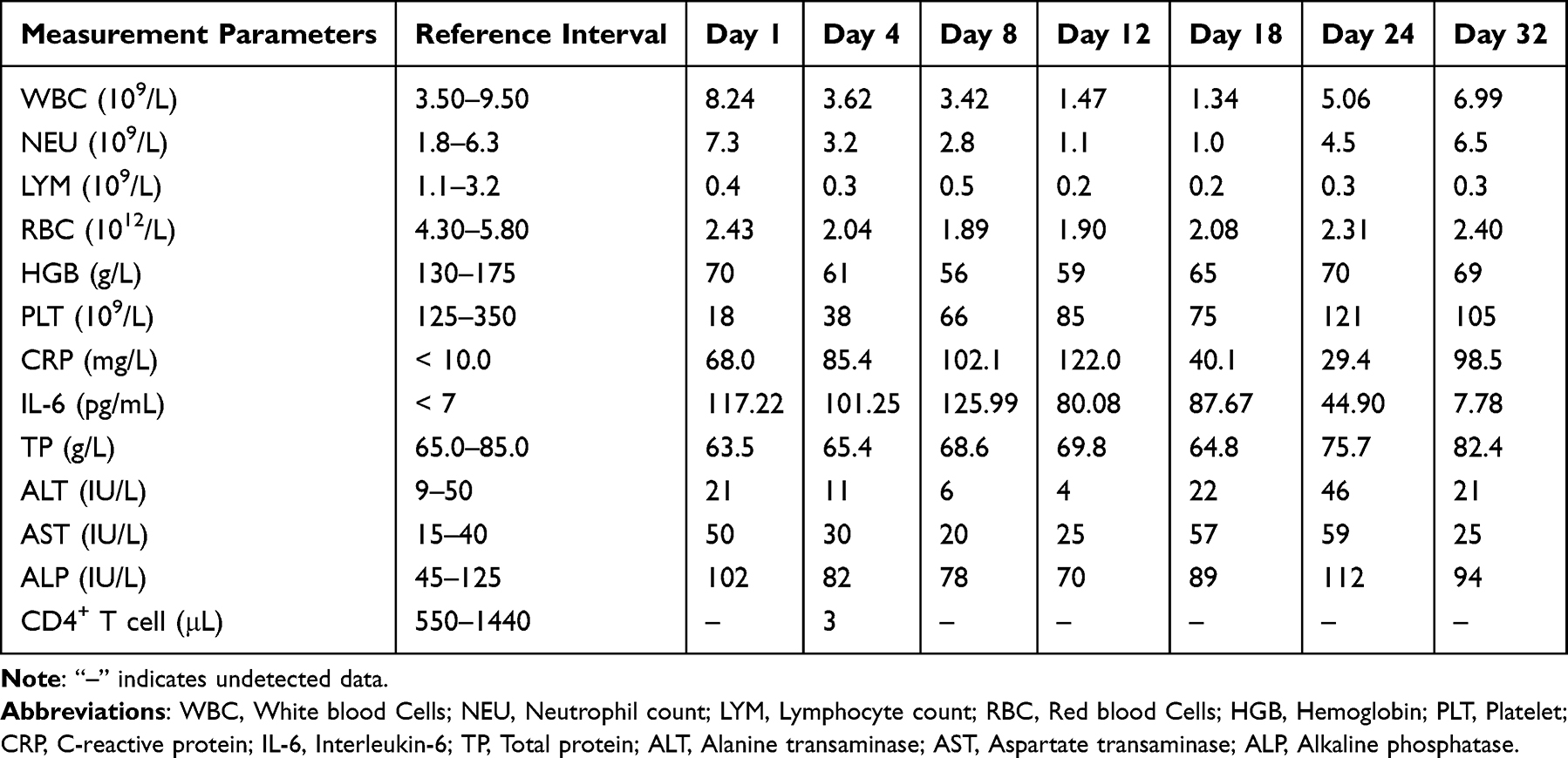 |
Table 1 Relevant Laboratory Test Results During Hospitalization at Jiaxing University Affiliated Hospital |
Of note, HIV viral load was not tested during this hospitalization due to logistical constraints. CD4⁺ T lymphocyte count was measured only once (Day 4: 3 cells/μL, Table 1) due to patient refusal of additional blood draws and sample quality issues. After discharge, regular follow-up included complete blood count and chest imaging examination, but serial HIV viral load and CD4⁺ T lymphocyte counts were not performed.
Diagnosis and Treatment Process in the First Affiliated Hospital of Zhejiang University School of Medicine (October 5–October 15, 2025)
During hospitalization, the patient was sequentially administered an intensified anti-MAC regimen, consisting of meropenem (1.0 g, every 8 hours), azithromycin (0.5 g, once daily, initiated on October 10), amikacin (0.5 g, once daily, started on October 10), and levofloxacin (0.5 g, once daily, administered from October 10 to 14 and subsequently discontinued due to the development of cardiac arrhythmia) (Figure 1). Simultaneously, comprehensive supportive care was provided, including somatostatin and proton pump inhibitors (PPIs) for the prophylaxis of potential bleeding, intravenous immunoglobulin pulse therapy, component blood transfusion, dexamethasone for antipyretic treatment, and foscarnet sodium for anti-cytomegalovirus treatment. The patient exhibited notable improvements in both clinical symptoms and laboratory indicators and was transferred to the First Hospital of Jiaxing for further management on October 15, 2025.
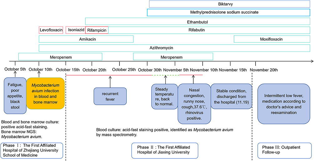 |
Figure 1 Diagnosis and treatment timeline of the patient. The figure presents the timeline of the patient’s clinical symptom evolution, key diagnostic examination results, and corresponding therapeutic interventions. |
Diagnostic Confirmation and Key Laboratory Findings
After the patient was transferred to the First Hospital of Jiaxing, the clinician emphasized to the microbiology laboratory the high possibility of NTM infection. The laboratory conducted a 5-day routine monitoring of the blood culture specimens submitted for testing, and the results were negative. Given the clear clinical high-risk indications of the patient, microbiological technicians adopted a blind subculture procedure to transfer the cultures from two negative aerobic culture bottles to blood agar plates and placed them in a 5% CO2 incubator (35.5°C) for extended cultivation for 15 days. Subsequently, small grayish-white colonies were visible on the plates (Figure 2A). Bacterial colony smears were taken and subjected to acid-fast staining under the microscope, revealing acid-fast positive bacteria (Figure 2B). Further identification by matrix-assisted laser desorption ionization time-of-flight mass spectrometry (MALDI-TOF MS, Brooke system) confirmed the presence of Mycobacterium avium, thereby achieving further laboratory confirmation of the pathogen (Figure 2C).
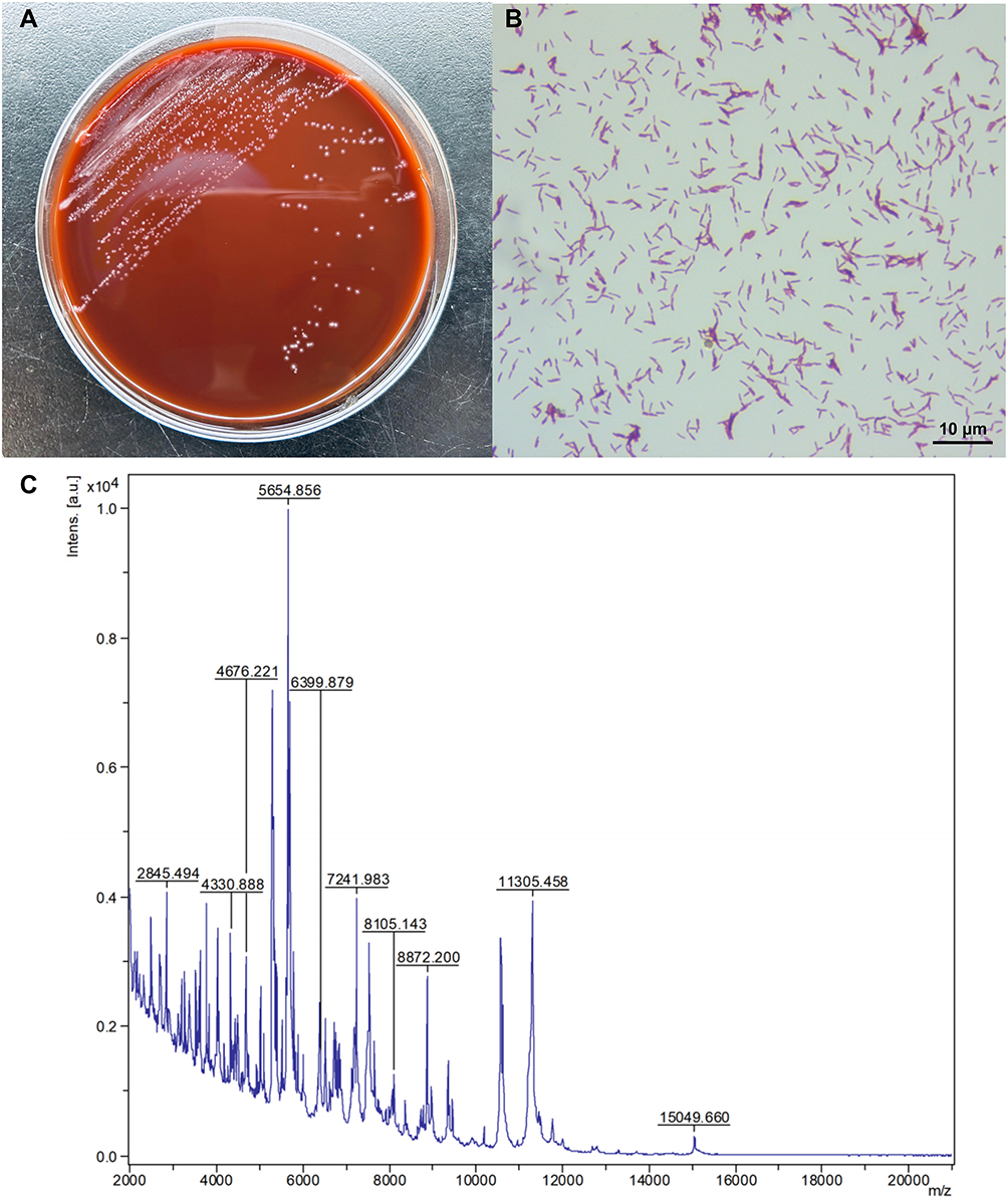 |
Figure 2 Microbiological test results of the patient. (A) The colony morphology of Mycobacterium avium was observed after blindly transferring negative blood culture specimens onto blood agar plates and continuously culturing for 15 days under 35.5°C and 5% CO2 conditions. (B) Bacterial colony smear grown on the blood plate, Ziehl-Neelsen acid-fast staining result (× 1000). (C) The strain was identified using Bruker MALDI-TOF MS and confirmed to be Mycobacterium avium, with an identification score of 2.32. |
Treatment Adjustment and Complex Disease Course Management (October 15–November 19, 2025, the Affiliated Hospital of Jiaxing University)
Initially, the treatment was based on the previous regimen, including meropenem, azithromycin, amikacin, and isoniazid. Due to the patient’s persistent fever, on October 18, the treatment was adjusted to include intravenous rifampicin and oral ethambutol in combination with azithromycin. On October 23, intravenous rifampicin was discontinued and replaced with oral rifabutin (two tablets, once daily) as the core treatment, which was continued in combination with azithromycin and ethambutol. On October 27, due to poor fever control, meropenem was reintroduced for intensified treatment (until November 12), and amikacin was discontinued. During this period, voriconazole was empirically administered for a short period (10.27–10.29) but was later discontinued due to the lack of evidence of fungal infection. On November 16th, because CRP (98.5 mg/L) was higher than before (Table 1), uncontrolled infection could not be ruled out. Moxifloxacin (1 tablet, once daily) was added for anti-infection treatment. The above information is specifically illustrated in Figure 1.
From October 15 to 30, 2025, the patient experienced persistent high fever (38.5–40.3°C), which could not be ruled out as being caused by an inflammatory cytokine response during anti-MAC therapy. Therefore, on October 30, methylprednisolone sodium succinate was administered to alleviate excessive systemic inflammation. The glucocorticoid dosage was dynamically adjusted according to the patient’s clinical course: 40 mg/day from October 30 to November 1, gradually decreased to 20 mg/day from November 2 to 12, and further decreased to 10 mg/day from November 13 to 17. Due to recurrent fever, the dose was increased to 20 mg/day on November 15, and finally escalated to 40 mg/day on November 18, which was maintained until discharge and continued during post-discharge follow-up. Meanwhile, antiretroviral therapy was officially resumed on November 1 using the Biktarvy regimen (bictegravir/emtricitabine/tenofovir alafenamide). After initial glucocorticoid intervention, the patient’s body temperature remained stable from November 2 to 6. However, after the dosage was reduced to 10 mg/day on November 13, high fever (39.0°C) recurred on November 15. Increasing the dose to 20 mg/day on November 15 was ineffective, and the dosage was subsequently adjusted to 40 mg/day on November 18. Notably, the patient’s fever fluctuations showed a significant dose-dependent correlation with glucocorticoid adjustments. Based on the temporal relationship between fever recurrence and ART reinitiation, the clinical presentation was highly suspected to represent MAC-associated IRIS. However, due to the lack of serial HIV viral load measurements and longitudinal CD4⁺ T lymphocyte count (only a single measurement was performed: 3 cells/μL on Day 4; Table 1) to validate objective immune reconstitution, the diagnosis remains uncertain. The detailed clinical process is shown in Figure 1.
The entire treatment process requires comprehensive management of multiple coexisting issues, including cytomegalovirus activity (using ganciclovir), arrhythmia caused by levofloxacin (timely discontinuation of medication), and human rhinovirus infection that occurred from November 7 to 10 (positive DNA test). The latter caused upper respiratory symptoms and temperature fluctuations and prompted cautious temporary adjustments and readjustments to the dosage of glucocorticoids. Continuous supportive treatment runs through, including platelet elevation, hemostasis, gastrointestinal protection, and nutritional support.
After the above comprehensive treatment, the patient’s body temperature was effectively controlled, and his overall condition and blood parameters improved. The patient was discharged with medication on November 19, 2025. The discharge instructions include oral anti-MAC drugs (rifabutin, ethambutol, azithromycin, and moxifloxacin) and a gradually decreasing glucocorticoid regimen. Outpatient follow-up until January 11, 2026, although the patient had an intermittent low-grade fever, the overall condition remained stable under close monitoring.
Discussion
Etiological Diagnosis Difficulty of MAC Infection in HIV-Positive Patients
HIV-positive patients complicated with MAC infection often lack characteristic clinical manifestations, posing significant challenges to early diagnosis.9 The patient in this case has a history of HIV infection for 10 years. Due to self-discontinuation of ART, the body developed severe immunodeficiency (CD4⁺ T lymphocyte counts of 3 cells/μL), providing a susceptible condition for MAC infection. Notably, advanced immunosuppression with a CD4⁺ T lymphocyte count < 50 cells/mm3 is a recognized high-risk factor for the development of disseminated MAC disease in HIV-positive individuals.9,11–13 Preliminary blood cultures of M. avium were obtained from the First Affiliated Hospital of Zhejiang University School of Medicine, and bone marrow NGS further confirmed the infection, providing important evidence for early diagnosis. Blood culture, as a fundamental method for pathogen diagnosis, can directly detect pathogenic bacteria in the blood.7 The combination of bone marrow sample detection and mNGS technology can further improve the accuracy of infection diagnosis, especially for cases with uncertain blood culture results or suspected disseminated infections.14 After being transferred to the Affiliated Hospital of Jiaxing University, due to the limitation of using only routine blood culture bottles for testing, the routine blood culture results were negative. It should be noted that MAC, as the main type of NTM, requires a longer incubation time for cultivation (usually 10–23 days, up to 57 days).7 Conventional blood culture bottles and shorter incubation times can easily lead to false negative results, which is also a common difficulty in clinical diagnosis.7,15 The pathogen was ultimately identified through blind subculture, prolonged culture, and MALDI-TOF MS analysis. This operation does not require additional reagents, special equipment, or additional costs and can be immediately adopted in any clinical microbiology laboratory, especially in environments where rapid molecular diagnostics (such as mNGS) are not available. This finding suggests that for HIV-positive patients suspected to be infected with NTM, if the initial blood culture is negative, blind subculture with extended incubation time or specialized NTM culture bottles should be considered to improve the detection rate of pathogens.15,16 MALDI-TOF MS has the advantage of rapid and accurate identification of NTM strains, which can provide a reliable basis for making individualized and targeted treatment plans in clinical practice.17
Challenges of MAC Infection Treatment in HIV-Positive Patients
The treatment of HIV-positive patients complicated with MAC infection has the characteristics of a long course of treatment, multidrug combination therapy, significant adverse drug reactions, and a high incidence of drug resistance, making clinical treatment difficult.18 The initial treatment plan for this case was to use meropenem, azithromycin, and amikacin, but the patient still experienced recurrent fever symptoms during treatment. It is speculated that the cause may be related to multiple factors such as pathogen drug resistance, severe autoimmune dysfunction, and concurrent infections. In response to the above treatment difficulties, the clinical anti-MAC treatment plan has been optimized and adjusted many times. The core adjustment measures include replacing rifampicin with rifabutin and strengthening the multidrug combination therapy strategy. Among them, compared with rifampicin, rifabutin has a lower risk of drug interaction and is more suitable for HIV-positive patients who need concurrent ART, which is consistent with clinical consensus and relevant guidelines.18 According to the guidelines for the diagnosis and treatment of HIV-related opportunistic infections, the treatment of MAC disease should follow the principle of at least two drug combinations.18,19 The common scheme is macrolide drugs (such as azithromycin and clarithromycin) combined with ethambutol to effectively prevent or delay the emergence of drug-resistant strains.17 In addition, the patient also experienced complications such as thrombocytopenia and bleeding during the treatment process, requiring timely symptomatic supportive treatment such as hematopoietic drugs and hemostatic drugs, further increasing the complexity of the overall treatment plan and the difficulty of clinical management.
Diagnostic Considerations for MAC-Associated IRIS
After ART reinitiation, the patient developed persistent high fever accompanied by generalized lymphadenopathy, with no other definitive infectious or non-infectious cause identified. According to the research of Surendra and Sharma, the following components are required for diagnosing MAC-associated IRIS.20 A temporal association must exist between ART initiation and subsequent symptom development (usually within 1–3 months), along with evidence of immune restoration (virological and immunological responses, such as a decrease in plasma HIV RNA level by more than 1 log10 copies/mL and an increase in CD4⁺ T lymphocyte count from baseline).20 Additionally, clinical signs and symptoms consistent with an inflammatory process must be present.20 In this case, the clinical deterioration occurred approximately two weeks after ART reinitiation, which was earlier than the typical 1–3 months onset window of typical IRIS. More importantly, there is no objective evidence of immune reconstitution, as no serial HIV viral load measurements or longitudinal CD4⁺ T lymphocyte counts were obtained during hospitalization. Without a documented decline in HIV viral load (eg, >1 log10 copies/mL) or a sustained rise in CD4⁺ T lymphocyte count, the fundamental criterion for IRIS cannot be verified. Although the patient presented with generalized lymphadenopathy (a typical inflammatory feature of IRIS), persistent fever and elevated C-reactive protein are nonspecific indicators that could also be caused by persistent or insufficiently controlled MAC infection or other occult infections. Given these limitations, we conservatively classify this as “suspected MAC-associated IRIS” rather than a definitive diagnosis. The possibility of paradoxical IRIS (ie, worsening of a previously diagnosed MAC infection after ART initiation) remains a consideration, but this should be framed cautiously given the absence of confirmatory immune monitoring.21
Glucocorticoids are an effective intervention to alleviate suspected excessive inflammation of IRIS.9,22 In this case, dose titration was highly individualized based on the severity of symptoms and immune status.8 The patient’s concurrent infection with human rhinovirus increases the complexity of treatment. Clinicians need to seek a balance between the anti-inflammatory benefits of suspected IRIS and the increased risk of viral infection exacerbated by excessive immune suppression.9 This highlights the value of implementing dynamic assessment and multi-objective comprehensive management in such complex situations.
For advanced AIDS patients with active disseminated MAC infection before ART, IRIS risk stratification before treatment and close monitoring after ART are important components of clinical management. Patients with severe immunosuppression (CD4⁺ T lymphocyte count <50 cells/mm3) have a higher risk of suspected IRIS during the basic immune reconstitution process.9 Therefore, it is necessary to strengthen monitoring and personalized intervention. We have also referenced published literature on IRIS associated with opportunistic fungal and mycobacterial infections to further illustrate the diagnostic challenges in similar immunocompromised populations.23
Prognostic Factors and Follow-Up Recommendations
The prognosis of HIV-positive patients with MAC infection is closely related to their immune function, severity of infection, treatment regimen rationality, and treatment compliance. The patient had a history of recurrent T. marneffei infection and voluntarily discontinued ART, resulting in sustained immune dysfunction. This may be the main reason for the recurrence of MAC infection and poor treatment response. During the follow-up period, the patient experienced recurrent fever on December 14 Based on available data, the cause of this fever cannot be definitively determined. In clinical practice, possible causes include incomplete control or early recurrence of disseminated MAC infection, recurrence of IRIS during corticosteroid reduction or ongoing immune recovery, or new co-infections (such as bacteria, viruses, or fungi). Suggest further improving laboratory and imaging examinations for differential diagnosis. Given the temporal correlation of fever and the lack of other clear causes, MAC recurrence or a low-grade IRIS flare is considered the most likely cause. This suggests that the current treatment regimen may need to be further extended or adjusted again to improve treatment response, control infection progression, and avoid disease recurrence. It is recommended that patients strictly follow the prescribed medication, regularly review relevant indicators (eg, complete blood count, inflammatory markers, HIV viral load, and CD4⁺ T lymphocyte count), and adjust the treatment plan accordingly based on the results. Concurrently, it is necessary to strengthen health education for patients, emphasize the importance of adhering to ART, discourage voluntary discontinuation or modification of medication, and thus prevent immune function deterioration and recurrent opportunistic infections.
This article has several limitations. First, as a single case report, the generalizability of the blind subculture requires validation through larger-scale studies. Second, the time to positivity in solid culture media may vary depending on the MAC strain type, inoculum size, and prior antibiotic exposure. Third, this case lacked serial CD4⁺ T lymphocyte counts and HIV viral load measurements. Only a single CD4⁺ count (3 cells/μL on Day 4) was available. Consequently, objective evidence of immune reconstitution is lacking, which precludes a definitive diagnosis of IRIS. Fourth, the interval between ART reinitiation and fever recurrence was approximately two weeks, which is shorter than the typical 1–3 months reported in the literature for IRIS. Notably, the patient did exhibit generalized lymphadenopathy at admission (involving the cervical, axillary, and inguinal regions), which represents specific inflammatory features consistent with IRIS. However, other clinical manifestations (fever and elevated CRP) were nonspecific. Therefore, we conservatively classify this as “suspected MAC-associated IRIS” rather than a definitive diagnosis. Fifth, larger prospective studies are needed to validate the diagnostic utility of blind subculture in NTM infection. Future case reports should include serial HIV viral load measurements and longitudinal CD4⁺ T lymphocyte counts to better characterize immune recovery and confirm IRIS diagnosis.
Conclusion
This case report detailed the diagnosis and treatment of an advanced AIDS patient with a complicated history of opportunistic infection who developed disseminated MAC infection and suspected MAC-associated IRIS. This study highlights that blind subculture of routinely negative blood culture bottles with extended incubation time is a simple, economical salvage strategy that improves NTM detection rates. Second, close clinical-laboratory collaboration is essential for optimizing the detection process and enabling timely and accurate identification of atypical mycobacterial infection in severely immunocompromised HIV patients. Third, for patients with suspected IRIS, early recognition of inflammatory fluctuations, cautious glucocorticoid intervention, and dynamic dose adjustments can effectively control excessive inflammatory responses and stabilize the patient’s clinical condition. Individualized anti-infection regimens, standardized complication management, and continuous health education are key factors for improving adherence and clinical prognosis. This case provides a practical reference for the clinical diagnosis and treatment of HIV-infected patients with complicated NTM infections and suspected IRIS.
Abbreviations
AIDS, Acquired immunodeficiency syndrome; HIV, Human immunodeficiency virus; ART, Antiretroviral therapy; M. avium, Mycobacterium avium; T. marneffei, Talaromyces marneffei; MAC, Mycobacterium avium Complex; IRIS, Immune reconstitution inflammatory syndrome; mNGS, Metagenomic next-generation sequencing; NTM, Nontuberculous mycobacteria; MALDI-TOF MS, Matrix-assisted laser desorption ionization time-of-flight mass spectrometry; WBC, White blood Cells; NEU, Neutrophil count; LYM, Lymphocyte count; RBC, Red blood Cells; HGB, Hemoglobin; PLT, Platelet; CRP, C-reactive protein; IL-6, Interleukin-6; TP, Total protein; ALT, Alanine transaminase; AST, Aspartate transaminase; ALP, Alkaline phosphatase.
Ethics Approval and Consent to Participate
Institutional approval was required and obtained for the publication of this case report. This retrospective case report was approved by the Ethics Committee of the Affiliated Hospital of Jiaxing University (approval No. 2025-KY-695). The patient has signed a written informed consent form, agreeing to the publication of the case details and all relevant imaging materials.
Acknowledgments
We thank anonymous reviewers whose comments and suggestions helped improve this manuscript.
Funding
This study was supported by the Science and Technology Plan Project of Jiaxing of Zhejiang Province of China (No. 2024AY10027), the Medical Outstanding Young Talents Project of Jiaxing City, Zhejiang Province (No. 2024-yxqnrc-47), the Hospital-level Project of the First Hospital of Jiaxing of Zhejiang Province of China (No. 2024-YB-044), Clinical Laboratory Medical Diagnostics Fund of the First Hospital of Jiaxing of Zhejiang Province of China (No. 2023-ZC-002), and Jiaxing Key Laboratory of Clinical Laboratory Diagnosis and Transformation Research (No. 2023-lcjyzdyzh).
Disclosure
The authors have no conflicts of interest to declare.
References
1. Ford N, Shubber Z, Meintjes G, et al. Causes of hospital admission among people living with HIV worldwide: a systematic review and meta-analysis. Lancet HIV. 2015;2(10):e438–10. doi:10.1016/S2352-3018(15)00137-X
2. Heidary M, Nasiri MJ, Mirsaeidi M, et al. Mycobacterium avium complex infection in patients with human immunodeficiency virus: a systematic review and meta-analysis. J Cell Physiol. 2019;234(7):9994–10001. doi:10.1002/jcp.27859
3. Wu HH, Zhao YK, Lai Y, et al. Disseminated Mycobacterium avium and Talaromyces marneffei coinfection in a patient of misdiagnosed tuberculosis and the challenges: a case report. BMC Infect Dis. 2025;25(1):1458. doi:10.1186/s12879-025-11895-2
4. Hariadi NI, Blackwood RA. Disseminated Mycobacterium avium complex in an adolescent with perinatally-acquired HIV infection. Infect Dis Rep. 2017;9(2):6884. doi:10.4081/idr.2017.6884
5. Jung Y, Song KH, Choe PG, et al. Incidence of disseminated Mycobacterium avium-complex infection in HIV patients receiving antiretroviral therapy with use of Mycobacterium avium-complex prophylaxis. Int J STD AIDS. 2017;28(14):1426–1432. doi:10.1177/0956462417713432
6. Baldolli A, Chocron R, Dargère S, et al. Mycobacterium genavense infections in immunocompromised patients without HIV: case series of solid organ transplant patients and literature review. Open Forum Infect Dis. 2022;9(10):ofac498. doi:10.1093/ofid/ofac498
7. National Institutes of Health; HIV Medicine Association of the Infectious Diseases Society of America Panel on Opportunistic Infections in Adults and Adolescents With HIV— A Working Group of the Office of AIDS Research Advisory Council (OARAC). Guidelines for the prevention and treatment of opportunistic infections in adults and adolescents with HIV. Department of Health and Human Services; 2024.
8. Lisek-Kubacka K, Cholewińska G, Mazurek L, et al. Disseminated mycobacteriosis and activation of latent opportunistic infections in the course of immune reconstitution inflammatory syndrome (IRIS) in a patient with AIDS: a case report. Przegl Epidemiol. 2025;79(3):319–332. doi:10.32394/pe/211175
9. Nikjeh O, Rejali S, Sasaninia K, et al. Overcoming treatment challenges in HIV-associated mycobacterial diseases: new therapeutic frontiers. Int J Mol Sci. 2025;26(21):10325. doi:10.3390/ijms262110325
10. Wu A, Gai W, Guo Y, et al. Clinical features of Talaromyces marneffei infection and colonization in HIV-negative patients: the role of mNGS in diagnosis. Front Med. 2025;12:1579522. doi:10.3389/fmed.2025.1579522
11. Marochi-Telles JP, Muniz R, Sztajnbok J, et al. Disseminated Mycobacterium avium on HIV/AIDS: historical and current literature review. AIDS Rev. 2020;22(1):9–15. doi:10.24875/AIDSRev.20000104
12. Chai J, Zhang S, Ma C, et al. Clinical analysis and risk factors associated with poor prognosis in nontuberculous mycobacterial infection. Virulence. 2025;16(1):2459313. doi:10.1080/21505594.2025.2459313
13. Hill E, Wooten D. A patient with mycobacteremia due to two different nontuberculous mycobacteria. Open Forum Infect Dis. 2022;9(10):ofac519. doi:10.1093/ofid/ofac519
14. Liu H, Xu T, Fu H, et al. Application of metagenomic next-generation sequencing in HIV-infected patients with bloodstream infections. Infect Drug Resist. 2025;18:2389–2399. doi:10.2147/IDR.S509665
15. Wang L, Chen Y, Wang Q, et al. Comparison of molecular testing methods for diagnosing non-tuberculous mycobacterial infections. Eur J Clin Microbiol Infect Dis. 2025;44(1):109–116. doi:10.1007/s10096-024-04981-x
16. Wijaya H, Kollins F, Lubis IN, et al. Norwegian scabies in human immunodeficiency virus and tuberculosis-infected child: a case report. Narra J. 2024;4(1):e661. doi:10.52225/narra.v4i1.661
17. Rindi L, Puglisi V, Franconi I, et al. Rapid and accurate identification of nontuberculous mycobacteria directly from positive primary MGIT cultures by MALDI-TOF MS. Microorganisms. 2022;10(7):1447. doi:10.3390/microorganisms10071447
18. Lopez P, Ali W, Donaldy W, et al. Disseminated extrapulmonary tuberculosis and pulmonary Mycobacterium avium complex co-infection in a newly diagnosed HIV patient: a case report. Cureus. 2025;17(9):e91532. doi:10.7759/cureus.91532
19. Asuphon O, Boonpattharatthiti K, Rattanachaisit N, et al. Comparing clinical outcomes of antibiotics for treating Mycobacterium avium complex infection in adults with HIV: a systematic review and network meta-analysis. Aids. 2026;40(1):16–23. doi:10.1097/QAD.0000000000004345
20. Sharma SK, Soneja M. HIV & immune reconstitution inflammatory syndrome (IRIS). Indian J Med Res. 2011;134(6):866–877. doi:10.4103/0971-5916.92632
21. Suryana K. A challenge in diagnosis of Tuberculosis-Associated Immune Reconstitution Inflammatory Syndrome (TB-IRIS). HIV AIDS. 2020;12:263–269.
22. Müller M, Wandel S, Colebunders R, et al. Immune reconstitution inflammatory syndrome in patients starting antiretroviral therapy for HIV infection: a systematic review and meta-analysis. Lancet Infect Dis. 2010;10(4):251–261. doi:10.1016/S1473-3099(10)70026-8
23. Thanh NT, Vinh LD, Liem NT, et al. Clinical features of three patients with paradoxical immune reconstitution inflammatory syndrome associated with Talaromyces marneffei infection. Med Mycol Case Rep. 2018;19:33–37. doi:10.1016/j.mmcr.2016.12.005
 © 2026 The Author(s). This work is published and licensed by Dove Medical Press Limited. The
full terms of this license are available at https://www.dovepress.com/terms
and incorporate the Creative Commons Attribution
- Non Commercial (unported, 4.0) License.
By accessing the work you hereby accept the Terms. Non-commercial uses of the work are permitted
without any further permission from Dove Medical Press Limited, provided the work is properly
attributed. For permission for commercial use of this work, please see paragraphs 4.2 and 5 of our Terms.
© 2026 The Author(s). This work is published and licensed by Dove Medical Press Limited. The
full terms of this license are available at https://www.dovepress.com/terms
and incorporate the Creative Commons Attribution
- Non Commercial (unported, 4.0) License.
By accessing the work you hereby accept the Terms. Non-commercial uses of the work are permitted
without any further permission from Dove Medical Press Limited, provided the work is properly
attributed. For permission for commercial use of this work, please see paragraphs 4.2 and 5 of our Terms.
Recommended articles

Nontuberculous Mycobacteria Lung Disease (NTM-LD): Current Recommendations on Diagnosis, Treatment, and Patient Management
Pathak K, Hart S, Lande L
International Journal of General Medicine 2022, 15:7619-7629
Published Date: 1 October 2022