Back to Journals » Risk Management and Healthcare Policy » Volume 17
Development and Validation of a Dynamic Nomogram for Predicting 3-Month Mortality in Acute Ischemic Stroke Patients with Atrial Fibrillation
Authors Yan X , Xia P, Tong H, Lan C, Wang Q , Zhou Y, Zhu H, Jiang C
Received 27 September 2023
Accepted for publication 11 January 2024
Published 16 January 2024 Volume 2024:17 Pages 145—158
DOI https://doi.org/10.2147/RMHP.S442353
Checked for plagiarism Yes
Review by Single anonymous peer review
Peer reviewer comments 4
Editor who approved publication: Dr Haiyan Qu
Xiaodi Yan,1,2 Peng Xia,3 Hanwen Tong,4 Chen Lan,1,2 Qian Wang,1,2 Yujie Zhou,5,* Huaijun Zhu,6 Chenxiao Jiang6,*
1Department of Pharmacy, Nanjing Drum Tower Hospital, School of Basic Medicine and Clinical Pharmacy, China Pharmaceutical University, Nanjing, Jiangsu, People’s Republic of China; 2School of Basic Medicine and Clinical Pharmacy, China Pharmaceutical University, Nanjing, Jiangsu, People’s Republic of China; 3Department of Pharmacy, Nanjing Drum Tower Hospital, School of Pharmacy, Nanjing Medical University, Nanjing, Jiangsu, People’s Republic of China; 4Department of Emergency Medicine, Nanjing Drum Tower Hospital, Affiliated Hospital of Medical School, Nanjing University, Nanjing, Jiangsu, People’s Republic of China; 5Department of Respiratory Critical Care Medicine, Nanjing Drum Tower Hospital, Affiliated Hospital of Medical School, Nanjing University, Nanjing, Jiangsu, People’s Republic of China; 6Department of Pharmacy, Nanjing Drum Tower Hospital, Affiliated Hospital of Medical School, Nanjing University, Nanjing, Jiangsu, People’s Republic of China
*These authors contributed equally to this work
Correspondence: Chenxiao Jiang; Yujie Zhou, Nanjing Drum Tower Hospital, Affiliated Hospital of Nanjing University Medical School, Zhongshan Road No. 321, Nanjing, Jiangsu, People’s Republic of China, Tel +86 177 6173 2923 ; +86 137 0140 9863, Email [email protected]; [email protected]
Background: Acute ischemic stroke (AIS) in patients with atrial fibrillation (AF) carries a substantial risk of mortality, emphasizing the need for effective risk assessment and timely interventions. This study aimed to develop and validate a practical dynamic nomogram for predicting 3-month mortality in AIS patients with AF.
Methods: AIS patients with AF were enrolled and randomly divided into training and validation cohorts. The nomogram was developed based on independent risk factors identified by multivariate logistic regression analysis. The prediction performance of the nomogram was evaluated using the area under the receiver operating characteristic curve (AUC-ROC), calibration plots, decision curve analysis (DCA), and Kaplan-Meier survival analysis.
Results: A total of 412 patients with AIS and AF entered final analysis, 288 patients in the training cohort and 124 patients in the validation cohort. The nomogram was developed using age, baseline National Institutes of Health Stroke Scale score, early introduction of novel oral anticoagulants, and pneumonia as independent risk factors. The nomogram exhibited good discrimination both in the training cohort (AUC, 0.851; 95% CI, 0.802– 0.899) and the validation cohort (AUC, 0.811; 95% CI, 0.706– 0.916). The calibration plots, DCA and Kaplan-Meier survival analysis demonstrated that the nomogram was well calibrated and clinically useful, effectively distinguishing the 3-month survival status of patients with AIS and AF, respectively. The dynamic nomogram can be obtained at the website: https://yanxiaodi.shinyapps.io/3-monthmortality/.
Conclusion: The dynamic nomogram represents the first predictive model for 3-month mortality and may contribute to managing the mortality risk of patients with AIS and AF.
Keywords: dynamic nomogram, mortality, acute ischemic stroke, atrial fibrillation
Introduction
Stroke, the most common type of which is acute ischemic stroke (AIS), is a major threat to health in China as it is the leading cause of death and the prevalence is on the rise.1 According to the Global Burden of Disease (GBD) Study 2019, China accounts for 2.19 million of the 6.55 million stroke-related deaths worldwide.2 Atrial fibrillation (AF) stands out as a primary risk factor for AIS, significantly elevating the risk of AIS occurrence and associated mortality.3,4 Previous studies indicated that patients with AF are five times more likely to experience AIS, often resulting in high mortality within the first 30 days following onset, with figures ranging from 22% to 27%.5–7 This represents a substantial public health concern. While anticoagulant therapy is recommended and acknowledged as an effective approach for reducing mortality in AIS patients with AF, a notable disparity exists between clinical practices and guideline recommendations.8–11 Consequently, the potential benefits of lowering mortality risk through anticoagulant therapy have not been fully realized.12,13 Therefore, the early identification of factors influencing mortality risk is of paramount importance to facilitate timely interventions and prolong patient longevity.
In the quest for early mortality risk assessment, researchers often turn to predictive models for guidance in treatment and management. Nomograms, as graphical tools, excel in this role by converting relevant risk factors into a continuous scoring system. They enable healthcare professionals to determine precise risk probabilities for specific outcomes in individual patients.14,15 In recent times, nomograms have been increasingly applied to predict mortality in stroke patients.16–18 Only limited nomograms were established for predicting functional outcome in patients with AIS and AF.19,20 However, these previous nomograms were developed based on the baseline characteristics and laboratory indicators, ignoring the important relationship between treatment regimens and mortality. Furthermore, numerous studies have explored mortality risk factors in AIS patients, shedding lights on the intricate interplay of variables.21–24 Nevertheless, the exact causes behind the elevated fatality rates in AIS patients with AF, as well as predictive nomograms, remain elusive. In summary, there is an urgent need to identify mortality risk factors and construct predictive nomograms for patients with AIS and AF.
Given the critical significance of accurately predicting mortality to inform sound medical interventions, it becomes imperative to investigate contributing risk factors and develop a user-friendly predictive nomogram. This study aims to construct and validate a practical dynamic nomogram, drawing on multiple independent risk factors, including treatment regimens, to predict 3-month mortality in AIS patients with AF.
Methods
Population and Study Design
This retrospective study was conducted at Nanjing Drum Tower Hospital from January 2019 to December 2021. Patients were enrolled according to the inclusion criteria: (1) diagnosed with AIS and confirmed by cranial computed tomography (CT) or magnetic resonance imaging (MRI) at admission; (2) diagnosed with AF by 24-hour dynamic electrocardiography (ECG) or had an AF history at admission; (3) age ≥18 years; (4) obtained the written informed consents. Exclusion criteria were as follows: (1) clinical data missing; (2) length of hospitalization over 1 year; (3) lost to follow-up. This study was conducted in accordance with the Declaration of Helsinki and approved by the Human Ethical Committee and Medical Research Council of Nanjing Drum Tower Hospital (Ethics Number: 2023-026-02). Written informed consents were obtained from all the patients.
Baseline Data Collection
Upon enrollment, patients’ characteristics including demographic, medical and medication histories, treatments after onset, stroke-associated infections (including pneumonia and urinary tract infection (UTI)), and baseline laboratory parameters were all collected. The antithrombotic regimens and the initiation time were determined based on the clinicians’ expertise in secondary prevention. Other medical interventions aligning with recommendations for stroke secondary prevention. According to the recommendation of the American Heart Association-American Stroke Association (AHA-ASA), the early introduction of NOACs (novel oral anticoagulants) was defined as NOACs treatment commencing within 14 days following AIS.25 Stroke-associated infections were defined as infections diagnosed during hospitalization. Each patient underwent physical examinations conducted by senior clinicians at admission to assess the National Institutes of Health Stroke Scale (NIHSS) score, CHA2DS2-VASc score and HAS-BLED score.
Outcome
The primary outcome was defined as 3-month all-cause mortality following the onset of AIS.26,27 Patients were tracked through phone calls or face-to-face interviews until the occurrence of the outcome event or up to 3 months. Patients were considered lost to follow-up when all follow-up methods failed. Subsequently, patients were categorized into two groups based on their survival status: the death group and the survival group.
Statistical Analysis
Continuous variables with normal distributions were presented as mean ± standard deviation (SD) and compared using the t-test. Non-normally distributed continuous variables were expressed as median (interquartile range, IQR) and analyzed via the Mann–Whitney U-test. The chi-square test or Fisher’s exact test was used to compare categorical variables, which were presented as frequency (percentage, %). The missing data was generated using the “mice” package by R software.
For the construction and validation of the nomogram, the study population was randomly divided into two cohorts: the training cohort (70%) and the validation cohort (30%). To generate the nomogram for the training cohort, a multivariate logistic regression analysis was performed using a forward stepwise method, incorporating all variables with P <0.1 from univariate logistic regression analysis. Variables with P <0.05 in the multivariate logistic regression analysis along with age (has a well-established association with mortality in stroke patients28) were reserved for nomogram development. The collinearity of variable combinations entered into the multivariate logistic regression analysis was assessed using variance inflation factors (VIF) (<2 considered not significant). The baseline CHA2DS2-VASc score was excluded from the multivariate logistic regression analysis, as it is calculated from age and medical histories. Regression coefficients and odds ratios (OR) with two-sided 95% confidence intervals (CI) were calculated for each variable in the model.
Discrimination of the nomogram was assessed using the area under the receiver operating characteristic curve (AUC-ROC). Calibration of the nomogram was evaluated by plotting the concordance between actual and predicted risk of 3-month mortality and applying the Hosmer-Lemeshow test (P >0.05). Clinical validity of the nomogram was assessed through decision curve analysis (DCA) by calculating the net benefit for a range of threshold probabilities. For further validation, an optimal cutoff value derived from the risk score in the training cohort was used to stratify patients into low-risk and high-risk groups. Subsequently, survival analysis was conducted by plotting Kaplan-Meier curves with Log rank tests to compare survival distributions between the low-risk and high-risk groups.
The statistical analyses were performed using STATA 17.0 (Stata Corporation, College Station, Texas) and R software (version 4.0.3). Statistical significance was defined as two-tailed P <0.05.
Results
Baseline Characteristics
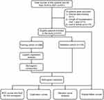
From January 2019 to December 2021, a total of 431 AIS patients with AF were enrolled in this study. After the exclusion of 19 patients, including 1 with clinical data missing, 3 with hospitalization over 1 year, and 15 lost to follow-up, a cohort of 412 patients were eligible for analysis. After random allocation, 288 patients comprised in the training cohort, and 124 patients formed the validation cohort (Figure 1).
 |
Figure 1 Flow diagram of the study. Abbreviations: AIS, acute ischemic stroke; AF, atrial fibrillation; ROC, receiver operating characteristic; AUC, area under the ROC curve. |
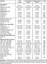
Table 1 presented the baseline characteristics of patients in both the training and validation cohorts. Among 412 patients, 209 (50.7%) were males, and the median age was 80 years. A total of 88 (21.4%) patients dead within 3 months after onset. During hospitalization, 146 (35.4%) patients developed pneumonia. Notably, the training cohort exhibited a higher incidence of pneumonia compared to the validation cohort (38.9% vs 27.4%, P=0.026). All other baseline characteristics and outcomes were well-balanced between the two cohorts (all P >0.05).
 |
Table 1 Comparison of Baseline Characteristics Between the Training and Validation Cohorts |
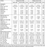
There were 66 (22.9%) patients and 22 (17.7%) patients dead in the training and validation cohorts, respectively. Table 2 demonstrated that patients in the death group were older and had higher baseline NIHSS score (all P <0.05). Pneumonia was notably more prevalent in the death group than in the survival group, both in the training and validation cohorts (P <0.001). In the training cohort, patients in the death group exhibited lower total cholesterol levels (P=0.016) and received thrombectomy therapy more frequently (P <0.001) than those in the survival group. Conversely, more patients in the survival group reported a history of drinking and received early introduction of NOACs (all P <0.05). In the validation cohort, the prevalence of hypertension was higher in the death group compared to the survival group (P=0.047).
 |
Table 2 Comparison of Baseline Characteristics Between the Death and Survival Groups in the Training and Validation Cohorts |
Predictive Nomogram Development
Univariate and multivariate logistic regression analysis were performed to identify risk factors for 3-month mortality in the training cohort (Table 3, Supplementary Table 1). No significant statistical collinearity was observed (all VIF <2) among the variables in the univariate logistic regression analysis. After adjusting for all confounders, age (adjusted odds ratio [aOR], 1.03; 95% CI, 0.99–1.07; P=0.130), baseline NIHSS score (aOR, 1.09; 95% CI, 1.04–1.13; P <0.001), early introduction of NOACs (aOR, 0.26; 95% CI, 0.09–0.71; P=0.008), and pneumonia (aOR, 3.13; 95% CI, 1.54–6.38; P=0.002) emerged as independent clinical predictors of 3-month mortality.
 |
Table 3 Univariate and Multivariate Logistic Regression Analysis for the Risk Factors Associated with 3-Month Mortality in the Training Cohort |
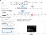
The predictive dynamic nomogram was subsequently developed based on these independent predictors (Figure 2A). To acquire the matching points on the “Points” scale, each predictor was highlighted on the nomogram, and a vertical line was drawn to the “Points” axis. The points of each predictor were summed to generate the total points, which was then located on the “Total points” axis to estimate the probability of 3-month mortality. Increasing total points in the nomogram were associated with a higher likelihood of 3-month mortality, while decreasing total points were related to a lower risk of 3-month mortality. The dynamic nomogram with an intuitive web-based interface was also developed so as to facilitate the use for clinicians in clinical practices (Figure 2B) (Dynamic Nomogram: https://yanxiaodi.shinyapps.io/3-monthmortality/).
 |
Figure 2 Dynamic nomogram for prediction of 3-month mortality in patients with AIS and AF. (A) The dynamic nomogram. Points were assigned to each predictor by drawing a vertical line to the “Points” axis and then calculated the total points as the sum of them. The probability of 3-month mortality can be easily obtained according to the total points. The example of using the nomogram was based on an 85-year-old patient, whose baseline NIHSS score was 15, got pneumonia, and without early introduction of NOACs, the probability of 3-month mortality was 52.7%. (B) The intuitive interface of the online dynamic nomogram (https://yanxiaodi.shinyapps.io/3-monthmortality/). **: P <0.05; ***: P <0.001. Abbreviations: NOACs, novel oral anticoagulants; NIHSS, National Institutes of Health Stroke Scale; AIS, acute ischemic stroke; AF, atrial fibrillation. |
Nomogram Validation
The discriminative capacity of the nomogram was verified based on the AUC values. The AUC values of the training (Figure 3A) and validation (Figure 3B) cohorts were 0.851 (95% CI, 0.802–0.899) and 0.811 (95% CI, 0.706–0.916), respectively, demonstrating that the established nomogram performed well. The optimal cutoff values of the training and validation cohorts were 0.790 (sensitivity, 86%; specificity, 72%) and 0.770 (sensitivity, 77%; specificity, 77%), respectively. Using the optimal cutoff value from the training cohort, patients in the training cohort, validation cohort and complete set were stratified into low-risk (below cutoff) and high-risk (above cutoff) groups. Kaplan-Meier survival curves revealed significantly lower 3-month mortality in the low-risk group compared to the high-risk group (all P <0.001) in the training cohort (Figure 4A), validation cohort (Figure 4B), and complete set (Figure 4C).
 |
Figure 4 Kaplan-Meier survival curves of patients in the low-risk and high-risk groups. (A) Training cohort; (B) Validation cohort; (C) Complete set. |
The calibration plots demonstrated high concordance between the predicted and actual probabilities of 3-month mortality both in the training (χ2=8.74, P=0.557) (Figure 5A) and validation (χ2=5.21, P=0.877) (Figure 5B) cohorts, signifying strong agreement between predicted and observed outcomes. The mean absolute errors of the calibration in training and validation cohorts, calculated via bootstrapping (resampling = 1000), were 0.017 and 0.029, respectively.
 |
Figure 5 Calibration plots of the nomogram in the training (A) and validation (B) cohorts. |
The clinical validity of the nomogram was further confirmed through DCA. The results indicated a substantial net benefit of the nomogram in predicting 3-month mortality in patients with AIS and AF across a range of threshold probability, spanning from 2% to 64% in the training cohort (Figure 6A) and 3% to 61% in the validation cohort (Figure 6B).
 |
Figure 6 Decision curve analysis for the training cohort (A) and the validation cohort (B). |
Discussion
In this study, we developed and validated a novel and practical dynamic nomogram for predicting 3-month mortality in patients with AIS and AF. The nomogram incorporated independent risk factors, including age, baseline NIHSS score, early introduction of NOACs, and pneumonia. The robust discriminative capacity of the nomogram, as evidenced by the AUC values and Kaplan-Meier survival analysis findings, suggested its potential to accurately predict 3-month survival in AIS patients with AF. Additionally, the nomogram demonstrated stability and reliability both in the training and validation cohorts, as indicated by the calibration plots. Moreover, the DCA results underscored the practicality and clinical applicability of the nomogram in identifying and managing the risk of 3-month mortality in patients with AIS and AF.
Our findings were consistent with previous studies, highlighted age and NIHSS score as primary independent risk factors for short- and long-term mortality in patients with ischemic stroke (IS) and AF.29–33 Age is closely intertwined with stroke incidence and severity.34 Previous researches reported that the mortality of patients with stroke rose with age.35,36 This condition may be caused by the fact that elderly patients often present with more comorbidities and poorer outcomes. Additionally, aging is associated with chronic inflammation and ischemic brain damage, both contributing to worse prognosis in stroke patients.37,38 Stroke severity, as assessed by the NIHSS score, is a critical determinant of post-stroke mortality.39–41 Besides, higher baseline NIHSS scores indicate severe neurological deficits, which are associated with an increased risk of mortality in IS patients.42–44 Therefore, the inclusion of age and baseline NIHSS score in our nomogram aligns with the established studies and strengthens its predictive capabilities.
Stroke-associated infections, particularly pneumonia, which accounts for 11.3% to 31.3% of all stroke-associated infections and arises virtually after stroke, represent a common complication in stroke patients.45,46 Traditionally, impaired levels of consciousness and severe dysphagia are considered as related risk factors for aspiration in stroke patients, leading to pneumonia.47,48 Moreover, patients experiencing an acute stroke are more susceptible to infections due to the systemic immunodepression, primarily triggered by excessive stimulation of the autonomous nervous system.49 In medical practice, the risk of mortality associated with pneumonia remains a significant concern for stroke patients. It has been reported that pneumonia can lead to an increase ranging in short-term mortality from 10.1% to 37.3% and in long-term mortality from 49.0% to 60.1% following AIS.50–52 The association between pneumonia and the increased risk of mortality has been well-documented, emphasizing the importance of monitoring and managing pneumonia in AIS patients with AF. Therefore, our inclusion of pneumonia as an independent predictor in the nomogram aligns with the clinical reality and enhances its prognostic accuracy.
AF predisposes patients to thromboembolic events by pooling blood in the atria, making anticoagulant therapy a critical component of stroke prevention.53 Both warfarin and NOACs have demonstrated their efficacy in reducing mortality among stroke patients with AF by inhibiting clot formation.54–58 Notably, both North American and European guidelines recommend anticoagulant therapy for stroke prevention in AF patients, favoring it over antiplatelet therapy.59 NOACs have shown superiority over warfarin in terms of both efficacy and safety in preventing stroke in AF patients. The ARISTOPHANES study, the largest observation study on NOACs and warfarin to date, found that NOACs significantly lower the risk of stroke/systemic embolism compared to warfarin in AF patients.60 However, the efficacy of early and delay NOACs treatment in patients with AIS and AF remains controversial. The definitions of early and delay NOACs treatment also vary among different studies. For instance, the TIMING study showed that early NOACs treatment (≤4 days) was noninferior to delay NOACs treatment (5–10 days) in reducing the risk of 90-day mortality in patients with AIS and AF.61 Similarly, Wilson et al found no significant difference in 90-day mortality between patients receiving early (≤4 days) and delayed (≥5 days) anticoagulant therapy in an observational study based on the data from CROMIS-2 study.62 In contrast, the RELAXED study reported that AIS patients with AF who initiated rivaroxaban within 14 days had a lower incidence of 90-day mortality compared to those started rivaroxaban ≥15 days after onset.63 These varying findings highlight the ongoing controversy surrounding the optimal timing of NOACs initiation in this patient population. Our study investigated the impact of the timing of NOACs initiation on 3-month mortality and revealed a significant association between early NOACs introduction (within 14 days) and reduced 3-month mortality. This finding provides valuable insights into the timing of anticoagulant therapy initiation in these high-risk patients.
The nomogram is regarded as a crucial decision-making prediction tool for investigating the relationship between the prognosis and baseline status.64,65 Previous nomograms designed to predict outcomes typically relied on baseline characteristics or predefined treatment regimens, often overlooking the impact of anticoagulant therapy on prognosis.66,67 For instance, Cappellari et al constructed a nomogram to predict functional outcomes based on the AF patients who received NOACs within 7 days after stroke.20 However, their study failed to explain the relationship between delayed NOACs treatment and functional outcomes or mortality. Our study developed a nomogram with favorable predictive capabilities, taking into account the significant influence of early introduction of NOACs on 3-month mortality. To the best of our knowledge, our study’s nomogram represents the first model tailored for predicting 3-month mortality in patients with AIS and AF. Early identification of mortality risk factors is paramount for enabling timely interventions. For instance, early assessment of NIHSS score increases the likelihood of detecting symptomatic fluctuations and enables more effective interventions to improve outcomes.68,69 Besides, patients have the potential to have better prognosis and lower mortality risk by minimizing the occurrence of pneumonia and treating with early NOACs therapy.9,10,47 Consequently, our dynamic nomogram is designed to be practical, reliable, and user-friendly. We established it based on readily available factors during hospitalization, offering clinicians valuable guidance for implementing early interventions to prevent pneumonia and administer anticoagulant therapy.
There were several limitations in our study. Firstly, this is a single-center retrospective study, which may limit the statistical power of the results. Secondly, the training cohort and the validation cohort had a significant difference in pneumonia, thus, further research centers and external validation are necessary to confirm the clinical utility and performance of the nomogram. Thirdly, our study did not include a comparative analysis of laboratory test results during follow-up. Despite these limitations, our study has successfully identified important 3-month mortality risk factors in patients with AIS and AF. Furthermore, the dynamic nomogram serves as a valuable tool for increasing the proportion of patients receiving early introduction of NOACs, ultimately improving the clinical outcomes of individuals with AIS and AF.
Conclusion
In summary, this study is the first attempt to develop and validate a user-friendly clinical dynamic nomogram, incorporating independent risk factors, for the prediction of 3-month mortality in patients with AIS and AF. This dynamic nomogram holds the potential to aid in the assessment of an individual’s 3-month mortality risk and serves as a compass for guiding future treatments aimed at prolonging the survival time of high-risk patients.
Abbreviations
GBD, Global Burden of Disease; AF, atrial fibrillation; AIS, acute ischemic stroke; CT, computed tomography; MRI, magnetic resonance imaging; ECG, electrocardiography; UTI, urinary tract infection; AHA-ASA, American Heart Association-American Stroke Association; NOACs, novel oral anticoagulants; NIHSS, National Institutes of Health Stroke Scale; SD, standard deviation; IQR, interquartile range; VIF, variance inflation factor; OR, odds ratio; CI, confidence interval; AUC-ROC, area under the receiver operating characteristic curve; DCA, decision curve analysis; aOR, adjusted Odds Ratio; IS, ischemic stroke.
Data Sharing Statement
Data presented in the study are included in the article. Data will be made available on request by contacting the corresponding authors.
Ethics Approval and Informed Consent
Ethical approval to report this work was obtained from the Ethics Committee of Nanjing Drum Tower Hospital (Ethics Number: 2023-026-02). Written informed consent was obtained from the patients for their anonymized information to be published in this article.
Acknowledgments
The authors thank all the patients and the participants.
Author Contributions
All authors made substantial contributions to conception, study design, execution, acquisition of data, analysis and interpretation, or in all these areas; took part in drafting the article or critically revising for important intellectual content; gave final approval of the version to be published; agreed to submit to the current journal; and agreed to be accountable for all aspects of the work.
Funding
This study was supported by the Clinical Trials from the Affiliated Drum Tower Hospital, Medical School of Nanjing University [grant number 2023-LCYJ-PY-24]; Project of China Hospital Reform and Development Research and Development Research Institute, Nanjing University [grant number NDYGN2023007]; Aid project of Nanjing Drum Tower Hospital Health, Education & Research Foundation; the Jiangsu Research Hospital Association for Precision Medication [grant number JY202120]; and the Jiangsu Pharmaceutical Association for Jinpeiying Project [grant number J2021001]. The funders had no involvement in the preparation or writing up of this research.
Disclosure
The authors report no conflict of interest in this work.
References
1. Tu WJ, Zhao Z, Yin P, et al. Estimated Burden of Stroke in China in 2020. JAMA Network Open. 2023;6(3):e231455. doi:10.1001/jamanetworkopen.2023.1455
2. Tu WJ, Wang LD; Special Writing Group of China Stroke Surveillance Report. China stroke surveillance report 2021. Mil Med Res. 2023;10(1):33. doi:10.1186/s40779-023-00463-x
3. Bjerkreim AT, Khanevski AN, Thomassen L, et al. Five-year readmission and mortality differ by ischemic stroke subtype. J Neurol Sci. 2019;403:31–37. doi:10.1016/j.jns.2019.06.007
4. Jung YH, Choi HY, Lee KY, et al. Stroke Severity in Patients on Non-Vitamin K Antagonist Oral Anticoagulants with a Standard or Insufficient Dose. Thromb Haemost. 2018;118(12):2145–2151. doi:10.1055/s-0038-1675602
5. Pikija S, Trkulja V, Malojcic B, Mutzenbach JS, Sellner J. A High Burden of Ischemic Stroke in Regions of Eastern/Central Europe is Largely Due to Modifiable Risk Factors. Curr Neurovasc Res. 2015;12(4):341–352. doi:10.2174/1567202612666150731105554
6. Fang MC, Go AS, Chang Y, et al. Thirty-day mortality after ischemic stroke and intracranial hemorrhage in patients with atrial fibrillation on and off anticoagulants. Stroke. 2012;43(7):1795–1799. doi:10.1161/STROKEAHA.111.630731
7. Choi SE, Sagris D, Hill A, Lip GYH, Abdul-Rahim AH. Atrial fibrillation and stroke. Expert Rev Cardiovasc Ther. 2023;21(1):35–56. doi:10.1080/14779072.2023.2160319
8. Giugliano RP, Ruff CT, Braunwald E, et al. Edoxaban versus warfarin in patients with atrial fibrillation. N Engl J Med. 2013;369(22):2093–2104. doi:10.1056/NEJMoa1310907
9. Steffel J, Collins R, Antz M, et al. 2021 European Heart Rhythm Association Practical Guide on the Use of Non-Vitamin K Antagonist Oral Anticoagulants in Patients with Atrial Fibrillation. Europace. 2021;23(10):1612–1676. doi:10.1093/europace/euab065
10. Kleindorfer DO, Towfighi A, Chaturvedi S, et al. 2021 Guideline for the Prevention of Stroke in Patients With Stroke and Transient Ischemic Attack: a Guideline From the American Heart Association/American Stroke Association. Stroke. 2021;52(7):e364–e467. doi:10.1161/STR.0000000000000375
11. Klijn CJ, Paciaroni M, Berge E, et al. Antithrombotic treatment for secondary prevention of stroke and other thromboembolic events in patients with stroke or transient ischemic attack and non-valvular atrial fibrillation: a European Stroke Organisation guideline. Eur Stroke J. 2019;4(3):198–223. doi:10.1177/2396987319841187
12. Yang X, Li Z, Zhao X, et al. Use of Warfarin at Discharge Among Acute Ischemic Stroke Patients With Nonvalvular Atrial Fibrillation in China. Stroke. 2016;47(2):464–470. doi:10.1161/STROKEAHA.115.011833
13. Gong X, Chen H, Wang J, et al. Undertreatment of Anticoagulant Therapy in Hospitalized Acute Ischemic Stroke Patients With Atrial Fibrillation. Front Cardiovasc Med. 2022;9:841020. doi:10.3389/fcvm.2022.841020
14. Cappellari M, Turcato G, Forlivesi S, et al. STARTING-SICH Nomogram to Predict Symptomatic Intracerebral Hemorrhage After Intravenous Thrombolysis for Stroke. Stroke. 2018;49(2):397–404. doi:10.1161/STROKEAHA.117.018427
15. Jehi L, Yardi R, Chagin K, et al. Development and validation of nomograms to provide individualised predictions of seizure outcomes after epilepsy surgery: a retrospective analysis. Lancet Neurol. 2015;14(3):283–290. doi:10.1016/S1474-4422(14)70325-4
16. Jin G, Hu W, Zeng L, Ma B, Zhou M. Prediction of long-term mortality in patients with ischemic stroke based on clinical characteristics on the first day of ICU admission: an easy-to-use nomogram. Front Neurol. 2023;14:1148185. doi:10.3389/fneur.2023.1148185
17. Li XD, Li MM. A novel nomogram to predict mortality in patients with stroke: a survival analysis based on the MIMIC-III clinical database. BMC Med Inform Decis Mak. 2022;22(1):92. doi:10.1186/s12911-022-01836-3
18. Szlachetka WA, Pana TA, Mamas MA, et al. Predicting 10-year stroke mortality: development and validation of a nomogram. Acta Neurol Belg. 2022;122(3):685–693. doi:10.1007/s13760-021-01752-9
19. Cappellari M, Seiffge DJ, Koga M, et al. A nomogram to predict unfavourable outcome in patients receiving oral anticoagulants for atrial fibrillation after stroke. Eur Stroke J. 2020;5(4):384–393. doi:10.1177/2396987320945840
20. Cappellari M, Turcato G, Forlivesi S, et al. Introduction of direct oral anticoagulant within 7 days of stroke onset: a nomogram to predict the probability of 3-month modified Rankin Scale score 2. J Thromb Thrombolysis. 2018;46(3):292–298. doi:10.1007/s11239-018-1700-8
21. Jhou HJ, Chen PH, Yang LY, Chang SH, Lee CH. Plasma Anion Gap and Risk of In-Hospital Mortality in Patients with Acute Ischemic Stroke: analysis from the MIMIC-IV Database. J Pers Med. 2021;11(10):1004. doi:10.3390/jpm11101004
22. Dabilgou AA, Dravé A, Kyelem JMA, Ouedraogo S, Napon C, Kaboré J. Frequency and Mortality Risk Factors of Acute Ischemic Stroke in Emergency Department in Burkina Faso. Stroke Res Treat. 2020;2020:9745206. doi:10.1155/2020/9745206
23. Huang ZX, Gu HQ, Yang X, Wang CJ, Wang YJ, Li ZX. Risk factors for in-hospital mortality among acute ischemic stroke patients in China: a nationwide prospective study. Neurol Res. 2021;43(5):387–395. doi:10.1080/01616412.2020.1866356
24. Ztriva E, Protopapas A, Mentizis P, et al. Hepatic Fibrosis Is a Risk Factor for Greater Severity and Worse Outcome of Acute Ischemic Stroke. J Clin Med. 2022;11(17):5141. doi:10.3390/jcm11175141
25. Powers WJ, Rabinstein AA, Ackerson T, et al. 2018 Guidelines for the Early Management of Patients With Acute Ischemic Stroke: a Guideline for Healthcare Professionals From the American Heart Association/American Stroke Association. Stroke. 2018;49(3):e46–e110. doi:10.1161/STR.0000000000000158
26. Zhang X, Yuan K, Wang H, et al. Nomogram to Predict Mortality of Endovascular Thrombectomy for Ischemic Stroke Despite Successful Recanalization. J Am Heart Assoc. 2020;9(3):e014899. doi:10.1161/JAHA.119.014899
27. Jin G, Hu W, Zeng L, et al. Development and verification of a nomogram for predicting short-term mortality in elderly ischemic stroke populations. Sci Rep. 2023;13(1):12580. doi:10.1038/s41598-023-39781-4
28. Dmytriw AA, Dibas M, Schirmer CM, et al. Age and Acute Ischemic Stroke Outcome in North American Patients With COVID-19. J Am Heart Assoc. 2021;10(14):e021046. doi:10.1161/JAHA.121.021046
29. Tracz J, Gorczyca-Głowacka I, Rosołowska A, Wożakowska-Kapłon B. Long-Term Outcomes after Stroke in Patients with Atrial Fibrillation: a Single Center Study. Int J Environ Res Public Health. 2023;20(4):3491. doi:10.3390/ijerph20043491
30. Li S, Zhao X, Wang C, et al. Risk factors for poor outcome and mortality at 3 months after the ischemic stroke in patients with atrial fibrillation. J Stroke Cerebrovasc Dis. 2013;22(8):e419–e425. doi:10.1016/j.jstrokecerebrovasdis.2013.04.025
31. Dutta D, Cannon A, Bowen E. Validation and comparison of two stroke prognostic models for in hospital, 30-day and 90-day mortality. Eur Stroke J. 2017;2(4):327–334. doi:10.1177/2396987317703581
32. Ding GY, Xu JH, He JH, Nie ZY. Clinical scoring model based on age, NIHSS, and stroke-history predicts outcome 3 months after acute ischemic stroke. Front Neurol. 2022;13:935150. doi:10.3389/fneur.2022.935150
33. Khanevski AN, Bjerkreim AT, Novotny V, et al. Recurrent ischemic stroke: incidence, predictors, and impact on mortality. Acta Neurol Scand. 2019;140(1):3–8. doi:10.1111/ane.13093
34. Samuthpongtorn C, Jereerat T, Suwanwela NC. Stroke risk factors, subtypes and outcome in elderly Thai patients. BMC Neurol. 2021;21(1):322. doi:10.1186/s12883-021-02353-y
35. Wang T, Li B, Gu H, et al. Effect of age on long-term outcomes after stroke with atrial fibrillation: a hospital-based follow-up study in China. Oncotarget. 2017;8(32):53684–53690. doi:10.18632/oncotarget.15729
36. Fromm A, Waje-Andreassen U, Thomassen L, Naess H. Comparison between Ischemic Stroke Patients <50 Years and ≥50 Years Admitted to a Single Centre: the Bergen Stroke Study. Stroke Res Treat. 2011;2011:183256. doi:10.4061/2011/183256
37. Dong P, Zhao J, Zhang Y, et al. Aging causes exacerbated ischemic brain injury and failure of sevoflurane post-conditioning: role of B-cell lymphoma-2. Neuroscience. 2014;275:2–11. doi:10.1016/j.neuroscience.2014.05.064
38. Finger CE, Moreno-Gonzalez I, Gutierrez A, Moruno-Manchon JF, McCullough LD. Age-related immune alterations and cerebrovascular inflammation. Mol Psychiatry. 2022;27(2):803–818. doi:10.1038/s41380-021-01361-1
39. Khanevski AN, Bjerkreim AT, Novotny V, et al. Recurrent ischemic stroke: incidence, predictors, and impact on mortality. Acta Neurol Scand. 2019;140(1):3–8.
40. Vinding NE, Kristensen SL, Rørth R, et al. Ischemic Stroke Severity and Mortality in Patients With and Without Atrial Fibrillation. J Am Heart Assoc. 2022;11(4):56.
41. Andersen KK, Andersen ZJ, Olsen TS. Predictors of early and late case-fatality in a nationwide Danish study of 26,818 patients with first-ever ischemic stroke. Stroke. 2011;42(10):2806–2812. doi:10.1161/STROKEAHA.111.619049
42. Purroy F, Vena A, Forné C, et al. Age- and Sex-Specific Risk Profiles and In-Hospital Mortality in 13,932 Spanish Stroke Patients. Cerebrovasc Dis. 2019;47(3–4):151–164. doi:10.1159/000500205
43. Bryndziar T, Matyskova D, Sedova P, et al. Predictors of Short- and Long-Term Mortality in Ischemic Stroke: a Community-Based Study in Brno, Czech Republic. Cerebrovasc Dis. 2022;51(3):296–303. doi:10.1159/000519937
44. Lehmann ALCF, Alfieri DF, de Araújo MCM, et al. Immune-inflammatory, coagulation, adhesion, and imaging biomarkers combined in machine learning models improve the prediction of death 1 year after ischemic stroke. Clin Exp Med. 2022;22(1):111–123. doi:10.1007/s10238-021-00732-w
45. Huang GQ, Lin YT, Wu YM, Cheng QQ, Cheng HR, Wang Z. Individualized Prediction Of Stroke-Associated Pneumonia For Patients With Acute Ischemic Stroke. Clin Interv Aging. 2019;14:1951–1962. doi:10.2147/CIA.S225039
46. Westendorp WF, Nederkoorn PJ, Vermeij JD, Dijkgraaf MG, van de Beek D. Post-stroke infection: a systematic review and meta-analysis. BMC Neurol. 2011;11:110. doi:10.1186/1471-2377-11-110
47. Warusevitane A, Karunatilake D, Sim J, Lally F, Roffe C. Safety and effect of metoclopramide to prevent pneumonia in patients with stroke fed via nasogastric tubes trial. Stroke. 2015;46(2):454–460. doi:10.1161/STROKEAHA.114.006639
48. Lakshminarayan K, Tsai AW, Tong X, et al. Utility of dysphagia screening results in predicting poststroke pneumonia. Stroke. 2010;41(12):2849–2854. doi:10.1161/STROKEAHA.110.597039
49. Westendorp WF, Dames C, Nederkoorn PJ, Meisel A. Immunodepression, Infections, and Functional Outcome in Ischemic Stroke. Stroke. 2022;53(5):1438–1448. doi:10.1161/STROKEAHA.122.038867
50. Barlas RS, Loke YK, Mamas MA, et al. Effect of Antiplatelet Therapy (Aspirin + Dipyridamole Versus Clopidogrel) on Mortality Outcome in Ischemic Stroke. Am J Cardiol. 2018;122(6):1085–1090. doi:10.1016/j.amjcard.2018.05.043
51. Tinker RJ, Smith CJ, Heal C, et al. Predictors of mortality and disability in stroke-associated pneumonia. Acta Neurol Belg. 2021;121(2):379–385. doi:10.1007/s13760-019-01148-w
52. Hannawi Y, Hannawi B, Rao CP, Suarez JI, Bershad EM. Stroke-associated pneumonia: major advances and obstacles. Cerebrovasc Dis. 2013;35(5):430–443. doi:10.1159/000350199
53. Kim YH, Roh SY. The Mechanism of and Preventive Therapy for Stroke in Patients with Atrial Fibrillation. J Stroke. 2016;18(2):129–137. doi:10.5853/jos.2016.00234
54. Baturova MA, Lindgren A, Carlson J, Shubik YV, Bertil Olsson S, Platonov PG. Non-permanent atrial fibrillation and oral anticoagulant therapy are related to survival during 10 years after first-ever ischemic stroke. Int J Cardiol. 2017;232:134–139. doi:10.1016/j.ijcard.2017.01.040
55. Arnao V, Agnelli G, Paciaroni M. Direct oral anticoagulants in the secondary prevention of stroke and transient ischemic attack in patients with atrial fibrillation. Intern Emerg Med. 2015;10(5):555–560. doi:10.1007/s11739-015-1226-4
56. Kapil N, Datta YH, Alakbarova N, et al. Antiplatelet and Anticoagulant Therapies for Prevention of Ischemic Stroke. Clin Appl Thromb Hemost. 2017;23(4):301–318. doi:10.1177/1076029616660762
57. Li X, Pathadka S, Man KKC, et al. Comparative Outcomes Between Direct Oral Anticoagulants, Warfarin, and Antiplatelet Monotherapy Among Chinese Patients with Atrial Fibrillation: a Population-Based Cohort Study. Drug Saf. 2020;43(10):1023–1033. doi:10.1007/s40264-020-00961-0
58. Goulart AC, Olmos RD, Santos IS, et al. The impact of atrial fibrillation and long-term oral anticoagulant use on all-cause and cardiovascular mortality: a 12-year evaluation of the prospective Brazilian Study of Stroke Mortality and Morbidity. Int J Stroke. 2022;17(1):48–58. doi:10.1177/1747493021995592
59. Jame S, Barnes G. Stroke and thromboembolism prevention in atrial fibrillation. Heart. 2020;106(1):10–17. doi:10.1136/heartjnl-2019-314898
60. Lip GYH, Keshishian A, Li X, et al. Effectiveness and Safety of Oral Anticoagulants Among Nonvalvular Atrial Fibrillation Patients. Stroke. 2018;49(12):2933–2944. doi:10.1161/STROKEAHA.118.020232
61. Oldgren J, Åsberg S, Hijazi Z, et al. Early Versus Delayed Non-Vitamin K Antagonist Oral Anticoagulant Therapy After Acute Ischemic Stroke in Atrial Fibrillation (TIMING): a Registry-Based Randomized Controlled Noninferiority Study. Circulation. 2022;146(14):1056–1066. doi:10.1161/CIRCULATIONAHA.122.060666
62. Wilson D, Ambler G, Banerjee G, et al. Early versus late anticoagulation for ischaemic stroke associated with atrial fibrillation: multicentre cohort study. J Neurol Neurosurg Psychiatry. 2019;90(3):320–325. doi:10.1136/jnnp-2018-318890
63. Yasaka M, Minematsu K, Toyoda K, et al. Rivaroxaban administration after acute ischemic stroke: the RELAXED study. PLoS One. 2019;14(2):e0212354. doi:10.1371/journal.pone.0212354
64. Tan WS, Ahmad A, Feber A, et al. Development and validation of a haematuria cancer risk score to identify patients at risk of harbouring cancer. J Intern Med. 2019;285(4):436–445.
65. Zou J, Chen H, Liu C, et al. Development and validation of a nomogram to predict the 30-day mortality risk of patients with intracerebral hemorrhage. Front Neurosci. 2022;16:942100. doi:10.3389/fnins.2022.942100
66. Chandriah H, Kumolosasi E, Islahudin F, Makmor-Bakry M. Effectiveness and safety of a 10mg warfarin initiation nomogram in Asian population. Pak J Pharm Sci. 2015;28(3):927–932.
67. Monkman K, Lazo-Langner A, Kovacs MJ. A 10 mg warfarin initiation nomogram is safe and effective in outpatients starting oral anticoagulant therapy for venous thromboembolism. Thromb Res. 2009;124(3):275–280. doi:10.1016/j.thromres.2008.12.001
68. Yamal JM, Grotta JC. National Institutes of Health Stroke Scale as an Outcome Measure for Acute Stroke Trials. Stroke. 2021;52(1):142–143. doi:10.1161/STROKEAHA.120.032994
69. Chalos V, van der Ende NAM, Lingsma HF, et al. National Institutes of Health Stroke Scale: an Alternative Primary Outcome Measure for Trials of Acute Treatment for Ischemic Stroke. Stroke. 2020;51(1):282–290. doi:10.1161/STROKEAHA.119.026791
 © 2024 The Author(s). This work is published and licensed by Dove Medical Press Limited. The full terms of this license are available at https://www.dovepress.com/terms.php and incorporate the Creative Commons Attribution - Non Commercial (unported, v3.0) License.
By accessing the work you hereby accept the Terms. Non-commercial uses of the work are permitted without any further permission from Dove Medical Press Limited, provided the work is properly attributed. For permission for commercial use of this work, please see paragraphs 4.2 and 5 of our Terms.

