
Back to Journals » Drug Design, Development and Therapy » Volume 20
Deciphering the Multi-Target Therapeutic Mechanisms of Traditional Chinese Medicine Against Alzheimer’s Disease: A Network Pharmacology Perspective
Authors Zhang Y, Qin G, Lian Q, Sun Y, Liu Z, Chen B, Li Z, Li N, Song R, Wang Y
Received 21 January 2026
Accepted for publication 6 July 2026
Published 22 July 2026 Volume 2026:20 597886
DOI https://doi.org/10.2147/DDDT.S597886
Checked for plagiarism Yes
Review by Single anonymous peer review
Peer reviewer comments 3
Editor who approved publication: Professor Manfred Ogris
Yiyi Zhang,1,* Gaofeng Qin,1,* Qinaqian Lian,2,* Yuanyuan Sun,1 Zhe Liu,1 Bing Chen,1 Zhenhua Li,1 Na Li,1 Rongqiang Song,1 Yujiu Wang1,3
1Department of Traditional Chinese Medicine, Shandong Medical and Pharmaceutical University Hospital, Binzhou, Shandong, People’s Republic of China; 2Department of Intravenous Medication Preparation, Shandong Medical and Pharmaceutical University Hospital, Binzhou, Shandong, People’s Republic of China; 3Department of Cardiovascular Surgery, Shandong Medical and Pharmaceutical University Hospital, Binzhou, Shandong, People’s Republic of China
*These authors contributed equally to this work
Correspondence: Rongqiang Song, Department of Traditional Chinese Medicine, Shandong Medical and Pharmaceutical University Hospital, Binzhou, Shandong, 256603, People’s Republic of China, Email [email protected] Yujiu Wang, Department of Cardiovascular Surgery, Shandong Medical and Pharmaceutical University Hospital, Binzhou, Shandong, People’s Republic of China, Email [email protected]
Abstract: Alzheimer’s disease (AD) poses a growing threat to global health, with no disease-modifying therapies currently available to cure or reverse its progression. Given its favorable safety profile and multi-target nature, Traditional Chinese Medicine (TCM) has attracted increasing attention as a potential strategy for AD prevention and treatment. In recent years, network pharmacology (NP) has emerged as a valuable predictive and analytical tool in TCM research, offering insights into the potential mechanisms underlying AD interventions. This review aims to systematically outline the methodology and commonly used databases in TCM NP, summarize the main TCM monomers and compounds studied for AD over the past decade, and identify five core signaling pathways, namely, the PI3K/Akt signaling pathway, the MAPK signaling pathway, the JAK-STAT signaling pathway, the AGE-RAGE signaling pathway, and the Nrf2 signaling pathway, implicated in pathogenesis and TCM action. To guide further exploration of TCM’s role in AD management and to support subsequent research on its disease mechanisms and therapeutic strategies.
Keywords: Alzheimer’s disease, traditional Chinese medicine, network pharmacology, PI3K/Akt signaling pathway, MAPK signaling pathway, JAK-STAT signaling pathway, AGE-RAGE signaling pathway, Nrf2 signaling pathway
Introduction
Alzheimer’s disease (AD) is a progressive neurodegenerative disorder characterized by a decline in cognitive abilities and a deterioration of daily living skills.1 According to the latest research reports on AD, more than 55 million people worldwide suffer from AD. This number doubles every five years. It is estimated that by 2050, this number will increase to 152 million, which will impose a huge economic and social burden on the world.2 The main typical pathological features of AD are the excessive formation of amyloid β-protein (Aβ) in senile plaques and the excessive deposition of phosphorylated Tau protein that forms neurofibrillary tangles, leading to severe neuronal degeneration and death.3 Traditional symptomatic treatments for AD include cholinesterase inhibitors and N-methyl-D-aspartate (NMDA) receptor antagonists.4,5 Represented by anti-Aβ monoclonal antibodies, AD disease-modifying therapies aim to improve AD by targeting and clearing Aβ plaques in the brain, but they carry the risk of Amyloid-Related Imaging Abnormalities (ARIA), which can be life-threatening in severe cases.6 Against this backdrop, it is of great significance to explore alternative or complementary therapies with multi-target potential and favorable safety profiles, such as Traditional Chinese Medicine (TCM).
TCM is a treasure of Chinese medicine. The latest research shows that TCM has demonstrated significant value and advantages in neurodegenerative diseases.7 Additionally, Chinese medicine monomers and compound preparations can effectively regulate multiple AD associated signaling pathways with their multi-targeted and broad-spectrum properties. Clear advantages and significant therapeutic effects in AD treatment have been demonstrated, especially in the processes of clearing Aβ deposition and suppressing Tau protein hyperphosphorylation.8
Network pharmacology (NP) is an emerging drug design method that integrates the advantages of disciplines such as biology, pharmacology, and computational technology. It uses data mining, bioinformatics tools, and computational models to predict the effects of drugs, discover new therapeutic targets.9 It employs high-throughput technology, molecular docking, and network analysis to convert complex network interactions into a visual “drug - component - target - disease” network model, which can be used to analyze the multi-dimensional treatment mechanisms of complex diseases.10 Further, NP can combine machine learning, neural networks, and molecular docking simulations to have great potential in developing multi-target synergistic treatment methods for the complex pathological processes of AD. More importantly, NP can also promote the development of drugs for common diseases that are difficult to treat, such as AD.11
AD is characterized by a highly complex pathogenesis that aligns closely with the multi-component, multi-target, and biological system-wide synergy inherent to TCM. Consequently, AD represents a paradigmatic fit for the methodological framework of NP. Leveraging NP to elucidate the intrinsic associations between TCM and AD, as well as to systematically characterize the actionable therapeutic targets of herbal medicines, holds considerable scientific and translational significance.12 This study employs NP to investigate the mechanisms of TCM in treating AD. It aims to elucidate the specific pathways and scientific rationale for identifying key therapeutic mechanisms and core pathways when TCM is integrated with modern NP, thereby providing a theoretical reference for subsequent research and clinical translation.
Method
This review examines the interplay between TCM, NP, and AD with a particular focus on how TCM acts on key AD-related signaling pathways such as Phosphoinositide 3-kinase /Protein kinase B (PI3K/Akt), Mitogen-Activated Protein Kinase(MAPK), Janus kinase-signal transducer and activator of transcription (JAK-STAT), Advanced Glycation End-products-Receptor for Advanced Glycation End-products (AGE-RAGE), and Nuclear factor erythroid 2–related factor 2(Nrf2) through NP-based screening. Furthermore, it comprehensively summarizes and discusses the underlying mechanisms through which TCM and NP contribute to mitigating AD pathology. We searched PubMed, Web of Science, Cochrane Library, and Scopus for articles published between January 1, 2015, and January 20, 2026. The search terms consisted of combinations of “Traditional Chinese medicine,” “Natural products,” “Network pharmacology,” “Pharmacology,” and “AD.” Additionally, the reference lists of relevant articles were manually screened to identify further eligible studies, with priority given to high-quality original research and peer-reviewed reviews that provide mechanistic insights into the interactions among TCM, NP, and AD pathology. The flow chart are illustrated in Figure 1.
 |
Figure 1 Flowchart of the study selection process. A total of 1,250 records were retrieved from four databases (PubMed, Web of Science, Cochrane Library, Scopus). After removing 122 duplicates, the remaining records underwent a two-stage screening based on their relevance to TCM, NP, and AD, resulting in 136 studies being included in the final review. |
Common Databases in NP
The Database for Collecting the Main Components of Drugs in NP
The TCM Systems Pharmacology Database and Analysis Platform (TCMSP), based on herbal systems pharmacology, offers comprehensive data on TCM compounds, including their chemical structures, pharmacokinetic properties, and therapeutic potential. It supports target prediction, network analysis, and pharmacokinetic/pharmacodynamic modeling, aiding in the elucidation of TCM mechanisms and optimization of drug delivery.10,13 It is freely accessible to the academic community. For the specific website address, please refer to Table 1.14
 |
Table 1 Commonly Used Databases in Network Pharmacology |
The Encyclopedia of Traditional Chinese Medicine (ETCM) constitutes a comprehensive data repository that systematically documents herbal attributes, including flavor profiles, meridian tropism, quality control standards, and formula compositions. It employs chemical fingerprint comparisons with established pharmaceuticals to predict putative gene targets and utilizes systems biology-driven visual networks to elucidate complex interrelationships. The detailed website can be found in Table 1. It advances TCM mechanistic research and modernization via integrated analysis.15 ETCM v2.0 enhances functionality by identifying the top 5 most similar TCM formulas, patent medicines, herbs, or components to submitted drugs. This feature enables the discovery of clinically analogous prescriptions and herbs, facilitates the summarization of application rules, and provides substitutes for endangered materials.8
The Herbal Encyclopedia for Research on Bioactivity (HERB) is a high-throughput experimental and reference-guided TCM database that links 12,933 targets and 28,212 diseases to 7,263 herbs and 49,258 components, providing six types of pairwise relationships, with its Chinese name as Ben-CaoZuJian. This robust framework supports TCM modernization and guides rational modern drug discovery.16 HERB 2.0 enriches data types and contents, enhances usability, and improves visualization to support TCM research and guide modern drug discovery.17 The detailed website can be found in Table 1.
Symptom Mapping (SymMap) is an integrative TCM database enhanced by symptom mapping. It enables cross-disciplinary integration of TCM and modern medicine at both phenotypic and molecular levels. By establishing systematic associations among symptoms, herbs, components, and target genes, the database facilitates evidence-based prioritization and filtering of drug discovery candidates for pharmaceutical scientists.18 The detailed website can be found in Table 1.
The TCM Integrated Database (TCMID) is a comprehensive repository that compiles TCM-related information through text mining from diverse resources. It establishes connections with common drug and disease databases, including DrugBank, OMIM, and PubChem. TCMID also visualizes integrative networks of relationships between herbs and treated diseases, as well as active components and their targets. This facilitates research on combination therapies and enhances the mechanistic understanding of TCM at the molecular level.19 The database is accessible at Table 1.
The Database Used for Collecting the Target Sites of Actions in NP
GeneCards serves as a comprehensive compendium of human genes, enabling researchers to efficiently navigate and interrelate a broad spectrum of human genes, diseases, variants, proteins, cells, and biological pathways.20 Enhanced by its extensive search functionality, it allows users to input Boolean expressions with disease-related keywords to identify the most relevant genes. To address the interpretation of variant-disease associations, GeneCards also provides VarElect, a leading phenotype-based gene prioritization tool. Unlike mere identification of potentially disruptive variants, gene prioritization here employs comprehensive information to recognize and rank genes that may be impaired in relation to one or more phenotypes. Therefore, the interpretation of novel variants in known disease genes is facilitated.21
OMIM is a seminal work pioneered by Victor McKusick. It provides the catalog of Mendelian phenotypes and associated genes online, and the detailed clinical features of over 7,500 Mendelian disorders and more than 16,400 genes. Based on the “phenotype series” function, OMIM can provide an overview of potential candidate genes, and also supports searching within the range of genomic coordinates to identify genes and phenotypes within specific intervals. As a dynamic knowledge base in medical genetics, it is also continuously updating entries and new phenotype-gene relationships. Currently, OMIM is still committed to coordinating existing and emerging gene-disease management, variant classification, and disease ontologies. It can adapt to and incorporate new discoveries, and record progress in understanding genetic variations and their role in Mendelian phenotypes.22
DrugBank, first released in 2006, has been actively enhancing both the quantity and quality of drug data in this knowledge repository. Currently, the database includes 4,563 FDA-approved drugs, 6,231 research drugs, 1,413,413 drug-drug interactions, and 2,475 drug-food interactions. It provides expanded information on drug indications, drug interactions, drug-food interactions, and numerous other related data types for a total of 11,891 drugs. Additionally, experimental and predicted MS/MS spectra, 1D/2D-NMR spectra, Collision cross-section (CCS), Cetention time (RT), and Retention index (RI) data are available for 9,464 of the 11,710 small-molecule drugs. Thousands of new, vividly colored, and richly annotated pathways describing drug mechanisms and metabolism have been added, establishing DrugBank as the “gold standard” knowledge resource for drugs, drug targets, and related pharmaceutical information.23
The Comparative Toxicogenomics Database (CTD) integrates data from published literature, linking chemicals, genes, phenotypes, diseases, and related elements. It harmonizes cross-species data on chemical exposures and their biological impacts, reporting on 17,100 chemicals, 54,300 genes, 6,100 phenotypes, 7,270 diseases, and 202,000 exposure statements. CTD uses the CTD tetramer to computationally generate information blocks that connect chemicals, genes, phenotypes, and diseases, constructing potential molecular pathways. By integrating terms of human biological media into Anatomy pages, CTD enables users to explore tissue chemical profiles and biomarker-phenotype correlations. CTD also provides a testable hypothesis through data integration, offering a molecular-level explanation mechanism and filling the knowledge gap in the field of environmental health.24
STRING Database Constructs a Protein-Protein Interaction Network
The STRING database collects and integrates the interactions between proteins, including physical interactions and functional associations.25 It gathers, evaluates, and integrates protein-protein association information extracted from experimental analysis, computational predictions, and prior knowledge, and provides supplementary tools such as network clustering and pathway enrichment analysis. As an online resource dedicated to exploring protein-protein interactions, STRING version 11.5 contains over 14,000 different biological species.26 The latest release, STRING 12.5, incorporates a novel regulatory network module, empowering users to independently visualize and interrogate three distinct network architectures: functional, physical, and regulatory interactions. Furthermore, each network category has been specifically optimized to address diverse research requirements.27
Construct and Analyze the TCM-Target-Disease Network Using Cytoscape
Cytoscape is an open-source bioinformatics tool that does not rely on specific models and is applicable to various different biological scenarios.28 It is data-centric and can seamlessly import and export data within the network, and can integrate the network with omics data and other large-scale datasets.29 The tool also enables high-performance rendering for large networks, supports rule-based, data-driven visual styling, and offers a comprehensive library of layout algorithms for network visualization. It also features robust network filtering and querying capabilities. With the collaborative effect of these features, researchers are empowered in the analysis, integration, visualization, and query of biological networks. Cytoscape is known as a versatile solution for a wide range of bioinformatics applications.30 With hundreds of built-in applications, Cytoscape is suitable for addressing research issues related to network visualization and integration, and supports a broad spectrum of biological research needs.31
Network Construction and Kyoto Encyclopedia of Genes and Genomes (KEGG) and Gene Ontology (GO)
The KEGG database is primarily used to elucidate the functions of genes and genomes in cellular organisms.32,33 In the KEGG database, the pathway maps are manually constructed based on published literature with the help of human expertise, instead of artificial intelligence or machine learning.34 These maps, which are of major significance to KEGG, depict the molecular interactions and reaction networks that underpin cellular and organismal functionalities.35 KEGG also provides mapping utilities that are used to integrate genomic sequences and other molecular datasets to facilitate functional analysis at both cellular and organismal levels.36
Gene Ontology (GO) uses linking omics-derived gene lists to depict gene product functions, so as to molecular pathways and functional categories. It provides structured, computationally accessible information about gene functions and standardizes annotations across all biological domains, thereby providing a solid foundation for the robust analysis of genome-wide datasets.37,38 Thanks to GO, the functional annotation of gene products across all domains of life is thus standardized, and the robust analysis of genome-wide experimental datasets is facilitated.39 A GO annotation describes the function of a specific gene product by linking it to a biological concept defined by a GO term.38 Based on these annotations, hypotheses can be proposed and combined with high-throughput data, which can lead to a better interpretation of the results.40
Molecular Docking
Molecular docking technology can identify the active sites between host molecules and guest molecules, select ligands and receptor structures from the database, and determine the most suitable binding conformation and interaction mode using software algorithms.41 This technology can develop new therapeutic targets and molecules through specific biological molecules, predicting target-ligand interactions, and analyzing the conformation of ligands at various binding sites, etc.42 Moreover, by using network tools and integrated platforms, the mechanisms of various drugs can be explored, and the multi-target characteristics of ligands can be identified.43 In summary, all these contribute to explaining the synergistic, additive and multi-target effects among TCMs, providing evidence support for the therapeutic effects of these medicines.44
Unraveling the Shared Therapeutic Mechanisms of TCM in AD Prevention and Management via NP
The PI3K/Akt Signaling Pathway and AD
PI3K possesses both serine/threonine kinase activity and phosphatidylinositol kinase activity. Relevant studies have shown that PI3K plays a crucial role in the signal transduction pathways activated by the binding of cell surface receptors to extracellular signals.45 Akt, a serine/threonine kinase, operates as a key downstream effector within the PI3K signaling pathway. Current research indicates that it is essential for cell survival, proliferation, metabolism, and angiogenesis.46 The PI3K/Akt signaling pathway participates in multiple physiological processes, including neuronal proliferation and differentiation, synaptic plasticity, autophagy, and neuroinflammation. Furthermore, in AD, it regulates downstream targets such as Glycogen Synthase Kinase 3β(GSK3β), Nuclear factor kappa-B (NF-κB), and mammalian Target of Rapamycin (mTOR), thereby influencing cell survival, proliferation, and metabolism.47,48 The PI3K/Akt pathway does not function independently. Extensive crosstalk occurs between PI3K/Akt and the MAPK pathway, particularly through the Rat sarcoma viral oncogene homolog-Rapidly Accelerated Fibrosarcoma kinase-Mitogen-activated protein kinase Kinase-Extracellular Signal-Regulated Kinase (Ras-Raf-MEK-ERK) cascade, which shares upstream regulators such as Epidermal Growth Factor Receptor(EGFR) and forms a synergistic signaling network to coordinately regulate neuronal survival, apoptosis, and Tau phosphorylation. In addition, the PI3K/Akt pathway positively modulates Nrf2 nuclear translocation and negatively regulates JAK-STAT inflammatory signaling, linking cell survival with antioxidant and anti-inflammatory responses.49,50
Recent studies have shown that the reduction in the activity of the PI3K/Akt signaling pathway weakens the inhibitory effect of Akt on GSK3β, leading to the overactivation of GSK3β, which accelerates the excessive phosphorylation of multiple sites on Tau protein, causing it to separate from microtubules and aggregate to form neurofibrillary tangles (NFTs), ultimately resulting in neuronal dysfunction and death, and accelerating the occurrence and development of AD.51 Further, the dysregulation of the PI3K/Akt pathway affecting insulin resistance also promotes the deposition of Aβ.52 Relevant studies have shown that the PI3K/Akt pathway phosphorylates IκB Kinase(IKK), thereby inhibiting the overactivation of NF-κB, preventing the degradation of Inhibitor of κB alpha (IκBα) and the nuclear translocation of NF-κB, and thus suppressing the transcription of pro-inflammatory genes.53 When the PI3K/Akt signaling pathway is impaired, NF-κB in microglia and astrocytes will lose control, activating downstream pro-inflammatory cytokines, further exacerbating neurotoxicity and promoting the progression of AD.54 In the early stage of AD, Aβ oligomers can inhibit the PI3K/Akt/mTOR pathway, thereby alleviating mTOR-mediated autophagy inhibition and activating the autophagy process to clear Aβ and abnormal Tau protein. However, in the late stage, excessive Aβ directly activates mTOR to inhibit autophagy and trigger neuronal apoptosis.55
NP indicates that AKT1, ACTB, TP53, CASP3, BCL2, IL-6, IL-1B, and TNF are the common core targets of Shenzhiling Oral Liquid (SZLD) and AD. Among them, Cysteine-aspartic proteases 3(Caspase3) and Bcl-2 family protein (Bcl-2) are indicators related to apoptosis and have a strong correlation, which is related to the PI3K/Akt signaling pathway. Molecular docking simulation shows that the core active ingredients of SZLD, such as Quercetin, Genistein, and Hexanal, have good binding affinity with the predicted key target proteins Akt and Caspase3. In the SH-SY5Y cell experiment induced by Aβ42, the drug-containing serum of SZLD can increase the protein expression levels of phosphorylated-PI3K (p-PI3K) and phosphorylated-Akt (p-Akt), reduce the protein levels of pro-inflammatory factors Tumor Necrosis Factor-alpha (TNF-α), Interleukin-6 (IL-6), and Interleukin-1 beta (IL-1β), and down-regulate the expression of pro-apoptotic protein Cleaved-Caspase 3, up-regulate the expression of anti-apoptotic protein Bcl-2, and simultaneously regulate the expression of Bcl-2 Associated X protein(Bax), thereby inhibiting cell apoptosis.56 NP analysis revealed that SRC, HSP90AA1, STAT3, PIK3R1, and MAPK3 are core targets connecting Gardeniae Fructus-Scutellariae Radix (ZZ-HQ), with AD, and are significantly enriched in the PI3K/Akt signaling pathway. Molecular docking simulations demonstrated that the key active constituents of ZZ-HQ, such as Baicalein and Norwogonin, possess exceptionally strong binding affinity for these core target proteins: SRC, HSP90AA1, STAT3, PIK3R1, and MAPK3. In the PC12 cell experiment induced by Aβ25-35, Baicalein could upregulate the phosphorylation levels of p-PI3K and p-Akt and restore the expression of autophagy-related proteins, indicating that it can activate the PI3K/Akt pathway and restore autophagy. When using the specific inhibitor of the PI3K/Akt pathway, LY294002, the protective effect of baicalein against cell damage induced by Aβ25-35 was significantly weakened. This proved that its neuroprotective effect depends on the activation of the PI3K/Akt pathway.57 NP revealed that the PI3K/Akt signaling pathway was identified as one of the key pathways significantly enriched for the Dihuang Yinzi (DHYZ) target sites. It was predicted that the core targets VEGF and EGFR were upstream activation factors of PI3K. When PI3K was activated, it could further activate the downstream Akt. The PI3K/Akt/CREB signaling pathway can increase the expression of synaptophysin and synapsin-1 in the brains of mice with cognitive dysfunction, thereby improving the spatial memory ability of the mice.58 NP analysis revealed that MAPK1, HRAS, EGFR, and MAPK2K1 are potential targets for Resveratrol in the treatment of AD, and they are related to the PI3K/Akt signaling pathway, Ras signaling pathway, and MAPK signaling pathway. Among them, the PI3K/Akt signaling pathway was identified as the core functional pathway. Resveratrol can activate upstream receptor tyrosine kinases to initiate the PI3K/Akt signaling cascade and inhibit the activity of Phosphatase and Tensin Homolog (PTEN), thereby increasing the level of p-Akt and enhancing the downstream signals. The activated PI3K/Akt pathway then regulates its downstream molecules mTOR and GSK3β, which are crucial for neuronal survival and synaptic plasticity. A meta-analysis included five randomized controlled trials, showing that compared with the placebo group, the Resveratrol treatment group had a significant improvement in the scores on the AD Cooperative Study Daily Living Activities Scale, and could significantly increase the levels of Aβ40 in cerebrospinal fluid and plasma. This might imply that Aβ40 is stabilizing and less prone to aggregation, thereby exerting an inhibitory effect on neuroinflammation.59 KEGG analysis clearly demonstrates that the potential targets of Resveratrol for AD treatment are significantly enriched in the PI3K/Akt signaling pathway. Molecular docking simulations further confirm that Resveratrol exhibits favorable binding affinity with three core target proteins: FCGRT, PLK4, and PRKAR2A. In the AD cell model, the mRNA expression changes of the core targets FCGRT and PRKAR2A were verified through RT-qPCR experiments, which were consistent with the results predicted by bioinformatics.60 In the neurotoxic environment simulating AD, Resveratrol activates the PI3K/Akt insulin signaling pathway, thereby inhibiting its downstream target GSK3β. At the same time, it also regulates the expressions of Tau, RELN, metalloproteinases and their inhibitors, which are closely related to the pathology of AD. These changes collectively support the neuroprotective effect of Resveratrol.61
Integrated NP and experimental studies have demonstrated that multiple active ingredients from TCM exert anti‑AD effects by modulating the PI3K/Akt signaling pathway, albeit with distinct or overlapping targets and downstream mechanisms. Both Ginsenoside Rg1, Rg5 and Rb1 regulate the expression of PI3K/Akt/GSK‑3β to confer neuroprotection. Nevertheless, Rg1 predominantly exhibits antioxidant, anti‑inflammatory, and anti‑apoptotic activities. What’s more, in LPS-treated BV2 microglia, Rg5 reduced the production of pro-inflammatory factors by inhibiting the activation of the PI3K/Akt pathway.62 In contrast, Rb1 has been shown to activate the PI3K/Akt pathway, reduces the phosphorylation levels of GSK3β and modulates the level of PP2A, and thereby more markedly inhibits excessive Tau hyperphosphorylation. However, in PC12 cells induced by Aβ25-35, Ginsenoside Rg2 activated the PI3K/Akt signaling pathway, upregulated the expression of anti-apoptotic protein Bcl-2, downregulated the expression of pro-apoptotic protein Bax, thereby inhibiting the activity of Caspase-3 and reducing cell apoptosis.63 In contrast, Icariin exhibits a broader regulatory spectrum. Based on the results of NP prediction and molecular docking, Icariin may improve AD by acting on core targets such as MAPK3 and AKT1, regulating the PI3K/Akt, MAPK and mTOR signaling pathways, and thereby modulating autophagy-related proteins. Among them, mTOR is regulated by the upstream PI3K/Akt pathway, and its excessive activation can lead to the accumulation of Aβ.64 Computer simulation strategies, combined with reverse docking technology and NP analysis, show that Icariin can activate the PI3K/Akt pathway, thereby inhibiting the activity of its downstream target GSK3β, and ultimately achieve the effect of reducing the excessive phosphorylation of Tau protein.65 These findings indicate that although different active compounds target the same core pathway, they may act on distinct key targets or upstream/downstream nodes, thereby exerting complementary and differentiated therapeutic effects.
NP reveals that Ginkgo biloba involves the PI3K/Akt pathway, and is associated with multiple key targets such as AChE, MAOB, GSK3β, NOS3, and MPO, collectively forming a network for the treatment of AD.66 Furthermore, molecular docking simulations demonstrated that specific bioactive constituents of Ginkgo biloba Extract, including quercetin and kaempferol, possess high binding affinities for both Acetylcholinesterase (AChE) and GSK3β. Importantly, GSK3β serves as a pivotal downstream effector within the PI3K/Akt signaling cascade.67 In vitro experiments confirmed that treatment of N2a-APP cells with 100 μM Genkwanin for 48 h affected core targets related to Tau pathology, such as CDK5 and GSK3β. Genkwanin is an active constituent of Ginkgo Folium.68 For the AD genes related to Quercetin, GO and KEGG analyses were conducted, and it was found that the PI3K/Akt signaling pathway was one of the significantly enriched pathways.69 NP analysis identified AKT1 as a core hub target for quercetin and quercetin-3-O-glucuronide (Q3OG) in the intervention of AD, suggesting their potential to modulate the downstream PI3K/Akt survival and anti-apoptotic pathway. Meanwhile, molecular docking results demonstrated that both quercetin and Q3OG can also stably bind to the pro-inflammatory target IL-1β, which may exert effects through the inhibition of its downstream signaling.70 Besides, NP analysis predicted that PIK3R1 was the key intersection target of Quercetin’s effect on AD, and this was confirmed by qPCR experiments in HT22 cells damaged by Aβ1-42. Quercetin can reverse the abnormal high expression of this target, and its mechanism of action involves the PI3K/Akt signaling pathway.71 Furthermore, molecular docking and kinetic simulations have also confirmed that Quercetin can stably bind to the common core targets of AD and type 2 diabetes (T2DM), such as MAPK1, AKT1, and TNF, influencing downstream signaling pathways such as MAPK and PI3K/Akt, thereby exerting anti-apoptotic, anti-inflammatory, improvement of insulin resistance, and neuroprotective effects.72
NP analysis revealed that the PI3K/Akt signaling pathway is a key enriched pathway for the common targets of Madhuca longifolia and AD, with AKT1 serving as the core hub target within this network. Subsequent molecular docking and molecular dynamics simulations further confirmed that its active components, particularly Quercetin, can bind to AKT1 with high affinity and stability. This indicates that Madhuca longifolia, and Quercetin specifically, may regulate the PI3K/Akt pathway by targeting AKT1, thereby exerting neuroprotective effects.73 NP analysis revealed that the PI3K/Akt signaling pathway is the key enriched pathway for the anti-AD effect of Betulin, with GSK3β as the core hub gene. The molecular docking results also confirmed that Betulin has a high binding affinity with GSK3β. Based on this, it is speculated that Betulin may exert its effect by regulating the PI3K/Akt /GSK3β signaling axis. In the AD cell model of HT22 hippocampal neurons induced by formaldehyde (FA), Betulin significantly increased the phosphorylation levels of p-PI3K and p-Akt, activated the PI3K/Akt signaling pathway, decreased the total level of GSK3β and its Tyr216 phosphorylation, and increased the phosphorylation at Ser9 site, thereby overall inhibiting the activity of GSK3β, reducing Tau phosphorylation, and upregulating Bcl-2 while downregulating Bax, exerting an anti-apoptotic effect.74 NP reveals that the PI3K/Akt signaling pathway is one of the significantly enriched pathways for AD and Yizhiqinxin Formula (YZQX), and AKT1 is one of the key hub targets. Molecular docking results indicate that YZQX does not act through a single component or a single target. Rather, its multiple active ingredients, such as Quercetin and Kaempferol, jointly act on multiple key targets including AKT1, thereby synergistically regulating the core PI3K/Akt signaling pathway.75
NP studies showed that the 10 main active components of Xixin Decoction (XXD), including Baicalein, Licochalcone A, and Gomisin B, can bind to core targets such as PIK3CA, AKT1, MAPKs, and TP53, thereby regulating the PI3K/Akt and MAPK signaling pathways. This demonstrates therapeutic potential in inhibiting neuronal apoptosis, protecting synaptic function, and alleviating neuroinflammation. Moreover, the study specifically pointed out that PIK3CA is a major target for the treatment of AD by XXD.76 NP analysis revealed that HSP90AA1, AKT1, and MAPK1 are potential targets of Shen Huang Chong Ji (SHCJ)for treating AD. Notably, AKT1 is considered a key node, suggesting the PI3K/Akt signaling pathway may be the core mechanism for its anti-AD effects. Among its components, Quercetin not only stably binds to 12 core target proteins, including HSP90AA1 and AKT1, but also interacts effectively with apoptosis-related proteins such as Caspase-3, Bax, and Bcl-2. This indicates that its mechanism may involve inhibiting cell apoptosis. In Aβ25-35-induced SH-SY5Y cell experiments, SHCJ significantly increased the phosphorylation levels of PI3K and Akt. It also reversed the abnormal expression of apoptosis-related proteins caused by Aβ25-35, up-regulating Bcl-2 while down-regulating cleaved Caspase-3 and Bax. The therapeutic effect of the compound was superior to that of single-component Quercetin, demonstrating the synergistic advantages of the multi-component, multi-target nature of the TCM formula.77
NP analysis revealed that the target sites of Guhan Yangshengjing (GHYSJ) include APP, MAPK3, PIK3CA, and EGFR, which are primarily associated with the regulation of Aβ aggregation and the PI3K/Akt signaling pathway. Molecular docking simulations showed that its key components, Glycyrrhizin and Ginsenoside Rh4, could strongly bind to Beta-Site Amyloid Precursor Protein Cleaving Enzyme 1(BACE1), a key enzyme responsible for generating toxic Aβ in AD. Furthermore, in Aβ25-35-induced SH-SY5Y cell experiments, the drug-containing serum of GHYSJ, as well as Liquiritigenin and Ginsenoside Rh4 individually, significantly enhanced the viability of damaged cells, inhibited apoptosis, and down-regulated the expression level of BACE1 protein.78 Concurrently, NP and KEGG pathway enrichment analysis indicated that the PI3K/Akt signaling pathway might be the central mechanism through which Banxia Xiexin Decoction (BXD), exerts its anti-AD effects. Molecular docking simulations demonstrated that the six main active components of BXD, including Stigmasterol, Wogonin, and Baicalein, all exhibited strong binding affinity for the core target protein Akt. This provides theoretical support for the direct action of these components on Akt to regulate the PI3K/Akt pathway. In Aβ42-induced SY5Y neuroblastoma cell experiments, BXD-containing serum significantly reversed the Aβ42-induced decrease in p-PI3K and p-Akt protein expression levels, reduced p-NF-κB expression, and markedly lowered the levels of key pro-inflammatory factors TNF-α, IL-6, and IL-1β, as well as NLRP3 inflammasome-related proteins ASC, Caspase-1, and pro-IL-1β. It also down-regulated the expression of pro-apoptotic proteins Cleaved-Caspase 3 and Bax while up-regulating the anti-apoptotic protein Bcl-2, thereby inhibiting neuronal apoptosis. In amyloid precursor protein/presenilin 1(APP/PS1) mice, four months of oral BXD administration improved learning and memory abilities and alleviated hippocampal neuron damage. These therapeutic effects align with the regulatory role of the PI3K/Akt pathway.79 NP analysis also identified IL-6, APP, VEGFA, BACE1, and GSK3B, among others, as common targets of Yuan-Zhi Decoction (YZD), and AD, with associations to signaling pathways such as PI3K/Akt. Molecular docking confirmed that various active components of YZD, including Tenuifolin, Ginsenoside Rg3, and β-Asarone, can strongly bind to core targets like APP, BACE1, and GSK3β. Animal experiments demonstrated that high-dose YZD treatment effectively up-regulated PI3K expression and p-Akt /Akt levels in APP/PS1 mice, while down-regulating p-GSK3β/GSK3β levels. It also significantly reduced the expression levels of β-Secretase BACE1 and Aβ protein, thereby improving the cognitive function of APP/PS1 mice.80
NP revealed that AKT1, Caspase 3, JUN, and VEGFA are key targets of Rhizoma Polygonati in treating AD, enriched in pathways related to nervous system development, lipid metabolism, and viral infection. Molecular docking further confirmed that diosgenin, the core active component, exhibits strong binding affinity with AKT1 and Caspase 3. In vitro cellular experiments validated these predictions, demonstrating that diosgenin effectively inhibits glutamate-induced neuronal apoptosis and oxidative stress by activating the AKT1/Caspase 3 signaling pathway, thereby reducing Aβ aggregation and exerting neuroprotective effects.81 NP revealed that GSK3β, GAPDH, and PPARG are the core intersecting targets between Kai Xin San (KXS) and AD. KEGG enrichment analysis identified the Aβ-GSK3β-Tau signaling axis as the key pathway involved. Molecular docking further confirmed that the major active components of KXS exhibit strong binding affinity to the central target GSK3β. In vivo experiments demonstrated that KXS significantly ameliorates cognitive deficits induced by Aβ1-42 in AD rats, through mechanisms involving downregulation of Aβ levels, inhibition of GSK3β activation, reversal of Tau protein hyperphosphorylation, and upregulation of protein phosphatases PP1A and PP2A, thereby exerting neuroprotective effects.82
As a central hub of the pathway, AKT1 serves as a direct interaction node for multiple components, such as Quercetin,70,72,73,75 Ginsenosides,62,63 Resveratrol,59 and the active ingredients of BXD.79 GSK3β, a key downstream effector, is negatively regulated by the PI3K/Akt pathway and is closely associated with Tau protein phosphorylation. It represents a critical target for interventions involving Icariin,64 Betulin,74 Ginkgo biloba extract,66,67 YZD,80 and others. A substantial body of evidence confirms that activation of the PI3K/Akt pathway consistently elicits anti-apoptotic effects, characterized by an upregulated Bcl-2/Bax ratio and suppressed Caspase-3 activity, alongside marked anti-inflammatory benefits through the inhibition of TNF-α, IL-6, and IL-1β. While the therapeutic outcome is consistent, distinct pharmaceuticals and bioactive constituents engage unique upstream initiators or downstream effectors. Specifically, DHYZ and Resveratrol emphasize the activation of upstream EGFR/VEGF signaling,58,59 whereas Ginsenoside Rb1 and Icariin primarily regulate Tau phosphorylation via GSK3β.63,65 Furthermore, the ZZ-HQ prioritizes the restoration of autophagic flux,57 while BXD exerts its therapeutic effects by activating the PI3K/Akt signaling pathway, suppressing neuroinflammation and apoptosis, thereby improving cognitive function. Mechanistically, YZD upregulates PI3K/p-Akt while downregulating p-GSK3β, consequently reducing the levels of BACE1 and Aβ.79,80 Please refer to Table 2 for specific details. The PI3K/Akt signaling pathway and the pathogenesis of AD are illustrated in Figure 2.
 |
Table 2 The PI3K/Akt Signaling Pathway and AD |
 |
Figure 2 The PI3K/Akt signaling pathway and AD. This schematic summarizes the central role of the PI3K/Akt cascade in integrating signals that influence neuronal survival, clearance, and inflammation. Activation of this pathway promotes cell survival and autophagy while suppressing apoptosis and neuroinflammation. Conversely, its impairment contributes to AD hallmarks: Tau hyperphosphorylation, Aβ accumulation, and neuronal damage. |
MAPK Signaling Pathway and AD
MAPK cascades are key signaling pathways that govern diverse cellular processes such as cell proliferation, differentiation, apoptosis, and stress responses. The c-Jun amino-terminal kinase (JNK) is encoded by the JNK1, JNK2, and JNK3 genes and is responsible for regulating cell proliferation, differentiation, and apoptosis. The JNK pathway is highly active in the central nervous system, and JNK3 shows specific expression in the brain, heart muscle, and testes.83 The JNK enhances Aβ deposition, aggravates Tau protein hyperphosphorylation, intensifies neuroinflammation, and induces synaptic damage and oxidative stress, thereby promoting the progression of AD. Extracellular signal-regulated kinase (ERK) is a serine/threonine protein kinase that mediates mitogenic signal transduction. In a resting state, it is located in the cytoplasm; upon activation, it moves to the nucleus to regulate transcription factors and the expression of downstream genes.84 The MAPK/ERK pathway exerts a bidirectional regulatory effect on AD. From a physiological standpoint, activation of testosterone, estrogen, and progesterone exerts neuroprotective effects by regulating APP metabolism, inhibiting neuronal apoptosis, and preserving synaptic plasticity. In contrast, abnormal hyperactivation of this pathway exacerbates Aβ deposition, Tau protein phosphorylation, and neuronal apoptosis, thereby accelerating AD progression. When the MAPK/ERK pathway is inadequately activated, the phosphorylation level of ERK1/2 is reduced, which suppresses the expression of the anti-apoptotic protein Bcl-2 while enhancing the activation of pro-apoptotic proteins Bax and Caspase-3. This subsequently induces mitochondrial damage in the hippocampus and cerebral cortex, triggering neuronal apoptosis. Further insufficient ERK1/2 activation also disrupts synaptic architecture, reduces the expression of synaptic-related proteins, impairs synaptic plasticity, and ultimately results in cognitive decline. Moreover, inhibition of the MAPK/ERK pathway not only downregulates BDNF expression, impairs Aβ clearance, and promotes excessive Tau phosphorylation but also exacerbates neuronal damage.85 P38 MAPK, as a key member of the MAPK family, has four subtypes: α, β, γ, and δ. P38α is the main pathogenic subtype, which can inhibit the lysosomal degradation of BACE1, thereby increasing the expression level of BACE1 and promoting the generation of Aβ. P38α can promote the excessive phosphorylation of Tau protein, thereby accelerating the formation of neurofibrillary tangles, disrupting microtubule stability, and damaging the cytoskeleton of nerve cells. In addition, it can upregulate the levels of IL-1β, TNF-α, Monocyte Chemoattractant Protein-1 (MCP-1), and reactive oxygen species, inhibit the long-term potentiation effect of the hippocampus, aggravate synaptic inhibition caused by Aβ, and ultimately lead to neuronal death and decline in cognitive ability. P38α also inhibits the induction process of long-term potentiation and enhances the long-term inhibitory effect, disrupts synaptic plasticity, and thus causes cognitive dysfunction in AD.86 The results of animal and cell experiments show that the activation of the p38 MAPK pathway will exacerbate Tau protein phosphorylation, synaptic damage, neuronal apoptosis, and cognitive dysfunction, and can serve as a potential therapeutic target for AD.87–94 The MAPK pathway is intricately integrated with the PI3K/Akt pathway, forming a coordinated network that regulates cell fate decisions. Moreover, specific MAPK subtypes, notably p38 and JNK, interact with the AGE‑RAGE axis to regulate oxidative stress and inflammatory signaling, and concurrently engage in crosstalk with Nrf2 to maintain redox homeostasis during the pathogenesis of AD.95–97
NP analysis revealed that APP, MAPK1, STAT3, KDR and PPARγ are the core targets for the treatment of AD by Curcumin. The enrichment analysis results indicated that these targets were significantly enriched in biological processes and pathways closely related to the pathology of AD, such as MAPK activity, protein phosphorylation, and inflammatory response. Molecular docking showed that Curcumin could stably bind to MAPK1, and the activation of the MAPK/ERK pathway is one of the important reasons for excessive phosphorylation of Tau protein. Therefore, Curcumin may intervene in Tau pathology by targeting this pathway.98 NP reveals that MAPK1, HRAS, EGFR and MAPK2K1 are the core targets of Resveratrol in the treatment of AD. MAPK1 and MAPK2K1, together with core targets such as HRAS and EGFR, are co-enriched in several closely interconnected pathways, including PI3K/Akt, Ras, and MAPK. This suggests that the therapeutic effects of resveratrol are likely mediated through an interwoven signaling network comprising the MAPK, PI3K/Akt, and Ras pathways, rather than through the independent modulation of a single pathway.59 Through NP analysis and content determination, it was speculated that Suffruticosol B and Trans-gnetin H are the main active components of the seed coat extracts of Paeonia suffruticosa (PSCE) that exert the effect of improving cognitive impairment. Enrichment analysis showed that they may improve cognitive dysfunction through the Tumor Protein 53 (p53) signaling pathway, Hypoxia-Inducible Factor 1(HIF-1) signaling pathway, MAPK signaling pathway and PI3K/Akt signaling pathway. The HIF-1 signaling pathway is the downstream signal of the PI3K/Akt signaling pathway and the MAPK signaling pathway. Therefore, PSCE may regulate the upstream PI3K/Akt and MAPK pathways, thereby affecting the downstream HIF-1 pathway, forming a synergistic signaling network, and jointly play a role in inhibiting neuronal apoptosis, improving oxidative stress and inflammatory injury states. In the mouse model of cognitive dysfunction induced by scopolamine, PSCE and Suffruticosol B can regulate the cholinergic system, enhance antioxidant capacity, and inhibit neuroinflammation to reverse the cognitive dysfunction induced by scopolamine in mice.99 NP reveals that MAPK14 and GSK3β are common targets of Icariin and AD, and are related to signaling pathways such as PI3K/Akt and MAPK. MAPK14 is a member of the p38 MAPK family, and the p38 MAPK signaling pathway is a key pathway for activating microglia, triggering neuroinflammation, and causing degeneration of dopaminergic neurons. Icariin can inhibit the activity of MAPK14, on the one hand, to enhance autophagy-lysosome function for the degradation of BACE1, and counteract the Aβ pathology of AD; on the other hand, it can inhibit neuroinflammation mediated by microglia. Furthermore, Icariin has been reported to be able to inhibit the protein expression of MAPK14, which provides experimental evidence to support the predictions made by NP.100 Various Ginsenoside components can exert neuroprotective effects by influencing the MAPK pathway. The NP analysis revealed that MAPK8 was identified as a common core target for both Rg1 and Rb1 in the treatment of AD, while MAPK1 was identified as one of the core targets for Rb1 in the treatment of AD. In the SK-N-SH cell model treated with Aβ, Rg1 significantly reduced the level of Tau protein hyperphosphorylation induced by Aβ by inhibiting the activation of the p38 MAPK signaling pathway. In the Parkinson’s disease model induced by LPS, Rg1 could inhibit the activation of the MAPK pathways, thereby alleviating neuroinflammation. In the PC12 cells treated with MPP+, Rb1 protected neurons by activating the ERK1/2 pathway and simultaneously inhibiting the activation of JNK and p38 pathways. In LPS-stimulated BV2 microglial cells, Ginsenoside Rg5 suppresses MAPK signaling cascade activation, thereby attenuating the production of pro-inflammatory mediators including TNF-α and IL-1β to elicit a potent anti-neuroinflammatory response. Collectively, these findings delineate the differential regulatory capacities of Ginsenosides across distinct MAPK sub-pathways, culminating in the attenuation of apoptosis and the conferment of neuroprotection.62 NP indicates that MAPK8 and MAPK9 are common targets for Ginsenosides and AD. Moreover, in the gene expression data of hippocampus and temporal lobe cortex from AD patients and healthy controls, it was found that the expressions of MAPK8 and MAPK9 were significantly downregulated in the brain tissues of AD patients. Through molecular docking and validation in in vitro cell models, Ginsenosides Rg3 and Ro were found to exert favorable neuroprotective effects and reduce the expression of Aβ1-42 in APPswe-SH-SY5Y cells. RT-qPCR experiments further confirmed that Ginsenoside Rg3 exerts potential anti-AD effects by targeting MAPK8, Flt1, and CCR5, while Ginsenoside Ro exerts such effects by targeting MAPK8, MAPK9, Flt1, and CCR5.101 NP indicates that AKT1, EGFR, MMP9, TNF, PTGS2, MMP2, IGF1R, MCL1, MET and PARP1 are common targets of Quercetin and its metabolite Q3OG for AD, enriched in metabolic pathways, PI3K/Akt signaling pathway and MAPK signaling pathway. Studies have shown that Quercetin and Q3OG can not only positively regulate the serine/threonine phosphorylation of insulin receptor substrate-1 (IRS-1) through the PI3K/Akt signaling pathway, and restore the downstream activation of Akt/eNOS, thereby inhibiting inflammation related to Reactive Oxygen Species(ROS), but also activate the JNK and MAPK signaling pathways, thereby improving the defect in basal synaptic transmission in the hippocampus.70 NP analysis revealed that MAPK1, AKT1, JUN, TNF, VEGFA, and EGFR are the key targets of Quercetin in the treatment of AD. KEGG pathway enrichment analysis indicated that these targets were significantly enriched in multiple key pathways such as the MAPK signaling pathway, PI3K/Akt signaling pathway, TNF signaling pathway, NF-κB signaling pathway, and AGE-RAGE signaling pathway, among which the MAPK pathway was the core pathway. Molecular docking confirmed that Quercetin has a good binding affinity with MAPK1. Molecular dynamics simulations verified the stable binding of Quercetin to core proteins, including MAPK1 and TNF.72 In the PC12 cell model, the expression of genes such as DYRK1A, FOXO1, NOS2, NGF, RORA and NQO1 was significantly regulated by Quercetin as verified by qRT-PCR. Among them, NOS2 is a classic inflammatory mediator, RORA is a nuclear receptor transcription factor involved in immune regulation, and NQO1 is an important antioxidant enzyme, which has been identified as a potential therapeutic target with significant therapeutic value.69 Therefore, Quercetin may exert a synergistic effect by simultaneously regulating these multiple pathways. It can act on multiple aspects such as TNF, NF-κB, AGE-RAGE, PI3K/Akt, MAPK, thereby collectively combating the complex pathology of AD. Please refer to Table 3 for specific details.
 |
Table 3 The MAPK Signaling Pathway and AD |
NP revealed that VEGFA, MAPK3, EGFR, AKT1, IL6, and TP53 are the core targets. Enrichment analysis indicated their association with signaling pathways such as PI3K/Akt, MAPK, and cAMP. Brain metabolomics further identified key differential metabolites such as nicotinic acid, norepinephrine, and 3-Methylthiopropanoic acid, which were enriched in nicotinic acid and nicotinamide metabolism, cAMP signaling pathway, cysteine and methionine metabolism. Animal experiments confirmed that DHYZ can significantly improve the learning and memory abilities of APP/PS1 mice, protect their hippocampal neuronal mitochondria and ultrastructure, and reverse the abnormal levels of the aforementioned brain metabolites.58 NP revealed that CASP3, EGFR, APP, CNR1, HIF1A, PTGS2, and MTOR are the core targets, with CASP3 and EGFR being the two most highly connected key targets. KEGG pathway enrichment analysis demonstrated that these targets are significantly enriched in pathways such as the MAPK signaling pathway, TNF signaling pathway, among others, with the MAPK signaling pathway considered one of the most critical pathways. Molecular docking demonstrated that the principal bioactive constituents of Chuanxiong Renshen Decoction (CRD), specifically Ferulic acid, Rutin, Ginsenoside Rg1, and Panaxydol, possess exceptional binding affinity for the predicted core target proteins, including CASP3, EGFR, and PTGS2. In 3xTg-AD model mice, animal experiments confirmed that CRD administration significantly ameliorated spatial learning and memory deficits, attenuated hippocampal neuronal damage, and reduced Aβ plaque deposition. Subsequent Western blot analysis verified that CRD markedly downregulated the expression levels of CASP3 and EGFR proteins within the hippocampal tissue of AD mice.102 Parallel NP analyses identified AChE and GSK3β as the core therapeutic targets of Gingko Biloba Extract against AD. Enrichment analysis associated these targets with signaling pathways governing neurodegenerative diseases, AD, cholinergic synapses, and key regulators including PI3K/Akt, cAMP, and MAPK. Molecular docking simulations confirmed that the principal bioactive constituents of Gingko Biloba Extract, specifically Quercetin, Kaempferol, and Isorhamnetin, exhibit high binding affinity for both AChE and GSK3β. Corroborating experimental studies have demonstrated that Gingko Biloba Extract exerts its anti-AD potential by concurrently inhibiting AChE and GSK3β, thereby ameliorating cholinergic deficiency while suppressing Tau hyperphosphorylation and neuroinflammation.67 NP identified HSP90AA1, ESR1, AKT1, VCAM1, EGFR, CDK1, MAPK1, CDK2, MYC, HSPB1, and HSPA5 as core targets. Enrichment analysis demonstrated that these targets are significantly enriched in key pathways, including the PI3K/Akt signaling pathway, MAPK signaling pathway, ubiquitin-mediated proteolysis, estrogen signaling pathway, and Transforming Growth Factor-beta (TGF-β) signaling pathway. Molecular docking demonstrated that the primary bioactive constituents of YZQX, including Quercetin, Kaempferol, Fumarine, Worenine, and Berberine, stably bind to multiple core targets such as HSP90AA1, ESR1, and AKT1. Complementary analyses comprising differentially expressed gene profiling of AD patient samples from the GEO database and protein-protein interaction network construction provided robust bioinformatics-level support for the NP predictions. Corroborating these findings, prior studies have established that specific YZQX components, namely Quercetin and Kaempferol, exert neuroprotective, anti-inflammatory, antioxidant, and cognitive-enhancing effects in AD models through modulation of key signaling pathways including PI3K/Akt and MAPK.75 NP identified TP53, PIK3CA, MAPK1, MAPK3, STAT3, AKT1, HSP90AA1, EGFR, and NFKB1 as core targets. Enrichment analysis showed that these targets are significantly enriched in key pathways, including the AD pathway, neuroactive ligand-receptor interaction, dopaminergic synapse, serotonergic synapse, and MAPK signaling pathway. Molecular docking confirmed that nine principal bioactive constituents of XXD, specifically Licochalcone A, Gomisin B, and Baicalein, exhibit exceptional binding affinity for eight core target proteins, namely TP53, MAPK3, STAT3, HSP90AA1, PIK3CA, AKT1, MAPK1, and EGFR. Prior experimental investigations have established that XXD inhibits Tau protein hyperphosphorylation, enhances synaptic functional protein expression, restores mitochondrial function, and improves cell viability in animal models. Key bioactive constituents within the formulation, including Ginsenoside Rg1, total alkaloids from Pinellia ternata, and extracts of Aconiti Lateralis Radix Praeparata, have demonstrated multifaceted neuroprotective effects encompassing the improvement of learning and memory, inhibition of acetylcholinesterase, and potent antioxidant activity.76 Among the potential active ingredients of XXD, Baicalein has been demonstrated to alleviate the neurotoxicity of PC12 cells induced by Aβ25-35 by increasing the expression of the MAPK pathway, indicating its neuroprotective effect.103 NP revealed that CASP3, TNF-α, VEGFA, and MAPK1 are the core targets of the Zhinao Capsule (ZNC) in the treatment of AD. Enrichment analysis demonstrated that these targets are significantly enriched in key pathways, including the T cell receptor (TCR) signaling pathway, TNF signaling pathway, and MAPK signaling pathway. Molecular docking demonstrated that the principal bioactive constituents of the ZNC, specifically Beta-sitosterol, Quercetin, and Baicalein, possess strong binding affinity for the core target proteins CASP3, TNF-α, VEGFA, and MAPK1. Experimental validation in a D-galactose and sodium nitrite-induced AD mouse model confirmed that the ZNC significantly downregulated hippocampal expression of the pro-apoptotic and inflammatory markers CASP3 and TNF-α, while concurrently upregulating the neuroprotective and reparative proteins VEGFA and MAPK1. Consequently, the formulation exerts its therapeutic efficacy through the modulation of the T cell receptor, TNF, and MAPK signaling pathways.104
MAPK1 predicted or validated to be targeted by Curcumin,98 Resveratrol,59 Quercetin,72 Ginsenoside Rb1,62 and various compound formulations. EGFR, an upstream receptor tyrosine kinase, serves as a common activation node for both the MAPK and PI3K/Akt pathways and represents a critical target for interventions involving resveratrol,59 CRD,102 and others. CASP3, the downstream executor of apoptosis, is a key output target of the MAPK pathway in regulating apoptosis and is prominently modulated in multiple formulations such as CRD102 and ZNC.104 In contrast to the PI3K/Akt pathway, which primarily orchestrates pro-survival and anti-apoptotic effects, the regulation of the MAPK pathway, particularly the p38 and JNK subfamilies, is more directly implicated in suppressing neuroinflammation and modulating stress-induced apoptosis. Concurrently, the ERK subfamily is critically involved in governing synaptic plasticity and Tau protein phosphorylation. A substantial body of research, including studies on resveratrol,59 PSCE,99 Ginsenosides,62 Quercetin,72 YZQX,75 and XXD,76 elucidates the close interaction and synergy between the MAPK and PI3K/Akt pathways. The inherent multi-component nature of TCM enables the simultaneous modulation of these two core signaling networks, thereby facilitating a more comprehensive regulation of cell fate. While the majority of investigations remain confined to NP analysis and molecular docking predictions, several pivotal studies, specifically those focusing on Ginsenosides,101 Ginkgo biloba,67 DHYZ,58 CRD,102 and ZNC,104 have provided robust in vitro and in vivo experimental validation. These studies clearly demonstrate the specific regulatory effects of TCM components on the phosphorylation status of MAPK sub-pathways and the expression of downstream key proteins. Please refer to Table 3 for specific details. The MAPK signaling pathway and the pathogenesis of AD are illustrated in Figure 3.
 |
Figure 3 The MAPK signaling pathway and AD. This schematic illustrates how AD-related stressors (Aβ, oxidative stress, inflammation) activate the MAPK cascade (via Ras, ERK, JUN, P38), which in turn promotes key AD pathologies: increased Aβ production, Tau hyperphosphorylation, and neuroinflammation, ultimately resulting in neuronal damage. |
The JAK-STAT Signaling Pathway and AD
Janus kinases (JAKs) are a family of non-receptor tyrosine kinases with four mammalian members: JAK1, JAK2, JAK3, and TYK2. JAK1, JAK2, and TYK2 are widely expressed in various tissues and cell types, while JAK3 is mainly found in the bone marrow and lymphoid system.105 In mammals, the signal transducer and activator of transcription (STAT) family includes seven members: STAT1, STAT2, STAT3, STAT4, STAT5A, STAT5B, and STAT6. All of the above structural domains can be activated by various cytokines and their associated JAKs.106 The JAK-STAT signaling pathway can transmit the activated signals from the cell membrane to the cell nucleus, regulating gene expression. This pathway can be activated by cytokines, interferons, and growth factors, regulating physiological processes such as cell proliferation, metabolism, and immune responses, and is also involved in the occurrence and development of diseases such as inflammation and tumors.107 The JAK-STAT signaling pathway can regulate the transformation of microglia between pro-inflammatory and anti-inflammatory phenotypes, influencing the metabolism of Aβ and Tau protein, and plays a dual role in the pathogenesis of AD. Specifically, JAK1, JAK3, STAT1, STAT3, and STAT5 can induce microglia to polarize towards the pro-inflammatory phenotype, thereby exacerbating Aβ deposition, Tau protein pathological damage, and neuroinflammatory responses; conversely, STAT6 can drive microglia to transform into the anti-inflammatory phenotype (M2), exerting neuroprotective effects.108 The JAK-STAT signaling pathway, activated by microglia and astrocytes, can trigger innate immunity, coordinate adaptive immunity, and regulate neuroinflammatory responses. It is a key hub for neuroinflammation in neurodegenerative diseases such as AD.109 Based on Genome-Wide Association Study (GWAS), clinical data, gene expression research, and animal model validation, the JAK-STAT signaling pathway plays a crucial role in the onset and progression of AD. The genetic association of this pathway increases the co-morbidity risk of AD and immune-related diseases, and it shows abnormal activation in the tissues of AD patients and the Aβ model. These research results indicate that this pathway can serve as a potential therapeutic target for AD, providing a new direction for anti-inflammatory treatment.110 The JAK-STAT pathway extensively interacts with the PI3K/Akt pathway in cytokine-mediated signal transduction, thereby contributing to neuroinflammation. Moreover, JAK-STAT signaling converges with the NF-κB and MAPK pathways, forming an inflammatory signaling hub that exacerbates Aβ deposition and Tau protein pathology.111,112
NP indicates that AKT1 and STAT3 are common targets for Ginsenosides and AD.63 NP analysis revealed that AKT1, JUN, STAT3, CASP3, and MTOR constitute the shared core targets between Madhuca longifolia and AD. Molecular docking simulations demonstrated that the principal bioactive components of Madhuca longifolia, namely Quercetin, Riboflavin, and Pantothenic acid, exert therapeutic effects through interaction with key targets including AKT1, JUN, and STAT3. Functionally, AKT1 enhances neuronal survival by mitigating neurotoxicity; JUN critically mediates neuroinflammatory responses and oxidative stress; and STAT3 contributes significantly to the neuroprotective phenotype.73 NP analysis identified SRC, PIK3R1, and STAT3 as core targets shared by Scutellaria baicalensis and AD. Enrichment analysis revealed significant associations with multiple pathophysiological processes, including the AD pathway, cancer pathways, atherosclerosis, insulin resistance, and endocrine resistance. Molecular docking and dynamics simulations demonstrated that key bioactive constituents of Scutellaria baicalensis, specifically baicalein, wogonin, and 5,2’-dihydroxy-6,7,8-trimethoxyflavone, effectively bind to SRC, PIK3R1, and STAT3 proteins. In vitro experiments confirmed that these compounds significantly downregulate the expression levels of SRC, PIK3R1, and STAT3 in N2a cells. Furthermore, they inhibit STAT3 expression by downregulating the PIK3R1/SRC signaling pathway.113 NP analysis identified TYK2, JAK2, and PARP1 as intersection targets between Dalbergia pinnata (Lour) Prain Essential Oil (DPEO) and AD. KEGG pathway enrichment indicated that the primary therapeutic mechanism of DPEO involves the JAK-STAT signaling pathway. Molecular docking validation demonstrated strong binding affinities between DPEO’s major active components and core targets, particularly TYK2 and JAK2. In vitro cellular experiments confirmed that DPEO significantly reduced levels of Aβ, GSK3β, and p-Tau in SH-SY5Y cell models, suppressed IL-1β, TNF-α, IL-6, and Cyclooxygenase-2 (COX-2), enhanced Superoxide Dismutase(SOD)activity, and decreased the activities of AChE.114 Studies have demonstrated that in N2a-APP cells and APP/PS1 mice, Folic acid dose-dependently elevates DNA methylation levels and significantly remodels the methylation profiles of the JAK-STAT signaling pathway, encompassing 24 focal genes including STAT3, IL4, and IL13, and the long-term depression signaling pathway, involving 12 focal genes including GNA11, Grm1, and Ppp2cb. Functional network and KEGG enrichment analyses indicate that Folic acid enhances DNA methylation within these pathways, thereby suppressing the expression of key genes such as STAT3, IL13, and Grm1. This mechanism attenuates cellular stress responses and restores impaired memory-associated signaling, suggesting that Folic acid may exert potential preventive and therapeutic effects on AD by epigenetically regulating the JAK-STAT and LTD pathways.115 NP analysis reveals that AKT1, STAT3, IL6, TNF, EGFR, and IL1B constitute the core targets shared by Mecasin and AD. KEGG pathway enrichment demonstrates significant involvement of these targets in signaling pathways closely related to neuroinflammation and cell survival, specifically the FoxO, JAK-STAT, MAPK, and TNF pathways. Molecular docking suggested high binding affinity between the core active components of Mecasin and the key targets AKT1 and TNF, validating their interaction potential at the structural level.116
STAT3 has been identified as a central hub within the JAK-STAT signaling cascade. As a transcription factor, it functions as the principal executor of the pathway’s functional output. The modulation of STAT3 is predominantly associated with the suppression of aberrantly activated inflammatory signaling and the elicitation of potential neuroprotective effects. Investigations focusing on Scutellaria baicalensis directly correlate STAT3 activity with anti-inflammatory outcomes, achieved through the downregulation of upstream transcription factors responsible for pro-inflammatory mediators. The JAK-STAT pathway does not operate in isolation. Substantial evidence strongly indicates its crosstalk with other critical signaling modules, notably the PI3K/Akt and SRC pathways. For instance, Ginsenosides may exert their regulatory effects on STAT3 by modulating its upstream activator,63 EGFR, whereas the bioactive constituents of Scutellaria baicalensis govern STAT3 activity via the PIK3R1/SRC axis.113 This mechanistic interplay exemplifies the characteristic multi-component synergy of TCM in targeting interconnected signaling networks. Please refer to Table 4 and for specific details. The JAK-STAT signaling pathway and the pathogenesis of AD are illustrated in Figure 4.
 |
Table 4 The JAK-STAT Signaling Pathway and AD |
 |
Figure 4 The JAK-STAT signaling pathway and AD. This schematic illustrates how cytokine-mediated activation of the JAK-STAT pathway promotes key AD pathological processes. The nuclear translocation of activated STAT complexes leads to altered gene expression, driving Aβ plaque deposition, tau protein hyperphosphorylation, and contributing to neuroinflammation, ultimately resulting in neuronal damage. |
AGE-RAGE Signaling Pathway and AD
Advanced Glycation End Products (AGEs) are a diverse group of irreversible compounds formed mainly by non-enzymatic glycation and glycooxidation reactions between reducing sugars and biological macromolecules like proteins and nucleic acids. Three main types of AGE receptors have been identified: full-length Receptor for Advanced Glycation End-products (RAGE), N-terminally truncated RAGE, and C-terminally truncated RAGE.117,118 The AGE-RAGE axis is linked to various pathological outcomes tied to neurodegeneration, such as blood-brain barrier disruption, neuroinflammation, extracellular matrix remodeling, polyol pathway dysregulation, and antioxidant enzyme imbalance. It is thus a core pathogenic factor in multiple neurodegenerative diseases.119 AGEs can explain many neuropathological and biochemical features of AD, such as extensive protein cross-linking, glial activation due to oxidative stress, and neuronal cell death.120 When AGEs bind to RAGE, the activation of Nicotinamide Adenine Dinucleotide Phosphate(NADPH)oxidase leads to the production of a large amount of ROS. Owing to the brain’s inherently limited antioxidant defense mechanisms and compromised neuronal repair capacity, ROS exert preferential neurotoxic effects, inducing irreversible cerebral damage. AGEs and ROS can also upregulate the expression of β-secretase, promoting the synthesis and deposition of Aβ; at the same time, they activate related kinases, inducing excessive phosphorylation of Tau protein, thereby forming neurofibrillary tangles. Moreover, ROS can promote the synthesis of Aβ, HMGB1, and S100B, which can bind to the RAGE, further generating more ROS, forming a vicious cycle, and exacerbating the pathological damage of AD.121 Persistent high blood sugar and oxidative stress drive the formation of AGEs through non-enzymatic glycation reactions. AGEs can directly promote the synthesis and aggregation of Aβ, and trigger neuroinflammation by binding to the RAGE.122 Activation of the AGE-RAGE axis triggers parallel downstream signaling, simultaneously stimulating NF‑κB‑driven inflammatory responses and inhibiting Nrf2‑mediated antioxidant defense, thereby coupling oxidative stress with chronic inflammation. Furthermore, AGE-RAGE signaling converges with the PI3K/Akt and MAPK pathways to promote Tau hyperphosphorylation and Aβ production, aggravating multiple pathological features of AD.123
NP revealed that AKT1, IL6, VEGFA, TP53, CASP3, JUN, IL1B, EGFR, PTGS2, and ESR1 are common targets shared by Erjingwan (EJW) and AD. Molecular docking demonstrated that the four core components of EJW, namely Diosgenin, Baicalein, Beta-sitosterol, and Quercetin, effectively bind to the aforementioned core target proteins, including JUN, AKT1, IL1B, CASP3, VEGFA, TP53, and IL6. In vivo animal experiments confirmed that EJW significantly reduces the mRNA and protein expression levels of AGE, RAGE, and NF-κB in the hippocampal tissues of APP/PS1 mice. Furthermore, the expression of downstream Tau protein was also markedly decreased. Consequently, the inhibition of the AGE-RAGE/NF-κB signaling pathway represents one of the key mechanisms underlying the therapeutic effects of EJW against AD.124
NP analysis revealed that NFKB1, CASP3, STAT3, MTOR, PPARG, CDK4, and others are common targets shared by Resveratrol and AD. Enrichment analysis showed that the effects of Resveratrol intervention in AD are significantly associated with pathways such as the AGE-RAGE signaling pathway, the PI3K-Akt signaling pathway, the Apelin signaling pathway, and the adipocytokine signaling pathway. Under conditions of type 2 diabetes and hyperglycemia, the accumulation of AGEs and RAGE can catalyze the generation of ROS, leading to irreversible neuronal damage and loss, and can activate the transcription factor NF-κB, triggering tissue damage and inflammatory responses. Resveratrol may reduce ROS production by interfering with the AGE-RAGE interaction in the brain, thereby alleviating synaptic damage and improving cognitive function.60 NP revealed that AKT1, JUN, MAPK, TNF, VEGFA, and EGFR are core targets shared by Quercetin and AD. Enrichment analysis showed significant associations with pathways such as the MAPK signaling pathway, the AGE-RAGE signaling pathway.72 NP revealed that IL-6, TNF, IL1B, CXCL8, IL10, CCL2, ICAM1, STAT3, and IL4 are core targets of Suanzaoren decoction (SZRD) in AD. Enrichment analysis showed significant associations with pathways such as Fluid shear stress and atherosclerosis, the AGE-RAGE signaling pathway, and Lipid and atherosclerosis. Research indicated that SZRD can improve cognitive function and reduce levels of IL-6, IL-1β, and TNF-α in APP/PS1 mice. Based on this, it is hypothesized that the core active components of SZRD may exert therapeutic effects by acting on key targets within the AGE-RAGE signaling pathway.125 NP revealed that ALB, APP, ACHE, PTGS2, and GABRA1 are the core targets of Jin-Si-Wei (JSW) in AD. Enrichment analysis showed that the therapeutic effects of JSW on AD are significantly associated with pathways such as the neuroactive ligand-receptor interaction pathway and the AGE-RAGE signaling pathway. Molecular docking and molecular simulation studies demonstrated that 10 key active components of JSW, including 2-Isopropyl-8-methylphenanthrene-3,4-dione, exhibit good binding activity to the aforementioned five core target proteins. In vivo animal studies demonstrated that JSW treatment downregulated the expression of the RAGE and upregulated low-density lipoprotein receptor-related protein 1, thereby promoting the efflux transport of Aβ from the brain. Furthermore, JSW decreased APP expression and increased neprilysin levels, which enhanced Aβ clearance. It also upregulated α-secretase expression while downregulating β- and γ-secretase expression, thereby shifting APP processing toward the non-amyloidogenic pathway and reducing the production of toxic Aβ fragments. Additionally, JSW was found to improve cholinergic system function. In vitro cell experiments, JSW-containing serum significantly enhanced the viability of Aβ1-42-injured SH-SY5Y cells, improved cell morphology, and reduced the ratio of the apoptosis-related protein Cleaved-Caspase3 to procaspase3. These findings suggest that JSW may exert its therapeutic effects in AD by inhibiting the AGE-RAGE signaling pathway, reducing the accumulation and neurotoxicity of Aβ in the brain.126 NP analysis identified STAT3, RELA, MAPK8, and AR as the core targets of Arctium lappa L. leaves in the context of AD. KEGG pathway enrichment analysis revealed that these targets are significantly enriched in key signaling pathways, including PI3K/Akt, AGE–RAGE, and MAPK. Molecular docking results further confirmed that the primary active components of Arctium lappa L. leaves exhibit high binding affinity with STAT3, RELA, and MAPK8. Analysis of the Gene Expression Omnibus (GEO) database indicated that, in the hippocampus and frontal cortex of AD patients, gene expression of STAT3 and RELA is significantly upregulated, whereas that of MAPK8 is markedly downregulated.127 NP analysis revealed that SPP1, MMP9, SERPINE1, RELA, etc. are the core targets of Coptis chinensis and AD. KEGG pathway enrichment analysis showed that these targets were significantly enriched in signaling pathways such as PI3K/Akt, and AGE-RAGE. Molecular docking results confirmed that the core active component of Coptis chinensis, berberine, exhibited high binding affinity with MMP9, RELA, MMP3, IL-1β, and MMP2.128 NP revealed that AKT1, TNF, JUN, IL1B, etc. are the core targets of Jiawei Qifuyin (JWQFY) and AD. KEGG pathway enrichment analysis showed that these targets were significantly enriched in signaling pathways such as AGE-RAGE and PI3K/Akt. In vitro experiments demonstrated that JWQFY significantly reduced the levels of inflammatory factors IL-2, IL-6, and TNF-α in LPS-induced BV2 microglial cells. In vivo experiments confirmed that JWQFY downregulated the mRNA expression of IL-1β, IL-6, RAGE, and NF-κB in brain tissue, reduced the deposition of Aβ in the brain, significantly changed the composition of the intestinal microbiota, and increased the content of short-chain fatty acids, indicating that it functions by regulating the gut-brain axis.129 NP analysis revealed that AKT1, CASP3, ESR1, HSP90AB1, etc. are the key targets of Acoritataninowii Rhizoma and AD. KEGG pathway enrichment analysis showed that these targets were significantly enriched in signaling pathways such as AGE-RAGE, PI3K/Akt, and MAPK. Molecular docking results confirmed that the core active component of Acoritataninowii Rhizoma, Cycloartenol, exhibited good binding affinity with ESR1, and Kaempferol exhibited good binding affinity with AKT1.130
AGEs, RAGE, and NF-κB are the three widely recognized key nodes of this pathway. TCM interventions primarily focus on reducing AGEs levels, downregulating RAGE expression, or inhibiting NF-κB activation. The main objective of modulating this pathway is to suppress chronic low-grade inflammation and oxidative stress triggered by metabolic toxic byproducts, which represents a crucial mechanism linking metabolic dysregulation to neurodegeneration. Its downstream effects are directly associated with reducing the release of inflammatory cytokines and alleviating neuronal damage/apoptosis. The AGE-RAGE pathway and the NF-κB pathway are classically linked in an upstream-downstream relationship. Simultaneously, it also intersects with pathways such as PI3K/Akt and MAPK, collectively forming an inflammatory and stress response network. Studies on the compound formulation JSW indicate that inhibition of this pathway acts synergistically with the direct modulation of Aβ metabolism, enabling a multi‑target interventional strategy.126 For both EJW and JSW, in vivo experiments have provided direct evidence of alterations in key pathway molecules such as RAGE and NF‑κB, as well as in pathological markers including Aβ and Tau.124,126 In contrast, research on single‑compound agents remains comparatively preliminary. Investigations into Resveratrol and Quercetin are still largely confined to NP predictions and mechanistic hypotheses, with a notable lack of direct experimental validation.60,72 Please refer to Table 5 for specific details. The AGE-RAGE signaling pathway and the pathogenesis of AD are illustrated in Figure 5.
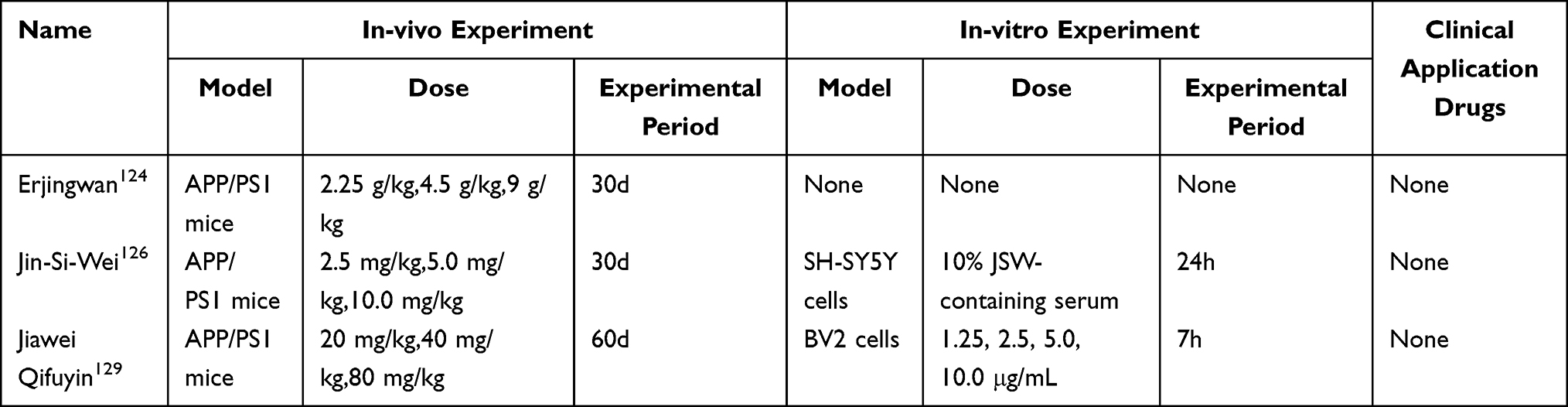 |
Table 5 The AGE-RAGE Signaling Pathway and AD |
 |
Figure 5 The AGE-RAGE signaling pathway and AD. This schematic illustrates the central role of the AGE-RAGE axis in AD pathology. Ligand binding to RAGE activates NADPH oxidase, increasing ROS production. This oxidative stress promotes β-secretase activity, leading to Aβ plaque deposition and Tau protein phosphorylation. The pathway also activates NF-κB, driving neuroinflammation via cytokine release. These combined pathological events result in neuronal damage. |
The Nrf2 Signaling Pathway and AD
Nrf2 is a key transcription factor that regulates target genes in response to stress stimuli. It helps reduce oxidative and electrophilic stress by lowering ROS and electrophilic compounds.131 Normally, Nrf2 is degraded by Keap1-Cul3-Rbx1. However, stress can change Keap1’s structure, allowing Nrf2 to move to the nucleus, bind to AREs, and activate protective genes like nqo1 and Heme Oxygenase-1 (HO-1).132 Nrf2 deficiency is a major factor in AD progression. Its reduced expression worsens AD through three main pathways: (1) suppressing the HO-1/GPX4 pathway, leading to astrocyte ferroptosis; (2) increasing ROS and NOX4 levels, which amplifies oxidative damage; (3) causing mitochondrial fragmentation, impairing astrocyte function.133 Activating the Nrf2 pathway can balance the expression levels of soluble and insoluble Aβ, reduce the generation of toxic Aβ peptide segments, and upregulate the expression of NDP52 protein. This, in turn, leads to the degradation of p-Tau through the autophagy pathway.134 This pathway can also enhance the antioxidant defense capacity, alleviate oxidative stress and inflammatory responses, and maintain protein stability. The Nrf2 pathway is regulated by multiple upstream signaling networks: PI3K/Akt enhances Nrf2 stability and nuclear translocation, whereas p38MAPK and GSK3β promote Nrf2 degradation. Meanwhile, Nrf2 negatively regulates the NF‑κB and AGE‑RAGE135 signaling axes, forming a negative feedback loop that cooperatively suppresses oxidative stress and neuroinflammation.136
NP studies revealed that the neuroprotective effects of Ginsenosides are primarily mediated through pathways such as anti-inflammation, anti-apoptosis, and anti-oxidative stress, with potential associations with signaling pathways including Nrf2. Notably, in vitro experiments confirmed that Ginsenosides Rg1 and Rb1 can downregulate the expression of ROS, Caspase-3, and Bax, while upregulating the expression of SOD, MMP, and Bcl-2. Mechanistically, these two Ginsenosides ameliorate neuronal function via the Nrf2 signaling pathway. In neuroblastoma cells overexpressing Aβ, Ginsenosides promote Nrf2 nuclear translocation and upregulate the expression of antioxidant enzymes, thereby reducing oxidative stress. In Aβ-induced cell models and APP/PS1 mice, Ginsenosides alleviate oxidative damage, mitochondrial dysfunction, and neuronal apoptosis, and improve cognitive deficits by activating the AMPK/Nrf2/HO-1 or Nrf2/HO-1 signaling pathways. Thus, NP predicts the multi‑target characteristics of Ginsenosides in AD treatment, while experimental studies validate their therapeutic effects through multiple core signaling pathways, including the Nrf2 pathway.62 Network analysis flags MAPT, APP, AChE, iNOS and COX-2 as core AD targets of Qi-Fu-Yin (QFY). Previous literature suggests that Panax ginseng and its active component Ginsenoside Rg3, which is identified as one of the main active compounds in QFY, can activate the Nrf2/HO-1 pathway. In vitro experiments confirm that QFY and its active components, especially Ginsenoside Rg3, can selectively downregulate the expression of iNOS in LPS-stimulated BV-2 microglial cells, thereby exerting anti-neuroinflammatory effects. Based on these findings, it is hypothesized that the mechanism of action is closely related to the activation of the Nrf2/HO-1 signaling pathway.137 NP reveals that PPARG, SLC40A1, LKB1, AMPK, Nrf2, etc. are the core targets of Rhizoma Anemarrhenae saponins (RAS) and AD. KEGG pathway enrichment analysis shows that these targets are significantly enriched in signaling pathways such as AMPK and Ferroptosis. Molecular docking results indicate that the key saponin components of RAS have good binding affinity with the selected core target proteins. In vitro experiments, RAS intervention significantly reversed the ferroptosis phenotype induced by Aβ, manifested by upregulation of GPX4, SLC7A11, and FPN protein expression, and downregulation of ACSL4 and TFR1 protein levels; at the same time, it effectively reduced intracellular iron accumulation and MDA content, increased GSH level and SOD activity, and inhibited lipid peroxidation damage. In APP/PS1 mice, RAS treatment significantly improved the iron death pathology in the brain, with protein regulation trends consistent with those in vitro: upregulation of GPX4, SLC7A11, and FPN expression, and downregulation of ACSL4 and TFR1 expression; and at the biochemical level, it reduced brain tissue iron content and MDA level, increased GSH and SOD activity, thereby alleviating neurodegenerative damage. Whether in vitro cells or in vivo animal models, RAS enhanced the interaction between LKB1 and AMPK, activated the AMPK/Nrf2 signaling pathway, thereby inhibiting neuronal ferroptosis, and ultimately achieving the effect of improving the pathological damage and cognitive impairment of AD.138 NP identified TP53, IL6, STAT3, Nrf2 and other genes as the core targets of Changpu-Yizhi-Wan (CYW) and AD. The enrichment test showed that these targets were mainly enriched in the Nrf2 signaling pathway and iron death-related pathways. Molecular docking revealed that the core components of CYW could have a good binding affinity with the Nrf2 protein. In vitro experiments confirmed that in the HT22 hippocampal neuron cell model induced by RSL3, CYW could significantly increase the survival rate of RSL3-damaged cells, significantly reduce Fe2⁺ level and lipid peroxidation products, and restore the oxidative-reductive balance of the cells. Western blot results showed that CYW could significantly up-regulate the expression of key proteins of the antioxidant system, such as Nrf2, SLC7A11, GPX4 and FTH1, confirming that CYW inhibits neuronal iron death by activating the Nrf2/SLC7A11/GPX4/FTH1 axis. These findings reveal that CYW may target Nrf2, regulate the downstream SLC7A11/GPX4/FTH1 pathway, thereby inhibiting neuronal iron death and achieving the purpose of treating AD.139 NP revealed that GSK3β, Nrf2, AKT, ERK are the intersectional targets of Norboldine and AD. The study found that Norboldine exerts neuroprotective effects by activating the AMPK/GSK3β/Nrf2 signaling pathway, thereby upregulating the expression of downstream antioxidant genes. In vitro experiments, Norboldine significantly increased the survival rate of damaged cells induced by Aβ, reduced cell apoptosis, lowered intracellular ROS levels, and restored mitochondrial membrane potential. When using AMPK inhibitors or gene knock down the protective effect of Norboldine disappeared. In 3×Tg mice, Norboldine reduced Aβ deposition in the brain (Aβ40/Aβ42) and inhibited excessive activation of glial cells. Western blot results showed that Norboldine decreased the levels of apoptosis-related proteins in brain tissue, increased the level of Bcl-2, and activated the AMPK/GSK3β/Nrf2 pathway.140 NP revealed that AKT1, MAPK3, IL6, TP53, VEGFA, TNF, SRC are the intersectional targets of Bu-Shen-Yi-Jing-Fang (BSYJF) and AD. Enrichment tests found that these targets were mainly enriched in the PI3K/Akt, Ras signaling pathways, and cell apoptosis signaling pathways. In the in vitro SKNMC cell model experiment, BSYJF significantly upregulated the protein expression levels of p-Akt and p-GSK3β, promoted the nuclear translocation of Nrf2, significantly upregulated the expression of its downstream antioxidant proteins HO-1 and NQO1, significantly upregulated the anti-apoptotic protein Bcl-2, and downregulated the expression of pro-apoptotic proteins Bax and cleaved Caspase-3/9. It also effectively reduced the intracellular levels of ROS, MDA, and Fe2⁺, while increasing the activities of SOD.141 KEGG analysis indicates that the PI3K/Akt/GSK3NRF2/HO-1 pathway may be involved in the regulation of cognitive impairment caused by Asafoetida. In vivo experiments, Asafoetida treatment can significantly improve cognitive dysfunction caused by scopolamine, enhance the learning and memory abilities of mice, and alleviate damage to hippocampal neurons, cholinergic system, oxidative stress and cell apoptosis in mice. In vitro experiments, Asafoetida can reduce pathological oxidative stress in PC12 cells induced by hydrogen peroxide, inhibit the production of ROS and MDA, and promote the activity of SOD, CAT and GSH. In addition, Asafoetida can significantly inhibit the increase in the expression levels of Caspase-3 and Bax induced by hydrogen peroxide, as well as the decrease in Bcl-2 expression.142
Nrf2 is the central regulatory target, and its downstream effector HO-1 serves as a key antioxidant and anti-inflammatory molecule, commonly used as a marker of pathway activation. Activating the Nrf2 pathway primarily aims to enhance endogenous cellular antioxidant defenses. This is achieved by upregulating a suite of antioxidant enzymes and Phase II detoxification enzymes, such as HO‑1, GST, and NQO1, to scavenge reactive oxygen species, mitigate oxidative damage, and indirectly exert anti-inflammatory and anti‑apoptotic effects, for instance through inhibiting iNOS. This mechanism closely aligns with the core pathological feature of oxidative stress in AD. The Nrf2 pathway exhibits a positive regulatory relationship with the AMPK pathway. The Nrf2 pathway exhibits a positive regulatory relationship with the AMPK pathway; for instance, ginsenosides activate Nrf2 via AMPK. Its antioxidant effects are interconnected with the inhibition of pro-inflammatory pathways such as NF-κB, working together to maintain redox balance and inflammatory homeostasis. Current research is highly focused on Panax ginseng and its ginsenoside components, particularly ginsenosides Rg1 and Rb1.62 Studies on these components are systematic and in-depth, spanning molecular, cellular, and animal levels, and have become an exemplary model for pharmacological research on the modulation of the Nrf2 pathway in TCM. In contrast, research on compound formulations such as QFY remains relatively preliminary, and direct evidence on how the formulation as a whole modulates the Nrf2 pathway is still lacking.137 Please refer to Table 6 for specific details. The Nrf2 signaling pathway and the pathogenesis of AD are illustrated in Figure 6.
 |
Table 6 The Nrf2 Signaling Pathway and AD |
 |
Figure 6 The Nrf2 signaling pathway and AD. This schematic illustrates the central mechanism by which Nrf2 activation counteracts AD pathogenesis. Under oxidative stress, Nrf2 is released from Keap1, translocates to the nucleus, and binds to AREs alongside Maf proteins, driving the expression of antioxidant genes. This reduces ROS levels and inhibits NOX4. Furthermore, Nrf2 activation suppresses pro-pathological signaling via GSK-3β, NF-κB, and p38 MAPK, thereby alleviating neuroinflammation, Tau hyperphosphorylation, and neuronal apoptosis. Collectively, this pathway mitigates oxidative stress, Aβ deposition, and neuronal/astrocyte damage. |
Integrated Crosstalk Network of Five Key Signaling Pathways
Interconnected Crosstalk Network of Five Core Signaling Pathways in AD.
The five core signaling pathways do not function in isolation but rather constitute an interconnected and interrelated network that collectively contributes to the therapeutic effects against AD. The PI3K/Akt and MAPK pathways share upstream regulators such as Ras, and they synergistically regulate neuronal survival, apoptosis, autophagy, and Tau phosphorylation.143 The Ras-Raf-MEK-ERK cascade often acts in parallel with the PI3K cascade, amplifying protective signals against AD pathology.144 The AGE-RAGE axis exacerbates oxidative stress and neuroinflammation by activating NF-κB and suppressing Nrf2, thereby promoting a pro-oxidative and pro-inflammatory state. In contrast, Nrf2 acts as a negative feedback regulator to counteract inflammation induced by AGE-RAGE.145 Furthermore, the JAK-STAT and PI3K/Akt pathways extensively interact during cytokine signal transduction, jointly controlling microglial polarization and the release of inflammatory factors, thereby regulating neuroinflammatory responses in AD.146 TCM through its multi-component and multi-target regulatory properties, simultaneously modulates the crosstalk among these five pathways, achieving systemic therapeutic effects including anti-apoptosis, anti-inflammation, antioxidant activity, suppression of Tau hyperphosphorylation, and reduction of Aβ deposition. Please refer to Figure 7 for specific details.
 |
Figure 7 Crosstalk and regulatory network of key signaling pathways in AD. This schematic illustrates how the five major signaling pathways are interconnected. Through the dysregulation of cellular processes, they collectively drive the core pathological hallmarks of AD, such as Aβ deposition, tauopathy, neuroinflammation, and neuronal loss. |
Discussion
AD is the primary cause of dementia in the elderly. Its prevalence continues to rise with the increase in life expectancy, imposing a heavy economic burden on global societies. Despite substantial financial investment and research efforts devoted to exploring its pathological mechanisms, the etiopathogenesis of AD remains not fully elucidated, and there are currently no effective therapeutic strategies to delay or halt its clinical progression.147,148 In recent years, TCM - based interventions for AD have garnered extensive attention, and notable achievements have been attained in multiple aspects, including reducing Aβ peptide deposition, clearing hyperphosphorylated Tau protein, regulating cholinergic neurotransmitters, improving vascular function and microcirculation, alleviating oxidative stress and inflammation, as well as inhibiting neuronal apoptosis.149 The therapeutic landscape for AD has fundamentally transformed, as the range of treatments approved by the US FDA now greatly exceeds the traditional handful of symptomatic options. Beyond established therapies such as cholinesterase inhibitors, memantine, and their combination formulations, the available treatments have been substantially augmented by the approval of Lecanemab (Leqembi) and Donanemab (Kisunla). These are disease-modifying therapies that actively clear amyloid plaques and have demonstrated the ability to slow clinical progression. Recent years have also seen the introduction of newer formulations, including transdermal patches and subcutaneous injections, alongside novel-mechanism agents such as Benzgalantamine (Zunveyl). Consequently, the management of AD has entered a diversified era, offering a spectrum of strategies from symptom control to disease modification, with varied mechanisms of action and routes of administration.150 TCM emphasizes the concept of holistic treatment. Chinese herbal medicines have the characteristics of “multiple components and multiple targets”, which are highly consistent with the “multiple targets and systematic regulation” diagnosis and treatment concept urgently needed by modern medicine.
The components of TCM are complex and include many types of bioactive components. NP, as a cutting-edge analytical tool, has revealed that TCM can exert its therapeutic effect on AD by regulating multiple molecular targets and signaling pathways. To explore the mechanism of TCM in preventing and treating AD based on NP, the following work was carried out in this study. Firstly, search for relevant literature in the PubMed database in the past 10 years with the key words of Chinese herbal medicine components, NP, and AD. Subsequently, systematically sort out and extract the key signaling pathways of TCM intervention in AD revealed by NP. Finally, summarize the frequently enriched pathways, and deeply explore the molecular mechanism of multi-target and multi-pathway treatment of AD by TCM, providing theoretical basis and research ideas for the subsequent development of new drugs against AD and clinical transformation. Despite its great potential in clarifying the complex mechanisms of TCM-based AD therapy, NP still faces limitations: outdated and limited information in existing databases, static NP models that cannot simulate drugs’ in vivo dynamics, and insufficient experimental validation. In the future, integrating multi-omics data, dynamic modeling, and efficient experimental validation will enable NP to advance TCM and precision medicine modernization, providing innovative strategies for AD.
Notably, the PI3K/Akt signaling pathway, MAPK signaling pathway, JAK-STAT signaling pathway, AGE-RAGE signaling pathway, and Nrf2 signaling pathway are among the core pathways targeted by TCM. This multi-dimensional action mode highlights the potential application of TCM as a treatment option for AD, especially considering the complex and multifactorial nature of the disease. Under physiological conditions, the PI3K/Akt pathway can inhibit the excessive activation of NF-κB by phosphorylating IKK, blocking the nuclear translocation of NF-κB, and thereby exerting an anti-inflammatory effect.53 The activity of the PI3K/Akt pathway is reduced, leading to excessive activation of NF-κB in microglia and astrocytes. This, in turn, promotes the transcription of pro-inflammatory cytokines such as IL-1β, TNF-α, and IL-6, ultimately triggering neuroinflammation and neuronal damage.54 The activation of the MAPK signaling pathway is one of the key factors that trigger AD. Specifically, the activated MAPK signal cascade reaction can promote neuronal apoptosis and regulate the phosphorylation process and stability of the APP through the JNK signaling axis. Moreover, this pathway can also regulate the transcriptional activation process and enzyme activity of β-secretase and γ-secretase, which are the key enzymes in the abnormal processing of APP.151 The JAK-STAT signaling pathway can regulate the function of microglia and thereby bidirectionally control the pathological process of AD. On one hand, it amplifies the pro-inflammatory response of microglia, which further aggravates the deposition of Aβ, the pathological damage of Tau protein, and neuroinflammation; on the other hand, it can also induce microglia to exhibit anti-inflammatory activity, providing protection for neurons.108 AGEs can upregulate the expression of APP, promote the generation of Aβ, and increase the accumulation of Aβ in the brain. This series of processes will induce the misfolding of Tau protein, exacerbating the occurrence and progression of neurological diseases. Moreover, the interaction between AGEs and RAGE will activate pathological processes related to neuroinflammation, oxidative stress, and excitotoxicity, thereby inducing neuronal cell death.152 In microglia, the activation of the Nrf2 signaling pathway upregulates the expression of key antioxidant enzymes, inhibits the production of pro-inflammatory cytokines, promotes the survival of microglia, and improves the bioenergetic function of mitochondria, thereby delaying the pathological process of AD. Additionally, the activation of Nrf2 in astrocytes, neurons, and vascular systems also helps to alleviate metabolic stress, oxidative damage, and vascular dysfunction.153 The core characteristic of TCM in treating AD lies in its regulation of crosstalk between signaling pathways, rather than targeting a single pathway. The PI3K/Akt and MAPK pathways form a synergistic survival-inflammation network through shared upstream regulators. The AGE-RAGE axis couples oxidative stress and inflammation via bidirectional regulation of NF-κB and Nrf2. Concurrently, JAK-STAT and PI3K/Akt pathways interact within cytokine signaling to modulate neuroinflammation. This extensive signal integration aligns perfectly with the multi-component, multi-target nature of TCM, explaining its holistic therapeutic advantage in addressing the complex pathogenesis of AD.
NP of TCM, by leveraging disciplines such as bioinformatics, systems biology, and computational biology, has built a bridge between traditional and modern medicine. It integrates the “TCM-disease target-gene” network and screens potential candidate drugs that can effectively combine with multiple targets of AD from this network.154 However, NP has inherent limitations, and TCM itself contains complex components. As merely a predictive tool, NP is prone to false positive results; its predictive methods are overly simplistic, failing to comprehensively evaluate the efficacy and toxicological characteristics of TCM. If outdated or un-updated databases are used, false positive and false negative results are likely to occur, which in turn can lead to deviations in the construction of biological networks.155 Molecular docking offers a viable solution to the above issues by providing reliable active component-target interaction data for NP, thereby reducing false positives in network construction. Molecular dynamics (MD) simulations compensate for the limitations of molecular docking in studying targets with unknown structures, thereby improving the reliability of predictions. However, both approaches are in silico virtual screening methods and cannot fully replicate the in vivo physiological environment; therefore, validation through in vitro and in vivo experiments remains essential. By employing up-to-date databases and effective algorithms to identify the latest targets, pathways, and biological processes, dynamically validating TCM-NP findings through more relevant in vitro and in vivo experiments, and incorporating the novel MTDL strategy, the shortcomings of TCM-NP in AD research can be partially addressed. This integrated approach will contribute to elucidating the mechanisms of AD pathogenesis and advancing the development of effective therapeutics.156,157 By selecting timely updated databases and effective algorithms to identify the latest targets, pathways, and biological processes, dynamically validating the results of TCM-NP through more relevant in vivo and in vitro experiments, and adopting the novel MTDL strategy, the shortcomings of TCM-NP in AD research can be addressed to a certain extent, facilitating the elucidation of AD pathogenesis and the development of effective drugs.158
This study summarized and concluded the mechanisms and common pathways of TCM in treating AD using the NP method. However, there are still some limitations that need to be further improved in the subsequent research. Firstly, NP is a predictive analytical method, and the reliability of its analysis results highly depends on the completeness of the existing databases. In some of the studies, the active components and the target sites identified have not yet been directly verified through experiments. Therefore, more in vitro experiments and in vivo studies are urgently needed for validation. Secondly, this study focuses on the overall regulation of TCM, but the synergistic or antagonistic effects between multiple active components are not yet clear. Further research should delve into the interaction patterns between core components. Thirdly, this study did not consider the influence of TCM processing methods, administration routes, and dosages on the biological activity of active components. Further relevant studies need to be supplemented. Finally, translating the findings into clinical practice requires well-designed randomized controlled trials to evaluate the safety and efficacy of TCM formulations, thereby promoting the modernization and internationalization of TCM in the treatment of neurodegenerative diseases.
NP primarily functions to predict the active components of TCM and their corresponding disease targets. Its fundamental purpose is to serve clinical translation by exploring therapeutic potential. In turn, clinical research on TCM interventions for AD provides objective efficacy evidence for such potential, thereby significantly enhancing the credibility of the conclusions. A systematic meta-analysis of 11 randomized controlled trials involving 798 patients demonstrated that DHYZ, administered either as monotherapy or as an adjunct to conventional Western medicine, significantly improved the overall clinical response rate, cognitive function and activities of daily living in patients with AD, with no serious adverse events reported.159 However, due to prevalent limitations such as inadequate allocation concealment and suboptimal blinding procedures across the included trials, the quality of evidence was graded as low to very low according to the GRADE framework, highlighting the necessity for more methodologically rigorous and high‑quality clinical trials to confirm these preliminary findings. A randomized, double‑blind, parallel‑group controlled trial enrolled 156 patients with mild‑to‑moderate depression and demonstrated that SZL achieved comparable efficacy to fluoxetine in reducing scores on both the 17‑item Hamilton Depression Rating Scale (HAM‑D17) and the Self‑Rating Depression Scale (SDS) after 8 weeks of treatment. Although SZL did not produce a significant improvement in working memory, it showed a distinct advantage in specifically lowering serum levels of apolipoprotein B (APOB) and apolipoprotein C3 (APOC3), as well as reducing the low‑density lipoprotein to high‑density lipoprotein ratio (LDL‑C/HDL‑C). These results suggest that the antidepressant mechanism of SZL may be closely linked to the modulation of lipid metabolic homeostasis.160 Furthermore, integrating NP with preclinical investigations to explore therapeutic targets for AD remains a key focus of our research.
Alterations in serum amino acid profiles, such as those of arginine, serine, and isoleucine, in patients with dementia are showing promise as biomarkers for auxiliary diagnosis or disease staging.161,162 This offers a new avenue for the early diagnosis, disease monitoring, and treatment response evaluation of AD. The signaling pathways reviewed here, such as PI3K/Akt, AGE-RAGE, and JAK-STAT, are implicated not only in core AD pathologies like Aβ deposition, Tau hyperphosphorylation, neuroinflammation, and oxidative stress, but their dysregulation may also be closely linked to systemic metabolic disturbances. Currently, NP research in TCM has largely focused on basic experimental studies, with a notable scarcity of clinical or preclinical validation. Strengthening such translational research is therefore a critical direction for future investigation. Linking readily measurable blood-based biomarkers with pathway activities predicted by NP can significantly enhance the translational relevance of this work, providing a more direct strategy for validating biomarkers in TCM-based AD interventions. In recent years, blood metabolomic studies seeking accessible AD biomarkers have supported this perspective. Clinical evidence indicates that serum levels of amino acids such as Arginine, Serine, and Isoleucine are specifically elevated in patients with AD or mild cognitive impairment (MCI).163,164 These metabolic changes are potentially connected to the pathway networks discussed. Elevated arginine levels, for instance, may relate to nitric oxide synthase (NOS) pathway dysregulation and exacerbated neuroinflammation, corresponding to the activation of pro-inflammatory pathways like AGE-RAGE and JAK-STAT.165,166 Furthermore, serine, particularly D-serine, which is an endogenous ligand of the N-methyl-D-aspartate receptor (NMDAR), may influence excitatory neurotransmission when dysregulated, thereby linking it to AD-related synaptic dysfunction and excitotoxicity.167,168 Disruptions in branched-chain amino acid metabolism, such as that of isoleucine, point to systemic impairments in energy metabolism and insulin signaling, processes closely tied to PI3K/Akt pathway function. Consequently, the therapeutic effects of TCM on these key pathways, as predicted by NP, may ultimately correlate with the normalization of such dysregulated amino acid metabolic profiles.169,170 In the future, serum amino acid profiles could serve as potential dynamic biomarkers for assessing the efficacy of TCM interventions in AD, thereby providing a crucial bridge to more closely integrate computationally predicted mechanisms with clinically observable phenotypic changes.
Conclusion
Given the complex pathology and lack of curative treatments for AD, TCM holds promise as a therapeutic strategy, largely due to its holistic, multi‑component, multi‑target philosophy. Based on a review of NP and TCM literature from the past decade, we summarize that TCM may exert its effects by modulating key signaling pathways, such as PI3K/Akt, MAPK, JAK‑STAT, AGE‑RAGE, and Nrf2. NP‑based analyses further reveal extensive crosstalk among these pathways, which may underlie the systemic benefits of TCM, including anti‑inflammatory, antioxidant, and anti‑apoptotic activities. However, it should be noted that current evidence still relies heavily on predictive NP approaches, which carry inherent limitations such as false‑positive findings. Thus, the proposed mechanisms require further experimental validation. Ultimately, rigorously designed randomized controlled trials (RCTs) will be essential to evaluate the clinical safety and efficacy of TCM, thereby supporting its modernization and broader international recognition.
Abbreviations
AD, Alzheimer’s disease; Aβ, amyloid β-protein; NP, Network Pharmacology; GSK3β, Glycogen Synthase Kinase 3β; mTOR, mammalian Target of Rapamycin; NF-κB, Nuclear factor kappa-B; PI3K/Akt,Phosphoinositide 3-kinase /Protein kinase B; MAPK, Mitogen-Activated Protein Kinase; JAK-STAT, Janus kinase/signal transducer and activator of transcription; AGE-RAGE, Advanced Glycation End-products-Receptor for Advanced Glycation End-products; Nrf2, Nuclear factor erythroid 2-related factor 2;NFTs, form neurofibrillary tangles; EGFR, Epidermal Growth Factor Receptor; IKK, IκB Kinase; IκBα, Inhibitor of κB alpha; Caspase3, Cysteine-aspartic proteases 3; Bcl-2, Bcl-2 family protein; Bax, Bcl-2 Associated X protein; PTEN, Phosphatase and Tensin Homolog; AChE, Acetylcholinesterase;BACE1, Beta-Site Amyloid Precursor Protein Cleaving Enzyme 1;JNK, c-Jun amino-terminal kinase; ERK, Extracellular signal-regulated kinase; APP/PS1, amyloid precursor protein/presenilin 1;MCP-1, Monocyte Chemoattractant Protein-1;HO-1, Heme Oxygenase-1;COX-2, Cyclooxygenase-2;SOD, Superoxide Dismutase; AGEs, Advanced Glycation End Products; RAGE, Receptor for Advanced Glycation End-products; JAKs, Janus kinases; STAT, signal transducer and activator of transcription.
Data Sharing Statement
The datasets used and/or analyzed during the current study are available from the corresponding author (Yujiu Wang) upon reasonable request.
Author Contributions
All authors made a significant contribution to the work reported, whether that is in the conception, study design, execution, acquisition of data, analysis and interpretation, or in all these areas; took part in drafting, revising or critically reviewing the article; gave final approval of the version to be published; have agreed on the journal to which the article has been submitted; and agree to be accountable for all aspects of the work.
Funding
This study was supported by Shandong Provincial Key Research and Development Program (Major Scientific and Technological Innovation Project): Development of Large-Diameter Artificial Blood Vessels (2023CXGC010504 to WYJ),Natural Science Foundation of Shandong Province (ZR2023QH030, ZR2024MH089 to QGF;ZR2023MH067 to WYJ), the Projects of Traditional Chinese medicine in Shandong Province (Z20244105 to SRQ), the Scientific Research Foundation of Binzhou Medical University (BY2021KYQD31 to QGF).
Disclosure
Yiyi Zhang, Gaofeng Qin, and Qinaqian Lian are co-first authors for this study. The authors declare that they have no known competing financial interests or personal relationships that could have appeared to influence the work reported in this paper.
References
1. Tenchov RA-O, Sasso JA-O, Zhou QA-OX. Alzheimer’s disease: exploring the landscape of cognitive decline. ACS Chem Neurosci. 2024;15(21):3800–34. doi:10.1021/acschemneuro.4c00339
2. Twarowski B, Herbet MA-O. Inflammatory processes in Alzheimer’s disease-pathomechanism, diagnosis and treatment: a review. Int J Mol Sci. 2023;24(7):6518. doi:10.3390/ijms24076518
3. Cai Y, Liu J, Wang B, et al. Microglia in the neuroinflammatory pathogenesis of Alzheimer’s disease and related therapeutic targets. Front Immunol. 2022;13(1):856376. doi:10.3389/fimmu.2022.856376
4. Soni U, Singh K, Jain D, et al. Exploring Alzheimer’s disease treatment: established therapies and novel strategies for future care. Eur J Pharmacol. 2025;998(1):177520. doi:10.1016/j.ejphar.2025.177520
5. Li J, Wang L, Zhang X, et al. Translating Alzheimer’s disease mechanisms into therapeutic opportunities. Biomolecules. 2025;15(9):1290. doi:10.3390/biom15091290
6. Rafii MA-O, Aisen PA-O. Amyloid-lowering immunotherapies for Alzheimer disease: current status and future directions. Nat Rev Neurol. 2025;21(9):490–498. doi:10.1038/s41582-025-01123-5
7. Ding MR, Qu Y-J, Hu B, et al. Signal pathways in the treatment of Alzheimer’s disease with traditional Chinese medicine. Biomed Pharmacother. 2022;152(1):113208. doi:10.1016/j.biopha.2022.113208
8. Zhang Y, Li X, Shi Y, et al. ETCM v2.0: an update with comprehensive resource and rich annotations for traditional Chinese medicine. Acta Pharm Sin B. 2023;13(6):2559–2571. doi:10.1016/j.apsb.2023.03.012
9. Joshi CP, Baldi A, Kumar N, et al. Harnessing network pharmacology in drug discovery: an integrated approach. Naunyn Schmiedebergs Arch Pharmacol. 2025;398(5):4689–4703. doi:10.1007/s00210-024-03625-3
10. Chen Q, Chen G, Wang Q. Application of Network pharmacology in the treatment of neurodegenerative diseases with traditional Chinese medicine. Planta Med. 2025;91(5):226–237. doi:10.1055/a-2512-8928
11. Alshabrmi FM, Alkhayl FFA, Rehman A. Novel drug discovery: advancing Alzheimer’s therapy through machine learning and network pharmacology. Eur J Pharmacol. 2024;976(1):176661. doi:10.1016/j.ejphar.2024.176661
12. Tsai ST, Huang HY. Network pharmacology implicates traditional Chinese medicine in regulating systemic homeostasis to benefit Alzheimer’s disease. Tzu Chi Med J. 2023;35(2):120–130. doi:10.4103/tcmj.tcmj_125_22
13. Yee JL, Huang C-Y, Yu Y-C, et al. Potential mechanisms of guizhi fuling wan in treating endometriosis: an analysis based on TCMSP and disgenet databases. J Ethnopharmacol. 2024;329(1):118190. doi:10.1016/j.jep.2024.118190
14. Zhao X, Xiu J, Yang H, et al. Network Pharmacology and bioinformatics study of six medicinal food homologous plants against colorectal cancer. Int J Mol Sci. 2025;26(3):930. doi:10.3390/ijms26030930
15. Xu HY, Zhang Y-Q, Liu Z-M, et al. ETCM: an encyclopaedia of traditional Chinese medicine. Nucleic Acids Res. 2019;47(D1):D976–D982. doi:10.1093/nar/gky987
16. Fang S, Dong L, Liu L, et al. HERB: a high-throughput experiment- and reference-guided database of traditional Chinese medicine. Nucleic Acids Res. 2021;49(D1):D1197–D1206. doi:10.1093/nar/gkaa1063
17. Gao K, Liu L, Lei S, et al. HERB 2.0: an updated database integrating clinical and experimental evidence for traditional Chinese medicine. Nucleic Acids Res. 2025;53(D1):D1404–D1414. doi:10.1093/nar/gkae1037
18. Wu Y, Zhang F, Yang K, et al. SymMap: an integrative database of traditional Chinese medicine enhanced by symptom mapping. Nucleic Acids Res. 2019;47(D1):D1110–D1117. doi:10.1093/nar/gky1021
19. Xue R, Fang Z, Zhang M, et al. TCMID: traditional Chinese medicine integrative database for herb molecular mechanism analysis. Nucleic Acids Res. 2013;41(Database issue):D1089–95. doi:10.1093/nar/gks1100
20. Stelzer G, Rosen N, Plaschkes I, et al. The genecards suite: from gene data mining to disease genome sequence analyses. Curr Protoc Bioinformatics. 2016;54(1):1.30.1–1.30.33. doi:10.1002/cpbi.5
21. Dahary D, Golan Y, Mazor Y, et al. Genome analysis and knowledge-driven variant interpretation with TGex. BMC Med Genomics. 2019;12(1):200. doi:10.1186/s12920-019-0647-8
22. Hamosh A, Amberger JS, Bocchini C, et al. Online Mendelian inheritance in man (OMIM®): victor McKusick ‘s magnum opus. Am J Med Genet A. 2021;185(11):3259–3265. doi:10.1002/ajmg.a.62407
23. Knox C, Wilson M, Klinger C, et al. DrugBank 6.0: the DrugBank knowledgebase for 2024. Nucleic Acids Res. 2024;52(D1):D1265–D1275. doi:10.1093/nar/gkad976
24. Davis AA-O, Wiegers TC, Johnson RJ, et al. Comparative Toxicogenomics Database (CTD): update 2023. Nucleic Acids Res. 2023;51(D1):D1257–D1262. doi:10.1093/nar/gkac833
25. Szklarczyk D, Kirsch R, Koutrouli M, et al. The STRING database in 2023: protein-protein association networks and functional enrichment analyses for any sequenced genome of interest. Nucleic Acids Res. 2023;51(D1):D638–D646. doi:10.1093/nar/gkac1000
26. Gable AL, Szklarczyk D, Lyon D, et al. Systematic assessment of pathway databases, based on a diverse collection of user-submitted experiments. Brief Bioinform. 2022;23(5):bbac355. doi:10.1093/bib/bbac355
27. Szklarczyk D, Nastou K, Koutrouli M, et al. The STRING database in 2025: protein networks with directionality of regulation. Nucleic Acids Res. 2025;53(D1):D730–D737. doi:10.1093/nar/gkae1113
28. Franz MA-O, Lopes CT, Fong D, et al. Cytoscape.js 2023 update: a graph theory library for visualization and analysis. Bioinformatics. 2023;39(1):btad031. doi:10.1093/bioinformatics/btad031
29. Doncheva NA-O, Morris JH, Holze H, et al. Cytoscape stringApp 2.0: analysis and visualization of heterogeneous biological networks. J Proteome Res. 2023;22(2):637–646. doi:10.1021/acs.jproteome.2c00651
30. Pratt D, Pillich RT, Morris JH. Translating desktop success to the web in the cytoscape project. Front Bioinform. 2023;3(1):1125949. doi:10.3389/fbinf.2023.1125949
31. Majeed A, Mukhtar S. Protein-Protein Interaction Network Exploration Using Cytoscape. Methods Mol Biol. 2023;2690(1):419–427. doi:10.1007/978-1-0716-3327-4_32
32. Jin Z, Sato Y, Kawashima M, et al. KEGG tools for classification and analysis of viral proteins. Protein Sci. 2023;32(12):e4820. doi:10.1002/pro.4820
33. Kanehisa MA-OX, Furumichi M, Sato Y, et al. KEGG for taxonomy-based analysis of pathways and genomes. Nucleic Acids Res. 2023;51(D1):D587–D592. doi:10.1093/nar/gkac963
34. Kanehisa MA-OX, Sato Y, Kawashima M. KEGG mapping tools for uncovering hidden features in biological data. Protein Sci. 2022;31(1):47–53. doi:10.1002/pro.4172
35. Kanehisa MA-OX, Furumichi M, Sato Y, et al. KEGG: biological systems database as a model of the real world. Nucleic Acids Res. 2025;53(D1):D672–D677. doi:10.1093/nar/gkae909
36. Kanehisa M, Furumichi M, Sato Y, et al. KEGG: integrating viruses and cellular organisms. Nucleic Acids Res. 2021;49(D1):D545–D551. doi:10.1093/nar/gkaa970
37. Ge SX, Jung D, Yao R. ShinyGO: a graphical gene-set enrichment tool for animals and plants. Bioinformatics. 2020;36(8):2628–2629. doi:10.1093/bioinformatics/btz931
38. Gaudet P, Logie C, Lovering RC, et al. Gene ontology representation for transcription factor functions. Biochim Biophys Acta Gene Regul Mech. 2021;1864(11–12):194752. doi:10.1016/j.bbagrm.2021.194752
39. Feuermann MA-O, Gaudet PA-O. Interpreting gene ontology annotations derived from sequence homology methods. Methods Mol Biol. 2024;2836(1):285–298. doi:10.1007/978-1-0716-4007-4_15
40. Zhu JA-O, Zhao Q, Katsevich E, et al. Exploratory gene ontology analysis with interactive visualization. Sci Rep. 2019;9(1):7793. doi:10.1038/s41598-019-42178-x
41. Li T, Guo R, Zong Q, et al. Application of molecular docking in elaborating molecular mechanisms and interactions of supramolecular cyclodextrin. Carbohydr Polym. 2022;276(1):118644. doi:10.1016/j.carbpol.2021.118644
42. Sahu D, Rathor LS, Dwivedi SD, et al. A review on molecular docking as an interpretative tool for molecular targets in disease management. Assay Drug Dev Technol. 2024;22(1):40–50. doi:10.1089/adt.2023.060
43. Pinzi L, Rastelli GA-O. Molecular docking: shifting paradigms in drug discovery. Int J Mol Sci. 2019;20(18):4331. doi:10.3390/ijms20184331
44. Rigby SP. Uses of molecular docking simulations in elucidating synergistic, additive, and/or multi-target (SAM) effects of herbal medicines. Molecules. 2024;29(22):5406. doi:10.3390/molecules29225406
45. Zhao Y, Qian Y, Sun Z, et al. Role of PI3K in the progression and regression of atherosclerosis. Front Pharmacol. 2021;12(1):632378. doi:10.3389/fphar.2021.632378
46. He Y, Sun MM, Zhang GG, et al. Targeting PI3K/Akt signal transduction for cancer therapy. Signal Transduct Target Ther. 2021;6(1):425. doi:10.1038/s41392-021-00828-5
47. Long HZ, Cheng Y, Zhou Z-W, et al. PI3K/AKT signal pathway: a target of natural products in the prevention and treatment of Alzheimer’s disease and Parkinson’s disease. Front Pharmacol. 2021;12(1):648636. doi:10.3389/fphar.2021.648636
48. Liu R, Chen Y, Liu G, et al. PI3K/AKT pathway as a key link modulates the multidrug resistance of cancers. Cell Death Dis. 2020;11(9):797. doi:10.1038/s41419-020-02998-6
49. Al-Rawashde FA-O, Al-wajeeh AS, Vishkaei MN, et al. Thymoquinone inhibits JAK/STAT and PI3K/Akt/ mTOR signaling pathways in MV4-11 and K562 myeloid leukemia cells. Pharmaceuticals. 2022;15(9):1123. doi:10.3390/ph15091123
50. Wu YA-O, Ling Y, Yang Y, et al. Soybean sprout peptides alleviate obesity via PI3K-Akt and JAK-STAT pathway modulation, gut microbiota regulation, and metabolic reprogramming. J Agric Food Chem. 2025;73(16):9662–9676. doi:10.1021/acs.jafc.5c00695
51. Yang W, Liu Y, Xu -Q-Q, et al. Sulforaphene ameliorates neuroinflammation and hyperphosphorylated tau protein via regulating the PI3K/Akt/GSK-3 β pathway in experimental models of Alzheimer’s disease. Oxid Med Cell Longev. 2020;2020(1):4754195. doi:10.1155/2020/4754195
52. Zhang Y, Huang N-Q, Yan F, et al. Diabetes mellitus and Alzheimer’s disease: GSK-3β as a potential link. Behav Brain Res. 2018;339(1):57–65. doi:10.1016/j.bbr.2017.11.015
53. Sun E, Motolani A, Campos L, et al. The pivotal role of NF-kB in the pathogenesis and therapeutics of Alzheimer’s disease. Int J Mol Sci. 2022;23(16):8972. doi:10.3390/ijms23168972
54. Zhu C, Liu J, Lin J, et al. Investigating the effects of Ginkgo biloba leaf extract on cognitive function in Alzheimer’s disease. CNS Neurosci Ther. 2024;30(9):e14914. doi:10.1111/cns.14914
55. Razani E, Pourbagheri-Sigaroodi A, Safaroghli-Azar A, et al. The PI3K/Akt signaling axis in Alzheimer’s disease: a valuable target to stimulate or suppress? Cell Stress Chaperones. 2021;26(6):871–887. doi:10.1007/s12192-021-01231-3
56. Qin GA-O, Song R, Sun J, et al. Investigating the therapeutic effects of Shenzhiling oral liquid on Alzheimer’s disease: a network pharmacology and experimental approach. 3 Biotech. 2025;15(1):14. doi:10.1007/s13205-024-04181-6
57. Ye JX, Wu JY, Zhu M, et al. Elucidating the role of gardeniae fructus and scutellariae radix herb pair in Alzheimer’s disease via network pharmacology: emphasis on oxidative stress, and the PI3K/Akt pathway. Curr Pharm Biotechnol. 2025;1(1):1–17. doi:10.2174/0113892010326797250422095516
58. Zhang J, Li Q, Yan B, et al. Integrated network pharmacology and brain metabolomics to analyze the mechanism of Dihuang Yinzi intervention in Alzheimer’s disease. Heliyon. 2024;10(5):e26643. doi:10.1016/j.heliyon.2024.e26643
59. Jin S, Guan X, Min D. Evidence of clinical efficacy and pharmacological mechanisms of resveratrol in the Treatment of Alzheimer’s disease. Curr Alzheimer Res. 2023;20(8):588–602. doi:10.2174/0115672050272577231120060909
60. Wu J, Tian Z, Wang B, et al. Exploring resveratrol against Alzheimer’s disease and Parkinson’s disease through integrating network pharmacology, bioinformatics, and experimental validation strategy in vitro. Heliyon. 2024;10(18):e37908. doi:10.1016/j.heliyon.2024.e37908
61. Ozpak LA-O, Bağca BG. Neuroprotective effects of resveratrol through modulation of PI3K/Akt/GSK-3β pathway and metalloproteases. IUBMB Life. 2024;76(12):1199–1208. doi:10.1002/iub.2902
62. Jiang M, Chi J, Qiao Y, et al. Ginsenosides Rg1, Rb1 and rare ginsenosides: promising candidate agents for Parkinson’s disease and Alzheimer’s disease and network pharmacology analysis. Pharmacol Res. 2025;212(1):107578. doi:10.1016/j.phrs.2025.107578
63. He S, Shi J, Chai H, et al. Mechanisms with network pharmacology approach of Ginsenosides in Alzheimer’s disease. Heliyon. 2024;10(5):e26642. doi:10.1016/j.heliyon.2024.e26642
64. Wang D, Zheng J, Sun X, et al. Study on the pharmacological mechanism of icariin for the treatment of Alzheimer’s disease based on network pharmacology and molecular docking techniques. Metabolites. 2023;14(1):1. doi:10.3390/metabo14010001
65. Cui Z, Sheng Z, Yan X, et al. In silico insight into potential anti-Alzheimer’s disease mechanisms of icariin. Int J Mol Sci. 2016;17(1):113. doi:10.3390/ijms17010113
66. Huang XY, Li -T-T, Zhou L, et al. Analysis of the potential and mechanism of Ginkgo biloba in the treatment of Alzheimer’s disease based on network pharmacology. Ibrain. 2021;7(1):21–28. doi:10.1002/j.2769-2795.2021.tb00060.x
67. Singh M, Jindal D, Kumar R, et al. Molecular docking and network pharmacology interaction analysis of Gingko Biloba (EGB761) extract with dual target inhibitory mechanism in Alzheimer’s disease. J Alzheimers Dis. 2023;93(2):705–726. doi:10.3233/JAD-221222
68. Zeng P, Fang M, Zhao H, et al. A network pharmacology approach to uncover the key ingredients in Ginkgo Folium and their anti-Alzheimer’s disease mechanisms. Aging. 2021;13(14):18993–19012. doi:10.18632/aging.203348
69. Wei C, Li S, Zhu Y, et al. Network pharmacology identify intersection genes of quercetin and Alzheimer’s disease as potential therapeutic targets. Front Aging Neurosci. 2022;14(1):902092. doi:10.3389/fnagi.2022.902092
70. Sabarathinam S. Unraveling the therapeutic potential of quercetin and quercetin-3-O-glucuronide in Alzheimer’s disease through network pharmacology, molecular docking, and dynamic simulations. Sci Rep. 2024;14(1):14852. doi:10.1038/s41598-024-61779-9
71. Qi P, Li J, Gao S, et al. Network pharmacology-based and experimental identification of the effects of quercetin on Alzheimer’s disease. Front Aging Neurosci. 2020;12(1):589588. doi:10.3389/fnagi.2020.589588
72. Zu G, Sun K, Li L, et al. Mechanism of quercetin therapeutic targets for Alzheimer disease and type 2 diabetes mellitus. Sci Rep. 2021;11(1):22959. doi:10.1038/s41598-021-02248-5
73. Khare N, Barot M, Singh S, et al. Network pharmacology reveals key targets and pathways of madhuca longifolia for potential Alzheimer’s disease treatment. Cell Biochem Biophys. 2024;82(3):2727–2746. doi:10.1007/s12013-024-01389-4
74. Wang N, Cui J, Sun Z, et al. Exploring the protective effect and molecular mechanism of betulin in Alzheimer’s disease based on network pharmacology, molecular docking and experimental validation. Mol Med Rep. 2024;30(6):232. doi:10.3892/mmr.2024.13356
75. Zhang T, Wei W, Chang S, et al. Integrated network pharmacology and comprehensive bioinformatics identifying the mechanisms and molecular targets of yizhiqingxin formula for treatment of comorbidity with Alzheimer’s disease and depression. Front Pharmacol. 2022;13(1):853375. doi:10.3389/fphar.2022.853375
76. Zhang Z, Xu J, Ma S, et al. Integration of network pharmacology and molecular docking technology reveals the mechanism of the therapeutic effect of xixin decoction on Alzheimer’s disease. Comb Chem High Throughput Screen. 2022;25(10):1785–1804. doi:10.2174/1386207325666220523151119
77. Tang LA-O, Liu J, Xu X, et al. Pharmacological mechanism of Shen Huang Chong Ji for treating Alzheimer’s disease based on network pharmacology and experimental validation. Evid Based Complement Alternat Med. 2022;2022(1):9243348. doi:10.1155/2022/9243348
78. Cheung S, Zhong Y, Wu L, et al. Mechanism interpretation of Guhan Yangshengjing for protection against Alzheimer’s disease by network pharmacology and molecular docking. J Ethnopharmacol. 2024;328(1):117976. doi:10.1016/j.jep.2024.117976
79. Qin G, Song R, Sun J, et al. Unveiling the therapeutic potential of Banxia Xiexin decoction in Alzheimer’s disease: insights from network pharmacology and experimental validation. Drug Des Devel Ther. 2025;19(1):2133–2155. doi:10.2147/DDDT.S499852
80. Wu Q, Li X, Jiang X-W, et al. Yuan-Zhi decoction in the treatment of Alzheimer’s disease: an integrated approach based on chemical profiling, network pharmacology, molecular docking and experimental evaluation. Front Pharmacol. 2022;13(1):893244. doi:10.3389/fphar.2022.893244
81. Zhou Q, Huang X, Chen Z, et al. Mechanism of Rhizoma Polygonati in the treatment of Alzheimer’s disease based on network pharmacology and molecular docking. Brain Res. 2025;1862(1):149680. doi:10.1016/j.brainres.2025.149680
82. Wang K, Yang R, Chen -T-T, et al. Therapeutic mechanism of Kai Xin San on Alzheimer’s disease based on network pharmacology and experimental validation. Chin J Integr Med. 2023;29(5):413–423. doi:10.1007/s11655-022-3589-5
83. De los reyes Corrales T, Losada-Pérez MA-O, Casas-Tintó SA-O. JNK pathway in CNS pathologies. Int J Mol Sci. 2021;22(8):3883. doi:10.3390/ijms22083883
84. Guo YJ, Pan -W-W, Liu S-B, et al. ERK/MAPK signalling pathway and tumorigenesis. Exp Ther Med. 2020;19(3):1997–2007. doi:10.3892/etm.2020.8454
85. Mitrovic M, Selakovic D, Jovicic N, et al. BDNF/proBDNF interplay in the mediation of neuronal apoptotic mechanisms in neurodegenerative diseases. Int J Mol Sci. 2025;26(10):4926. doi:10.3390/ijms26104926
86. Falcicchia CA-O, Tozzi F, Arancio O, et al. Involvement of p38 MAPK in synaptic function and dysfunction. Int J Mol Sci. 2020;21(16):5624. doi:10.3390/ijms21165624
87. Xue YY, Zhang Z-S, Lin -R-R, et al. CD2AP deficiency aggravates Alzheimer’s disease phenotypes and pathology through p38 MAPK activation. Transl Neurodegener. 2024;13(1):64. doi:10.1186/s40035-024-00454-5
88. Luo QA-O, Schnöder L, Hao W, et al. p38α-MAPK-deficient myeloid cells ameliorate symptoms and pathology of APP -transgenic Alzheimer’s disease mice. Aging Cell. 2022;21(8):e13679. doi:10.1111/acel.13679
89. Ha Y, Jo H-S, Kwon TW, et al. Korean black ginseng extract alleviates Alzheimer’s disease-related cognitive impairment by activating the Nrf2/HO-1 pathway and suppressing the p38 MAPK/NF-κB/STAT3 pathways and NLRP3 inflammasome via TLR2 and TLR4 modulation. J Ginseng Res. 2025;49(3):294–305. doi:10.1016/j.jgr.2025.02.002
90. Iwamoto M, Nakamura Y, Takemura M, et al. TLR4-TAK1-p38 MAPK pathway and HDAC6 regulate the expression of sigma-1 receptors in rat primary cultured microglia. J Pharmacol Sci. 2020;144(1):23–29. doi:10.1016/j.jphs.2020.06.007
91. Muraleva NA-O, Stefanova NA, Kolosova NA-O. SkQ1 suppresses the p38 MAPK signaling pathway involved in Alzheimer’s disease-like pathology in OXYS rats. Antioxidants. 2020;9(8):676. doi:10.3390/antiox9080676
92. Kim SH, Ju IG, Kim JH, et al. Linderae radix ameliorates cognitive dysfunction by inhibiting neuroinflammation and synaptic damage in Alzheimer’s disease models. Mol Neurobiol. 2023;60(12):7196–7207. doi:10.1007/s12035-023-03544-z
93. Son SH, Lee N-R, Gee MS, et al. Chemical knockdown of phosphorylated p38 mitogen-activated protein kinase (MAPK) as a novel approach for the treatment of Alzheimer’s disease. ACS Cent Sci. 2023;9(3):417–426. doi:10.1021/acscentsci.2c01369
94. Tang L, Xiang Q, Xiang J, et al. Tripterygium glycoside ameliorates neuroinflammation in a mouse model of Aβ25-35-induced Alzheimer’s disease by inhibiting the phosphorylation of IκBα and p38. Bioengineered. 2021;12(1):8540–8554. doi:10.1080/21655979.2021.1987082
95. Chen L, Wang J, Ren Y, et al. Artesunate improves glucose and lipid metabolism in db/db mice by regulating the metabolic profile and the MAPK/PI3K/Akt signalling pathway. Phytomedicine. 2024;126(1):155382. doi:10.1016/j.phymed.2024.155382
96. Luo J, Qin X, Chen J, et al. Novel glycyrrhizinic acid derivative YCY-20 inhibits cerebral ischemia/reperfusion induced apoptosis via the AGE-RAGE/MAPK pathway. CNS Neurosci Ther. 2026;32(2):e70792. doi:10.1002/cns.70792
97. Morgos DT, Stefani C, Miricescu D, et al. Targeting PI3K/AKT/mTOR and MAPK Signaling Pathways in Gastric Cancer. Int J Mol Sci. 2024;25(3):1848. doi:10.3390/ijms25031848
98. Vijh D, Imam MA, Haque MMU, et al. Network pharmacology and bioinformatics approach reveals the therapeutic mechanism of action of curcumin in Alzheimer disease. Metab Brain Dis. 2023;38(4):1205–1220. doi:10.1007/s11011-023-01160-3
99. Wang Y, Wu X, Yang K, et al. Integrating network pharmacology analysis and pharmacodynamic evaluation for exploring the active components and molecular mechanism of moutan seed coat extract to improve cognitive impairment. Front Pharmacol. 2022;13(1):952876. doi:10.3389/fphar.2022.952876
100. Wang S, Ma J, Zeng Y, et al. Icariin, an up-and-coming bioactive compound against neurological diseases: network pharmacology-based study and literature review. Drug Des Devel Ther. 2021;15(1):3619–3641. doi:10.2147/DDDT.S310686
101. Liu JA-O, Yu W, Ma C, et al. Network pharmacology and mechanism studies of the protective effect of ginseng against Alzheimer’s disease based on Aβ pathogenesis. Planta Med. 2023;89(10):990–1000. doi:10.1055/a-2014-6061
102. Shen ZJ, Fu YB, Hou JL, et al. Integrating network pharmacology, UPLC-Q-TOF-MS and molecular docking to investigate the effect and mechanism of Chuanxiong Renshen decoction against Alzheimer’s disease. Chin Med. 2022;17(1):143. doi:10.1186/s13020-022-00698-1
103. Ji Y, Han J, Lee N, et al. Neuroprotective effects of Baicalein, Wogonin, and Oroxylin A on amyloid beta-induced toxicity via NF-κB/MAPK pathway modulation. Molecules. 2020;25(21):5087. doi:10.3390/molecules25215087
104. Ma YA-O, Huang S, Jiang H, et al. Mechanism of Zhinao capsule in treating Alzheimer’s Disease based on network pharmacology analysis and molecular docking validation. J Healthc Eng. 2022;2022(1):5708769. doi:10.1155/2022/5708769
105. Liu JA-O, Wang FA-O, Luo FA-O. The role of JAK/STAT pathway in fibrotic diseases: molecular and cellular mechanisms. Biomolecules. 2023;13(1):119. doi:10.3390/biom13010119
106. Hu Q, Bian Q, Rong D, et al. JAK/STAT pathway: extracellular signals, diseases, immunity, and therapeutic regimens. Front Bioeng Biotechnol. 2023;11(1):1110765. doi:10.3389/fbioe.2023.1110765
107. Xue C, Yao Q, Gu X, et al. Evolving cognition of the JAK-STAT signaling pathway: autoimmune disorders and cancer. Signal Transduct Target Ther. 2023;8(1):204. doi:10.1038/s41392-023-01468-7
108. Zuppe H, Reed E. Common cytokine receptor gamma chain family cytokines activate MAPK, PI3K, and JAK/STAT pathways in microglia to influence Alzheimer’s disease. Front Mol Neurosci. 2024;17(1):1441691. doi:10.3389/fnmol.2024.1441691
109. Rusek MA-O, Smith J, El-Khatib K, et al. The role of the JAK/STAT signaling pathway in the pathogenesis of Alzheimer’s disease: new potential treatment target. Int J Mol Sci. 2023;24(1):864. doi:10.3390/ijms24010864
110. Nevado-Holgado AJ, Ribe E, Thei L, et al. Genetic and real-world clinical data, combined with empirical validation, nominate jak-stat signaling as a target for Alzheimer’s disease therapeutic development. Cells. 2019;8(5):425. doi:10.3390/cells8050425
111. Yang H, Luo Y, Lai X. Il-34 regulates MAPKs, PI3K/Akt, JAK and NF-κB pathways and induces the expression of inflammatory factors in RA-FLS. Clin Exp Rheumatol. 2022;40(9):1779–1788. doi:10.55563/clinexprheumatol/6t1d4i
112. Nookala AR, Shah A, Noel RJ, et al. HIV-1 Tat-mediated induction of CCL5 in astrocytes involves NF-κB, AP-1, C/EBPα and C/EBPγ transcription factors and JAK, PI3K/Akt and p38 MAPK signaling pathways. PLoS One. 2013;8(11):e78855. doi:10.1371/journal.pone.0078855
113. Peng Y, Zhou C. Network pharmacology and molecular docking identify the potential mechanism and therapeutic role of scutellaria baicalensis in Alzheimer’s disease. Drug Des Devel Ther. 2024;18(1):1199–1219. doi:10.2147/DDDT.S450739
114. Qin S, Fang J, He X, et al. The therapeutic potential of dalbergia pinnata (Lour.) prain essential oil in Alzheimer’s disease: EEG signal analysis In Vivo, SH-SY5Y cell model in vitro, and network pharmacology. Biology. 2024;13(7):544. doi:10.3390/biology13070544
115. Li W, Liu H, Yu M, et al. Folic acid alters methylation profile of JAK-STAT and long-term depression signaling pathways in Alzheimer’s disease models. Mol Neurobiol. 2016;53(9):6548–6556. doi:10.1007/s12035-015-9556-9
116. Jo H, Shin J, Lee H, et al. Network pharmacology-based characterization of mecasin (KCHO-1) as a multi-target modulator of neuroinflammatory pathways in Alzheimer’s disease. Nutrients. 2025;18(1):8. doi:10.3390/nu18010008
117. Prasad K. AGE-RAGE stress and coronary artery disease. Int J Angiol. 2021;30(1):4–14. doi:10.1055/s-0040-1721813
118. Prasad K, Khan AS, Bhanumathy KK. Does AGE-RAGE stress play a role in the development of coronary artery disease in obesity? Int J Angiol. 2022;30(1):1–9. doi:10.1055/s-0042-1742587
119. Bhattacharya R, Alam MR, Kamal MA, et al. AGE-RAGE axis culminates into multiple pathogenic processes: a central road to neurodegeneration. Front Mol Neurosci. 2023;16(1):1155175. doi:10.3389/fnmol.2023.1155175
120. Naz S, Mahmood T, Gupta R, et al. Clinical manifestation of AGE-RAGE axis in neurodegenerative and cognitive impairment disorders. Drug Res. 2023;73(6):309–317. doi:10.1055/a-2004-3591
121. Prasad KA-O. AGE-RAGE stress: a changing landscape in pathology and treatment of Alzheimer’s disease. Mol Cell Biochem. 2019;459(1–2):95–112. doi:10.1007/s11010-019-03553-4
122. Aaseth JA-O, Skalny AV, Roos PM, et al. Copper, iron, selenium and lipo-glycemic dysmetabolism in Alzheimer’s disease. Int J Mol Sci. 2021;22(17):9461. doi:10.3390/ijms22179461
123. Tang Y, Jiang Y-H, Wu C-Y, et al. Quercetin alleviates CCl(4)-induced liver fibrosis via regulating gut microbiota and the AGE-RAGE/PI3K/Akt signaling axis. Biochem Biophys Rep. 2026;46(1):102540. doi:10.1016/j.bbrep.2026.102540
124. Xu Y, Zhang J, Li X. Erjingwan and Alzheimer’s disease: research based on network pharmacology and experimental confirmation. Front Pharmacol. 2024;15(1):1328334. doi:10.3389/fphar.2024.1328334
125. Chen T, Lei Y, Li M, et al. Network pharmacology to unveil the mechanism of suanzaoren decoction in the treatment of Alzheimer’s with diabetes. Hereditas. 2024;161(1):2. doi:10.1186/s41065-023-00301-z
126. Zhi J, Yin L, Zhang Z, et al. Network pharmacology-based analysis of Jin-Si-Wei on the treatment of Alzheimer’s disease. J Ethnopharmacol. 2024;319(Pt 3):117291. doi:10.1016/j.jep.2023.117291
127. Mu SC, Xue D-F, Qin X-M, et al. Exploring the mechanism of Arctium Lappa L. Leaves in the treatment of Alzheimer’s disease based on chemical profile, network pharmacology and molecular docking. Adv Biol. 2023;7(8):e2300084. doi:10.1002/adbi.202300084
128. Qian QA-O, Luo WL. A network pharmacology method explores the molecular mechanism of Coptis chinensis for the treatment of Alzheimer’s disease. Medicine. 2024;103(5):e37103. doi:10.1097/MD.0000000000037103
129. Cheng JA-OX, Li W, Wang L, et al. Jiawei Qifuyin enhances immunity and improves cognitive impairment in APP/PS1 mice through modulation of neuroinflammatory pathways. J Inflamm Res. 2024;17(1):9021–9040. doi:10.2147/JIR.S479899
130. Qiu ZK, Zhou B-X, Pang J, et al. The network pharmacology study and molecular docking to investigate the potential mechanism of Acoritataninowii Rhizoma against Alzheimer’s disease. Metab Brain Dis. 2023;38(6):1937–1962. doi:10.1007/s11011-023-01179-6
131. Ghafouri-Fard SA-OX, Shoorei H, Bahroudi Z, et al. Nrf2-related therapeutic effects of curcumin in different disorders. Biomolecules. 2022;12(1):82. doi:10.3390/biom12010082
132. Helou DG, Martin SF, Pallardy M, et al. Nrf2 involvement in chemical-induced skin innate immunity. Front Immunol. 2019;10(1):1004. doi:10.3389/fimmu.2019.01004
133. Tang Z, Chen Z, Guo M, et al. NRF2 deficiency promotes ferroptosis of astrocytes mediated by oxidative stress in Alzheimer’s disease. Mol Neurobiol. 2024;61(10):7517–7533. doi:10.1007/s12035-024-04023-9
134. Bahn G, Jo DA-O. Therapeutic approaches to Alzheimer’s disease through modulation of NRF2. Neuromolecular Med. 2019;21(1):1–11. doi:10.1007/s12017-018-08523-5
135. Yu W, Xu H, Yuan X, et al. Cinnamaldehyde promotes diabetic wound healing via synergetic effects of AGE/RAGE-mediated macrophage polarization affecting fibroblast activation and angiogenesis, and Nrf2-dependent antioxidants. Biochem Biophys Res Commun. 2025;781(1):152519. doi:10.1016/j.bbrc.2025.152519
136. Kaur K, Narang RA-OX, Singh SA-O. Role of Nrf2 in oxidative stress, neuroinflammation and autophagy in Alzheimer’s disease: REGULATION of Nrf2 by different signaling pathways. Curr Mol Med. 2025;25(4):372–387. doi:10.2174/1566524023666230726145447
137. Ngo FY, Wang W, Chen Q, et al. Network pharmacology analysis and molecular characterization of the herbal medicine formulation Qi-Fu-Yin for the inhibition of the neuroinflammatory biomarker iNOS in microglial BV-2 cells: implication for the treatment of Alzheimer’s disease. Oxid Med Cell Longev. 2020;2020(1):5780703. doi:10.1155/2020/5780703
138. Peng JA-O, Lu Z, Sai F, et al. Unraveling the mechanisms of active saponins from rhizoma anemarrhenae against ferroptosis in Alzheimer’s disease: integrating network pharmacology, cell membrane chromatography, and experimental validation. Am J Chin Med. 2025;53(4):1119–1154. doi:10.1142/S0192415X25500429
139. Xiong RA-O, Liu H, Zhang S, et al. Integrating network pharmacology and experimental verification to reveal the ferroptosis-associated mechanism of Changpu-Yizhi-Wan in the treatment of Alzheimer’s disease. Metab Brain Dis. 2025;40(1):106. doi:10.1007/s11011-024-01504-7
140. Zeng Y, Xiong L, Tang H, et al. Norboldine improves cognitive impairment and pathological features in Alzheimer’s disease by activating AMPK/GSK3β/Nrf2 signaling pathway. J Ethnopharmacol. 2024;333(1):118498. doi:10.1016/j.jep.2024.118498
141. Hu Y, Hao R, Li D, et al. Experimental verification about treatment of Bu-Shen-Yi-Jing-Fang in Alzheimer’s disease by the analysis of the feasible signaling pathway of network pharmacology. BMC Complement Med Ther. 2024;24(1):222. doi:10.1186/s12906-024-04527-w
142. Huang Q, Zhang C, Dong S, et al. Asafoetida exerts neuroprotective effect on oxidative stress induced apoptosis through PI3K/Akt/GSK3β/Nrf2/HO-1 pathway. Chin Med. 2022;17(1):83. doi:10.1186/s13020-022-00630-7
143. Shen S, Sun T, Ding X, et al. The exoprotein Gbp of Fusobacterium nucleatum promotes THP-1 cell lipid deposition by binding to CypA and activating PI3K-AKT/MAPK/NF-κB pathways. J Adv Res. 2024;57(1):93–105. doi:10.1016/j.jare.2023.04.007
144. Liu J, Jiang J, Huang J, et al. Bufalin inhibits hepatocellular carcinoma progression by blocking EGFR-mediated RAS-RAF-MEK-ERK pathway activation. J Exp Clin Cancer Res. 2025;44(1):260. doi:10.1186/s13046-025-03531-3
145. Peng Y, Kim J-M, Park H-S, et al. AGE-RAGE signal generates a specific NF-κB RelA “barcode” that directs collagen I expression. Sci Rep. 2016;6(1):18822. doi:10.1038/srep18822
146. Steelman LS, Pohnert SC, Shelton JG, et al. JAK/STAT,Raf/MEK/ERK, PI3K/Akt and BCR-ABL in cell cycle progression and leukemogenesis. Leukemia. 2004;18(2):189–218. doi:10.1038/sj.leu.2403241
147. Sighencea MG, Popescu R, Trifu SA-O. From fundamentals to innovation in Alzheimer’s disease: molecular findings and revolutionary therapies. Int J Mol Sci. 2024;25(22):12311. doi:10.3390/ijms252212311
148. Ivanova AV, Kutuzova AD, Kuzmichev IA, et al. Alzheimer’s disease: from molecular mechanisms to promising therapeutic strategies. Int J Mol Sci. 2025;26(19):9444. doi:10.3390/ijms26199444
149. Zhu J, Liang Q, He S, et al. Research trends and hotspots of neurodegenerative diseases employing network pharmacology: a bibliometric analysis. Front Pharmacol. 2023;13(1):1109400. doi:10.3389/fphar.2022.1109400
150. Lozupone M, Dibello V, Resta E, et al. Uncertainties in anti-amyloid monoclonal antibody therapy for Alzheimer’s disease: the challenges ahead. Expert Rev Neurother. 2025;25(6):649–659. doi:10.1080/14737175.2025.2500752
151. Chakraborty J, Chakraborty S, Chakraborty S, et al. Entanglement of MAPK pathways with gene expression and its omnipresence in the etiology for cancer and neurodegenerative disorders. Biochim Biophys Acta Gene Regul Mech. 2023;1866(4):194988. doi:10.1016/j.bbagrm.2023.194988
152. Raghavan CT. Advanced glycation end products in neurodegenerative diseases. J Mol Neurosci. 2024;74(4):114. doi:10.1007/s12031-024-02297-1
153. Chu CA-O, Uruno A, Katsuoka F, et al. Role of NRF2 in Pathogenesis of Alzheimer’s Disease. Antioxidants. 2024;13(12):1529. doi:10.3390/antiox13121529
154. Zhang PA-OX, Zhang D, Zhou W, et al. Network pharmacology: towards the artificial intelligence-based precision traditional Chinese medicine. Brief Bioinform. 2023;25(1):bbad518. doi:10.1093/bib/bbad518
155. Zhao W, Wang B, Li S. Network pharmacology for traditional Chinese medicine in era of artificial intelligence. Chin Herb Med. 2024;16(4):558–560. doi:10.1016/j.chmed.2024.08.004
156. Jiashuo WU, Fangqing Z, Zhuangzhuang LI, et al. Integration strategy of network pharmacology in traditional Chinese medicine: a narrative review. J Tradit Chin Med. 2022;42(3):479–486. doi:10.19852/j.cnki.jtcm.20220408.003
157. Jiao X, Jin X, Ma Y, et al. A comprehensive application: molecular docking and network pharmacology for the prediction of bioactive constituents and elucidation of mechanisms of action in component-based Chinese medicine. Comput Biol Chem. 2021;90(1):107402. doi:10.1016/j.compbiolchem.2020.107402
158. Yuan Z, Pan Y, Leng T, et al. Progress and prospects of research ideas and methods in the network pharmacology of traditional Chinese medicine. J Pharm Pharm Sci. 2022;25(1):218–226. doi:10.18433/jpps32911
159. Zhang Z, Zhu Y, Zhu C, et al. Effects of Dihuang Yinzi decoction on Alzheimer’s disease: a systematic review and meta-analysis. Complement Med Res. 2023;30(5):440–452. doi:10.1159/000531931
160. Hu Y, Wang Y, Chen C, et al. A randomized, placebo-controlled, double-blind study on the effects of SZL on patients with mild to moderate depressive disorder with comparison to fluoxetine. J Ethnopharmacol. 2021;281(1):114549. doi:10.1016/j.jep.2021.114549
161. Mai Y, Huang F, Mi H, et al. Metabolomics and lipidomics study on serum metabolite signatures in Alzheimer’s disease and mild cognitive impairment. Neurotherapeutics. 2025;22(6):e00756. doi:10.1016/j.neurot.2025.e00756
162. Chu XA-O, Guo Y, Fu Y, et al. An amino acid and carnitine metabolite profile for the early detection and differential diagnosis of Alzheimer’s disease. J Neurochem. 2025;169(10):e70234. doi:10.1111/jnc.70234
163. Socha E, Kośliński P, Koba M, et al. Serum amino acid profiles in patients with mild cognitive impairment and in patients with mild dementia or moderate dementia. Amino Acids. 2021;53(1):97–109. doi:10.1007/s00726-020-02928-y
164. Socha E, Kośliński P, Koba M, et al. Amino acid levels as potential biomarker of elderly patients with dementia. Brain Sci. 2020;10(12):914. doi:10.3390/brainsci10120914
165. Jahan H, Choudhary MI. Gliclazide alters macrophages polarization state in diabetic atherosclerosis in vitro via blocking AGE-RAGE/TLR4-reactive oxygen species-activated NF-kβ nexus. Eur J Pharmacol. 2020;894(1):173874. doi:10.1016/j.ejphar.2021.173874
166. Moskowitz AA-OX, Stuver RA-O, Horwitz SA-O. Current and upcoming treatment approaches to common subtypes of PTCL (PTCL, NOS; ALCL; and TFHs). Blood. 2024;144(18):1887–1897. doi:10.1182/blood.2023021789
167. Botto RA-O, Callai N, Cermelli A, et al. Anxiety and depression in Alzheimer’s disease: a systematic review of pathogenetic mechanisms and relation to cognitive decline. Neurol Sci. 2022;43(7):4107–4124. doi:10.1007/s10072-022-06068-x
168. Escamilla S, Badillos R, Comella JX, et al. Synaptic and extrasynaptic distribution of NMDA receptors in the cortex of Alzheimer’s disease patients. Alzheimers Dement. 2024;20(12):8231–8245. doi:10.1002/alz.14125
169. Liu Y, Dong W, Shao J, et al. Branched-chain amino acid negatively regulates KLF15 expression via PI3K-AKT pathway. Front Physiol. 2017;8(1):853. doi:10.3389/fphys.2017.00853
170. Xiong H, Liu R, Xu K, et al. Branched-chain amino acid and cancer: metabolism, immune microenvironment and therapeutic targets. J Transl Med. 2025;23(1):636. doi:10.1186/s12967-025-06664-3
 © 2026 The Author(s). This work is published and licensed by Dove Medical Press Limited. The
full terms of this license are available at https://www.dovepress.com/terms
and incorporate the Creative Commons Attribution
- Non Commercial (unported, 4.0) License.
By accessing the work you hereby accept the Terms. Non-commercial uses of the work are permitted
without any further permission from Dove Medical Press Limited, provided the work is properly
attributed. For permission for commercial use of this work, please see paragraphs 4.2 and 5 of our Terms.
© 2026 The Author(s). This work is published and licensed by Dove Medical Press Limited. The
full terms of this license are available at https://www.dovepress.com/terms
and incorporate the Creative Commons Attribution
- Non Commercial (unported, 4.0) License.
By accessing the work you hereby accept the Terms. Non-commercial uses of the work are permitted
without any further permission from Dove Medical Press Limited, provided the work is properly
attributed. For permission for commercial use of this work, please see paragraphs 4.2 and 5 of our Terms.
Recommended articles

Metabolomics Combined with Network Pharmacology-Based Strategy to Reveal the Underlying Mechanism of Zhenhuang Submicron Emulsion in Treating Oropharyngeal Mucositis Complications of Radiation Therapy for Head and Neck Cancer
Chen W, Li C, Jin D, Shi Y, Zhang M, Bo M, Qian D, Wang M, Li G
Drug Design, Development and Therapy 2022, 16:3169-3182
Published Date: 17 September 2022
Network and Experimental Pharmacology to Decode the Action of Wendan Decoction Against Generalized Anxiety Disorder
Jin Q, Li J, Chen GY, Wu ZY, Liu XY, Liu Y, Chen L, Wu XY, Liu Y, Zhao X, Song YH
Drug Design, Development and Therapy 2022, 16:3297-3314
Published Date: 27 September 2022
Network Pharmacology Analysis of the Mechanisms Underlying the Therapeutic Effects of Yangjing Zhongyu Tang on Thin Endometrium
Zhang L, Li H, Zhang L, Zu Z, Xu D, Zhang J
Drug Design, Development and Therapy 2023, 17:1805-1818
Published Date: 17 June 2023
Integration of Network Pharmacology, Transcriptomics, and Metabolomics Strategies to Uncover the Mechanism of Chaihuang Qingfu Pill in Treating Sepsis-Induced Liver Injury
Zhang C, Chen F, Jiang Y, Deng J, Yan X, Yin X, Su B, Liu W
Drug Design, Development and Therapy 2025, 19:4665-4688
Published Date: 2 June 2025
Potential Applications of Natural Components of Traditional Chinese Medicine Delivery via Nanoparticle Drug Delivery Systems in the Treatment of Alzheimer’s Disease
Lai G, Wu H, Yang K, Hu K, Zhao W, Chen X, Li J, Wang H, Lv Z, Xie G, Wu X
International Journal of Nanomedicine 2025, 20:7781-7810
Published Date: 17 June 2025