Back to Journals » Drug Design, Development and Therapy » Volume 17
Network Pharmacology Analysis of the Mechanisms Underlying the Therapeutic Effects of Yangjing Zhongyu Tang on Thin Endometrium
Authors Zhang L, Li H, Zhang L, Zu Z, Xu D, Zhang J
Received 14 March 2023
Accepted for publication 12 June 2023
Published 17 June 2023 Volume 2023:17 Pages 1805—1818
DOI https://doi.org/10.2147/DDDT.S409659
Checked for plagiarism Yes
Review by Single anonymous peer review
Peer reviewer comments 2
Editor who approved publication: Dr Qiongyu Guo
Lei Zhang,1,* Honglin Li,1,* Liang Zhang,2 Zhihui Zu,1 Dinglin Xu,1 Jianwei Zhang1,2
1The First Clinical College, Shandong University of Traditional Chinese Medicine, Jinan, People’s Republic of China; 2Gynecology, Obstetrics and Reproductive Center, Affiliated Hospital of Shandong University of Traditional Chinese Medicine, Jinan, People’s Republic of China
*These authors contributed equally to this work
Correspondence: Jianwei Zhang, Affiliated Hospital of Shandong University of Traditional Chinese Medicine, 42, Wenhua West Road, Jinan, Shandong Province, People’s Republic of China, Tel +8653168901404, Email [email protected]
Purpose: Yangjing Zhongyu Tang (YJZYT) is a classic Chinese prescription for infertility treatment and exerts therapeutic effects via activity on the thin endometrium (TE). However, the major components and underlying mechanisms of YJZYT actions remain to be established. The main objectives of this study were to clarify the effects of YJZYT on the TE and provide insights into the related mechanisms based on network pharmacology and molecular docking analyses.
Methods: Network pharmacology was employed to explore the main bioactive components and targets of YJZYT. TE-related genes were obtained from the Genecards database and screened for intersections with YJZYT. The Cytoscape 3.8.2 was used to build a “compounds-disease-targets” network and molecular docking analysis performed on key targets. The mechanism of action of YJZYT was further validated in vivo using a rat model.
Results: A total of 98 YJZYT active ingredients, 2409 thin endometrium-associated genes, and 186 common targets were obtained. Through topological analysis, 10 core objectives were screened. Data from the PPI network suggest that AKT1, TNF, VEGFA, IL-6, TP53, INS, ESR1, MMP9, ALB, and ACTB serve as key targets in the action of YJZYT on TE. PI3K-Akt, TNF, apoptosis, IL-17 and MAPK were established as the main functional pathways. Molecular docking analysis revealed high affinity of the active ingredients of YJZYT, specifically, ursolic acid, palbinone, stigmasterol, and beta-sitosterol, for TNF, VEGFA, IL-6, AKT, and MMP9. YJZYT improved endometrial recovery, promoted endometrial angiogenesis, and upregulated protein expression of VEGF, PI3K, AKT, and p-AKT in the TE rat model.
Conclusion: Network pharmacological and animal studies facilitated the prediction and validation of the active components and key targets of YJZYT potentially contributing to TE. Preliminary evidence from in vivo experiments showed that YJZYT promotes angiogenesis and thin endometrial repair via regulation of the PI3K/AKT pathway, providing a reference for further research.
Keywords: network pharmacology, molecular docking, Yangjing Zhongyu Tang, thin endometrium, PI3K/AKT signaling pathway
Introduction
Thin endometrium (TE), a recent hot topic of research in the field of reproductive medicine, is a key factor affecting the clinical pregnancy outcomes of assisted reproductive technology (ART). This condition is not a disorder but a special type of imaging manifestation detected via ultrasound during the development of ART. The 2019 Canadian Fertility and Andrology Society (CFAS) guidelines stipulate that endometrial thickness <8 mm on the day of human chorionic gonadotropin (HCG) administration and <7 mm in the frozen transplant cycle represent TE, and cancelling of the cycle is recommended.1 The pathogenesis of TE is significantly related to a number of factors, including inflammation, drugs, and iatrogenic manipulation. Insufficient endometrial receptivity in patients often results in embryo implantation failure.2 However, the associated mechanisms are unclear and proposed to be related to thinning of the inner membrane of the functional layer, which leads to a much smaller distance between the embryo and inner membrane of the basal layer and damage to the embryo caused by high concentrations of oxygen.3,4 A previous study demonstrated that in addition to differences in uterine thickness, TE patients have vascular dysplasia, which leads to a significant increase in uterine artery blood flow resistance and consequent suppression of endometrial receptivity.5
Traditional Chinese medicine (TCM) classifies TE in the categories of “infertility”, “oligomenorrhea”, and “amenorrhea”. The endometrium is metaplasted by sperm blood and the root cause of thinness lies in the deficiency of sperm blood. Yangjing Zhongyu Tang (YJZYT), a treatment for “thin infertility” in the seed chapter of “Fuqingzhu female department”, is a commonly used therapeutic agent. YJZYT consists of four TCMs, specifically, cooked shu-di-huang (SDH) (Rehmannia glutinosa), dang-gui (DG) (Angelica sinensis), shan-zhu-yu (SZY) (Cornus Officinalis), and bai-shao (BS) (Paeonia Albiflora), and has several beneficial effects in terms of nourishing kidneys, filling essence, nourishing blood, and regulating flushes. The compound is clinically used for the treatment of infertility, polycystic ovary syndrome, thin endometrium, repeated implant failure, and premature ovarian insufficiency.6–8 While clinical studies to date have confirmed that YJZYT increases endometrial thickness, improves endometrial receptivity, and augments the clinical pregnancy rate, its specific mechanisms of action are yet to be established.9,10 Despite considerable research, the pharmacological interpretation of TCM prescriptions still faces significant challenges, which greatly hinders the widespread clinical application of TCM worldwide.11 In the current study, we comprehensively explored the active ingredients, potential targets and molecular mechanisms of YJZYT actions based on network pharmacology and molecular docking experiments along with preliminary in vivo experiments to validate the key pathways.
Materials and Methods
Prediction of Active Compounds and Targets of YJZYT
All chemical components and targets of YJZYT were searched in the TCMSP (https://tcmsp-e.com/),12 ETCM (http://www.tcmip.cn/ETCM/),13 and BATMAN-TCM databases (http://bionet.ncpsb.org/batman-tcm/).14 Oral bioavailability (OB) ≥30% and drug-likeness (DL) ≥0.18 were used as screening criteria to identify active ingredients based on recommendations in the TCMSP database.15 The ETCM database was screened as moderate or good according to “Druglikeness Grading” and targets with a comprehensive prediction score >0.8 retained. Compounds with a “score cutoff” >100 were screened from the BATMAN-TCM database according to the target prediction method. The active compounds obtained from all three major databases were removed from duplicates and the literature consulted to supplement the compounds with high contents and significant medicinal effects. Screened target proteins were imported into the UniProt database (http://www.uniprot.org/) for protein name conversion gene ID16 and a network for herbs-active compounds constructed.
Prediction of TE-Related Genes
Setting the keyword as “thin endometrium”, human genes were screened in the Genecards (https://www.genecards.org/),17 OMIM (https://omim.org/),18 and DisGeNET (http://www.disgenet.org/) databases19 with the aim of identifying TE-related genes. All key genes identified in the databases were combined and duplicate and false-positive genes removed to obtain a final disease target dataset.
Network Construction and Pathway Enrichment
A Venn diagram (http://bioinformatics.psb.ugent.be/webtools/Venn/) was generated to determine the common targets of YJZYT and TE. The protein–protein interaction (PPI) networks of common targets were plotted using the STRING database (https://string-db.org/),20 with species set to “Homo Sapiens” and the protein interaction confidence score threshold set to 0.4.21 Cytoscape 3.8.2 software was used for network topology analysis. The “Analyze network” plug-in was used to find the key targets and obtain the network diagram of the key targets.22
Gene ontology and Kyoto Encyclopedia of Genes and Genomes (GO and KEGG, respectively) pathway enrichment analyses were performed using the DAVID database (http://www.david.niaid.nih.gov).23 DAVID software was used for GO function enrichment analysis of target proteins potentially involved in the therapeutic effects of YJZYT on TE. For GO analysis, the three categories of biological processes (BP), cellular composition (CC), and molecular function (MF) were selected. KEGG pathway enrichment analysis retention of the first 20 critical pathways associated with TE.
Molecular Docking
The protein structure was obtained from the RCSB PDB database (https://www.rcsb.org/), along with structures of compounds from the PubChem database (https://pubchem.ncbi.nlm.nih.gov/).24 AutoDockTool 1.5.6 software was employed for docking key proteins with compounds to obtain the molecular docking score. Visualization of docking results was performed with PyMOL 2.5.25
Experimental Animals and Groups
Forty specific pathogen-free Sprague-Dawley (SD) female rats aged 56–62 days and weighing 200–220 g were housed in the Experimental Animal Center of the Affiliated Hospital of Shandong University of Traditional Chinese Medicine in a barrier environment (12-h light/12-h dark, temperature of 20–22°C, humidity of 40–60%). Our experiments conformed to the 3R principles of the Guide for the Care and Use of Laboratory Animals.
Thirty female rats with regular estrus cycles were randomly divided into control (n = 10), model (n = 10), and YJZYT (n = 10) groups. Except for the control group, rats were anesthetized by intraperitoneal injection of 3% sodium pentobarbital 0.1 mL/100 g. The researcher slowly injected 95% absolute ethanol until the uterus was full while holding the needle still after which the ethanol remained in the uterine cavity for 5 min. The uterus could be observed to change from red to white. As described previously, a total of 1 mL of 95% absolute ethanol was infused into the uterine cavity on both sides.26,27 Rats were observed for two estrus cycles for successful establishment of the TE model. Rats in the YJZYT group received the prescription via gavage for three estrus cycles while those in the other two groups received the same volume of normal saline via gavage.
Sample Collection
The rats were sacrificed by inhalation of CO2. The uterus of rats was weighed and one-quarter of it was fixed in the 4% paraformaldehyde solution. The rest of the uterus was collected on ice and stored at −80°C.
Experimental Drugs
YJZYT was purchased from Guangdong Yifang Pharmaceutical Co., Ltd (Guangzhou, China). The composition of YJZYT was as follows: SDH 30 g (voucher No. 1101983), DG 15 g (voucher No. 1103203), BS 15 g (voucher No. 1100093), and SZY 15 g (voucher No. 1092873). The drug quality of Intelligent Free decoction granules was assessed by the pharmacy department. Adults are generally recommended 75 g YJZYT per day, equivalent to 18 g of formula granules. Accordingly, the daily dosage for rats was calculated as 1.674 g/kg. In view of our previous finding that the medium dose was optimal, rats were administered 1 mL/100 g per day via gavage. YJZYT formula granules were prepared as a suspension at a concentration of 0.334 g/mL.
Reagents and Instruments
The reagents used for study included RIPA buffer (high) (R0010, Solarbio, Beijing, China), BCA protein assay kit (P0010, Beyotime Biotechnology, Shanghai, China), phosphatase inhibitor (M7528, AbMole, Shanghai, China), anti-CD34 antibody (GB121693, Servicebio, Wuhan, China), anti-PI3K antibody (4249, Cell Signaling Technology (CST), USA), anti-AKT antibody (4691, CST, USA), anti-pAKT antibody (4060, CST, USA), anti-VEGF antibody (A5708, ABclonal, Wuhan, China), GAPDH antibody (AC033, ABclonal, Wuhan, China), goat anti-rabbit IgG (AS014, ABclonal, Wuhan, China) and goat anti-mouse IgG (AS003, ABclonal, Wuhan, China).
The instruments employed used included a light microscope (AxioScope.A1, Zeiss, Germany), photomicroscope (AxioVert.A1, Zeiss, Germany), slice scanner (KF-PRO-005-EX, Ningbo, China), high-speed low-temperature tissue grinding instrument (KZ-III-F, Servicebio, Wuhan, China) and electrophoresis system (Mini-Protean system, BioRad, USA).
Hematoxylin and Eosin (H&E) Staining
Paraffin-fixed tissues were cut into 4-μm-thick sections. After dewaxing, rehydration, and embedding, paraffin sections were dewaxed to water, stained in hematoxylin-eosin solution, rinsed with running water, dehydrated with gradient alcohol, and sealed with neutral gum. Endometrial thickness was measured with K-Viewer software (Version 1.0.2) after scanning. The number of uterine glands per unit area was counted using Fiji-ImageJ software (Version 2.1.0). Five fields of view were randomly selected from each slice for counting the number of endometrial glands. Counting was performed three times for each slice to obtain an average value.
Immunofluorescence (IF)
After dewaxing and hydration, paraffin sections were repaired, washed with PBS, and treated with BSA at room temperature for 30 min. Following rinsing with PBS, primary CD34 (1:500) antibody was added to the wet box and incubated overnight at 4°C. Sections were washed with PBS, followed by incubation with FITC-labeled goat anti-rabbit antibody for 50 min away from light. After further washes with PBS, DAPI staining of the nucleus was performed and sections were sealed with anti-fluorescence quenching sealant. Relative fluorescence intensity was evaluated via microscopy using Fiji ImageJ software.
Western Blot (WB) Analysis
Uterine tissue was processed with an electric grinder and fully homogenized by adding RIPA lysis buffer. Lysates were centrifuged at 12,000 rpm at 4°C for 5 min and the supernatant fraction used for determination of protein concentrations with the BCA assay. After mixing with 5X Loading Buffer, the uterine protein supernatant sample was placed in a metal bath at 96°C and boiled for 8 minutes. Proteins were separated via 7.5% sodium dodecyl sulfate-polyacrylamide gel electrophoresis (SDS-PAGE) and transferred to polyvinylidene fluoride (PVDF) membrane. The PVDF membrane was blocked with rapid blocking solution for 15 min, incubated with antibodies (VEGF 1:2000, AKT 1:1000, pAKT 1:2000, PI3K 1:1000, GAPDH 1:50000) specific for target proteins overnight at 4°C, and subsequently washed five times with Tris-Buffered Saline containing Tween-20 for 5 min. Following incubation with horseradish peroxidase (HRP)-conjugated goat anti-mouse or rabbit IgG (H + L) (1:5000) for 1 h, the membrane was visualized using an enhanced chemiluminescence (ECL) method. The gray value of the protein band was measured with Fiji-ImageJ software and the target protein/GAPDH value calculated as relative expression.
Statistical Analysis
Statistical analyses and graph plotting were performed using GraphPad Prism (Version 9.4). Results are presented as mean ± SEM. Data that conformed to normality and homogeneity of variance were analyzed via one-way ANOVA. Otherwise, a non-parametric test (Mann–Whitney U) was used. Differences were accepted as significant at P < 0.05 (highlighted with different letters and asterisks).
Results
Active Components and Targets of YJZYT
A total of 110 active components were obtained from three major Chinese medicine databases and earlier literature, including 9 processed Rehmanniae radix, 30 Cornus officinalis, 20 Radix paeoniae radix and 51 Radix sinensis radix. After removal of repeat components, 98 active components remained, which yielded a total of 679 protein targets. After removal of duplicates, 477 protein targets remained. Based on the data, a network of YJZYT active components was constructed (Figure 1, Supplementary Table S1).
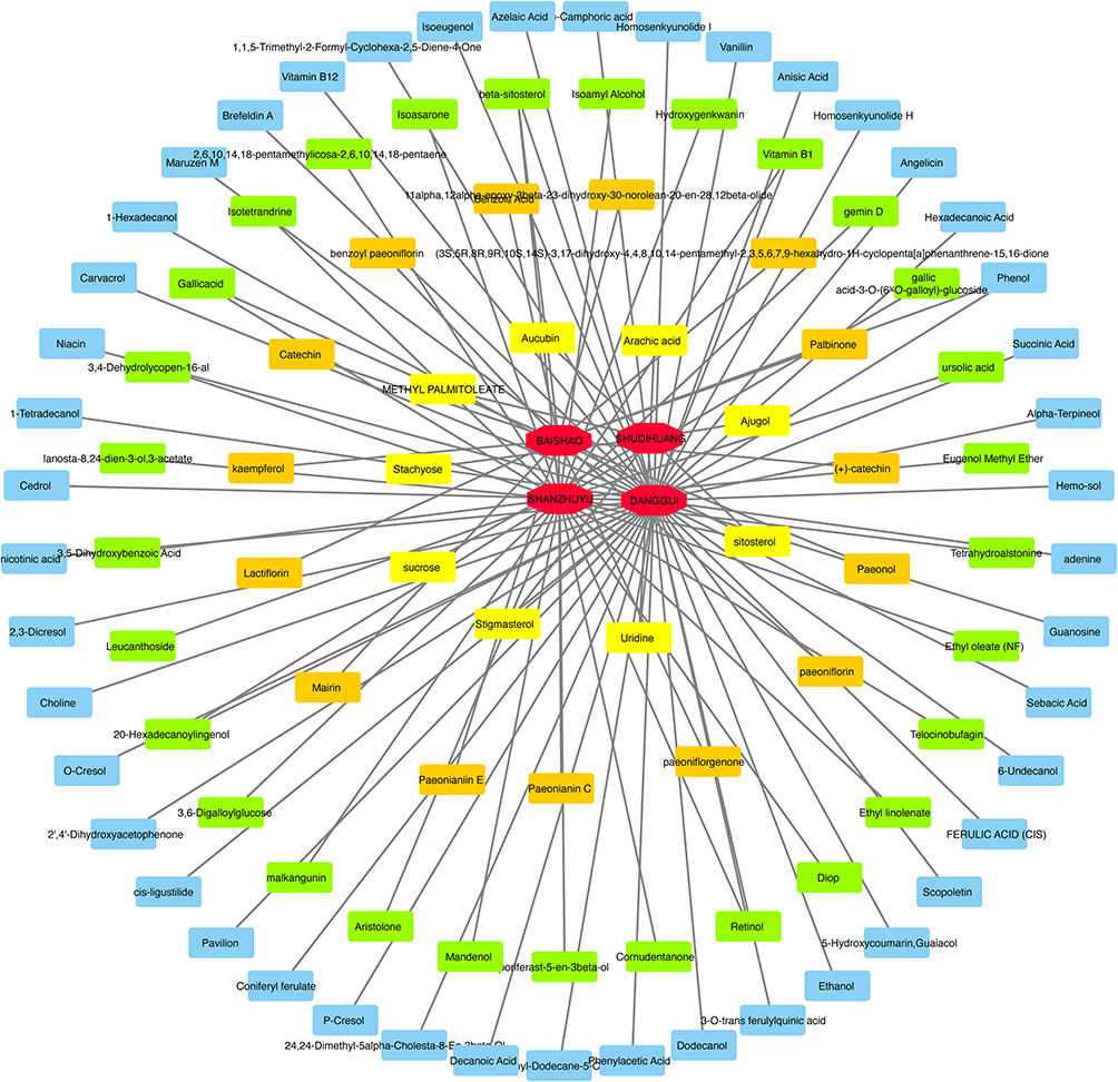 |
Figure 1 Yangjing Zhongyu Tang (YJZYT) active component network. |
Potential Targets of YJZYT Involved in Therapeutic Effects on TE
A total of 2409 disease genes for TE were retrieved from the Genecards database while none were retrieved from the OMIM and DisGeNET databases, as of December 31, 2022. A Venn diagram of TE and YJZYT genes was generated, leading to the identification of 186 potential TE therapeutic targets of YJZYT (Figure 2A). These targets were entered into the STRING database to obtain PPI (Figure 2B). A network analyzer was used to evaluate network topology characteristics, sorted according to the degree value of nodes, and 40 core nodes subsequently determined with a degree greater than twice the average of the PPI network (Figure 2C). Taking Degree, Betweenness Centrality, and Closeness Centrality as further screening parameters, a total of 10 key targets were obtained, specifically, AKT1, tumor necrosis factor (TNF), VEGFA, interleukin 6 (IL-6), tumor protein 53 (TP53), insulin (INS), estrogen receptor 1 (ESR1), matrix metalloprotein 9 (MMP9), albumin (ALB), and beta actin (ACTB) as shown in Figure 2D and E. A network diagram of active YJZYT compounds and TE targets was further generated (Figure 3). The graph included 289 nodes and 546 edges, whereby the nodes represent target genes and edges represent lines connecting the nodes.
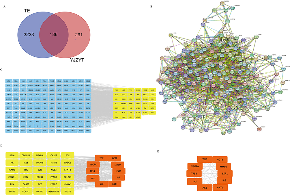 |
Figure 2 Protein–protein interaction (PPI) network and cluster analysis of disease targets. (A) Venn diagram of TE and YJZYT target genes. (B) PPI network of potential targets of YJZYT contributing to therapeutic effects on TE. (C) PPI network of significant proteins extracted from (B) based on network analysis. (D) Forty core proteins implicated in the effects of YJZYT on TE were extracted from (C). (E) A total of 10 key targets were obtained. |
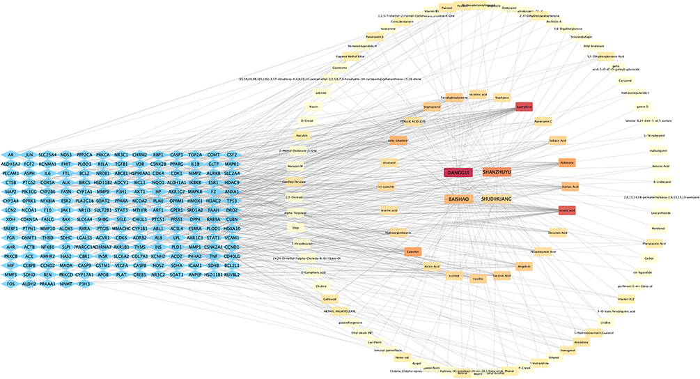 |
Figure 3 Network of YJZYT active compounds and TE targets. Notes: The degree value is from large to small, and the color gradually changes from red to orange. |
GO and KEGG Pathway Enrichment Analysis for Potential Targets of YJZYT in TE Treatment
GO functional enrichment analysis showed that items with P < 0.01, minimum count of 3, and enrichment factor >1.5 were screened, resulting in enrichment of a total of 956 items. The top 10 entries for BP, CC, and MF are presented (Figure 4A, Supplementary Table S2). The biological processes mainly included regulation of response to lipopolysaccharide, regulation of cell apoptosis, response to endogenous stimuli, active regulation of cell proliferation, and responses to various compounds and drugs. MF hint, enzyme binding, RNA polymerase II transcription factor activity, identical protein binding, steroid binding, protein homodimerization activity, iron ion binding, protein binding, steroid hormone receptor activity, oxidoreductase activity, and transcription factor binding. Therefore, YJZYT may exert beneficial effects on TE through mediating signal transduction, affecting cell proliferation and apoptosis, and regulating hormone levels, metabolic processes, and oxidative stress responses.
 |
Figure 4 Gene Ontology (GO) and Kyoto Encyclopedia of Genes and Genomes (KEGG) functional analysis. (A) Top 10 GO enrichment results. (B) Top 20 key signaling pathways from KEGG analysis. Abbreviations: BP, biological process; CC, cellular composition; MF, molecular function. |
KEGG pathway enrichment analysis of the 186 potential targets of YJZYT revealed 175 signaling pathways with significance (P < 0.01), which were related to cell proliferation and apoptosis, inflammatory response, hormone regulation, and angiogenesis (Figure 4B, Supplementary Table S3). Irrelevant pathways, such as “Lipid and atherosclerosis”, were subsequently removed. The top 20 pathways are presented in Figure 4B, among which the major pathways identified were PI3K-Akt, TNF, apoptosis, IL-17, MAPK, and estrogen signaling.
Molecular Docking
To further explore the potential effects of the seven active compounds of YJZYT on eight key hypothetical targets (AKT1, TNF, VEGFA, IL-6, TP53, INS, ESR1, and MMP9), we performed molecular docking analysis (Supplementary Table S4). Binding free energies are shown in Table 1. PyMoL software was used to visualize target proteins and small molecules with strong binding affinities (Figure 5). Our docking results showed that binding between the compound and target protein is mainly mediated by hydrophobic interactions. The free binding energies between the components of YJZYT, specifically, ursolic acid, palbinone, beta-sitosterol and stigmasterol, and their corresponding targets, AKT1, VEGFA, TNF and ESR1, were relatively high. Thus, these compounds may represent the key pharmacological components of YJZYT with therapeutic activity.
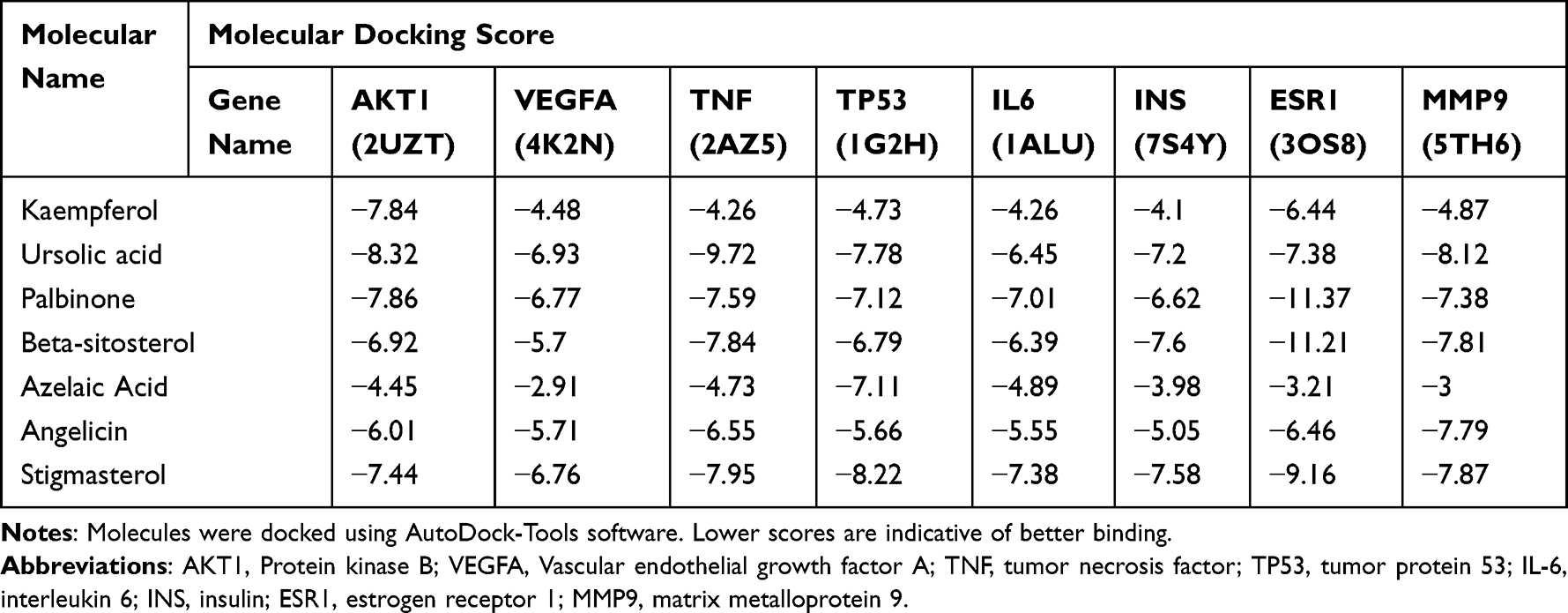 |
Table 1 Molecular Docking Scores of the Seven Candidate Compounds and Eight Targets |
 |
Figure 5 Docking results of seven active compounds of YJZYT and eight key TE targets. (A1) AKT1 and palbinone; (A2) AKT1 and kaempferol; (A3) AKT1 and ursolic acid; (B1) VEGFA and palbinone; (B2) VEGFA and stigmasterol; (B3) VEGFA and ursolic acid; (C1) TNF and beta-sitosterol; (C2) TNF and stigmasterol; (C3) TNF and ursolic acid; (D1) TP53 and palbinone; (D2) TP53 and stigmasterol; (D3) TP53 and ursolic acid; (E1) IL6 and palbinone; (E2) IL6 and stigmasterol; (E3) IL6 and ursolic acid; (F1) INS and beta-sitosterol; (F2) INS and stigmasterol; (F3) INS and ursolic acid; (G1) ESR and beta-sitosterol; (G2) ESR and stigmasterol; (G3) ESR and palbinone; (H1) MMP9 and beta-sitosterol; (H2) MMP9 and stigmasterol; (H3) MMP9 and ursolic acid. Abbreviations: AKT1, protein kinase B; VEGFA, Vascular endothelial growth factor A; TNF, tumor necrosis factor; TP53, tumor protein 53; IL-6, interleukin 6; INS, insulin; ESR1, estrogen receptor 1; MMP9, matrix metalloprotein 9. |
Histological Analysis of Rat Uterus
In control rats, the uterine cavity was morphologically intact, with normal distribution of glands and blood vessels in the stroma (Figure 6A and D). In rats from the model group, the uterine cavity was larger, the whole layer of the uterine wall became thinner and wavy, and the structure was incomplete, with few glands and sparse capillaries (Figure 6B and E). The YJZYT treatment group displayed slightly greater endometrial thickness, mainly intact glandular and luminal epithelial cells, continuous endometrium, increased number of glands, and higher number of capillaries showing neat arrangement (Figure 6C and F). Compared with the model group, endometrial thickness and number of glands were significantly increased in the control and YJZYT groups (P<0.05, P<0.01; Figure 6G and H, respectively). The uterine wet weight and uterine organ index of the control and YJZYT groups were higher than those of the model group, with significant differences (P<0.05, P<0.01; Figure 6I and J, respectively).
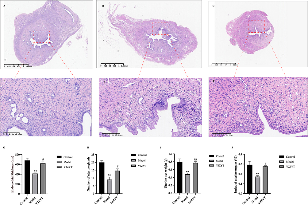 |
Figure 6 Histological analysis of rat uterus. (A–C) H&E staining results of uterine in the control, model and YJZYT groups (magnification: 40×). (D–F) The red areas represent magnification of H&E staining results. (G–J) Rat uterine wet weight, uterine organ index, thickness of the endometrium and number of glands. Data are presented as mean ± SEM; **Compared to the Control group, **P < 0.01. #Compared to the Model group, #P < 0.05, ##P < 0.01. |
Expression of CD34 in Rats with TE
CD34, a widely used marker of hematopoietic stem and progenitor cells, is mainly expressed in monolayer epithelium, including the cytoplasm of luminal and glandular epithelial cells.28 Immunofluorescence experiments revealed expression in the cytoplasm of endometrial cavity epithelial and glandular epithelial cells of rats from all groups (Figure 7A–I). Expression of CD34 in the model group was significantly decreased relative to that in the YJZYT and control groups (Figure 7J).
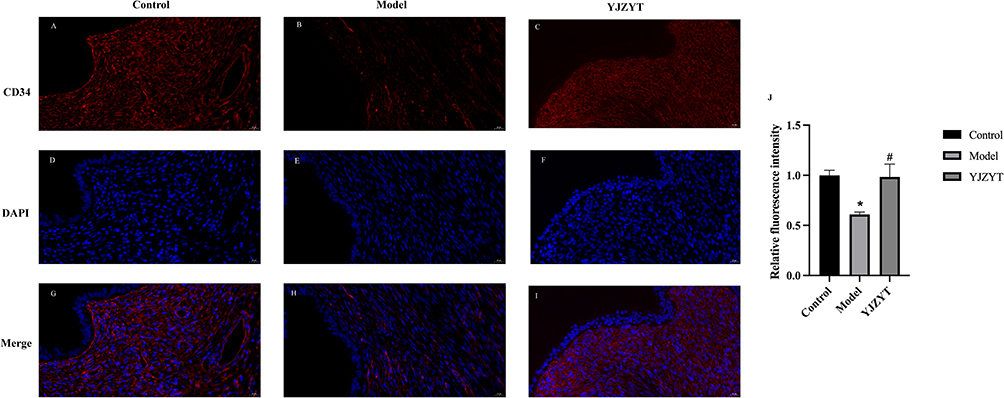 |
Figure 7 Changes in angiogenesis of rat uterus. (A–I) Immunofluorescence of the uterine receptivity marker, CD34 (red), and nuclear marker, DAPI (blue) (Magnification: 400×). (J) Relative fluorescence intensity of rats in control, model, and YJZYT groups. Data are presented as mean ± SEM; *Compared to the Control group, *P < 0.05. #Compared to the Model group, #P < 0.05. |
Protein Expression of PI3K, AKT, p-AKT and VEGF in Rats
Compared with the control group, PI3K, AKT, p-AKT and VEGF protein levels in the uterus of rats in the model group were significantly decreased. Relative to model rats, PI3K, AKT, p-AKT and VEGF protein levels in the uterus of rats in the YJZYT group were increased, with significant differences between the groups (P<0.05, P<0.01; Figure 8A–E).
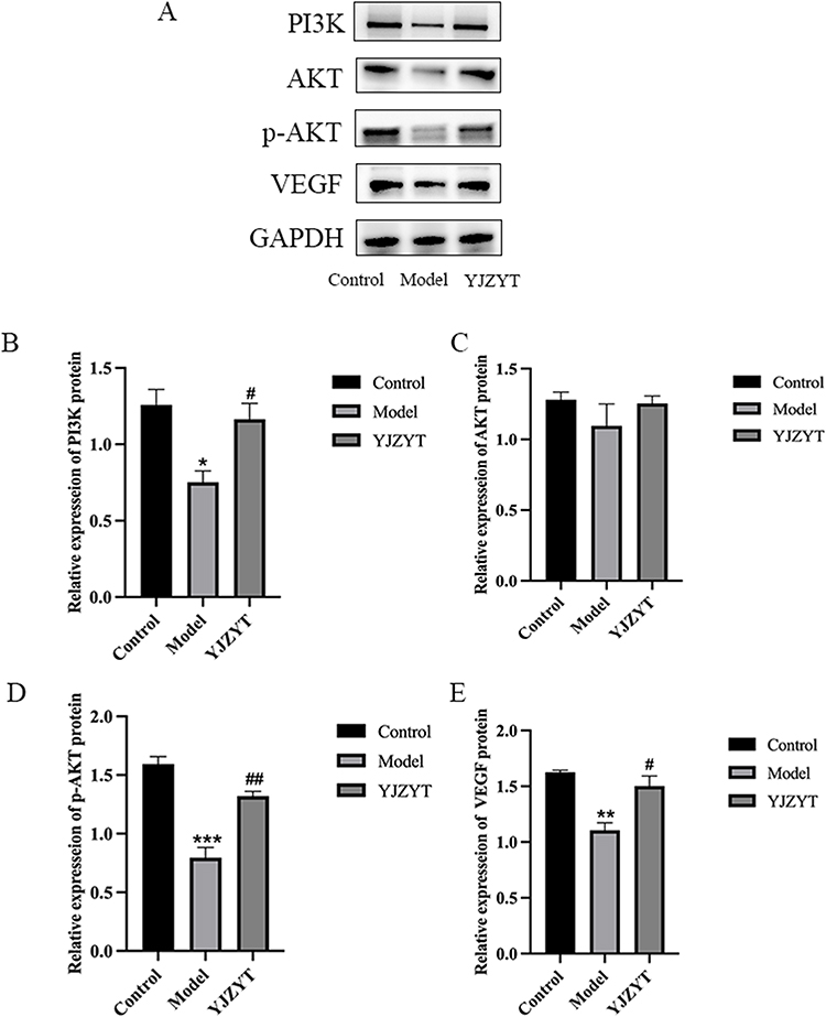 |
Figure 8 Protein expression patterns of rats in control, model, and YJZYT groups. (A) Western blot analysis. (B–E) Protein expression of PI3K, AKT, p-AKT and VEGF using GAPDH as an internal reference. Data are presented as mean ± SEM; *Compared to the Control group, *P < 0.05, **P < 0.01, ***P < 0.001. #Compared to the Model group, #P < 0.05, ##P < 0.01. |
Discussion
TE is the main cause of embryo implantation failure that can lead to long-term infertility and poor family outcomes.29 Periodic changes in thickness of the lining of the uterus occur due to changes in steroid hormones.30 At present, no uniform diagnostic standards for thin endometrium are available. Clinical research reports suggest that the minimal endometrial thickness that can facilitate pregnancy is 3.7 mm.31 Further studies have consistently highlighted that at endometrial thickness <6 mm, the possibility of pregnancy is extremely low.32,33 Appropriate endometrial thickness is therefore an important condition for successful pregnancy. The physiopathology and molecular mechanisms of thin endometrium remain ambiguous and current therapeutic options are limited and controversial due to its complex pathogenesis. Several treatments for thin uterine lining have been attempted,34 such as high estrogen doses, tamoxifen to promote endometrial growth,34–36 supplementation of growth hormone,37 intrauterine perfusion of platelet-rich plasma38 and mesenchymal stem cells.39,40 However, endometrial thickness and implantation rates have only slightly improved, especially in patients with severe endometrial damage. Treatment of TE thus remains a considerable clinical challenge.
TCM has become a research hotspot in recent years, and clinical intervention with YJZYT is reported to play a positive role in improving pregnancy outcomes of patients with TE. In this study, the network pharmacology method was used to explore the main potential targets and pathways of YJZYT involved in its therapeutic effects on TE. Screening of active ingredients and target prediction showed that Cornus Officinalis had the most active components and binding targets, indicative of an important therapeutic role. The mechanism mainly involved active ingredients, such as ursolic acid, palbinone, beta-sitosterol and stigmasterol, and many targets detected in the network of “YJZYT-active compounds-targets” could serve as key therapeutic ingredients for thin endometrium. A number of studies have demonstrated that inflammatory activation and abnormal autophagy are critical pathological links that cause endometrial epithelial thinning.41 Endometrial vascular dysplasia has been identified as a major cause of endometrial thinning due to decreased VEGF in the glandular epithelium.42 Therefore, induction of angiogenesis may serve as an effective strategy for clinical treatment of thin endometrium. The related compounds and pathways identified in this study, including IL-17, MAPK and estrogen, and target proteins, such as MMP9, ESR, TNF, INS, and Akt, warrant further research. In addition, the multi-target and multi-channel regulation properties of YJZYT support its potential as a therapeutic agent for other gynecological disorders, such as endometrial cancer, pelvic inflammatory and inflammatory diseases.
Previous experiments have also confirmed that Bushen Huoxue recipe can induce endometrial damage in rats with TE.43 Icariin produced an increase in endometrial thickness, and it may regulate the expression of VEGF and CD31.44 Dingkun Dan combined with estradiol valerateon was found to increase the thickness of the endometrium via up-regulation of VEGF.45 Our experimental results show that after intrauterine injection of 95% ethanol in rats, the endometrium was significantly damaged, significantly thinner, gland number was greatly reduced, and integrity was destroyed, all of which are consistent with previous findings.46–48 Rats with TE were subject to YJZYT intervention for three estrous cycles. Notably, the thickness and number of glands in rats with TE that were treated with YJZYT increased with a concomitant reduction in the degree of endometrial damage. Our data suggest that YJZYT can effectively repair damaged endometrium, promote endometrial growth, and restore its shape and structure.
Angiogenesis is an important factor for repair and proliferation of endometrium after injury. Expression of CD34 in rats in the YJZYT group was increased, indicating a positive effect of Chinese medicine on angiogenesis. WB results showed that compared with the model group, expression of PI3K, p-Akt, and VEGF proteins in the treatment group was significantly upregulated. Our data suggest that the PI3K/Akt signaling pathway occurs in both normal and thin endometrial tissues, but a significant difference in expression levels exists between the two groups. PI3K/Akt is a classic pathway that plays important roles in many aspects of cellular activity through phosphorylation or dephosphorylation, in particular, regulation of cell growth, proliferation, migration and vascular permeability. Previous studies confirmed that Ligustrazine, a TCM, induces the repair of TE in rats via up-regulation of the expression of VEGF and VEGFR-2 and activation of the PI3K/AKT signaling pathway.49 Our experiments support the theory that YJZYT activates the PI3K/Akt signaling pathway to promote angiogenesis, cell proliferation, and endometrial repair, providing new perspectives in terms of the utility of YJZYT as a therapeutic option therapy for TE and guidance on the value of TCM in clinical applications. In addition, combination of network pharmaceutical forecasting and experimental verification can be effectively employed to elucidate the multi-target and multi-target pharmacological mechanisms of action of traditional Chinese medicine formulae.
This study has a number of limitations that should be taken into consideration. First, while we examined the effects of YJZYT on angiogenesis in rats with TE, the study did not focus on analysis of pregnancy in rats. Future studies will thus aim to clarify the effects of YJZYT on biological mechanisms associated with pregnancy in the TE rat model. Second, evaluation of YJZYT fingerprint maps and pharmacology is lacking and further high-performance liquid chromatography (HPLC) experiments on the chemical composition of this TCM are warranted. In addition, because tissue samples of rats were obtained after the three estrous cycle treatments, evaluation of dynamic changes in the PI3K/AKT pathway and angiogenesis after administration of YJZYT is necessary. Both short- and long-term effects of YJZYT require further clarification.
Conclusion
The comprehensive strategy of network pharmacological analysis and experimental validation in the current study led to the successful identification of active ingredients and molecular mechanisms underlying the beneficial effects of YJZYT on thin endometrium. The key targets and signaling channels were established through network pharmacology and molecular docking experiments in vitro and further validated in a rat model in vivo. Our collective findings indicate that YJZYT promotes generic vessels of the uterine endometrium, associated with activation of the PI3K/AKT signaling pathway and upstream VEGF protein expression. In the future, the active ingredients and targets of YJZYT will be explored, and more mechanisms of YJZYT in the treatment of TE will be verified.
Ethics Statement
As network pharmacology studies only involved human databases, the ethics approval had been waived by the Reproductive Medicine Ethics Committee of the Affiliated Hospital of Shandong University of Traditional Chinese Medicine. The animal protocol was approved by the Experimental Animal Management Committee of the Affiliated Hospital of Shandong University of Traditional Chinese Medicine (No. 2021-70).
Acknowledgments
The experiment was carried out in the central Laboratory, affiliated Hospital of Shandong University of Traditional Chinese Medicine. Thanks to Dr Yuehua Jiang, Dr Zhiyong Liu, Dr Denglu Zhang, Dr Lei Qi, Dr Chenchen Ma and laboratory technology teachers for their help.
Funding
This research was supported by the Youth Science Fund Project of National Natural Science Foundation of China (82205174), Shandong Provincial Natural Science Foundation of China (ZR2020MH363), and Science and Technology Development Plans of TCM of Shandong Province (2021M176).
Disclosure
The authors have no conflicts of interest to declare in this work.
References
1. Liu KE, Hartman M, Hartman A. Management of thin endometrium in assisted reproduction: a clinical practice guideline from the Canadian fertility and andrology society. Reprod Biomed Online. 2019;39(1):49–62. doi:10.1016/j.rbmo.2019.02.013
2. Alfer J, Happel L, Dittrich R, et al. Insufficient angiogenesis: cause of abnormally thin endometrium in subfertile patients? Geburtshilfe Frauenheilkd. 2017;77(7):756–764. doi:10.1055/s-0043-111899
3. Kunicki M, Łukaszuk K, Liss J, Skowrońska P, Szczyptańska J. Granulocyte colony stimulating factor treatment of resistant thin endometrium in women with frozen-thawed blastocyst transfer. Syst Biol Reprod Med. 2017;63(1):49–57. doi:10.1080/19396368.2016.1251505
4. Yoshii A, Kitahara S, Ueta H, Matsuno K, Ezaki T. Role of uterine contraction in regeneration of the murine postpartum endometrium. Biol Reprod. 2014;91(2):32. doi:10.1095/biolreprod.114.117929
5. Khan MS, Shaikh A, Ratnani R. Ultrasonography and Doppler study to predict uterine receptivity in infertile patients undergoing embryo transfer. J Obstet Gynaecol India. 2016;66(Suppl 1):377–382. doi:10.1007/s13224-015-0742-5
6. Zhang L, Zhang J, Wu H. Effects of Yangjing Zhongyu decoction on endometrial receptivity in deficiency of kidney yin patients with repeated implantation failure. Chin J Tradit Chin Med Pharm. 2019;34(08):3842–3845. Chinese.
7. Zhang P, Xu L, Wang J. Study on the mechanism of modified Yangjing Zhongyu decoction and conventional western medicine in treating PCOS-induced infertility of kidney deficiency and liver depression pattern. West J Tradit Chin Med. 2021;34(09):13–17. Chinese.
8. Lin J, Ma H, Li H, et al. The treatment of complementary and alternative medicine on female infertility caused by endometrial factors. Evid Based Complement Alternat Med. 2022;2022:4624311. doi:10.1155/2022/4624311
9. Wang X, Wu W. An analysis of mechanism of treating female infertility with the Yangjing Zhongyu decoction. Clin J Chin Med. 2017;9(11):122–123. Chinese.
10. Liu L, Yu T, Chen L, Yuan CSM, Zhang L. Effects of Yangjing Zhongyu Tang supplemented IVF-ET on endometrial receptivity. Acta Chin Med. 2017;32(04):623–626. Chinese.
11. Zhou Z, Chen B, Chen S, et al. Applications of network pharmacology in traditional Chinese medicine research. Evid Based Complement Alternat Med. 2020;2020:1646905. doi:10.1155/2020/1646905
12. Ru J, Li P, Wang J, et al. TCMSP: a database of systems pharmacology for drug discovery from herbal medicines. J Cheminform. 2014;6:13. doi:10.1186/1758-2946-6-13
13. Xu HY, Zhang YQ, Liu ZM, et al. ETCM: an encyclopaedia of traditional Chinese medicine. Nucleic Acids Res. 2019;47(D1):D976–D982. doi:10.1093/nar/gky987
14. Liu Z, Guo F, Wang Y, et al. BATMAN-TCM: a bioinformatics analysis tool for molecular mechanism of traditional Chinese medicine. Sci Rep. 2016;6:21146. doi:10.1038/srep21146
15. Wu N, Yuan T, Yin Z, et al. Network pharmacology and molecular docking study of the Chinese Miao Medicine Sidaxue in the treatment of rheumatoid arthritis. Drug Des Devel Ther. 2022;16:435–466. doi:10.2147/dddt.S330947
16. UniProt C. UniProt: a worldwide hub of protein knowledge. Nucleic Acids Res. 2019;47(D1):D506–D515. doi:10.1093/nar/gky1049
17. Rebhan M, Chalifa-Caspi V, Prilusky J, Lancet D. GeneCards: integrating information about genes, proteins and diseases. Trends Genet. 1997;13(4):163. doi:10.1016/s0168-9525(97)01103-7
18. Amberger JS, Bocchini CA, Schiettecatte F, Scott AF. OMIM.org: Online Mendelian Inheritance in Man (OMIM(R)), an online catalog of human genes and genetic disorders. Nucleic Acids Res. 2015;43(Database issue):D789–D798. doi:10.1093/nar/gku1205
19. Pinero J, Ramirez-Anguita JM, Sauch-Pitarch J, et al. The DisGeNET knowledge platform for disease genomics: 2019 update. Nucleic Acids Res. 2020;48(D1):D845–D855. doi:10.1093/nar/gkz1021
20. Szklarczyk D, Gable AL, Lyon D, et al. STRING v11: protein-protein association networks with increased coverage, supporting functional discovery in genome-wide experimental datasets. Nucleic Acids Res. 2019;47(D1):D607–D613. doi:10.1093/nar/gky1131
21. Newman DJ. Modern traditional Chinese medicine: identifying, defining and usage of TCM components. Adv Pharmacol. 2020;87:113–158. doi:10.1016/bs.apha.2019.07.001
22. Bader GD, Hogue CW. An automated method for finding molecular complexes in large protein interaction networks. BMC Bioinform. 2003;4:2. doi:10.1186/1471-2105-4-2
23. Huang da W, Sherman BT, Lempicki RA. Systematic and integrative analysis of large gene lists using DAVID bioinformatics resources. Nat Protoc. 2009;4(1):44–57. doi:10.1038/nprot.2008.211
24. Kim S, Thiessen PA, Bolton EE, et al. PubChem substance and compound databases. Nucleic Acids Res. 2016;44(D1):D1202–D1213. doi:10.1093/nar/gkv951
25. Trott O, Olson AJ. AutoDock Vina: improving the speed and accuracy of docking with a new scoring function, efficient optimization, and multithreading. J Comput Chem. 2010;31(2):455–461. doi:10.1002/jcc.21334
26. Zhao J, Gao H, Li Y. Development of an animal model for thin endometrium using 95% ethanol. J Fert In Vitro. 2012;2(4):109.
27. Zhao J, Zhang Q, Wang Y, Li Y. Uterine infusion with bone marrow mesenchymal stem cells improves endometrium thickness in a rat model of thin endometrium. Reprod Sci. 2015;22(2):181–188. doi:10.1177/1933719114537715
28. Sidney LE, Branch MJ, Dunphy SE, Dua HS, Hopkinson A. Concise review: evidence for CD34 as a common marker for diverse progenitors. Stem Cells. 2014;32(6):1380–1389. doi:10.1002/stem.1661
29. Lin Y, Dong S, Ye X, et al. Synergistic regenerative therapy of thin endometrium by human placenta-derived mesenchymal stem cells encapsulated within hyaluronic acid hydrogels. Stem Cell Res Ther. 2022;13(1):66. doi:10.1186/s13287-022-02717-2
30. Critchley HOD, Maybin JA, Armstrong GM, Williams ARW. Physiology of the endometrium and regulation of menstruation. Physiol Rev. 2020;100(3):1149–1179. doi:10.1152/physrev.00031.2019
31. Achache H, Tsafrir A, Prus D, Reich R, Revel A. Defective endometrial prostaglandin synthesis identified in patients with repeated implantation failure undergoing in vitro fertilization. Fertil Steril. 2010;94(4):1271–1278. doi:10.1016/j.fertnstert.2009.07.1668
32. Liu KE, Hartman M, Hartman A, Luo ZC, Mahutte N. The impact of a thin endometrial lining on fresh and frozen-thaw IVF outcomes: an analysis of over 40 000 embryo transfers. Hum Reprod. 2018;33(10):1883–1888. doi:10.1093/humrep/dey281
33. Mouhayar Y, Franasiak JM, Sharara FI. Obstetrical complications of thin endometrium in assisted reproductive technologies: a systematic review. J Assist Reprod Genet. 2019;36(4):607–611. doi:10.1007/s10815-019-01407-y
34. Lebovitz O, Orvieto R. Treating patients with “thin” endometrium - an ongoing challenge. Gynecol Endocrinol. 2014;30(6):409–414. doi:10.3109/09513590.2014.906571
35. Sharma S, Rani G, Bose G, Saha I, Bathwal S, Chakravarty BN. Tamoxifen is better than low-dose clomiphene or gonadotropins in women with thin endometrium (<7 mm) after clomiphene in intrauterine insemination cycles: a prospective study. J Hum Reprod Sci. 2018;11(1):34–39. doi:10.4103/jhrs.JHRS_9_17
36. Ke H, Jiang J, Xia M, Tang R, Qin Y, Chen ZJ. The effect of tamoxifen on thin endometrium in patients undergoing frozen-thawed embryo transfer. Reprod Sci. 2018;25(6):861–866. doi:10.1177/1933719117698580
37. Cui N, Li AM, Luo ZY, et al. Effects of growth hormone on pregnancy rates of patients with thin endometrium. J Endocrinol Invest. 2019;42(1):27–35. doi:10.1007/s40618-018-0877-1
38. Eftekhar M, Neghab N, Naghshineh E, Khani P. Can autologous platelet rich plasma expand endometrial thickness and improve pregnancy rate during frozen-thawed embryo transfer cycle? A randomized clinical trial. Taiwan J Obstet Gynecol. 2018;57(6):810–813. doi:10.1016/j.tjog.2018.10.007
39. Ye MX, Yu L, Wang SF, Fan WS, Meng YG. Efficacy of gamma-irradiated adipose-derived stem cells for treatment of thin endometrium in rats. Nan Fang Yi Ke Da Xue Xue Bao. 2017;37(5):575–580. doi:10.3969/j.issn.1673-4254.2017.05.02
40. Yi KW, Mamillapalli R, Sahin C, Song J, Tal R, Taylor HS. Bone marrow-derived cells or C-X-C motif chemokine 12 (CXCL12) treatment improve thin endometrium in a mouse model. Biol Reprod. 2019;100(1):61–70. doi:10.1093/biolre/ioy175
41. Meng Y, Yannan Z, Ren L, Qi S, Wei W, Lihong J. Adverse reproductive function induced by maternal BPA exposure is associated with abnormal autophagy and activating inflammation via mTOR and TLR4/NF-κB signaling pathways in female offspring rats. Reprod Toxicol. 2020;96:185–194. doi:10.1016/j.reprotox.2020.07.001
42. Lei L, Lv Q, Jin Y, et al. Angiogenic microspheres for the treatment of a thin endometrium. ACS Biomater Sci Eng. 2021;7(10):4914–4920. doi:10.1021/acsbiomaterials.1c00615
43. Yin XD, Xue XO, Wang JS, et al. Effect of Bushen Huoxue recipe on women with thin endometrial ovulation disorder and a rat model of thin endometrium resulted from kidney deficiency-related blood stasis. Gynecol Endocrinol. 2021;37(5):433–437. doi:10.1080/09513590.2020.1781079
44. Le AW, Wang ZH, Dai XY, et al. An experimental study on the use of icariin for improving thickness of thin endometrium. Genet Mol Res. 2017;16(1). doi:10.4238/gmr16019126
45. Zheng J, Tan Y. Influence of Dingkun Dan combined with estradiol valerateon Wnt/β-catenin signaling pathway in rats with thin endometrium with Kidney-Yang deficiency. Sichuan Da Xue Xue Bao Yi Xue Ban. 2021;52(2):235–240. Chinese. doi:10.12182/20210160508
46. Xi J, Cheng J, Jin CC, et al. Electroacupuncture improves pregnancy outcomes in rats with thin endometrium by promoting the expression of pinopode-related molecules. Biomed Res Int. 2021;2021:6658321. doi:10.1155/2021/6658321
47. Xia L, Meng Q, Xi J, et al. The synergistic effect of electroacupuncture and bone mesenchymal stem cell transplantation on repairing thin endometrial injury in rats. Stem Cell Res Ther. 2019;10(1):244. doi:10.1186/s13287-019-1326-6
48. Xie Y, Tian Z, Qi Q, et al. The therapeutic effects and underlying mechanisms of the intrauterine perfusion of granulocyte colony-stimulating factor on a thin-endometrium rat model. Life Sci. 2020;260:118439. doi:10.1016/j.lfs.2020.118439
49. Ye Q, Zhang Y, Fu J, et al. Effect of ligustrazine on endometrium injury of thin endometrium rats. Evid Based Complement Alternat Med. 2019;2019:7161906. doi:10.1155/2019/7161906
 © 2023 The Author(s). This work is published and licensed by Dove Medical Press Limited. The
full terms of this license are available at https://www.dovepress.com/terms
and incorporate the Creative Commons Attribution
- Non Commercial (unported, 3.0) License.
By accessing the work you hereby accept the Terms. Non-commercial uses of the work are permitted
without any further permission from Dove Medical Press Limited, provided the work is properly
attributed. For permission for commercial use of this work, please see paragraphs 4.2 and 5 of our Terms.
© 2023 The Author(s). This work is published and licensed by Dove Medical Press Limited. The
full terms of this license are available at https://www.dovepress.com/terms
and incorporate the Creative Commons Attribution
- Non Commercial (unported, 3.0) License.
By accessing the work you hereby accept the Terms. Non-commercial uses of the work are permitted
without any further permission from Dove Medical Press Limited, provided the work is properly
attributed. For permission for commercial use of this work, please see paragraphs 4.2 and 5 of our Terms.
Recommended articles
Anti-Inflammatory Effects and Molecular Mechanisms of Shenmai Injection in Treating Acute Pancreatitis: Network Pharmacology Analysis and Experimental Verification
He Y, Hu C, Liu S, Xu M, Liang G, Du D, Liu T, Cai F, Chen Z, Tan Q, Deng L, Xia Q
Drug Design, Development and Therapy 2022, 16:2479-2495
Published Date: 2 August 2022
Network and Experimental Pharmacology to Decode the Action of Wendan Decoction Against Generalized Anxiety Disorder
Jin Q, Li J, Chen GY, Wu ZY, Liu XY, Liu Y, Chen L, Wu XY, Liu Y, Zhao X, Song YH
Drug Design, Development and Therapy 2022, 16:3297-3314
Published Date: 27 September 2022
A Novel Approach Based on Gut Microbiota Analysis and Network Pharmacology to Explain the Mechanisms of Action of Cichorium intybus L. Formula in the Improvement of Hyperuricemic Nephropathy in Rats

Amatjan M, Li N, He P, Zhang B, Mai X, Jiang Q, Xie H, Shao X
Drug Design, Development and Therapy 2023, 17:107-128
Published Date: 20 January 2023
Network Pharmacology and Experimental Validation to Explore That Celastrol Targeting PTEN is the Potential Mechanism of Tripterygium wilfordii (Lév.) Hutch Against IgA Nephropathy

Zhao J, Liu H, Xia M, Chen Q, Wan L, Leng B, Tang C, Chen G, Liu Y, Zhang L, Liu H
Drug Design, Development and Therapy 2023, 17:887-900
Published Date: 23 March 2023
Investigating the Mechanism of Action of Schisandra chinensis Combined with Coenzyme Q10 in the Treatment of Heart Failure Based on PI3K-AKT Pathway

Wen S, Yang K, Bai Y, Wu Y, Liu D, Wu X, Zhang X, Sun J
Drug Design, Development and Therapy 2023, 17:939-957
Published Date: 27 March 2023