Back to Journals » Journal of Hepatocellular Carcinoma » Volume 11
Construction and Validation of a Novel Nomogram Predicting Recurrence in Alpha-Fetoprotein-Negative Hepatocellular Carcinoma Post-Surgery Using an Innovative Liver Function-Nutrition-Inflammation-Immune (LFNII) Score: A Bicentric Investigation
Authors Zhang BL, Liu J, Diao G, Chang J, Xue J, Huang Z, Zhao H , Yu L, Cai J
Received 29 November 2023
Accepted for publication 29 February 2024
Published 6 March 2024 Volume 2024:11 Pages 489—508
DOI https://doi.org/10.2147/JHC.S451357
Checked for plagiarism Yes
Review by Single anonymous peer review
Peer reviewer comments 2
Editor who approved publication: Dr David Gerber
Bo-Lun Zhang,1 Jia Liu,2 Guanghao Diao,2 Jianping Chang,1 Junshuai Xue,1 Zhen Huang,1 Hong Zhao,1 Lingxiang Yu,2 Jianqiang Cai1
1Department of Hepatobiliary Surgery, National Cancer Center/National Clinical Research Center for Cancer/Cancer Hospital, Chinese Academy of Medical Sciences and Peking Union Medical College, Beijing, People’s Republic of China; 2Department of Hepatobiliary Surgery, the Fifth Medical Center of the PLA General Hospital, Beijing, People’s Republic of China
Correspondence: Jianqiang Cai, Department of Hepatobiliary Surgery, National Cancer Center/National Clinical Research Center for Cancer/Cancer Hospital, Chinese Academy of Medical Sciences and Peking Union Medical College, 17 Panjiayuan Nanli, Chaoyang District, Beijing, 100021, People’s Republic of China, Tel +86-10-87787100, Fax +86-10-67709698, Email [email protected]
Purpose: We developed a nomogram based on the liver function, nutrition, inflammation, and immunity (LFNII) score to predict recurrence-free survival (RFS) post-resection in patients with hepatocellular carcinoma (HCC) exhibiting alpha-fetoprotein (AFP) negativity (AFP ≤ 20 ng/mL).
Patients and Methods: Clinical data of 661 patients diagnosed with alpha-fetoprotein-negative hepatocellular carcinoma (AFP-NHCC) who underwent surgical resection at two medical centers between 2012 and 2021 were collected. A total of 462 and 199 patients served as the training and validation sets, respectively. Pre-operative blood markers were collected and analyzed for LFNII. The LFNII score was formulated using the least absolute shrinkage and selection operator Cox regression model. A nomogram model was developed using the training set to incorporate other relevant clinicopathological indicators and predict postoperative recurrence. Model discrimination was assessed using the receiver operating characteristic curve, calibration was evaluated using a calibration curve, and clinical applicability was assessed using clinical decision curve analysis. A comparison with liver cancer staging was performed using the nomogram model. Finally, a cohort study was conducted to validate our findings.
Results: We derived the LFNII scores from nine indicators. Elevated LFNII scores correlated with unfavorable clinicopathological features. The LFNII score area under the curve revealed superior predictive efficacy at 1-, 2-, and 5-year RFS intervals, with values of 0.675, 0.658, and 0.633, respectively. Multivariate Cox analysis revealed that a high LFNII score independently increased RFS risk in patients with AFP-NHCC. The C-index of the LFNII-nomogram model was 0.686 (95% confidence interval [CI], 0.651– 0.721). The nomogram model’s clinical application value surpassed that of standard HCC staging systems.
Conclusion: The LFNII score-derived nomogram effectively predicted the RFS of patients with AFP-NHCC after curative resection.
Keywords: hepatocellular carcinoma, alpha-fetoprotein-negative, immunity, inflammation, nutrition, recurrence-free survival
Introduction
Primary liver cancer is the sixth most common malignancy and the third leading cause of cancer-related deaths worldwide.1 Hepatocellular carcinoma (HCC) is the most common form of liver cancer, accounting for 75–90% of primary liver cancer cases.2–5 Surgical excision is largely recognized as the most effective therapy for HCC.6 However, a significant challenge arises, as approximately 70% of patients with HCC are ineligible for surgery upon detection. Diagnosing and treating HCC is challenging, especially in advanced stages, which are characterized by a high recurrence rate and low postoperative survival.
Alpha-fetoprotein (AFP) is a crucial biomarker in HCC detection.7 The importance of AFP as a critical tumor marker in HCC diagnosis is insufficiently emphasized, given its association with an unfavorable prognosis in cases of elevation.8,9 However, 30–40% of patients with HCC had low serum AFP levels (AFP ≤20 ng/mL), termed alpha-fetoprotein-negative HCC (AFP-NHCC).10,11 Despite the relatively more favorable prognosis in AFP-NHCC than in high AFP cases,12 the significant postoperative recurrence rate underscores the need for postoperative monitoring and prognostic assessment. However, effective biomarkers for postoperative monitoring of patients with AFP-NHCC are lacking. Researchers have explored AFP-NHCC biomarkers, such as PIVKA-II and AFP-L3.13,14 Simultaneously, others have developed novel scoring models to predict the prognosis of patients with AFP-NHCC. Huang et al constructed a nomogram model for postoperative recurrence of AFP-NHCC by combining eight indicators such as gender, cirrhosis, alkaline phosphatase level, microvascular invasion, and tumor size.15 Another study from China analyzed 419 patients with AFP-NHCC, including patients with advanced-stage condition, and constructed a nomogram model to predict overall survival by incorporating indicators such as BMI, tumor stage, and blood biochemical indicators.16 In recent studies, PIVKA-II and Tumor Burden Score were combined to construct a new tumor burden score (TPS) prediction model to predict the postoperative prognosis of AFP-NHCC.17 All the aforementioned models showed certain predictive ability; however, validation of their predictive power is necessary, as most data are based on single centers, and the clinical applicability of these indicators remains limited. These findings indicate the urgent need for monitoring postoperative recurrence in patients with AFP-NHCC.
Prior investigations have established correlations between immune response, inflammatory processes, and nutritional factors in relation to tumor prognosis.18–21 Pre-operative hematological indicators, such as platelet-to-lymphocyte ratio (PLR), neutrophil-to-lymphocyte ratio (NLR), lymphocyte-to-monocyte ratio (LMR), prognostic nutritional index (PNI), systemic immune-inflammation index (SII), serum immune-to-inflammation ratio (SIRI), have been validated for their relevance in the prognosis of HCC, reflecting immunity, inflammation, and nutrition.22–26 Furthermore, studies have indicated that biomarkers linked to liver function, inflammation, nutrition, and immunity, such as gamma-glutamyl transpeptidase to alkaline phosphatase ratio (GAPR), gamma-glutamyl transpeptidase to aspartate aminotransferase ratio (GAR), aspartate aminotransferase to alanine aminotransferase ratio (AAR), gamma-glutamyltransferase-to-lymphocyte ratio (GLR), albumin-bilirubin score (ALBI), modified albumin-bilirubin score (PALBI), monocyte-to-lymphocyte ratio (MLR), and neutrophil times gamma-glutamyl transpeptidase-to-lymphocyte ratio (NrLR), demonstrate diagnostic and prognostic relevance in AFP-NHCC.27–31 Nevertheless, a solitary indicator does not comprehensively represent the nutrition, immunity, and inflammation statuses of the body. Previous studies have underscored the prognostic advantages of combining these indicators in intrahepatic cholangiocarcinoma. However, whether such a combination can predict AFP-NHCC recurrence remains unknown. This study introduced the liver function-nutrition-inflammation-immune (LFNII) score, which is calculated by integrating liver function indicators with factors related to immunity, inflammation, and nutrition. A novel nomogram integrating the LFNII score with clinicopathological factors was developed to predict post-resection AFP-NHCC recurrences. This study aimed to compare the novel model with standard HCC staging systems and examine its correlation with clinicopathological factors.
Materials and Methods
Study Cohort
The study data were obtained from the Hepatobiliary Surgery Departments at the Cancer Hospital of the Chinese Academy of Medical Sciences and the Fifth Medical Center of the PLA General Hospital. Patients diagnosed with hepatocellular carcinoma who underwent curative resection between 2012 and 2021 were identified from the electronic medical records of both institutions. In total, 1853 and 1689 patients were recruited from both centers, respectively. The inclusion criteria were as follows: 1) HCC confirmed through postoperative pathology; 2) alpha-fetoprotein level of ≤20 ng/mL; 3) no history of other malignant tumors; 4) no preoperative anti-tumor therapy, including preoperative targeted therapy, immunotherapy, chemotherapy, interventional therapy, radiotherapy, radiofrequency ablation, and traditional Chinese medicine with definite anti-tumor effect; and 5) Child-Pugh A/B grade. The exclusion criteria encompassed patients with pre-operative AFP >20 ng/mL and those meeting the following criteria: (1) receipt of other antineoplastic therapies preoperatively, (2) presence of other malignancies, (3) insufficient clinical or follow-up information, and (4) surgical treatment for tumor recurrence. The final study cohort comprised 661 patients with AFP-NHCC (Figure 1). The patients included in the study were randomized to the training or validation set at a 7:3 ratio. In addition, 157 patients with AFP-positive HCC who underwent hepatectomy at Cancer Hospital, Chinese Academy of Medical Sciences, during the same period were included to verify the predictive ability of the LFNII score in patients with different AFP levels. The inclusion and exclusion criteria were the same as before, except that the AFP levels exceeded 20 ng/mL. The researchers obtained ethical permission from the Ethics Committees of both centers (approval number 21/198-2869). This study was conducted in accordance with the principles outlined in the Declaration of Helsinki, and the research protocols were conducted in accordance with applicable standards and laws.
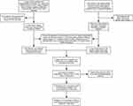 |
Figure 1 Flow chart of the retrospective study. |
Clinicopathologic Variables and Follow-Up
Patient clinicopathological characteristics were classified into the following groups: (1) General patient details: age, sex, family history of malignant tumors, history of diabetes, alcoholism, smoking, and hepatitis B and C virus infection, and body mass index; (2) Surgical factors: the surgery duration, intraoperative blood loss, and intraoperative blood transfusion; (3) tumor pathology details: the number of tumors, liver capsule invasion, tumor pathological grade (Edmondson Steiner grade), microvascular invasion (MVI), maximum diameter of the largest tumor, satellite nodules, liver fibrosis grade (Scheuer scoring system), AFP level, AJCC-TNM stage (eighth edition), the Barcelona Clinic Liver Cancer system (BCLC) stage, and alpha-feto protein tumor burden score (ATS);29 (4) Pre-operative hematological indicators: including AFP, white blood cell count, neutrophil (NEUT), lymphocyte (LYMPH), monocyte (MONO), hemoglobin, blood platelet count (PLT), alanine aminotransferase (ALT), aspartate aminotransferase (AST), alkaline phosphatase (ALP), total bilirubin (TBIL), albumin (ALB), and gamma-glutamyl transpeptidase (GGT).
The following formulas were applied to calculate liver function, inflammation, immunity, and nutrition indicators: AAPR = ALB (g/L) / ALP (U/L), AAR (DeRitis ratio) = AST (U/L) / ALT (U/L), NLR = NEUT / LYMPH, LMR = LYMPH / MONO, PLR = PLT / LYMPH, GAR = GGT (U/L) / AST (U/L), GAPR = GGT (U/L) / ALP (U/L), ANRI = AST (U/L) / NEUT, ALRI = AST (U/L) / LYMPH. PNI = ALB (g/L) + 5 × LYMPH, SIRI = NEUT × MONO/LYMPH, and SII = PLT × NEUT/LYMPH. ALBI = log10 (TBIL; mol/L) × 0.66 – ALB (g/L) × 0.085; the ALBI grade was stratified as follows: ≤ −2.60 (ALBI grade 1), > −2.60 ≤ −1.39 (ALBI grade 2), and ≥ −1.39 (ALBI grade 3). The ATS score was computed using the formula: ATS2 = (lnAFP)2 + (maximum tumor diameter)2 + (number of tumors)2.32
The optimal cutoff values for hematological indicators were determined using the surv_cutpoint function in the R (4.2.1) survminer package. The patients underwent follow-up one month post-resection and subsequently every three months. Follow-up assessments included general examination, hematological indices, enhanced CT of the whole body, and liver-enhanced MRI. RFS was determined by calculating the time from the resection procedure to the identification of tumor recurrence in accordance with the diagnostic and treatment criteria for primary liver cancer in China.33
LFNII Score Construction
In the preliminary phase, univariate Cox regression analysis was conducted on the entire cohort of 661 individuals to identify pre-operative markers associated with liver function, immunology, inflammation, and nutrition. Indicators with P values < 0.05 were retained for subsequent analysis. Kaplan–Meier (KM) curves were then used to assess the influence of various parameters on survival duration. Subsequently, least absolute shrinkage and selection operator (LASSO) Cox regression analysis was used to evaluate the predictive relevance of the indicators mentioned above in relation to tumor recurrence. The LFNII score was then calculated using variables with non-zero coefficients.
Correlation Between LFNII Score and Clinicopathological Features
To determine the optimal threshold value for LFNII scores and examine the differences in clinicopathological characteristics between high- and low-LFNII scores groups, we used the survminer package in R (version 4.2.1). Samples were classified into high- and low-LFNII score groups based on the selected threshold. The predictive accuracy of the LFNII score for 1-, 2-, and 5-year RFS in patients with AFP-NHCC was assessed and compared with that of individual component indices using receiver operating characteristic (ROC) curves.
Construction and Validation of a Nomogram
We conducted univariate Cox regression analysis of clinicopathological factors in the training dataset that included the LFNII score. Variables with a p-value <0.05 were further evaluated for potential inclusion in a multivariate Cox regression analysis, using the stepwise backward approach to identify independent prognostic indicators for RFS. These indicators were used to develop the nomogram model. The nomogram’s prediction accuracy for 1-, 2-, and 5-year RFS was assessed in the training and validation datasets using ROC and calibration curves. Decision curve analysis (DCA) was performed to compare the nomogram with each constituent index to assess the clinical utility at various time intervals. Patients were then classified into high- and low-risk categories based on their nomogram-derived risk scores. KM curves were constructed to assess and evaluate the disparity in survival rates across the groups. The predictive efficacy of the nomogram model in conjunction with the TNM and BCLC stages was assessed and compared using DCA and the time-dependent area under the curve (AUC).
Comparison of LFNII Score with Other Prognostic Indicators
We screened three previously reported indicators and compared them with AFPN-HCC prognosis indicators according to data availability, to determine whether the predictive power of the LFNII score exceeded that of previous studies. These three indicators were neutrophil times γ-glutamyl transpeptidase to lymphocyte ratio (NrLR),30 γ-glutamyl transpeptidase to lymphocyte count ratio (GLR),28 and platelet-albumin-bilirubin (PALBI) score.31 The predictive ability of the four indicators for postoperative recurrence and the degree of clinical benefit were compared using time-dependent AUC analysis and clinical decision assessment. Simultaneously, to broaden the clinical application of the LFNII score, we incorporated it into the AFP-positive cohort for analysis. The prognostic ability of LFNII score in AFP-positive patients was evaluated using KM curve and time-dependent AUC.
Statistical Analysis
Categorical variables, such as frequency with percentage, were compared using chi-squared or Fisher’s exact tests. The surv_cutpoint function within the R (4.2.1) survminer package was used to determine optimal cutoff values. Univariate and multivariate Cox regression analyses, LASSO Cox regression, and the generation of ROC, calibration, DCA, and KM curves were performed using the R software packages “compare groups”, “glmnet”, “survival”, “forest plot”, “survival ROC”, “stdca. R”, “rms”, “survminer”, “timeROC”, and “ggplot2”. Statistical significance was set at P < 0.05.
Results
Basic Clinical Information
This study included 661 patients who were diagnosed with AFP-NHCC. The cohort comprised 588 males (89.0%) and 73 females (11.0%). Patients were recruited from two medical centers. The median follow-up period for the study participants was 60 months (range, 1–122 months). The RFS rates at 1, 2, and 5 years were 80.6%, 70.9%, and 49.5%, respectively. Table 1 summarizes the basic characteristics of the cohort and subsequent segmentation analysis. The baseline clinicopathological characteristics of the training and validation sets did not differ significantly after segmentation (p > 0.05). Simultaneously, we enrolled 157 patients with AFP-positive HCC: 118 men (75.2%) and 39 women (24.8%). The 1 -, 2 -, and 5-year RFS were 79.6%, 70.2%, and 35.8%, respectively (Supplementary Table 1).
 |
Table 1 Comparison of Clinicopathological Characteristics in Training and Validation Sets |
LFNII Score
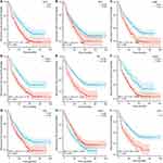
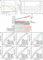
Univariate Cox regression analysis was performed on all computed indices related to liver function, nutrition, immunology, and inflammation. The results revealed a significant correlation between postoperative recurrence in AFP-NHCC and elevated levels of ALRI, ANRI, SII, GAPR, AAR, and ALBI and reduced levels of AAPR, SIRI, and PNI (p < 0.05; Supplementary Figure 1). These findings were consistent with the outcomes derived from the KM analysis (Figure 2A-I). Subsequently, the aforementioned nine indicators were subjected to LASSO Cox regression analysis, confirming their connection with postoperative recurrence in patients with AFP-NHCC (Figure 3A and B). Using coefficients from the LASSO Cox regression, the LFNII score was calculated using the formula: LFNII = ALRI × 0.144 - SIRI × 0.069 + ANRI × 0.064 + SII × 0.404 + PNI × 0.164 + GAPR × 0.461 + AAR × 0.253 - AAPR × 0.037 + ALBI × 0.264. An optimal cutoff of 0.89 was used to classify all samples into high- and low-LFNII groups to examine the connection between the score and clinicopathological features. Comparisons of various indicators between these groups revealed that elevated LFNII scores were associated with unfavorable clinicopathological features, including a larger tumor burden, liver capsule invasion, increased tumor diameter, microvascular invasion, higher alpha-fetoprotein levels, advanced tumor stage, greater intraoperative blood loss, and substantial intraoperative blood transfusion (Figure 3C). Comprehensive comparisons between the baseline groups are presented in Supplementary Table 2. To evaluate the predictive efficacy of the LFNII score compared to the individual component indices, the AUC for 1-, 2-, and 5-year RFS were 0.675, 0.658, and 0.633, respectively. This performance surpassed that of any single component index in the overall cohort (Figure 3D-I).
Independent RFS Prognostic Factor: LFNII Score
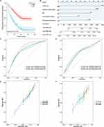
Univariate Cox regression analysis was performed on the training set, considering intraoperative conditions and postoperative pathological indicators. The results revealed that, in addition to the ATS score and factors such as liver fibrosis grade, MVI, invasion of the hepatic capsule, intraoperative blood loss, intraoperative blood transfusion, and hepatitis B Virus (HBV) infection, a high LFNII score was a significant risk factor for RFS (Table 2). The KM curve showed a strong correlation between the LFNII score and postoperative recurrence (Figure 4A). Multivariate (backward approach) Cox regression analysis included indicators with a univariate p-value of < 0.05. This analysis confirmed that a significantly elevated LFNII score was a distinct risk factor for RFS (hazard ratio [HR], 1.858; 95% confidence interval [CI], 1.397–2.471; Table 2). Simple LFNII score computation can be accessed at the following URL: https://wyy2023.shinyapps.io/LFNII-1/.
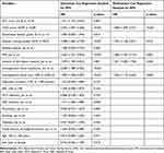 |
Table 2 Results of Univariate and Multivariate Cox Regression Analysis for RFS in the Training Set |
Construction and Validation of the LFNII-Nomogram Model
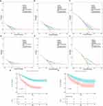
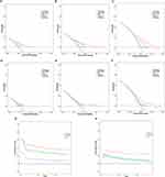
Following multivariate Cox regression analysis using the backward method, an RFS nomogram was developed (Figure 4B) by integrating the LFNII score, liver fibrosis grade, MVI, invasion of the hepatic capsule, and intraoperative blood loss. A concordance index of 0.686 (95% CI: 0.651–0.721) was obtained using the nomogram model. The AUCs of the 1-, 2-, and 5-year training sets were 0.769, 0.728, and 0.714, respectively (Figure 4C). The ROC curve areas of the validation set for 1, 2, and 5 years were 0.639, 0.645, and 0.671, respectively, indicating robust discriminatory capability (Figure 4D). Calibration curves for the training and validation sets revealed substantial convergence between the predicted and observed probabilities of RFS at 1, 2, and 5 years, closely aligned with the standard lines, further validating the reliability (Figure 4E and F). The DCA curve highlighted the clinical value of the nomogram model at 1, 2, and 5 years, demonstrating its superiority over any single component index (Figure 5A-F).
A nomogram score of 188 was used to classify the training and validation sets into high- and low-scoring groups. Patients with high nomogram scores exhibited significantly poorer prognoses in both sets (p <0.001; Figure 5G and H). These collective findings demonstrate that the nomogram model, based on the LFNII score, possesses excellent discrimination, calibration, and clinical applicability.
Comparison of LFNII-Nomogram Model with Common Tumor Staging System
Performance assessment included the LFNII nomogram, TNM staging system (eighth edition), and BCLC staging system. In the training and validation sets, the DCA for the RFS nomogram model at 1, 2, and 5 years showed that the LFNII model had better clinical application than the recognized TNM staging and BCLC staging systems (Figure 6A-F). Figure 6G shows that the nomogram model outperformed the TNM and BCLC staging systems over time, as evidenced by its greater AUC. Similar results were observed in the validation group (Figure 6H).
LFNII Score and Different Prognostic Indicators
The time-dependent AUC revealed that, compared with NrLR, GLR, and PALBI, the LFNII score was the best predictor of postoperative recurrence in the training set for 1–5 years post-surgery [C-index: LFNII 0.626 (0.610–0.643) vs NrLR 0.533 (0.516–0.550) vs GLR 0.561 (0.545–0.577) vs PALBI 0.504 (0.483–0.526)]. These findings were consistent in the validation set (Supplementary Figure 2A and B). In addition, DCA revealed that the LFNII score was superior to these three indicators in terms of clinical benefit at 2 and 5 years post-surgery in the training cohort, and this trend was corroborated in the validation cohort (Supplementary Figure 2C-F). In patients with AFP-positive HCC, KM curve revealed that the LFNII score and AFP were associated with postoperative recurrence (p values <0.05) (Supplementary Figure 3A and B). In patients with AFP-positive HCC, the KM curve showed that both the LFNII score and AFP were associated with postoperative recurrence (p values <0.05) (Supplementary Figure 3A and B). Furthermore, the time-dependent AUC showed that the AUC value of the LFNII score was lower than that of AFP in predicting the ability of recurrence at 1, 2, 3, 4, and 5 years post-surgery, suggesting that the predictive ability of LFNII did not match that of AFP (Supplementary Figure 3C).
Discussion
Inflammation, nutrition, and immunity are well known to be closely linked to tumor onset and progression.21,34–37 Recent studies have further elucidated the intricate interplay between inflammation, immunity, and nutrition, underscoring their association with the occurrence and unfavorable prognosis of liver cancer.38–41 AFP-NHCC generally exhibits a more favorable prognosis than high AFP HCC;42 however, its early postoperative recurrence rate remains notably elevated, ranging from 20–52.6%.24,28,29,34 Our retrospective study similarly recorded a 29.8% recurrence rate within 2 years, underscoring the critical significance of postoperative patient surveillance. Notably, previous research has highlighted the limited applicability of AFP levels in postoperative monitoring,43 emphasizing the urgent need for simple and readily available serological markers for the effective postoperative surveillance of these patients.
Currently, studies are exploring biomarkers for AFP-negative proteins;44–46 however, standardized clinical practice criteria are lacking. Numerous inflammatory, nutritional, and immune indicators, including GAR, GAPR, AAR, ALBI, LMR, NrLR, CRP, and PALBI, have been linked to the prognosis of patients with AFP-NHCC.24,27–29,31 However, these individual indices are insufficient for patient risk stratification. Previous studies have demonstrated the prognostic efficacy of a composite score combining inflammation, immunity, and nutrition in intrahepatic cholangiocarcinoma;47 however, such a study is lacking in HCC. Therefore, we sought to use amalgamate as an indicator of the comprehensive liver function, inflammatory, nutritional, and immune statuses of patients. Our study revealed significant correlations between ALRI, ANRI, SII, GAPR, AAR, ALBI, AAPR, SIRI, and PNI with postoperative recurrence. The newly formulated LFNII scores incorporated these nine indicators. Earlier studies identified ANRI, ALRI, AAPR, SII, and SIRI as indicators of inflammatory immune status in patients.48–57 GAPR, AAR, and ALBI grading have been used to assess liver function.27,58 The PNI reflects a patient’s nutritional status.59,60 Therefore, the LFNII score comprehensively integrates liver function, inflammation, immunity, and nutritional status, providing a holistic reflection of the patient’s overall condition. The predictive power of the newly devised LFNII surpassed that of any single indicator. Simultaneously, the LFNII-based nomogram exhibited a robust early recurrence prediction ability. The areas under the ROC curves for the training and validation sets demonstrated superior prediction efficacy compared with the traditional staging methods.
Our investigation revealed that patients in the high LFNII score group had a higher postoperative recurrence rate, which was associated with a larger tumor burden, liver capsule invasion, increased tumor diameter, microvascular invasion, elevated alpha-fetoprotein levels, advanced tumor stage, greater intraoperative blood loss, and massive intraoperative blood transfusion. This finding suggests that the LFNII score can preoperatively predict adverse pathological features.
Among the nine metrics that comprise the LFNII score, the present study found that high ALRI, ANRI, SII, GAPR, AAR, and ALBI levels, as well as low AAPR and PNI levels, were associated with worse prognoses in patients with AFP-negative hepatocellular carcinomas, which is consistent with the findings of previous studies. Interestingly, we found that high SIRI was associated with a better prognosis, a finding that is contrary to those of previous studies.54–57 SIRI is obtained from neutrophil, monocyte, and lymphocyte counts — both neutrophils and lymphocytes, which have tumor-suppressing and tumor-promoting properties. Neutrophils can secrete large amounts of inflammatory factors, causing cellular damage, promoting epithelial-mesenchymal transformation of tumor cells, and promoting neovascularization, which promotes tumor growth and metastasis.61 However, they can induce tumor detachment from the basal lamina, which prevents tumor growth.62 In the tumor microenvironment, different lymphocyte subpopulations play different roles; CD4+Th1, CD8+T, and NK cells recognize, kill, and clear tumor cells, whereas CD4+T Th2, Treg cells play tumor-promoting roles. The association between serum immune cells and the level of immune cells in the tumor microenvironment is unclear; therefore, whether serum immune cells truly reflect the anti-tumor immune status of patients is currently unknown and requires further study. However, it is now clearer that persistent non-specific inflammatory responses may promote tumor growth. Given that the LFNII score primarily comprises indicators reflecting inflammatory immune status, liver function, and nutrition, poor prognoses in patients with a high LFNII score may stem from impaired liver function, compromised nutritional status, and an exaggerated non-specific immune response.
The inclusion of other indicators, such as microvascular invasion, degree of hepatic fibrosis, and significant intraoperative blood loss in the column chart model, are all recurrence risk factors of frequent clinical concern. Notably, this study included the indicator of tumor invasion of the hepatic peritoneum, which is not currently considered to be a high risk of recurrence or a factor that can lead to a poor prognosis. According to the AJCC-TNM staging criteria, hepatocellular carcinoma invasion of the hepatic peritoneum is a peritoneal invasion and should be considered a locally advanced condition, which is in line with our findings; however, the reliability of the results requires further multicenter and large-sample studies. In addition to our constructed LFNII score, the ATS score, a noteworthy prognostic index, was associated with the early recurrence of AFP-NHCC. Tsilimigras et al revealed a favorable synergistic effect of AFP and tumor burden on the long-term survival outcomes of patients with HCC undergoing curative hepatectomy.63 The ATS score, developed by Ding et al, is a concise and user-friendly prognostic tool that reflects tumor biology and is calculated based on tumor size, number, and AFP.32 Despite the low AFP levels in the enrolled patients, the ATS score remained associated with early recurrence, and the LFNII score was positively correlated with the ATS score. This suggests that tumor size and number are crucial for early recurrence in patients with HCC with low AFP levels.
In this study, we compared the LFNII score with three previously reported prognostic indicators for AFP-negative HCC, namely NrLR, GLR, and PALBI. We found that the LFNII score had the best efficacy for postoperative recurrence, possibly because of the fact that the other three indicators primarily represent a single facet of the inflammatory state, whereas the LFNII score integrates various factors such as immunity, inflammation, and nutrition, resulting in a stronger correlation with prognosis.30 Further, elevated inflammation-related markers were associated with lower differentiation, larger tumor burden, more severe intraoperative injury and worse liver function. This concordance with the LFNII score reflects the worse clinicopathological features of the patients and serves as important evidence for the poor prognosis of such patients. At present, many indicators related to the prognosis of AFPN-HCC have been reported, including AFP-L3 and PIVKA-II, but most of them have not been included in routine tests, making it challenging to obtain such data in clinical practice. Therefore, for comparison, we selected indicators that are easily available in the laboratory. In contrast, markers based on routine hematology tests are more readily available and inexpensive. The LFNII score is solely based on routine laboratory indicators, making it easily implementable and applicable in clinical practice. We have also created a web calculator to make this LFNII score easier to access. Moreover, because each center often combines its own experience in the screening of surgical patients, there are differences in the baseline status of patients. Therefore, such a comparison also has some limitations, and more data from more centers are needed to prove the application value of these indicators. This study is grounded in data from two large-scale liver cancer centers in China, which also had high reliability.
Neoadjuvant therapy for HCC is rapidly evolving, offering improved prognoses for patients at high risk of recurrence.64 Despite this progress, a reliable index for predicting the efficacy of neoadjuvant therapy is lacking, making it challenging to stratify the degree of benefit and identify suitable candidates. The LFNII score may serve as a valuable indicator of high-risk recurrence of AFP-NHCC preoperatively, suggesting that individuals with elevated LFNII scores may derive potential benefits from neoadjuvant therapy. However, this study also has some limitations. First, although it is based on data from two large centers, external validation in a larger population is needed to prove the effectiveness of the score. Second, the laboratory indicators of peripheral blood are affected by many factors, leading to some fluctuation, although these are substantial at the overall level. In the future, postoperative and perioperative dynamic changes should be evaluated. Third, as this study is a retrospective study, there may be some bias. Further prospective cohort studies are warranted to determine whether these patients benefit from pre-operative neoadjuvant therapy. In addition to the differences in clinicopathological features, the potential association of the score with the genome and transcriptome at the molecular level warrants further evaluation.
Conclusion
The study findings indicated that the LFNII score had robust predictive capabilities for postoperative recurrence in patients with AFP-NHCC. Furthermore, a nomogram based on the LFNII score has been effectively developed to predict RFS in AFP-NHCC patients following surgery, providing a valuable resource for clinical decision-making and patient management.
Abbreviations
AAR, aspartate aminotransferase to alanine aminotransferase; AFP, alpha-fetoprotein; AFP-NHCC, alpha-fetoprotein-negative hepatocellular carcinoma; ALB, albumin; ALBI, albumin-bilirubin score; ALP, alkaline phosphatase; ALT, alanine aminotransferase; AST, aspartate aminotransferase; ATS, alpha-feto protein tumor burden score; AUC, area under the curve; BCLC, Barcelona Clinic Liver Cancer system; DCA, decision curve analysis; GAPR, gamma-glutamyl transpeptidase to alkaline phosphatase; GAR, gamma-glutamyl transpeptidase to aspartate aminotransferase; GGT, gamma-glutamyl transpeptidase; GLR, gamma-glutamyltransferase-to-lymphocyte ratio; HCC, hepatocellular carcinoma; KM, Kaplan–Meier; LASSO, least absolute shrinkage and selection operator; LFNII, liver function-nutrition-inflammation-immune; LMR, lymphocyte-to-monocyte ratio; LYMPH, lymphocyte; MLR, monocyte-to-lymphocyte ratio; MONO, monocyte; MVI, microvascular invasion; NEUT, neutrophil; NLR, neutrophil-to-lymphocyte ratio; NrLR, neutrophil times gamma-glutamyl transpeptidase-to-lymphocyte ratio; PALBI, modified albumin-bilirubin score; PLR, platelet-to-lymphocyte ratio; PLT, blood platelet count; RFS, recurrence-free survival; ROC, receiver operating characteristic; SII, systemic immune-inflammation index; SIRI, serum immune-to-inflammation ratio index; TBIL, total bilirubin.
Data Sharing Statement
All data generated or analyzed during this study are included in this article and the Supplementary Materials. Further inquiries can be directed to the corresponding authors.
Ethics Approval
The researchers obtained ethical permission from the Ethics Committees of the Cancer Hospital of the Chinese Academy of Medical Sciences and the Fifth Medical Center of the PLA General Hospital, with approval number 21/198-2869. This study was conducted in accordance with the principles outlined in the Declaration of Helsinki, and the research protocols were conducted in accordance with applicable standards and laws. Informed consent was obtained from all participants prior to their inclusion in the study.
Acknowledgments
The authors would like to thank the patients who participated in this study and the staff at the Department of Hepatobiliary Surgery of the Cancer Hospital, Chinese Academy of Medical Sciences, and The Fifth Medical Center of the PLA General Hospital for their support and cooperation.
Author Contributions
All authors made a significant contribution to the work reported, whether that is in the conception, study design, execution, acquisition of data, analysis, and interpretation, or in all these areas; took part in drafting, revising, or critically reviewing the article; gave final approval of the version to be published; have agreed on the journal to which the article has been submitted; and agree to be accountable for all aspects of the work.
Funding
This study was supported by the CAMS Innovation Fund for Medical Sciences (CIFMS; grant number 2021-I2M-1-066); CAMS Initiative for Innovative Medicine (grant number 2021-I2M-1-015); National Natural Science Foundation of China (grant numbers 81972311, 82141127); and Sanming Project of Medicine in Shenzhen (grant number SZSM202011010).
Disclosure
The author(s) report no conflicts of interest in this work.
References
1. Sung H, Ferlay J, Siegel RL, et al. Global cancer statistics 2020: GLOBOCAN estimates of incidence and mortality worldwide for 36 cancers in 185 countries. CA Cancer J Clin. 2021;71(3):209–249. doi:10.3322/caac.21660
2. Llovet JM, Zucman-Rossi J, Pikarsky E, et al. Hepatocellular carcinoma. Nat Rev Dis Primers. 2016;2(1):16018. doi:10.1038/nrdp.2016.18
3. McGlynn KA, Petrick JL, El-Serag HB. Epidemiology of hepatocellular carcinoma. Hepatology. 2021;73(9 1):4–13. doi:10.1002/hep.31288
4. Rumgay H, Ferlay J, de Martel C, et al. Global, regional, and national burden of primary liver cancer by subtype. Eur J Cancer. 2022;161:108–118. doi:10.1016/j.ejca.2021.11.023
5. Zhang CH, Cheng Y, Zhang S, Fan J, Gao Q. Changing epidemiology of hepatocellular carcinoma in Asia. Liver Int. 2022;42(9):2029–2041. doi:10.1111/liv.15251
6. Yoh T, Seo S, Taura K, et al. Surgery for recurrent hepatocellular carcinoma: achieving long-term survival. Ann Surg. 2021;273(4):792–799. doi:10.1097/SLA.0000000000003358
7. Galle PR, Foerster F, Kudo M, et al. Biology and significance of alpha-fetoprotein in hepatocellular carcinoma. Liver Int. 2019;39(12):2214–2229. doi:10.1111/liv.14223
8. He H, Chen S, Fan Z, et al. Multi-dimensional single-cell characterization revealed a suppressive immune microenvironment in AFP-positive hepatocellular carcinoma. Cell Discov. 2023;9(1):60. doi:10.1038/s41421-023-00563-x
9. Bai DS, Zhang C, Chen P, Jin SJ, Jiang GQ. The prognostic correlation of AFP level at diagnosis with pathological grade, progression, and survival of patients with hepatocellular carcinoma. Sci Rep. 2017;7(1):12870. doi:10.1038/s41598-017-12834-1
10. Beale G, Chattopadhyay D, Gray J, et al. AFP, PIVKAII, GP3, SCCA-1, and follisatin as surveillance biomarkers for hepatocellular cancer in non-alcoholic and alcoholic fatty liver disease. BMC Cancer. 2008;8:200. doi:10.1186/1471-2407-8-200
11. Agopian VG, Harlander-Locke MP, Markovic D, et al. Evaluation of patients with hepatocellular carcinoma that do not produce α-fetoprotein. JAMA Surg. 2017;152(1):55–64. doi:10.1001/jamasurg.2016.3310
12. Yan WT, Li C, Yao LQ, et al. Predictors and long-term prognosis of early and late recurrence for patients undergoing hepatic resection of hepatocellular carcinoma: a large-scale multicenter study. Hepatobiliary Surg Nutr. 2023;12(2):155–168. doi:10.21037/hbsn-21-288
13. Ji J, Wang H, Li Y, et al. Diagnostic evaluation of des-gamma-carboxy prothrombin versus α-fetoprotein for hepatitis B virus-related hepatocellular carcinoma in China: a large-scale, multicenter study. PLoS One. 2016;11(4):e0153227. doi:10.1371/journal.pone.0153227
14. Best J, Bilgi H, Heider D, et al. The GALAD scoring algorithm based on AFP, AFP-L3, and DCP significantly improves the detection of BCLC early-stage hepatocellular carcinoma. Z Gastroenterol. 2016;54(12):1296–1305. doi:10.1055/s-0042-119529
15. Huang J, Liu FC, Li L, Zhou WP, Jiang BG, Pan ZY. Nomograms to predict the long-time prognosis in patients with alpha-fetoprotein negative hepatocellular carcinoma following radical resection. Cancer Med. 2020;9(8):2791–2802. doi:10.1002/cam4.2944
16. Wang X, Mao M, He Z, et al. Development and validation of a prognostic nomogram in AFP-negative hepatocellular carcinoma. Int J Biol Sci. 2019;15(1):221–228. doi:10.7150/ijbs.28720
17. Qiu ZC, Wu YW, Qi WL, et al. Pivka-II combined with Tumor Burden Score to predict long-term outcomes of AFP-negative hepatocellular carcinoma patients after liver resection. Cancer Med. 2023.
18. Wiseman MJ. Nutrition and cancer: prevention and survival. Br J Nutr. 2019;122(5):481–487. doi:10.1017/S0007114518002222
19. Engelhard VH, Rodriguez AB, Mauldin IS, Woods AN, Peske JD, Slingluff CL. Immune cell infiltration and tertiary lymphoid structures as determinants of antitumor immunity. J Immunol. 2018;200(2):432–442. doi:10.4049/jimmunol.1701269
20. Demaria O, Cornen S, Daëron M, Morel Y, Medzhitov R, Vivier E. Harnessing innate immunity in cancer therapy. Nature. 2019;574(7776):45–56. doi:10.1038/s41586-019-1593-5
21. Zitvogel L, Pietrocola F, Kroemer G. Nutrition, inflammation, and cancer. Nat Immunol. 2017;18(8):843–850. doi:10.1038/ni.3754
22. Xu L, Yu S, Zhuang L, et al. Systemic inflammation response index (SIRI) predicts prognosis in hepatocellular carcinoma patients. Oncotarget. 2017;8(21):34954–34960. doi:10.18632/oncotarget.16865
23. Huang PY, Wang CC, Lin CC, et al. Predictive effects of inflammatory scores in patients with BCLC 0-A hepatocellular carcinoma after hepatectomy. J Clin Med. 2019;8(10):1676. doi:10.3390/jcm8101676
24. Hu B, Yang XR, Xu Y, et al. The systemic immune-inflammation index predicts the prognosis of patients after curative resection for hepatocellular carcinoma. Clin Cancer Res. 2014;20(23):6212–6222. doi:10.1158/1078-0432.CCR-14-0442
25. Wang D, Bai N, Hu X, et al. Preoperative inflammatory markers of NLR and PLR as indicators of poor prognosis in resectable HCC. PeerJ. 2019;7:e7132. doi:10.7717/peerj.7132
26. Wang BL, Tian L, Gao XH, et al. Dynamic change of the systemic immune inflammation index predicts the prognosis of patients with hepatocellular carcinoma after curative resection. Clin Chem Lab Med. 2016;54(12):1963–1969. doi:10.1515/cclm-2015-1191
27. Li J, Tao H, Zhang E, Huang Z. Diagnostic value of gamma-glutamyl transpeptidase to alkaline phosphatase ratio combined with gamma-glutamyl transpeptidase to aspartate aminotransferase ratio and alanine aminotransferase to aspartate aminotransferase ratio in alpha-fetoprotein-negative hepatocellular carcinoma. Cancer Med. 2021;10(14):4844–4854. doi:10.1002/cam4.4057
28. Li S, Xu W, Liao M, et al. The significance of gamma-glutamyl transpeptidase to lymphocyte count ratio in the early post-operative recurrence monitoring and prognosis prediction of AFP-negative hepatocellular carcinoma. J Hepatocell Carcinoma. 2021;8:23–33. doi:10.2147/JHC.S286213
29. Mao S, Yu X, Shan Y, Fan R, Wu S, Lu C. Albumin-bilirubin (ALBI) and monocyte-to-lymphocyte ratio (MLR)-based nomogram model to predict tumor recurrence of AFP-Negative hepatocellular carcinoma. J Hepatocell Carcinoma. 2021;8:1355–1365. doi:10.2147/JHC.S339707
30. Wu Q, Zeng J, Zeng J. Inflammation-Related marker NrLR predicts prognosis in AFP-negative HCC patients after curative resection. J Hepatocell Carcinoma. 2023;10:193–202. doi:10.2147/JHC.S393286
31. Yang C, Wu X, Liu J, et al. Nomogram based on platelet-albumin-bilirubin for predicting tumor recurrence after surgery in alpha-fetoprotein-negative hepatocellular carcinoma patients. J Hepatocell Carcinoma. 2023;10:43–55. doi:10.2147/JHC.S396433
32. Ding HF, Yang T, Lv Y, Zhang XF, Pawlik TM, International Hepatocellular Carcinoma Study Group. Development and validation of an α-fetoprotein tumor burden score model to predict post-recurrence survival among patients with hepatocellular carcinoma. J Am Coll Surg. 2023;236(5):982–992. doi:10.1097/XCS.0000000000000638
33. Guo Z. Department of medical administration, National Health and Health Commission of the People’s Republic of China. Zhonghua Gan Zang Bing Za Zhi. 2020;28(2):112–128.
34. Taniguchi K, Karin M. NF-κB, inflammation, immunity, and cancer: coming of age. Nat Rev Immunol. 2018;18(5):309–324. doi:10.1038/nri.2017.142
35. Grivennikov SI, Greten FR, Karin M. Immunity, inflammation, and cancer. Cell. 2010;140(6):883–899. doi:10.1016/j.cell.2010.01.025
36. Key TJ, Bradbury KE, Perez-Cornago A, Sinha R, Tsilidis KK, Tsugane S. Diet, nutrition, and cancer risk: what do we know and what is the way forward? BMJ. 2020;368:m511. doi:10.1136/bmj.m511
37. Narimatsu H, Yaguchi YT. The role of diet and nutrition in cancer: prevention, treatment, and survival. Nutrients. 2022;14(16):3329. doi:10.3390/nu14163329
38. Ringelhan M, Pfister D, O’Connor T, Pikarsky E, Heikenwalder M. The immunology of hepatocellular carcinoma. Nat Immunol. 2018;19(3):222–232. doi:10.1038/s41590-018-0044-z
39. Leone V, Ali A, Weber A, Tschaharganeh DF, Heikenwalder M. Liver inflammation and hepatobiliary cancers. Trends Cancer. 2021;7(7):606–623. doi:10.1016/j.trecan.2021.01.012
40. De Oliveira S, Houseright RA, Graves AL, et al. Metformin modulates innate immune-mediated inflammation and early progression of NAFLD-associated hepatocellular carcinoma in zebrafish. J Hepatol. 2019;70(4):710–721. doi:10.1016/j.jhep.2018.11.034
41. George ES, Sood S, Broughton A, et al. The Association between diet and hepatocellular carcinoma: a systematic review. Nutrients. 2021;13(1):172. doi:10.3390/nu13010172
42. Zheng Y, Zhu M, Li M. Effects of alpha-fetoprotein on the occurrence and progression of hepatocellular carcinoma. J Cancer Res Clin Oncol. 2020;146(10):2439–2446. doi:10.1007/s00432-020-03331-6
43. Zhang XF, Qi X, Meng B, et al. Prognosis evaluation in alpha-fetoprotein negative hepatocellular carcinoma after hepatectomy: comparison of five staging systems. Eur J Surg Oncol. 2010;36(8):718–724. doi:10.1016/j.ejso.2010.05.022
44. Luo P, Wu S, Yu Y, et al. Current status and perspective biomarkers in AFPnegative HCC: towards screening for and diagnosing hepatocellular carcinoma at an earlier stage. Pathol Oncol Res. 2020;26(2):599–603. doi:10.1007/s12253-019-00585-5
45. She S, Xiang Y, Yang M, et al. C-reactive protein is a biomarker of AFP-negative HBV-related hepatocellular carcinoma. Int J Oncol. 2015;47(2):543–554. doi:10.3892/ijo.2015.3042
46. Wang T, Zhang KH. New blood biomarkers for the diagnosis of AFP-negative hepatocellular carcinoma. Front Oncol. 2020;10:1316. doi:10.3389/fonc.2020.01316
47. Zhu J, Wang D, Liu C, et al. Development and validation of a new prognostic immune-inflammatory-nutritional score for predicting outcomes after curative resection for intrahepatic cholangiocarcinoma: a multicenter study. Front Immunol. 2023;14:1165510. doi:10.3389/fimmu.2023.1165510
48. Zheng Z, Guan R, Zou Y, et al. Nomogram based on inflammatory biomarkers to predict the recurrence of hepatocellular carcinoma-A multicenter experience. J Inflam Res. 2022;15:5089–5102. doi:10.2147/JIR.S378099
49. Zheng J, Seier K, Gonen M, et al. Utility of serum inflammatory markers for predicting microvascular invasion and survival for patients with hepatocellular carcinoma. Ann Surg Oncol. 2017;24(12):3706–3714. doi:10.1245/s10434-017-6060-7
50. Young S, Cam I, Gencturk M, et al. Inflammatory scores: comparison and utility in HCC patients undergoing transarterial chemoembolization in a North American cohort. J Hepatocell Carcinoma. 2021;8:1513–1524. doi:10.2147/JHC.S335183
51. Wu W, Wang Q, Han D, et al. Prognostic value of pre-operative inflammatory markers in patients with hepatocellular carcinoma who underwent curative resection. Cancer Cell Int. 2021;21(1):500. doi:10.1186/s12935-021-02204-3
52. Tian G, Li G, Guan L, Yang Y, Li N. Pre-treatment albumin-to-alkaline phosphatase ratio as a prognostic indicator in solid cancers: a meta-analysis with trial sequential analysis. Int J Surg. 2020;81:66–73. doi:10.1016/j.ijsu.2020.07.024
53. Zhang F, Lu S, Tian M, et al. Albumin-to-alkaline phosphatase ratio is an independent prognostic indicator in combined hepatocellular and cholangiocarcinoma. J Cancer. 2020;11(17):5177–5186. doi:10.7150/jca.45633
54. Dziedzic EA, Gąsior JS, Tuzimek A, Dąbrowski M, Jankowski P. The association between serum vitamin D concentration and new inflammatory biomarkers-systemic inflammatory index (SII) and systemic inflammatory response (SIRI)-in patients with ischemic heart disease. Nutrients. 2022;14(19):4212. doi:10.3390/nu14194212
55. Wang D, Hu X, Xiao L, et al. The prognostic nutritional index and systemic immune-inflammatory index predict the prognosis of patients with HCC. J Gastrointest Surg. 2021;25(2):421–427. doi:10.1007/s11605-019-04492-7
56. Wang RH, Wen WX, Jiang ZP, et al. The clinical value of the neutrophil-to-lymphocyte ratio (NLR), systemic immune-inflammation index (SII), platelet-to-lymphocyte ratio (PLR), and systemic inflammation response index (SIRI) for predicting the occurrence and severity of pneumonia in patients with intracerebral hemorrhage. Front Immunol. 2023;14:1115031. doi:10.3389/fimmu.2023.1115031
57. Deng-Xiong L, Qing-Xin Y, De-Chao F, et al. Systemic immune-inflammation index (SII) during induction has a higher predictive value than preoperative SII in non-muscle-invasive bladder cancer patients receiving intravesical bacillus Calmette -Guerin. Clin Genitourin Cancer. 2023;21(3):e145–e152. doi:10.1016/j.clgc.2022.11.013
58. Johnson PJ, Berhane S, Kagebayashi C, et al. Assessment of liver function in patients with hepatocellular carcinoma: a new evidence-based approach-The ALBI grade. J Clin Oncol. 2015;33(6):550–558. doi:10.1200/JCO.2014.57.9151
59. Raposeiras Roubín S, Abu Assi E, Cespón Fernandez M, et al. Prevalence and prognostic significance of malnutrition in patients with acute coronary syndrome. J Am Coll Cardiol. 2020;76(7):828–840. doi:10.1016/j.jacc.2020.06.058
60. Wang Z, Wang Y, Zhang X, Zhang T. Pretreatment prognostic nutritional index as a prognostic factor in lung cancer: a review and meta-analysis. Clin Chim Acta. 2018;486:303–310. doi:10.1016/j.cca.2018.08.030
61. Cristinziano L, Modestino L, Loffredo S, et al. Anaplastic thyroid cancer cells induce the release of mitochondrial extracellular DNA traps by viable neutrophils. J Immunol. 2020;204(5):1362–1372. doi:10.4049/jimmunol.1900543
62. Gershkovitz M, Caspi Y, Fainsod-Levi T, et al. TRPM2 mediates neutrophil killing of disseminated tumor cells. Cancer Res. 2018;78(10):2680–2690. doi:10.1158/0008-5472.CAN-17-3614
63. Tsilimigras DI, Hyer JM, Diaz A, et al. Synergistic impact of alpha-fetoprotein and tumor burden on long-term outcomes following curative-intent resection of hepatocellular carcinoma. Cancers. 2021;13(4):747. doi:10.3390/cancers13040747
64. Fan Y, Xue H, Zheng H. Systemic therapy for hepatocellular carcinoma: current updates and outlook. J Hepatocell Carcinoma. 2022;9:233–263. doi:10.2147/JHC.S358082
 © 2024 The Author(s). This work is published and licensed by Dove Medical Press Limited. The full terms of this license are available at https://www.dovepress.com/terms.php and incorporate the Creative Commons Attribution - Non Commercial (unported, v3.0) License.
By accessing the work you hereby accept the Terms. Non-commercial uses of the work are permitted without any further permission from Dove Medical Press Limited, provided the work is properly attributed. For permission for commercial use of this work, please see paragraphs 4.2 and 5 of our Terms.

