
Back to Journals » International Journal of General Medicine » Volume 19
Comparative Prognostic Value of ASPECTS and CTP Core Infarct Volume in Acute Ischemic Stroke Patients Undergoing Endovascular Therapy
Authors Wang Z, Zhang L, Wang X, Yu X, Tang J, Dai X, Qiu T
Received 19 November 2025
Accepted for publication 2 April 2026
Published 21 April 2026 Volume 2026:19 582848
DOI https://doi.org/10.2147/IJGM.S582848
Checked for plagiarism Yes
Review by Single anonymous peer review
Peer reviewer comments 2
Editor who approved publication: Dr Redoy Ranjan
Zezhao Wang,1,2,* Liyuan Zhang,1,2,* Xi Wang,2,3 Xueji Yu,2 Jie Tang,2 Xiaoyan Dai,4 Tao Qiu2
1Department of Neurology,The Affiliated Hospital of North Sichuan Medical College, Nanchong, Sichuan, 637000, People’s Republic of China; 2Department of Neurology, Zigong First People’s Hospital, Zigong, Sichuan, 643000, People’s Republic of China; 3Department of Neurology, Affiliated Hospital of Southwest Medical University, Luzhou, Sichuan, 646000, People’s Republic of China; 4Outpatient Department, Zigong First People’s Hospital, Zigong, Sichuan, 643000, People’s Republic of China
*These authors contributed equally to this work
Correspondence: Tao Qiu, Department of Neurology, Zigong First People’s Hospital, No. 42, Shangyihao 1st Branch Road, Ziliujing District, Zigong, Sichuan, 643000, People’s Republic of China, Email [email protected] Xiaoyan Dai, Outpatient Department, Zigong First People’s Hospital, No. 42, Shangyihao 1st Branch Road, Ziliujing District, Zigong, Sichuan, 643000, People’s Republic of China, Email [email protected]
Background: Rapid and accurate imaging assessment is critical for selecting acute ischemic stroke (AIS) patients for endovascular therapy. While CT perfusion (CTP) provides quantitative evaluation of infarct core, the ASPECTS score based on non-contrast CT (NCCT) is widely used for its speed and simplicity. However, their comparative predictive value and consistency in clinical practice remain unclear.
Objective: To evaluate the prognostic value of NCCT-based ASPECTS and CTP-derived core infarct volume in AIS patients undergoing endovascular treatment, and to explore their correlation and clinical implications.
Methods: In this retrospective single-center study, we analyzed 82 patients with acute ischemic stroke who underwent endovascular therapy within 24 hours of symptom onset. Preoperative ASPECTS scores and CTP-defined core infarct volumes (processed via Shukun software) were recorded. Prognostic outcome was defined by the 90-day modified Rankin Scale (mRS). Statistical analysis included logistic regression, ROC curve analysis, and Spearman correlation.
Results: Patients with poor outcomes had significantly larger CTP-derived infarct core volumes and lower ASPECTS scores (both P< 0.001). In the overall cohort, CTP (AUC=0.74) and ASPECTS (adjusted AUC=0.71) demonstrated comparable predictive performance, with no statistically significant difference. In subgroup analyses stratified by onset-to-treatment time, both modalities remained predictive, and no significant differences in predictive accuracy were observed between ASPECTS and CTP in either the ≤ 6-hour or 6– 24-hour groups. A moderate negative correlation was observed between ASPECTS and core infarct volume (r=– 0.61, P< 0.001).
Conclusion: Both ASPECTS and CTP-derived core infarct volume predict functional outcomes in AIS patients undergoing endovascular therapy, with comparable performance. ASPECTS offers a rapid assessment, whereas CTP provides quantitative evaluation. The two modalities may serve complementary roles in clinical decision-making.
Keywords: ASPECTS, CT perfusion, acute ischemic stroke, endovascular therapy, prognosis
Introduction
Acute ischemic stroke (AIS), accounting for over 80% of all stroke cases, results from the abrupt reduction or cessation of cerebral blood flow, leading to irreversible ischemic necrosis of brain tissue.1 It remains one of the leading causes of death and disability worldwide.2 Timely and effective reperfusion therapy—either intravenous thrombolysis or endovascular thrombectomy—can significantly reduce infarct volume and improve neurological outcomes.3,4 The pathophysiology of AIS involves a central infarct core surrounded by an ischemic penumbra. The penumbra is potentially reversible with prompt reperfusion, making rapid and accurate identification of salvageable tissue critical for treatment decisions.5 In clinical practice, neuroimaging plays a central role in this process by facilitating diagnosis, stratifying patients, and guiding reperfusion strategies.
Currently, non-contrast computed tomography (NCCT), CT angiography (CTA), and CT perfusion (CTP) are widely used for the emergency evaluation of AIS.6 Among these, the Alberta Stroke Program Early CT Score (ASPECTS) based on NCCT is a semi-quantitative, rapid method for assessing early ischemic changes, widely adopted due to its accessibility and speed.7 CTP, on the other hand, provides quantitative assessments of cerebral hemodynamics, including infarct core, ischemic penumbra, and collateral circulation.5 Recent studies have further highlighted the important role of CT perfusion imaging in guiding reperfusion therapy and prognostic evaluation in acute ischemic stroke.8 Landmark randomized trials have further clarified the role of imaging-based infarct assessment in selecting patients for endovascular therapy. The HERMES meta-analysis demonstrated that patients with ASPECTS ≥6 derive substantial benefit from thrombectomy in the early treatment window,3 whereas the DAWN9 and DEFUSE-310 trials established that quantitative infarct core volume measured by CT perfusion or diffusion-weighted MRI can identify patients who benefit from thrombectomy in the extended time window of 6–24 hours after symptom onset.
According to current guidelines, ASPECTS remains the initial imaging modality of choice in most AIS cases, while CTP is recommended for patients within the extended window (6–24 hours) or with unclear onset.5 ASPECTS and CTP are both routinely used to estimate infarct core volume in AIS, serving as critical tools for therapeutic decision-making. However, despite their widespread application, accumulating evidence indicates notable discrepancies between the two modalities in defining infarct extent and predicting outcomes.11,12 ASPECTS, while simple, is subject to interobserver variability and limited sensitivity to subtle early changes, whereas CTP offers quantitative accuracy but may be influenced by technical and post-processing variability.13,14
Recent studies have demonstrated only moderate correlation between ASPECTS and CTP-derived core volumes, and sometimes marked inconsistency.15 For example, CTP-defined large core infarctions (≥70 mL) appear to be more predictive of poor prognosis than ASPECTS-defined large cores (ASPECTS ≤5), particularly in patients treated within 6 hours of onset.16 Although both modalities were employed in landmark randomized trials such as SELECT2 and ANGEL-ASPECT,5 criteria and predictive performances varied. Until now, direct comparisons of their prognostic performance across different therapeutic time windows remain limited. In particular, whether these two imaging approaches provide consistent predictive value in both early (<6 hours) and extended (6–24 hours) treatment windows has not been fully clarified.
Therefore, this study aimed to compare the prognostic value of ASPECTS and CTP-derived infarct core volume in patients with AIS undergoing endovascular therapy. We also assessed the agreement between these two modalities and their predictive performance in early and extended therapeutic windows. Given that advanced perfusion imaging is not universally available in all clinical settings, particularly in resource-limited hospitals, clarifying the relative utility of these imaging approaches may help guide more practical and efficient imaging strategies in acute stroke care.
Materials and Methods
Study Design and Population
This retrospective study included patients diagnosed with acute ischemic stroke (AIS) who were treated through the stroke emergency green channel at the First People’s Hospital of Zigong between January 2023 and December 2024. Patients were eligible for inclusion if they met the following criteria: age ≥18 years, underwent both non-contrast computed tomography (NCCT) and CT perfusion (CTP) imaging within 24 hours of symptom onset in the emergency department, and had infarcts involving the anterior circulation with diagnosis and treatment conducted in accordance with the Chinese Guidelines for Endovascular Treatment of Acute Ischemic Stroke (2023 edition). Exclusion criteria included infarction involving the posterior circulation, bilateral hemispheric infarctions, intracranial hemorrhage, incomplete clinical or imaging data, or poor image quality that hindered accurate assessment.
Clinical and Imaging Data Collection
Demographic, clinical, and imaging data were extracted from the hospital’s electronic medical records and imaging archiving system. Additional follow-up data were collected through structured telephone interviews. The recorded variables included age, sex, time from symptom onset to hospital admission (categorized as ≤6 hours or 6–24 hours), history of smoking, alcohol use, hypertension, hyperglycemia, hyperlipidemia, atrial fibrillation, prior stroke, baseline ASPECTS score based on NCCT, and core infarct volume determined by CTP using fully automated processing software (Shukun Technology, Beijing, China). Neurological impairment at admission was assessed using the National Institutes of Health Stroke Scale (NIHSS), and functional outcome was evaluated using the modified Rankin Scale (mRS) at 90 days post-treatment.
Imaging Protocol and Assessment Criteria
All patients underwent NCCT scanning upon admission, and ASPECTS was used to semi-quantitatively assess early ischemic changes in the territory of the middle cerebral artery. ASPECTS is scored out of 10, with one point subtracted for each area showing early ischemic changes such as focal parenchymal hypoattenuation or loss of gray-white matter differentiation. A score of 0 indicates diffuse ischemia throughout the MCA territory.
CTP images were processed using Shukun automated software to generate perfusion maps and calculate infarct core volume. In this study, the infarct core was defined as the volume of brain tissue exhibiting a cerebral blood flow (CBF) reduction of more than 70%, ie, CBF <30% of the contralateral normal value. This threshold has been widely adopted in clinical trials for delineating irreversibly damaged brain tissue.
Outcome Evaluation
Short-term neurological improvement was evaluated using the NIHSS score 7±2 days after treatment. A reduction of more than 4 points in the NIHSS score compared to baseline was considered indicative of significant neurological recovery. Long-term functional outcome was assessed at 90 days post-treatment using the mRS. A favorable outcome was defined as an mRS score of less than 2, while a poor outcome was defined as an mRS score greater than 2. An mRS score of 4 or higher was interpreted as severe disability or death.
Statistical Analysis
All statistical analyses were conducted using SPSS version 27.0 (IBM Corp., Armonk, NY, USA). For continuous variables following a normal distribution, data were presented as mean ± standard deviation (SD). For skewed data, the median and interquartile range (IQR) were reported. Categorical variables were summarized as frequencies and percentages. For univariate analysis, independent sample t-tests or Mann–Whitney U-tests were used to compare continuous variables, and χ2-tests were employed for categorical data. Multivariate logistic regression analysis was conducted to identify independent predictors of endovascular treatment outcomes. Receiver operating characteristic (ROC) curve analysis was performed to evaluate the predictive performance of imaging parameters for poor functional outcome (mRS > 2). Because higher ASPECTS scores indicate smaller infarct burden and a lower likelihood of poor outcome, a directionally aligned variable (10 − ASPECTS) was used in the ROC analysis to ensure consistent interpretation when predicting unfavorable outcomes. The area under the ROC curve (AUC) was calculated and compared across models. Spearman correlation analysis was used to examine the association between ASPECTS scores and CTP-derived core infarct volumes. A two-sided P-value <0.05 was considered statistically significant.
Results
Patient Characteristics
A total of 82 patients with acute ischemic stroke (AIS) who underwent endovascular therapy via the emergency green channel between January 2023 and December 2024 were included according to the predefined inclusion and exclusion criteria. The patients ranged in age from 38 to 89 years, with 55 patients (67.07%) aged 70 years or older and 27 patients (32.93%) younger than 70 years. The cohort comprised 47 females and 35 males.
Among these patients, 72 (87.8%) had at least one comorbid condition. Hypertension was the most prevalent (69.5%, 57/82), followed by hyperglycemia (34.1%, 28/82) and atrial fibrillation (29.3%, 24/82). A total of 23 patients (28.0%) had a history of prior stroke, underlining the importance of secondary prevention in this population.
The preoperative neurological status, as measured by the NIHSS, showed a median score of 12 (interquartile range [IQR]: 8.75–17), indicating moderate-to-severe neurological deficits in a substantial portion of patients. In terms of imaging findings, the median ASPECTS score was 9 (IQR: 7–10), while the median core infarct volume based on CTP was 20 mL (IQR: 1.93–41.5), suggesting that most patients had relatively preserved ischemic penumbra and moderate core infarction. Detailed baseline characteristics are summarized in Tables 1 and 2.
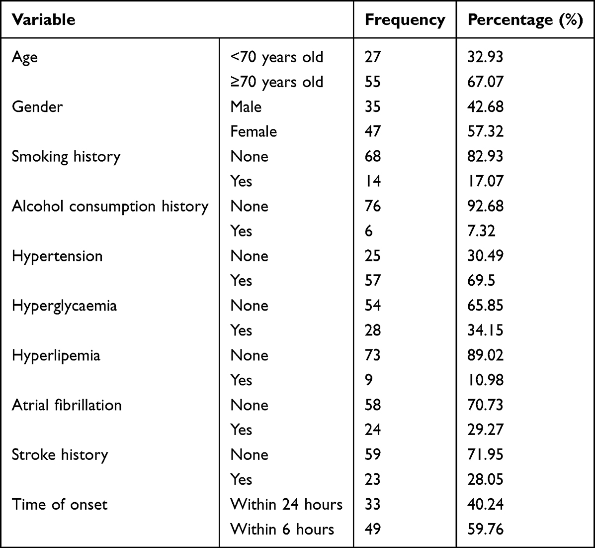 |
Table 1 Baseline Demographic and Clinical Characteristics of the Patients |
 |
Table 2 General Basic Information of Patients |
Prognostic Analysis
Based on the 90-day modified Rankin Scale (mRS) (Table 3), patients were categorized into two groups: favorable prognosis (mRS ≤2, n = 37) and poor prognosis (mRS >2, n = 45). Patients in the poor prognosis group exhibited higher rates of smoking (64.29% vs. 35.71%) and alcohol consumption (66.67% vs. 33.33%) than those in the favorable group; however, these differences were not statistically significant (p > 0.05). Similarly, the prevalence of hypertension, hyperglycemia, hyperlipidemia, atrial fibrillation, and stroke history was higher in the poor prognosis group, though none of these differences reached statistical significance.
 |
Table 3 Prognostic Variable in Acute Ischemic Stroke Patients Undergoing Endovascular Therapy |
The median preoperative NIHSS score was slightly higher in the poor prognosis group (13 [IQR: 9–18.5]) compared to the favorable group (12 [IQR: 7–15]), but this difference was not statistically significant (Z = –1.682, p = 0.093) (Table 4). However, significant differences were observed in imaging-based predictors. The median core infarct volume in the poor prognosis group was substantially larger (32 mL [IQR: 15.5–53.6]) than in the favorable group (9 mL [IQR: 0–21.5]; Z = –3.729, p < 0.001). Conversely, the ASPECTS score was significantly lower in the poor prognosis group (median 8 [IQR: not stated]) compared to the favorable group (median 10 [IQR: 9–10]; Z = –3.392, p < 0.001).
 |
Table 4 Prognostic Value of CTP and ASPECTS in Acute Ischemic Stroke Patients Undergoing Endovascular Therapy |
Furthermore, postoperative neurological improvement assessed at 7±2 days, defined by a decrease in NIHSS score ≥4 points, was significantly associated with favorable long-term outcomes (p < 0.001, χ2-test), indicating early symptom improvement as a strong predictor of prognosis.
Prognostic Comparison Between ≤6 Hours and 6–24 Hours Subgroups
A multivariate logistic regression analysis was conducted including variables such as age, time from symptom onset, ASPECTS score, CTP core infarct volume, and preoperative NIHSS score. The results identified age ≥70 years and ASPECTS score as independent predictors of poor prognosis following endovascular therapy in patients with AIS.
To compare the predictive performance of ASPECTS and CTP core infarct volume for unfavorable outcomes, receiver operating characteristic (ROC) curve analysis was performed. In the overall cohort, both ASPECTS and CTP core volume showed predictive value, with CTP displaying superior accuracy (AUC = 0.74, 95% CI: 0.63–0.85) compared to ASPECTS (AUC = 0.29, 95% CI: 0.17–0.41). Because higher ASPECTS scores indicate smaller infarct burden and a lower likelihood of poor outcome, a directionally aligned variable (10 − ASPECTS) was used in the ROC analysis predicting poor functional outcome (mRS > 2). After this adjustment, the recalculated AUC for 10-ASPECTS was 0.71, reflecting its predictive value for unfavorable outcomes. The difference between them was not statistically significant (p = 0.62; Figure 1).
 |
Figure 1 Comparison of ROC Curves for CTP Core Infarct Volume and ASPECTS Score in Predicting 90-Day Outcomes. |
When stratified by time from onset, predictive performance remained significant in patients treated within 6 hours. In this subgroup, ASPECTS showed better accuracy (AUC = 0.80, 95% CI: 0.66–0.94) compared to CTP (AUC = 0.76, 95% CI: 0.61–0.90), with no statistically significant difference (p = 0.50; Figure 2). In patients treated between 6 and 24 hours after onset, the predictive value of CTP approached significance (p = 0.053; AUC = 0.70, 95% CI: 0.51–0.88), whereas ASPECTS demonstrated greater prognostic accuracy in this subgroup (AUC = 0.57, 95% CI: 0.37–0.77; Figure 3).
 |
Figure 2 ROC Curves for CTP and ASPECTS in Predicting 90-Day Outcomes Within 6 Hours of Stroke Onset. |
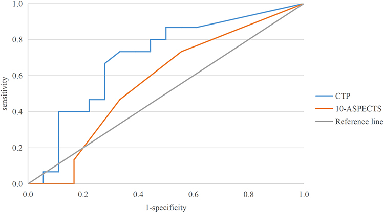 |
Figure 3 ROC Curves for CTP and ASPECTS in Predicting 90-Day Outcomes in Patients Presenting 6 to 24 Hours After Onset. |
Correlation Between ASPECTS Score and Core Infarct Volume
A Spearman correlation analysis was performed to assess the relationship between ASPECTS scores and CTP-derived core infarct volumes in the entire cohort. A significant inverse correlation was observed (r = –0.61, p < 0.001), indicating that lower ASPECTS scores were associated with larger infarct cores.
When stratified by onset-to-treatment time, the negative correlation remained statistically significant in both subgroups. For patients imaged within 6 hours of symptom onset, the correlation coefficient was –0.67 (p < 0.001), while in the 6–24 hour group, the coefficient was –0.47 (p < 0.05), suggesting a consistent inverse relationship between these two imaging markers irrespective of time window.
Discussion
This study demonstrates that both ASPECTS and CTP-derived core infarct volume are significantly associated with functional outcomes in patients with acute ischemic stroke undergoing endovascular therapy. The two imaging modalities showed comparable prognostic performance, with a moderate inverse correlation between them. These findings suggest that ASPECTS and CTP provide complementary information for outcome prediction in clinical practice.
Our findings are more consistent with the view that ASPECTS and CTP-derived core infarct volume provide comparable but complementary prognostic information in acute ischemic stroke patients undergoing endovascular therapy, rather than indicating a clear superiority of one modality over the other. In our cohort, both imaging markers were associated with 90-day outcome, whereas CTP showed only a numerically higher discriminative performance than ASPECTS, without a statistically significant difference. This interpretation is supported by prior studies showing only a moderate correlation between ASPECTS and CTP-defined core volume, with substantial variability of core volume within the same ASPECTS strata, suggesting that the two methods are related but not interchangeable.15 Haussen et al reported that ASPECTS and automated CTP core volume were independently and comparably associated with good outcome after thrombectomy, despite considerable heterogeneity between the two measures.17 Likewise, Nannoni et al demonstrated a moderate inverse correlation between ASPECTS and CTP core volume and suggested that ASPECTS may serve as a surrogate marker of infarct core in selected late-presenting patients, particularly those with large-vessel occlusion.15
At the same time, some studies suggest that CTP may offer additional value in specific scenarios, especially when defining large-core infarction. For example, Sui et al showed that a CTP-defined large core (≥70 mL) was more strongly associated with poor outcome than an ASPECTS-defined large core (ASPECTS ≤5), particularly within 6 hours after onset.18 However, this advantage appears to depend on the clinical context and threshold definition rather than reflecting uniform superiority across all patient groups. By contrast, McDonough et al found that, among patients with low ASPECTS, a model combining ASPECTS and collateral status outperformed a CTP-based model for predicting favorable outcome, underscoring the continuing clinical value of simpler imaging approaches.19 In addition, Chu et al reported that ASPECTS across NCCT, CTA-SI, and CTP-CBV remained associated with final infarction, although CTP-CBV-based ASPECTS showed somewhat better reliability for delineating early ischemic changes.20 Our findings are in line with this literature: although CTP provides quantitative characterization of infarct core, ASPECTS remains clinically important because of its speed, accessibility, and retained prognostic significance in multivariable analysis. This may also explain why ASPECTS remained an independent predictor in our model, whereas CTP core volume did not, possibly because of collinearity between the two imaging variables and the limited sample size.
Multiple studies have further addressed the performance variability of automated imaging platforms. For example, Mallon et al14 compared the outputs of Brainomix and RAPID software and found discrepancies in infarct volume estimations and treatment eligibility, underscoring the need for careful interpretation of automated outputs. Likewise, Muehlen et al21 cautioned that CTP summary maps may underperform in patients with poor revascularization, where large infarcts are present. Notably, the correlation between ASPECTS and CTP core volume in our study (r = –0.61) is comparable to previous reports. Nannoni et al15 demonstrated a similar moderate negative correlation (ρ ≈ –0.49), with stronger associations in patients presenting later or with large vessel occlusion. This supports our subgroup analyses showing time-window–independent correlation. Conversely, Siegler et al5 reported instances where ASPECTS outperformed perfusion imaging in detecting irreversible damage in late-presenting patients, calling for careful clinical contextualization of imaging metrics. Additionally, our analysis confirmed that the negative correlation between ASPECTS and CTP core volume persisted across both early (<6 hours) and extended (6–24 hours) time windows, reinforcing the reliability of this relationship across different clinical scenarios. These findings align with previous work that emphasized the clinical utility of ASPECTS in both early and late presentations.7 Nevertheless, the limited sensitivity of NCCT in detecting early ischemic changes has been a recurring concern in the literature,15 and many authors recommend complementing ASPECTS evaluation with perfusion imaging whenever feasible.14
This study suggests that both CTP-derived core infarct volume and NCCT-based ASPECTS are clinically informative for prognostic assessment in AIS patients undergoing endovascular treatment, with overall comparable predictive performance in our cohort. Although CTP provides quantitative estimation of infarct core, ASPECTS remains an important and practical tool because of its simplicity, rapid availability, and broad accessibility, particularly in emergency settings and resource-limited hospitals. The significant inverse correlation between ASPECTS and CTP-defined core infarct volume (r = –0.61, p < 0.001), which was observed in both the <6-hour and 6–24-hour subgroups, indicates that despite their methodological differences—ASPECTS being a semi-quantitative regional score and CTP providing voxel-based volumetric measurement—both approaches reflect the extent of ischemic injury and may offer complementary information in clinical decision-making. In practice, ASPECTS may serve as a rapid first-line imaging marker to facilitate timely treatment decisions, whereas CTP may provide additional value by more precisely quantifying infarct core when available. This interpretation is consistent with our finding that ASPECTS remained an independent predictor in multivariable analysis, while the effect of CTP core volume may have been attenuated by collinearity or limited sample size. In addition, older age was associated with poorer outcome, underscoring the importance of integrating imaging findings with key clinical variables when evaluating prognosis. Overall, these findings support a pragmatic imaging strategy in which ASPECTS and CTP are used in a complementary manner according to local resources, workflow efficiency, and clinical context.
This study has several limitations that should be acknowledged. First, it employed a retrospective, single-center design with a relatively small sample size, which may limit the generalizability of the findings, reduce statistical power in certain analyses—particularly subgroup analyses—and increase the risk of selection bias. Because the study was based on retrospectively collected clinical data, no formal prospective sample size or power calculation was performed. Larger, multicenter prospective studies are needed to validate these results and enhance their external applicability. Second, the imaging assessment focused primarily on ASPECTS scores and CTP-derived core infarct volumes, without incorporating other important parameters such as collateral circulation status, mismatch between perfusion and diffusion, or degree of reperfusion post-intervention. In addition, ASPECTS scores and CTP-derived core infarct volume both reflect the extent of ischemic injury and may therefore exhibit some degree of collinearity when included simultaneously in multivariable models. These additional imaging markers may provide a more comprehensive understanding of the pathophysiology and treatment response in AIS. Future studies should consider integrated evaluations involving multimodal imaging, larger cohorts, and standardized software platforms to improve the accuracy, reliability, and clinical applicability of imaging-based prognostic tools in acute stroke care.
Conclusions
Both ASPECTS and CTP-derived core infarct volume predict functional outcomes in AIS patients undergoing endovascular therapy, with comparable performance. ASPECTS offers a rapid assessment, whereas CTP provides quantitative evaluation. The two modalities may serve complementary roles in clinical decision-making.
Data Sharing Statement
The datasets used and/or analyzed during the current study are available from the corresponding author Tao Qiu upon reasonable request.
Ethical Approval
This study was approved by the Ethics Committee of the First People’s Hospital of Zigong [(M)2025-036] and was conducted in accordance with the principles of the Declaration of Helsinki. Written informed consent was obtained from all participants.
Author Contributions
All authors made a significant contribution to the work reported, whether that is in the conception, study design, execution, acquisition of data, analysis and interpretation, or in all these areas; took part in drafting, revising or critically reviewing the article; gave final approval of the version to be published; have agreed on the journal to which the article has been submitted; and agree to be accountable for all aspects of the work.
Disclosure
All authors declare that they have no conflict of interest.
References
1. Walter K. What is acute ischemic stroke? JAMA. 2022;327(9):885. doi:10.1001/jama.2022.1420
2. Ma QF, Li R, Wang LJ, et al. Temporal trend and attributable risk factors of stroke burden in China, 1990—2019: an analysis for the global burden of disease study 2019. Lancet Public Health. 2021;6(12):e897–10. doi:10.1016/S2468-2667(21)00228-0
3. Goyal M, Menon BK, van Zwam WH, et al. Endovascular thrombectomy after large-vessel ischaemic stroke: a meta-analysis of individual patient data from five randomised trials. Lancet. 2016;387(10029):1723–1731. doi:10.1016/S0140-6736(16)00163-X
4. Hacke W, Kaste M, Bluhmki E, et al. Thrombolysis with alteplase 3 to 4.5 hours after acute ischemic stroke. N Engl J Med. 2008;359(13):1317–1329. doi:10.1056/NEJMoa0804656
5. Lee NT, Savvidou I, Selan C, et al. Development of endothelial-targeted CD39 as a therapy for ischemic stroke. J Thromb Haemost. 2024;22(8):2331–2344. doi:10.1016/j.jtha.2024.04.023
6. Ting FNL, Shen N. A deep learning and radiomics based Alberta stroke program early CT score method on CTA to evaluate acute ischemic stroke. J Xray Sci Technol. 2023.
7. Costalat V, Lapergue B, Albucher JF, et al. Evaluation of acute mechanical revascularization in large stroke (ASPECTS ≤5). Int J Stroke. 2024;19(1):114–119. doi:10.1177/17474930231191033
8. Bobe RC, Coroiu RE, Cirstian AE, et al. The role of CT perfusion in the evaluation and management of acute ischemic stroke-a systematic review. Life. 2025;15(11):1693. doi:10.3390/life15111693
9. Nogueira RG, Jadhav AP, Haussen DC, et al. Thrombectomy 6 to 24 hours after stroke with a mismatch between deficit and infarct. N Engl J Med. 2018;378(1):11–21. doi:10.1056/NEJMoa1706442
10. Albers GW, Marks MP, Kemp S, et al. Thrombectomy for stroke at 6 to 16 hours with selection by perfusion imaging. N Engl J Med. 2018;378(8):708–718. doi:10.1056/NEJMoa1713973
11. Bivard F, Gollop P, Campbell BCV, et al. Association of noncontrast computed tomography and perfusion modalities with outcomes in patients undergoing late-window stroke thrombectomy. JAMA Network Open. 2022.
12. Huo X, Ma G, Tong X, et al. Trial of endovascular therapy for acute ischemic stroke with large infarct. N Engl J Med. 2023;388(14):1272–1283. doi:10.1056/NEJMoa2213379
13. Shifeng X, Yu S, Shanshan L. Application of computed tomography perfusion imaging-guided mechanical thrombectomy in ischemic stroke patients with large vessel occlusion beyond the therapeutic time window. Curr Med Imaging. 2023.
14. Mallon DH, Kavanagh EC, O’Hare A, et al. Comparison of automated ASPECTS and CTP software outputs for infarct core quantification in stroke triage. J Stroke Cerebrovasc Dis. 2022;31(10):106702. doi:10.1016/j.jstrokecerebrovasdis.2022.106702
15. Nannoni S, de Havenon A, Strambo D, et al. Correlation between ASPECTS and core volume on CT perfusion: impact of time since stroke onset and presence of large-vessel occlusion. AJNR Am J Neuroradiol. 2021;42(3):422–428. doi:10.3174/ajnr.A6959
16. Sui Y, Liu H, Zhang J, et al. Prognostic value of CTP-defined large core versus ASPECTS-defined core in patients with large-vessel occlusion undergoing thrombectomy. Stroke. 2024;55(5):1227–1234. doi:10.1161/STROKEAHA.123.045091
17. Haussen DC, Dehkharghani S, Rangaraju S, et al. Automated CT perfusion ischemic core volume and noncontrast CT ASPECTS (Alberta Stroke Program Early CT Score): correlation and clinical outcome prediction in large vessel stroke. Stroke. 2016;47(9):2318–2322. doi:10.1161/STROKEAHA.116.014117
18. Sui Y, Chen W, Chen C. CTP-defined large core is a better predictor of poor outcome for endovascular treatment than ASPECTS-defined large core. Stroke. 2024;55(5):1227–1234.
19. McDonough R, Elsayed S, Faizy TD, et al. Computed tomography-based triage of extensive baseline infarction: ASPECTS and collaterals versus perfusion imaging for outcome prediction. J Neurointerv Surg. 2021;13(10):869–874. doi:10.1136/neurintsurg-2020-016848
20. Chu Y, Ma G, Xu XQ, et al. Total and regional ASPECT score for non-contrast CT, CT angiography, and CT perfusion: inter-rater agreement and its association with the final infarction in acute ischemic stroke patients. Acta Radiol. 2022;63(8):1093–1101. doi:10.1177/02841851211029080
21. Muehlen I, Borutta M, Siedler G. Prognostic accuracy of CTP summary maps in patients with large vessel occlusive stroke and poor revascularization after mechanical thrombectomy-comparison of three automated perfusion software applications. Tomography. 2022;8(3):1350–1362. doi:10.3390/tomography8030109
 © 2026 The Author(s). This work is published and licensed by Dove Medical Press Limited. The
full terms of this license are available at https://www.dovepress.com/terms
and incorporate the Creative Commons Attribution
- Non Commercial (unported, 4.0) License.
By accessing the work you hereby accept the Terms. Non-commercial uses of the work are permitted
without any further permission from Dove Medical Press Limited, provided the work is properly
attributed. For permission for commercial use of this work, please see paragraphs 4.2 and 5 of our Terms.
© 2026 The Author(s). This work is published and licensed by Dove Medical Press Limited. The
full terms of this license are available at https://www.dovepress.com/terms
and incorporate the Creative Commons Attribution
- Non Commercial (unported, 4.0) License.
By accessing the work you hereby accept the Terms. Non-commercial uses of the work are permitted
without any further permission from Dove Medical Press Limited, provided the work is properly
attributed. For permission for commercial use of this work, please see paragraphs 4.2 and 5 of our Terms.
Recommended articles
Lymphocyte to Monocyte Ratio is Independently Associated with Futile Recanalization in Acute Ischemic Stroke After Endovascular Therapy
Guan J, Wang Q, Zhao Q
Neuropsychiatric Disease and Treatment 2023, 19:2585-2596
Published Date: 28 November 2023