
Back to Journals » Infection and Drug Resistance » Volume 19

Clinical and Epidemiologic Characteristics of Anaerobic Bacteria Originating from Blood Cultures: A Five-Year Retrospective Study
Authors Peng Y, Qin J, Bai L, Bai L, Jiang X, Li H, Liao K, Wang S, Guo P
Received 30 March 2026
Accepted for publication 8 July 2026
Published 14 July 2026 Volume 2026:19 611452
DOI https://doi.org/10.2147/IDR.S611452
Checked for plagiarism Yes
Review by Single anonymous peer review
Peer reviewer comments 3
Editor who approved publication: Dr Hazrat Bilal
Yaqin Peng,1,* Jiamin Qin,1,* Lu Bai,1 Lihong Bai,2 Xiaowen Jiang,1 Huiting Li,1 Kang Liao,1 Shuai Wang,3 Penghao Guo1
1Department of Clinical Laboratory, The First Affiliated Hospital, Sun Yat-sen University, Guangzhou, Guangdong, People’s Republic of China; 2Department of Pulmonary and Critical Care Medicine, The First Affiliated Hospital, Sun Yat-sen University, Guangzhou, Guangdong, People’s Republic of China; 3Institute for Biological Product Control, National Institutes for Food and Drug Control (NIFDC), Beijing, People’s Republic of China
*These authors contributed equally to this work
Correspondence: Shuai Wang, Email [email protected] Penghao Guo, Email [email protected]
Background: With the rise of anaerobic bacteremia and resistance to anaerobes for commonly used agents, the resistance patterns of many anaerobes have changed significantly among different geographic areas and medical facilities. This study investigated the clinical and epidemiologic characteristics of anaerobic bacteremia for therapeutic optimization at the local level.
Methods: This retrospective observational study included all positive blood cultures (BC) from 2020 to 2024. For patients with anaerobic bacteria originating from BCs, clinical and microbiological data were collected.
Results: In total, 239 anaerobic strains were isolated from 223 patients. Among BC-positive bacteria, the proportion of anaerobes accounted for 4.4% (239/5450), with a gradual increase from 3.0% in 2020 to 6.1% in 2024. The most common anaerobes were Bacteroides fragilis (34.7%) and Cutibacterium acnes (20.5%). The resistance rates of gram-negative anaerobes to penicillin, imipenem, and meropenem were 94.4%, 21.1%, and 35.2%, respectively, which were much higher than those of gram-positive anaerobes (16.7%, 0%, 0%). Among the 223 cases, abdominal disorders were the most frequent underlying condition (54.7%). In the empirical antibiotic therapy, 94.6% of the patients received β-lactam antibiotics, and the infection symptoms improved in 74.9% of the patients. For Bacteroides spp. bacteremia, the improvement rate in patients treated with carbapenems and β-lactam/β-lactamase inhibitor combinations was 78.1% and 40.0%, respectively.
Conclusion: The study presents valuable data for monitoring and improving anaerobic bacteremia treatment in Southern of China.
Keywords: anaerobic bacteria, blood culture, epidemiology, antimicrobial susceptibility test, empirical antibiotic therapy
Introduction
Anaerobic bacteremia is a severe but often underestimated condition with a high mortality rate of 15–50%, while the proportion of anaerobes in bacteremia is only 4% on average (range: 0.5–12%).1 Remarkably, the prevalence of anaerobic bacteremia has been increasing, along with the increasing number of complex and invasive procedures, immunosuppressed patients, and patients requiring hospitalization in tertiary-care hospitals.2,3
Anaerobes are abundant in commensal microbiota throughout different body sites.4 The entire spectrum of anaerobic species has been reported as a pathogen causing bacteremia.5 However, most anaerobic infections are still based on empirical therapies such as β-lactam antibiotics, metronidazole, or clindamycin.6–8 There are several reasons. Firstly, it is usually time-consuming to establish the etiological diagnosis because of the long generation time and rigorous growth requirements of these anaerobes, even with the use of Matrix-Assisted Laser Desorption-Ionization Time-of-Flight Mass Spectrometry (MALDI-TOF MS).4,9 Secondly, antibiotic susceptibility testing (AST) for these anaerobes is not routinely performed in most of clinical laboratories, especially in Asian countries.10 In recent years, with the development of techniques such as rapid microbroth dilution panels, the main reason why many laboratories do not perform AST for anaerobic bacteria is cost-related, rather than technical barriers.10 In addition, anaerobic bacteremia is considered predictable based on several predisposing factors such as malignant neoplasms, hematologic disorders, organ transplantation, recent gastrointestinal or obstetric/gynecologic surgery, and diabetes mellitus.1
Of note, inappropriate empirical therapy that lacks anaerobic coverage has been repeatedly linked to poor outcomes.5 However, current knowledge of anaerobic bacteremia is inadequate and fragmented with literature.11,12 Epidemiological patterns vary considerably with geography, patient characteristics, antibiotic policies, and hospital treatment levels.13 Hence, updated local epidemiological data are urgently needed to guide appropriate antimicrobial diagnostic and management strategies.
Literature on anaerobic bacteremia is very limited in China. A recent study from Northwest China reported that anaerobes accounted for 3% of positive blood cultures (BC).12 Owing largely to economic constraints, few laboratories in China routinely perform ASTs for anaerobes, leading to a scarcity of susceptibility data. As a result, antibiotic selection relies primarily on clinical experience, yet there is almost a complete lack of real-world efficacy data.
In this retrospective observational study, we aimed to investigate carefully the clinical and epidemiological characteristics of anaerobic bacteria originating from BCs at a tertiary hospital over five years. To the best of our knowledge, this is the first study on anaerobic bacteremia in Southern China, which would provide a rational basis for optimizing empirical antibiotic therapy in this local setting.
Materials and Methods
Study Population
In this retrospective observational study, BC-positive bacteria identified in our bacteriology laboratory were included between January 2020 and December 2024 from the First Affiliated Hospital of Sun Yat-sen University, a 2800-bed teaching hospital in Southern China. If duplicate bacteria were isolated from a patient with multiple positive BCs within 30 days, only the first isolate was retained for analysis. For patients with positive BCs for anaerobes, clinical and laboratory data were collected through the electronic medical record and laboratory information management system. A flow chart of the study population and data analysis is shown in Figure 1.
 |
Figure 1 A flow chart of the study population and data analysis. Abbreviations: G+, gram-positive; G−, gram-negative. |
This study was approved by the institutional review board of the First Affiliated Hospital of Sun Yat-sen University (Ethics Number: [2026]205).
BC Procedure and Species Identification
For each patient with a suspected bloodstream infection, BCs were taken aseptically at the request of the attending physicians prior to the initiation of empirical antibiotic therapy whenever possible. At least two sets of BCs were collected from two separate venipuncture sites of each patient. Each set consisted of two bottles (aerobic and anaerobic). Inoculated bottles were immediately transported to the clinical microbiology laboratory at ambient temperature and were loaded into the BacT/ALERT® VIRTUO system (bioMérieux, France) within two hours of collection. Each bottle containing 8–10 mL of blood was incubated at 37 °C for a maximum of six days.
Once the system showed a positive signal, gram staining and sub-culturing were performed according to routine clinical practice.14 If an anaerobic bottle was positive, blood samples were transferred to two blood agar plates (Autobio, China). One plate was incubated under aerobic conditions (5% CO2) and the other in a GENbag anaer (bioMérieux, France). After incubation at 35°C for a maximum of 5 days, the pathogens were identified using the VITEK® MS system in IVD mode following the manufacturer’s instructions (bioMérieux, France). Briefly, a single colony isolated from subcultures was thinly smeared onto a MALDI-TOF target plate, and then overlaid with 1 μL CHCA matrix. After the matrix dried at room temperature, the mass spectra were acquired and automatically analyzed by the IVD knowledge base V3.2. A (log)score value of ≥ 2.0 was considered acceptable; otherwise, species-level identification was confirmed by PCR sequencing (Sangon Biotech, China) as described previously.9
ASTs
The AST data of anaerobic strains were collected through laboratory information management system, excluding those missing one or more antibiotics.
AST was performed for three β-lactam antibiotics: penicillin, imipenem, and meropenem, using the E-test gradient diffusion method according to the manufacturer’s instructions. Bacterial colonies were picked up and suspended in 0.45% NaCl, with turbidity adjusted to 0.5 McFarland. Then the suspension was uniformly streaked onto Mueller Hinton blood agar plates (Autobio, China). After application of E-test strips (Autobio, China), plates were incubated under anaerobic conditions at 35 °C until visible growth was observed. AST results were interpreted in accordance with the guidelines provided by the Clinical and Laboratory Standards Institute (CLSI M100 ED35:2025).15
The strains of Bacteroides fragilis (ATCC 25285) and Bacteroides thetaiotaomicron (ATCC 29741) were used as quality controls weekly. Due to laboratory constraints, AST was not performed for other anti-anaerobic agents (eg, metronidazole, clindamycin) in routine work.
Antibiotic Treatment Response
A variety of medical interventions were immediately administered to all the patients with suspected bloodstream infections, which included empirical antibiotic therapy, adequate supportive care, and source control (eg, drainage of abscesses, surgical debridement), if necessary. The antibiotic treatment response of each patient was evaluated by two independent trained physicians according to the medical records.
This study focused primarily on empirical antibiotic selection, considering that it was hard to control for all confounding variables that might influence clinical outcomes. Therefore, the data regarding the association between empirical antibiotic regimens and clinical improvement should be interpreted as descriptive observations rather than definitive evidence of differential antibiotic efficacy.
Definitions
Bacteremia was classified as community-acquired if blood cultures were obtained within 48 hours of hospital admission or during an outpatient visit.7 A positive BC was considered mono-microbial if a single bacterium was isolated, and poly-microbial if ≥ 2 germs were isolated within 7 days of the first positive BCs.7
Based on a review of the patient’s medical history, an episode was considered as a true bloodstream infection if it met any of the following criteria: (1) anaerobic bacteremia was confirmed by the treating physicians in the medical records; (2) the same anaerobic species was isolated from ≥ 2 BC sets drawn within 48 hours; (3) a single positive BC for gram-negative (G−) anaerobe; (4) for gram-positive (G+) anaerobe isolated from one BC, the patient had one or more of systemic infection symptoms (eg, fever > 38.0 °C or hypothermia < 36.0 °C, chills, hypotension) and no other obvious source of infection was identified; for Cutibacterium acnes as a common skin commensal, additional criteria was required:16,17 foreign intravascular device present > 48 hours prior to blood sample.
The empirical antibiotic therapy was defined as the first antibiotic regimen administered after blood collection but before the availability of gram stain or species identification results. The clinical improvement was considered if it met any of the following criteria: (1) the medical records clearly documented that the patient responded favorably to the empirical antibiotic regimen; (2) within 72 hours of empirical antibiotic therapy, the patient’s infection-related symptoms improved, and the following infection parameters returned to normal levels or showed a progressively decreasing trend: temperature, white blood cell count, procalcitonin, and C-reactive protein. If clinical improvement occurred only after antibiotic adjustment guided by BC results, the case was not classified as a success of empirical antibiotic therapy.
Statistics
Statistical analysis was performed with SAS 9.4. The statistical analysis adopted complete-case analysis, which automatically excluded participants with any missing values. In this retrospective observational study, the data was missed completely at random with 8.2% of covariate missingness. Given missingness < 10%, the complete-case analysis was statistically defensible for this single-center hospital clinical observational cohort.18
Categorical variables were reported as frequencies and percentages and analyzed using the Chi-square test. Continuous variables were expressed as the mean and SD or median and interquartile range (IQR) where appropriate. The t test was used to analyze normally distributed continuous variables, whereas the Wilcoxon Rank-Sum test was used to analyze nonnormally distributed continuous variables. To assess the independent factors, the comparison between G+ and G− anaerobes was further performed by multivariable logistic regression analysis, with adjustment for potential confounders. Statistical significance was set at P < 0.05.
Results
Anaerobic Strains Isolated from Positive BCs
A total of 5450 bacterial strains were isolated from positive BCs over a period of five years in our tertiary hospital, among which 239 (4.4%) detected the presence of anaerobic bacteria. Among BC-positive bacteria, the proportion of anaerobic strains increased from 3.0% (27/901) in 2020 to 6.1% (69/1126) in 2024 (Figure 2).
 |
Figure 2 The annual proportion of anaerobic strains among blood culture-positive bacteria between 2020 and 2024. |
Of 239 anaerobic bacteria, 19 genera and 40 species were identified. As shown in Table 1, G− rods (53.1%; 127/239) were the predominant isolates, most of which were Bacteroides spp. (44.4%; 106/239). There were 45.2% (108/239) of G+ anaerobes. Except Clostridium spp. (7.9%, 19/239) and Robinsonella peoriensis (0.4%, 1/239), the remaining strains were non-spore-forming. The five top species were B. fragilis (34.7%, 83/239), C. acnes (20.5%, 49/239), Eggerthella lenta (8.8%, 21/239), B. thetaiotaomicron (6.7%, 16/239), and Parvimonas micra (3.8%, 9/239).
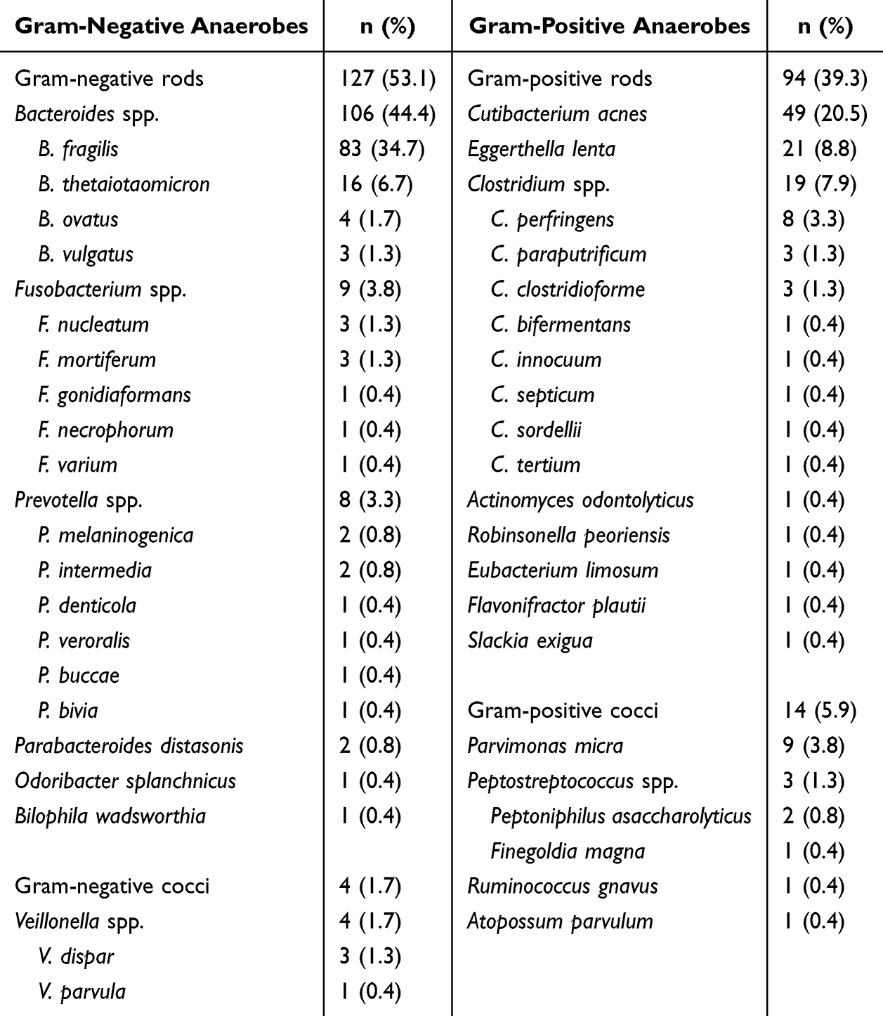 |
Table 1 The Distribution of Anaerobes from Blood Cultures in Our Tertiary Hospital from 2020 to 2024 |
Figure 3 presents the resistance rates of anaerobic strains to the three main β-lactams, showing a significant difference between G+ and G− anaerobes. Compared with G+ anaerobes (16.7%), the resistance rate of G− anaerobic bacteria to penicillin reached 94.4%, mostly because of the intrinsic resistance of Bacteroides spp. The resistance rates of G− anaerobes to imipenem and meropenem were 21.1% and 35.2%, respectively. None of the G+ strains was resistant to imipenem or meropenem.
 |
Figure 3 The resistance rate of anaerobic strains to three mainly β-lactams including penicillin, imipenem and meropenem. |
Clinical Characteristics and Strain Distribution Among Patients
A total of 239 anaerobic strains were isolated from 223 patients. The median age of these patients was 55 years (IQR: 44 −67), and 61.9% (138/223) were males. In total, 35.4% (79/223) of patients were admitted to the ICU.
Among the 223 cases, abdominal disorders were the most frequent underlying conditions (54.7%, 122/223), followed by urogenital (18.8%, 42/223), hematological (8.1%, 18/223), cardiovascular (4.9%, 11/223), and respiratory (4.0%, 9/223) disorders (Figure 4a). Additionally, 51.6% (115/223) of patients had malignant tumors, with abdominal cancers accounting for 61.7% (71/115) (Figure 4b).
 |
Figure 4 The clinical characteristics and strain distributions among the 223 patients with blood culture-positive anaerobes. (a) the distribution of underlying conditions among the 223 patients. (b) the distribution of tumor types among the patients with cancers; abdominal cancers included stomach, colorectal, liver, pancreatic, gallbladder, and esophageal cancers. (c) the distribution of anaerobic strains among patients with mainly underlying conditions respectively. |
Figure 4c presents the distribution of anaerobic strains isolated from patients with the three top underlying conditions. Among the cases with abdominal and urogenital disorders, Bacteroides spp. were the most prevalent (both > 50.0%). In contrast, the proportion of C. acnes accounted for 61.1% (11/18) among cases with hematological conditions.
Comparison Between Patients with Mono-Microbial BCs Involving G+ and G− Anaerobes
Among 223 cases, 76.7% (171/223) were mono-microbial BCs. Table 2 shows the main characteristics of 171 patients with mono-microbial BCs.
 |
Table 2 Patient Characteristics and Laboratory Data Among Patients with Mono-Microbial Blood Cultures (BC) |
The presence of neutrophilic deficiency was more frequently observed in patients due to G+ versus G− anaerobes (11.3% vs 1.1%, P = 0.005), whereas the receipt of surgery within the past 30 days (P = 0.005) and recent use of broad-spectrum antibiotics (P = 0.014) were more frequently associated with patients due to G− versus G+ anaerobes. A significant association with G+ anaerobes versus G− anaerobes was observed for male (P = 0.008) and ICU (P = 0.026). In terms of infection-related indicators, no significant differences were observed between patients with G− and G+ anaerobes, except for temperature (P = 0.002) (Table 2). After adjusting for sex, ICU, temperature, neutropenia, recent surgery and antibiotic exposure, ICU remained independently associated with the G+ (vs G−) group (OR: 3.35, 95% CI: 1.38–8.13, P = 0.0074).
Table S1 presents the comparison between the two top species (ie, B. fragilis and C. acnes). Multivariable logistic regression analysis showed that ICU (OR: 6.01, 95% CI: 1.57–23.02, P = 0.0088) and C-reactive protein (OR: 0.99, 95% CI: 0.98–1.000, P = 0.0078) were two independent factors between the two groups.
Empirical Antibiotic Therapy and Antibiotic Treatment Response
In the empirical antibiotic therapy, 94.6% (211/223) of the patients received β-lactam antibiotics, among which 61.9% (138/223) were treated with carbapenems. Only 14 and one case received nitroimidazoles and clindamycin, respectively, all of which were in combination with β-lactams. Infection symptoms improved in 74.9% (167/223) of the patients after empirical antibiotic therapy.
The empirical antibiotic therapy and clinical response of the 171 patients with mono-microbial BCs are presented in Figure 5. The improvement rate of these 171 patients was 74.9% (128/171) after empirical antibiotic therapy. For Bacteroides spp. bacteremia, the improvement rate in patients treated with carbapenems and β-lactam/β-lactamase inhibitor combinations (eg, piperacillin/tazobactam, cefoperazone/sulbactam, ampicillin/sulbactam, and ceftazidime/avibactam) was 78.1% and 40.0%, respectively. In addition, for patients without improvement after the empirical antibiotic therapy, 44.2% (19/43) received correspondingly adjusted antibiotic treatments according to the identification results by MALDI-TOF MS, 89.5% (17/19) of which improved.
 |
Figure 5 The antibiotic treatment response of patients with mono-microbial blood cultures through empirical antibiotic therapy. Carbapenem included imipenem and meropenem. Abbreviations: G+/G−, gram-positive/negative; TZP, piperacillin/tazobactam; SCF, cefoperazone/sulbactam; SAM, ampicillin/sulbactam; CZA, ceftazidime/avibactam; VA, vancomycin; LZD, linezolid; TCP, Teicoplanin. |
Discussion
In clinical practice, empirical antibiotic therapy has traditionally been considered safe for anaerobic bacteremia because of its rather low incidence, predictability of infection risk and antimicrobial susceptibility of anaerobes.1,5 However, this traditional pattern has faced increasing challenges over the past decade.
First, the incidence of anaerobic bacteremia has been reported with an increasing trend in multiple studies.2,5 Similarly, our study also demonstrated a year-over-year rise in the detection rate of anaerobes originating from BCs over the past five years (Figure 2). Malignant tumors are considered as a primary risk factor for anaerobic bacteremia.13,19 Studies have shown that the incidence of malignant tumors, especially gastrointestinal cancers, has increased rapidly in recent years.20,21 In this study, malignant tumors accounted for 51.6% (115/223) of the cases, most of which were abdominal cancers (61.7%, 71/115), suggesting that anaerobic coverage should be included in the empirical treatment when suspected bloodstream infections in these patients.
The wide application of MALDI-TOF MS in clinical microbiology laboratories over the past decade has made the identification of anaerobes faster, simpler, and more cost-effective, benefiting for accurate microbiological diagnostics and local epidemiological assessment. Similar to other findings, Bacteroides spp. (44.4%) was the most frequently isolated genus, most of which originated from patients with abdominal and urogenital comorbidities.2,7,13 The distribution of the different species among Bacteroides spp. was in line with that previously published: B. fragilis was the most frequent, followed by B. thetaiotaomicron, B. ovatus and B. vulgatus.12 Remarkably, the detection rate of Clostridium spp. (7.9%) was slightly lower than that of Eggerthella lenta (8.8%), which was different from most of studies.2,5,13 A similar phenomenon was observed among patients with abdominal disorders (Figure 4c). The reason of this phenomenon is unclear, which might be associated with a variety of factors such as patient populations, antibiotic practices, and disease spectra. Another interesting phenomenon was observed that compared with G− anaerobes, G+ anaerobes were more frequently isolated from ICU patients (OR: 3.35, 95% CI: 1.38–8.13, P = 0.0074). Further studies are needed to explore the underlying factors.
It is noteworthy that a re-evaluation of clinical significance may be warranted for anaerobes originating from single-set BCs in immunocompromised patients, especially for skin commensals that are most often considered as BC contaminants. In this study, C. acnes, often considered as a contaminant when present in BCs, was the second most common blood isolate (20.5%; 49/239). To the best of our knowledge, this has been directly overlooked and has not been reported in many clinical laboratories limited by cognitive reasons. Boman et al proposed that definitions of Cutibacterium bacteremia with a demand that more than one BC must be positive might miss true infections.16 According to the definition of true infection described in previous studies,16,17 the proportion of cases represented bloodstream infections here reached up to 22.4% (11/49) through microbiological and medical records studied retrospectively. However, only one case of true bacteremia was diagnosed by clinicians. In addition, C. acnes was present in a surprisingly high proportion (61.1%, 11/18) in patients with hematologic disorders (Figure 4c). To our knowledge, this phenomenon has not been previously reported in China. We propose that bloodstream infections caused by C. acnes may be significantly underdiagnosed in immunocompromised patients. More data is required to explore this phenomenon.
In recent years, AST data from the United States and Europe have demonstrated a clear increase in the resistance to anaerobes for several frequently used agents (eg, beta-lactams, clindamycin, and metronidazole).5,22,23 AST of anaerobic bacteria is considered critically important; however, it has not been routinely performed in most clinical laboratories of Asian countries limited by technical and financial limitations.4,10 For example, at our hospital, only three β-lactams were tested for antimicrobial susceptibility to anaerobes, mostly due to financial reasons, showing a significant difference between G+ and G− anaerobes (Figure 3). An extremely high resistance rate to imipenem (21.1%) and meropenem (35.2%) was observed respectively among G− anaerobes in this study, which was far exceeded those presented in other studies.8,11 It suggests that the high resistance rate of G− anaerobes to carbapenem should warrant close attention in this region. However, the E-test here, as an alternative method, is not the CLSI reference method (agar dilution).15 Although laboratory quality control was performed weekly, this result should be interpreted with caution due to the lack of external quality assessment for anaerobic susceptibility testing in China. More data are needed to further support this observation.
It is necessary to investigate the effectiveness of empirical antibiotic therapy based on antibiotic policies and treatment levels of local healthcare institutions. In this study, most patients (94.6%) received β-lactams, whereas the use of nitroimidazoles or clindamycin was very low (6.7%) all combined with β-lactams. Some differences were observed in the anti-infection improvement rates among patients isolated from different genera during the initial empirical treatments (Figure 5). Surprisingly, for Bacteroides spp. bacteremia, the infection symptoms improved in only 40.0% of the patients treated with β-lactam/β-lactamase inhibitor combinations, suggesting high resistance rates to these β-lactams in this genus. It should be noted that this data reflected the overall clinical response and might be attributable to a combination of empirical antibiotic therapy and other concurrent medical interventions. Future prospective studies incorporating comprehensive data on all treatment modalities are needed to evaluate the true determinants of clinical outcomes in anaerobic bacteremia.
Several other limitations nevertheless deserve mention. In most clinical assessments, anaerobic bacteria (especially G+ strains) are considered contaminants if they are obtained from a single BC.8 Here also faced this issue. Due to incomplete medical records for some outpatient cases in this retrospective study, the proportion of true bacteremia was not assessed, which might have been largely underestimated in clinical practice. Some patients with G+ anaerobes isolated from single BC bottles had more than two bacterial species detected, making it difficult to accurately determine the clinical significance of these anaerobes and the efficacy of empirical therapy against them. Hence, an analysis of these poly-microbial cases in separate were not performed. Additionally, limited by our laboratory conditions, the anaerobic strains were not collected for AST. In the future, we will pay more attention to the clinical judgement of true anaerobic bacteremia and the resistance of anaerobes.
In summary, our study tried to present the epidemiology and clinical characteristics of anaerobe originating from BCs over the past five years. It provides valuable data for monitoring and improving anaerobic bacteremia treatment in Southern of China.
Abbreviations
MALDI-TOF MS, Matrix-Assisted Laser Desorption-Ionization Time-of-Flight Mass Spectrometry; AST, antimicrobial susceptibility testing; BC, blood culture; G+, gram-positive; G−, gram-negative; ICU, Intensive Care Unit; TZP, piperacillin/tazobactam; SCF, cefoperazone/sulbactam; SAM, ampicillin/sulbactam; CZA, ceftazidime/avibactam; VA, vancomycin; LZD, linezolid; TCP, Teicoplanin.
Ethical Statement
This study was performed in accordance with the principles of the Declaration of Helsinki. Patient confidentiality was strictly maintained throughout this study: all data were anonymized prior to analysis, and no identifying patient information is included in this paper. Approval was granted by the Ethics Committee of the First Affiliated Hospital of Sun Yat-sen University (Ethics Number: [2026]205). Informed consent was not sought due to its retrospective study, and an informed consent waiver was approved by the institutional ethical committee of the First Affiliated Hospital of Sun Yat-sen University.
Funding
There is no funding to report.
Disclosure
All authors declare no competing interests in this work.
References
1. Brook I. The role of anaerobic bacteria in bacteremia. Anaerobe. 2010;16(3):183–11. doi:10.1016/j.anaerobe.2009.12.001
2. Mortensen KK, Nielsen HL, Sogaard KK. Clinical and microbiological characteristics of anaerobic bacteremia during 1994-2019: a Danish population-based cohort study. Anaerobe. 2024;89:102898. doi:10.1016/j.anaerobe.2024.102898
3. Nagaoka K, Iwanaga N, Takegoshi Y, et al. Mortality risk factors and fulminant sub-phenotype in anaerobic bacteremia: a 10-year retrospective, multicenter, observational cohort study. Eur J Clin Microbiol Infect Dis. 2024;43:459–467. doi:10.1007/s10096-023-04743-1
4. Reissier S, Penven M, Guérin FO, et al. Recent trends in antimicrobial resistance among anaerobic clinical isolates. Microorganisms. 2023;11:1474. doi:10.3390/microorganisms11061474
5. Gajdacs M, Urban E. Relevance of anaerobic bacteremia in adult patients: a never-ending story? Eur J Microbiol Immunol. 2020;10(2):64–75. doi:10.1556/1886.2020.00009
6. Di Bella S, Antonello RM, Sanson G, et al. Anaerobic bloodstream infections in Italy (ITANAEROBY): a 5-year retrospective nationwide survey. Anaerobe. 2022;75:102583. doi:10.1016/j.anaerobe.2022.102583
7. Zouggari Y, Lelubre C, Lali SE, Cherifi S. Epidemiology and outcome of anaerobic bacteremia in a tertiary hospital. Eur J Intern Med. 2022;105:63–68. doi:10.1016/j.ejim.2022.08.024
8. Ligero-Lopez J, Rubio-Mora E, Ruiz-Bastian MD, Quiles-Melero MI, Cacho-Calvo J, Cendejas-Bueno E. Antimicrobial susceptibility testing of anaerobic bacteria causing bacteremia: a 13-year (2010-2022) retrospective study in a tertiary hospital. Anaerobe. 2023;84:102803. doi:10.1016/j.anaerobe.2023.102803
9. D’Inzeo T, Fiori B, Liotti FM, et al. Direct matrix-assisted laser desorption ionization-time of flight mass spectrometry testing from positive blood cultures for rapid identification of bloodstream infection-causing anaerobic bacteria. J Clin Microbiol. 2021;59(7):e0052121. doi:10.1128/JCM.00521-21
10. Sood A, Ray P, Angrup A. Antimicrobial susceptibility testing of anaerobic bacteria: in routine and research. Anaerobe. 2022;75:102559. doi:10.1016/j.anaerobe.2022.102559
11. Boattini M, Bianco G, Bastos P, et al. Diagnostic and epidemiological landscape of anaerobic bacteria in Europe, 2020-2023 (ANAEuROBE). Int J Antimicrob Agents. 2025;65:107478. doi:10.1016/j.ijantimicag.2025.107478
12. Yang R, Wang W, Yin G, et al. An eleven-year retrospective survey of anaerobic bloodstream infection in adults in a general hospital. Infect Drug Resistance. 2024;17:4521–4530. doi:10.2147/IDR.S482698
13. Dumont Y, Bonzon L, Michon AL, et al. Epidemiology and microbiological features of anaerobic bacteremia in two French University hospitals. Anaerobe. 2020;64:102207. doi:10.1016/j.anaerobe.2020.102207
14. Peng Y, Xie R, Luo Y, et al. Clinical evaluation of a multiplex droplet digital PCR for diagnosing suspected bloodstream infections: a prospective study. Front Cell Infect Microbiol. 2024;14:1489792. doi:10.3389/fcimb.2024.1489792
15. CLSI. Performance Standards for Antimicrobial Susceptibility Testing.
16. Boman J, Nilson B, Sunnerhagen T, Rasmussen M. True infection or contamination in patients with positive Cutibacterium blood cultures-a retrospective cohort study. Eur J Clin Microbiol Infect Dis. 2022;41(7):1029–1037. doi:10.1007/s10096-022-04458-9
17. Rasmussen M, Mohlin AW, Nilson B. From contamination to infective endocarditis-a population-based retrospective study of Corynebacterium isolated from blood cultures. Eur J Clin Microbiol Infect Dis. 2020;39(1):113–119. doi:10.1007/s10096-019-03698-6
18. Ross RK, Breskin A, Westreich D. When is a complete-case approach to missing data valid? The importance of effect-measure modification. AM J Epidemiol. 2020;189(12):1583–1589. doi:10.1093/aje/kwaa124
19. Justesen US, Ellebaek MB, Qvist N, et al. Colorectal cancer and association with anaerobic bacteraemia: a Danish nationwide population-based cohort study. J Infect. 2024;89(2):106212. doi:10.1016/j.jinf.2024.106212
20. Li Q, Xia C, Li H, et al. Disparities in 36 cancers across 185 countries: secondary analysis of global cancer statistics. Front Med. 2024;18(5):911–920. doi:10.1007/s11684-024-1058-6
21. Cao W, Chen HD, Yu YW, Li N, Chen WQ. Changing profiles of cancer burden worldwide and in China: a secondary analysis of the global cancer statistics 2020. Chin Med J. 2021;134(7):783–791. doi:10.1097/CM9.0000000000001474
22. Brook I. Overview of anaerobic infections in children and their treatment. J Infect Chemother. 2024;30(11):1104–1113. doi:10.1016/j.jiac.2024.07.014
23. Buhl M, Sunnerhagen T, Join-Lambert O, et al. Antimicrobial resistance surveillance of Bacteroides fragilis isolated from blood cultures, Europe, 2022 (ReSuBacfrag). Int J Antimicrob. 2024;64(3):107241. doi:10.1016/j.ijantimicag.2024.107241
 © 2026 The Author(s). This work is published and licensed by Dove Medical Press Limited. The
full terms of this license are available at https://www.dovepress.com/terms
and incorporate the Creative Commons Attribution
- Non Commercial (unported, 4.0) License.
By accessing the work you hereby accept the Terms. Non-commercial uses of the work are permitted
without any further permission from Dove Medical Press Limited, provided the work is properly
attributed. For permission for commercial use of this work, please see paragraphs 4.2 and 5 of our Terms.
© 2026 The Author(s). This work is published and licensed by Dove Medical Press Limited. The
full terms of this license are available at https://www.dovepress.com/terms
and incorporate the Creative Commons Attribution
- Non Commercial (unported, 4.0) License.
By accessing the work you hereby accept the Terms. Non-commercial uses of the work are permitted
without any further permission from Dove Medical Press Limited, provided the work is properly
attributed. For permission for commercial use of this work, please see paragraphs 4.2 and 5 of our Terms.