
Back to Journals » International Medical Case Reports Journal » Volume 19
Cardiac Tamponade Secondary to Massive Pericardial Effusion in Severe Primary Hypothyroidism: A Case Report
Authors Okema JN  , Oriba DL , Okot J , Obal R, Pitua I , Odong C , Otika D , Bongomin F , Olum R
Received 20 March 2026
Accepted for publication 17 May 2026
Published 22 May 2026 Volume 2026:19 610746
DOI https://doi.org/10.2147/IMCRJ.S610746
Checked for plagiarism Yes
Review by Single anonymous peer review
Peer reviewer comments 2
Editor who approved publication: Dr Xudong Zhu
James Nelson Okema,1,2 Dan Langoya Oriba,2 Jerom Okot,1 Ronald Obal,2 Ivaan Pitua,3 Christopher Odong,4 Donald Otika,1 Felix Bongomin,1,5,6 Ronald Olum7
1Faculty of Medicine, Gulu University, Gulu, Uganda; 2Department of Internal Medicine, St. Mary’s Hospital Lacor, Gulu, Uganda; 3School of Medicine, College of Health Sciences, Makerere University, Kampala, Uganda; 4Department of Internal Medicine, Mulago National Referral Hospital, Kampala, Uganda; 5Department of Medical Microbiology and Immunology, Gulu University, Gulu, Uganda; 6Department of Internal Medicine, Gulu Regional Referral Hospital, Gulu, Uganda; 7Department of Epidemiology, Johns Hopkins Bloomberg School of Public Health, Baltimore, MD, USA
Correspondence: James Nelson Okema, Department of Internal Medicine, St. Mary’s Hospital Lacor, Gulu, Uganda, Email [email protected]
Background: Pericardial effusion is a recognized manifestation of overt hypothyroidism, but progression to cardiac tamponade is rare. In tuberculosis-endemic settings, diagnostic focus often favors tuberculous pericarditis, risking delayed recognition of reversible endocrine causes.
Case Presentation: A 65-year-old Black African woman from Northern Uganda presented with five months of progressive dyspnea, orthopnea, and generalized edema, followed by acute pleuritic chest pain. She had a history of hypertension and type 2 diabetes mellitus. No prior thyroid disease, tuberculosis, malignancy, or rheumatologic condition was identified. Examination revealed marked sinus bradycardia (42 beats/min), elevated jugular venous pressure, muffled heart sounds, pulsus paradoxus, and Woltman’s sign. The thyroid gland was diffusely enlarged without palpable nodules or cervical lymphadenopathy. Electrocardiography showed low-voltage QRS complexes. Transthoracic echocardiography demonstrated a large circumferential pericardial effusion (maximal posterior separation 3.2 cm) with right ventricular diastolic collapse and Doppler evidence of tamponade physiology, and a mildly reduced left ventricular ejection fraction of approximately 40%, with diffuse hypokinesis and no regional wall motion abnormalities, making ischemic cardiomyopathy less likely. Urgent pericardiocentesis drained 520 mL of clear, straw-colored fluid. Pericardial fluid analysis showed elevated total protein (4.2 g/dL; serum protein 7.0 g/dL; fluid-to-serum ratio 0.6), moderately elevated lactate dehydrogenase (below 1000 U/L), low adenosine deaminase (< 5 U/L), and negative mycobacterial studies, making tuberculous pericarditis unlikely. Thyroid function tests confirmed severe primary hypothyroidism (thyroid-stimulating hormone 88.08 mIU/L; free thyroxine 4.0 pmol/L) with positive anti-thyroid peroxidase antibodies, consistent with autoimmune thyroiditis. Low-dose levothyroxine was initiated with gradual titration. Hemodynamic status improved immediately after pericardiocentesis. Follow-up echocardiography showed complete resolution of the effusion, and the patient achieved biochemical euthyroid within three months.
Conclusion: Severe primary hypothyroidism should be routinely considered in patients with unexplained pericardial effusion, even in tuberculosis-endemic settings. Paradoxical bradycardia in tamponade, Woltman’s sign, low-voltage electrocardiography, and a protein-rich pericardial effusion with low adenosine deaminase are important diagnostic clues. Prompt pericardiocentesis treats hemodynamic compromise, while cautious levothyroxine replacement prevents recurrence.
Keywords: hypothyroidism, cardiac tamponade, pericardial effusion, levothyroxine, Woltman’s sign, Uganda
Key Clinical Message
Severe hypothyroidism can present with large pericardial effusion and even cardiac tamponade, mimicking tuberculous pericarditis in endemic regions. Bradycardia, Woltman’s sign, low-voltage electrocardiography, and protein-rich pericardial fluid with low adenosine deaminase offer crucial diagnostic clues. Prompt pericardiocentesis and cautious thyroid hormone replacement are essential to ensure rapid recovery.
Introduction
Pericardial effusion occurs in 3–37% of patients with overt hypothyroidism, but progression to cardiac tamponade is uncommon1 Hypothyroidism-related pericardial effusions typically accumulate slowly, allowing progressive pericardial stretching and accommodation of large fluid volumes before hemodynamic compromise develops.1,2
In sub-Saharan Africa and other tuberculosis-endemic regions, tuberculous pericarditis remains the leading cause of pericardial effusion, and diagnostic pathways are often oriented toward infectious etiologies.3,4 This orientation can delay recognition of noninfectious, reversible endocrine causes particularly when thyroid function testing is not routinely included in the diagnostic workup. Unawareness and lack of clinical suspicion are among the principal drivers of diagnostic delay.
We report a 65-year-old woman from Northern Uganda with severe primary hypothyroidism presenting with massive pericardial effusion and early cardiac tamponade. This case highlights the diagnostic importance of paradoxical bradycardia in tamponade, the clinical value of delayed relaxation of deep tendon reflexes as a clue to hypothyroidism, and the need for cautious initiation and titration of levothyroxine in older patients with cardiac compromise.1,3,5
Case Presentation
Clinical History
A 65-year-old Black African woman from Northern Uganda presented with five months of progressive exertional dyspnea, bilateral lower limb edema, and facial puffiness. Orthopnea developed two weeks before admission. On the night prior to presentation, she experienced acute pleuritic retrosternal chest pain with worsening breathlessness. She had a five-year history of hypertension managed with amlodipine 10 mg daily and had been diagnosed with type 2 diabetes mellitus eight months prior to presentation, managed with metformin 500 mg twice daily; both conditions had been sub optimally controlled due to financial constraints limiting consistent medication procurement. There was no prior history of thyroid disease, thyroid function testing, tuberculosis treatment, malignancy, rheumatologic disease, thoracic trauma, or cardiac surgery. She reported no history of fever, night sweats, chronic cough, weight loss, or hemoptysis. She did not smoke or consume alcohol.
Physical Examination
On admission, she appeared frail with periorbital puffiness and generalized pitting edema (Grade 3+, with indentation persisting >30 seconds), predominantly affecting the bilateral lower extremities and sacral region. The blood pressure was 220/110 mmHg with appropriate antihypertensive therapy commenced on admission (see Management section). She was markedly bradycardic with a regular heart rate of 42 beats per minute. The respiratory rate was 24 breaths per minute, oxygen saturation was 94% on room air, and axillary temperature was 36.0 °C.
The thyroid gland was diffusely enlarged (consistent with a goiter), firm in consistency, and non-tender, without palpable nodules. There was no cervical lymphadenopathy.
Cardiovascular examination revealed elevated jugular venous pressure (estimated at approximately 6 cm above the sternal angle, corresponding to 8–10 cmH2O), muffled heart sounds, and a pulsus paradoxus of 12 mmHg, consistent with hemodynamically significant pericardial effusion. The liver edge was palpable 2 to 3 cm below the costal margin, suggesting systemic venous congestion.
Neurological examination demonstrated delayed relaxation of the Achilles tendon reflexes bilaterally, consistent with Woltman’s sign, a classic but often underrecognized feature of hypothyroidism. A video of this sign was not available for attachment. Mental status examination revealed mildly slowed mentation with preserved orientation (Glasgow Coma Scale score 14: E4V5M6). Respiratory examination showed reduced breath sounds at both lung bases, consistent with small pleural effusions.
Investigations
ECG and Chest Radiograph
The 12-lead electrocardiogram (Figure 1) showed sinus bradycardia (42 beats/min) with low-voltage QRS complexes in limb and precordial leads, compatible with large pericardial effusion. While low-voltage QRS is also a recognized feature of pericardial effusion alone, its presence in this context alongside the clinical and biochemical profile of hypothyroidism offered an additional supportive clue rather than a pathognomonic finding.
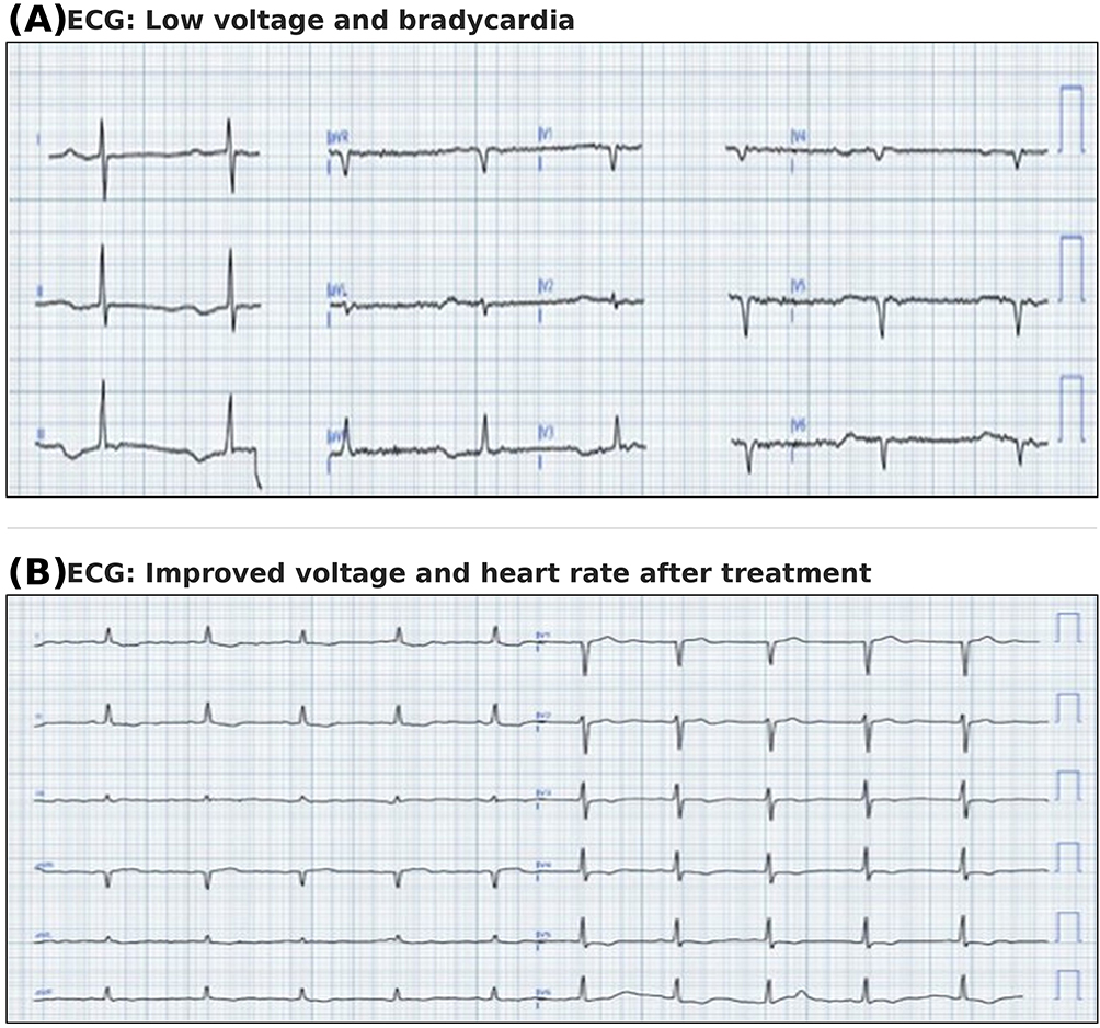 |
Figure 1 (A) Twelve-lead ECG demonstrating low voltage and bradycardia. (B) Twelve-lead ECG showing improvement in QRS voltage and heart rate following treatment. |
Chest radiograph revealed cardiomegaly with a “water bottle” cardiac silhouette and small bilateral pleural effusions (Figure 2).
 |
Figure 2 Serial chest radiographs demonstrating resolution of pericardial effusion. (A) Initial chest radiograph (posteroanterior view) showing a markedly widened cardiac silhouette with a cardiothoracic ratio greater than 0.55, consistent with a massive pericardial effusion. R = right side of patient. (B) Follow-up chest radiograph demonstrating significant reduction in the cardiac silhouette, indicating near-complete resolution of the pericardial effusion following treatment. |
Echocardiography
Transthoracic echocardiography demonstrated a large circumferential pericardial effusion with a maximal posterior separation of 3.2 cm at end diastole (Figure 3). There was right ventricular diastolic collapse occupying more than 30% of diastole. Doppler assessment showed exaggerated respiratory variation in transmitral E-wave velocity (>30%) and transtricuspid inflow variation (>60%). The inferior vena cava measured 2.1 cm in diameter with less than 20% inspiratory collapse, indicating elevated right atrial pressure. Left ventricular systolic function was mildly reduced, with an estimated ejection fraction of approximately 40%; wall motion assessment demonstrated diffuse hypokinesis without regional wall motion abnormalities, making ischemic cardiomyopathy less likely as an explanation for the reduced ejection fraction. These findings were consistent with early tamponade physiology. In the echocardiographic images (Figure 3), the arrows indicate: the white arrow identifies the echo-free pericardial effusion posterior to the left ventricle; the yellow arrow highlights right ventricular diastolic collapse; and the red arrow denotes the inferior vena cava with absence of inspiratory collapse, indicating elevated right atrial pressure.
 |
Figure 3 Transthoracic echocardiography demonstrating pericardial effusion with tamponade physiology. (A) Apical 4-chamber view showing large circumferential pericardial effusion (arrows); maximum posterior separation 3.2 cm. (B) Parasternal long-axis view showing posterior echo-free space (arrow) with right ventricular diastolic collapse. |
Laboratory Tests
Thyroid function tests showed markedly elevated thyroid-stimulating hormone at 88.08 mIU/L (reference range 0.4–4.0 mIU/L) and low free thyroxine at 4.0 pmol/L (reference range 9–22 pmol/L). Anti–thyroid peroxidase antibodies were strongly positive, confirming severe primary hypothyroidism due to autoimmune thyroiditis. Additional laboratory results included hemoglobin 10.2 g/dL with normocytic indices, white blood cell count 6.8 × 10⁹/L, serum creatinine 1.1 mg/dL, and random plasma glucose 287 mg/dL. Serum protein was 7.0 g/dL and serum lactate dehydrogenase were within normal limits.
Management and Course
Acute Management
Given echocardiographic and clinical evidence of early tamponade, urgent echo-guided subxiphoid pericardiocentesis was performed, yielding 520 mL of clear, straw-colored pericardial fluid. Hemodynamic parameters improved immediately thereafter, with resolution of pulsus paradoxus and normalization of heart rate trend over the subsequent days. Blood pressure management was initiated with intravenous labetalol followed by transition to oral antihypertensives (amlodipine 10 mg daily was continued, with addition of a renin-angiotensin system agent once renal function was confirmed stable), targeting a blood pressure below 140/90 mmHg. Glycemic management was maintained with metformin 500 mg twice daily; insulin sliding scale was added during the acute admission due to the elevated admission glucose, with subsequent optimization once oral intake and hemodynamics stabilized.
Pericardial Fluid Analysis
Pericardial fluid analysis showed elevated total protein at 4.2 g/dL, with a fluid-to-serum protein ratio of 0.6, fulfilling one of Light’s criteria when applied to pericardial fluid. Lactate dehydrogenase was moderately elevated but remained below 1000 U/L. Glucose concentration was similar to serum levels. White blood cell count was low with lymphocyte predominance. Gram stain, bacterial cultures, acid-fast bacilli smear, and mycobacterial cultures were all negative. Cytological analysis showed no malignant cells. Pericardial fluid adenosine deaminase was below 5 U/L. These findings taken together with the absence of constitutional symptoms and negative mycobacterial studies made tuberculous pericarditis unlikely.
Thyroid Hormone Replacement
Levothyroxine was initiated at 25 µg once daily on an empty stomach 30–60 minutes before breakfast, consistent with guidelines recommending conservative low-dose initiation in patients older than 65 years, particularly when cardiac disease is present.6,7 It should be noted that the low-dose strategy is principally advocated in the presence of coronary artery disease or atrial fibrillation in elderly patients; in cases with full-blown hypothyroid features but without these comorbidities, the appropriateness of this approach warrants careful clinical judgment. In this patient, the decision to start low-dose was guided by her age, recent cardiac compromise, and the need to avoid precipitating ischemia. Dose was titrated every 4–6 weeks based on thyroid-stimulating hormone and free thyroxine levels (Table 1). Clinical improvement was evident beginning at day 4–7 after pericardiocentesis, with biochemical euthyroid achieved by 12 weeks (thyroid-stimulating hormone 2.1 mIU/L, free thyroxine 16 pmol/L). Complete resolution of pericardial effusion was documented on follow-up echocardiography.
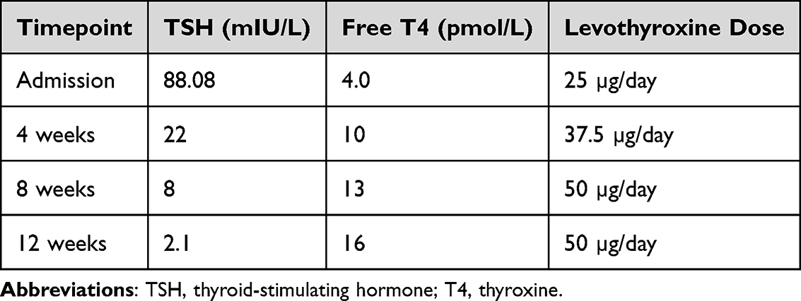 |
Table 1 Levothyroxine Dose Titration Guided by Thyroid-Stimulating Hormone (TSH) and Free Thyroxine (fT4) Levels Over 12 Weeks |
Follow-Up
At one month, repeat echocardiography showed complete resolution of pericardial effusion with no residual fluid visible in any view. Thyroid-stimulating hormone had decreased to 22 mIU/L and free thyroxine was 10 pmol/L. At three months, she was clinically well with thyroid-stimulating hormone 2.1 mIU/L and free thyroxine 16 pmol/L, both within the target range. Echocardiography showed no recurrence of effusion. She remained on 50 µg levothyroxine daily with excellent adherence. A follow-up chest radiograph demonstrated interval resolution of cardiomegaly and bilateral pleural effusions, confirming the radiographic improvement that paralleled the echocardiographic and biochemical response.
Discussion
This case illustrates a clinically important but uncommon presentation: cardiac tamponade attributable to severe primary hypothyroidism in a tuberculosis-endemic region. The primary learning objectives of this report are threefold: (1) to demonstrate that large hypothyroid pericardial effusions can progress to life-threatening tamponade; (2) to highlight the clinical clues that should prompt early consideration of hypothyroidism in this setting; and (3) to describe the integrated management of pericardiocentesis and cautious thyroid hormone replacement.
Pericardial effusion in hypothyroidism results from altered capillary permeability and impaired lymphatic drainage rather than active inflammation.1,3 Slow fluid accumulation permits substantial pericardial distension, which explains why large effusions may develop with relatively mild symptoms until late hemodynamic compromise occurs.2,3 Although progression to tamponade is uncommon, published cases of hypothyroid-related tamponade have described a consistent pattern: elderly or middle-aged women, severe biochemical hypothyroidism, large circumferential effusions, and marked bradycardia preceding or accompanying hemodynamic decompensation.1,2,4 This case is consistent with that reported phenotype and further illustrates that delayed diagnosis allows life-threatening deterioration despite the reversible nature of the underlying condition.
In tuberculosis-endemic settings, diagnostic reasoning is often anchored on tuberculous pericarditis when pericardial effusion is identified.3,4 In this context, a protein-rich pericardial fluid may reinforce an infectious diagnosis. However, hypothyroidism-related effusions are frequently protein-rich and may fulfill biochemical thresholds traditionally labeled exudative when Light’s criteria originally developed for pleural fluid are applied to pericardial fluid.8,9 Because protein concentration alone does not reliably distinguish inflammatory from noninflammatory pericardial etiologies, elevated pericardial fluid protein should not be interpreted as evidence of infection or malignancy without corroborating clinical or microbiological findings. Failure to recognize this limitation can lead to diagnostic delay and inappropriate empiric treatment.
In this patient, a pericardial fluid adenosine deaminase below 5 U/L, negative mycobacterial studies, and the absence of constitutional symptoms made tuberculous pericarditis unlikely.10 While no single test definitively excludes tuberculosis, integrated assessment of epidemiology, fluid biochemistry, microbiology, and clinical findings supported severe primary hypothyroidism as the most plausible etiology.
Several practical clinical lessons emerge from this case. First, cardiac tamponade typically provokes tachycardia; marked bradycardia in this setting reflects hypothyroidism blunting the expected compensatory adrenergic response and should prompt immediate thyroid function testing.1,2 Second, delayed relaxation of deep tendon reflexes (Woltman’s sign), although relatively insensitive, remains a valuable bedside clue, particularly in resource-limited environments where thyroid function assays are not always reflexively ordered.5 Third, routine inclusion of thyroid function tests in the evaluation of unexplained pericardial effusion is a low-cost strategy that can prevent missed diagnoses in tuberculosis-endemic regions.3,4
Regarding management, pericardiocentesis provides rapid hemodynamic stabilization, while cautious initiation and gradual titration of levothyroxine corrects the underlying endocrine disorder and prevents recurrence.1,2,6 The concurrent management of hypertension and diabetes in this patient was equally important; omission of these aspects would risk incomplete hemodynamic recovery and ongoing cardiovascular risk.
Conclusion
Severe primary hypothyroidism should be routinely considered in patients with unexplained pericardial effusion, even in tuberculosis-endemic settings, as large effusions may develop with minimal systemic symptoms and mimic infectious or inflammatory etiologies. Paradoxical bradycardia in tamponade, Woltman’s sign, and a protein-rich pericardial effusion with low adenosine deaminase should prompt early thyroid function testing. Low-voltage electrocardiography, while also encountered in pericardial effusion alone, adds a supportive diagnostic clue when present with other hypothyroid features. Timely pericardiocentesis and cautious levothyroxine replacement effectively reverse hemodynamic compromise and prevent recurrence.
Patient Perspective
The patient reported marked improvement in breathlessness and fatigue within weeks of treatment and was able to resume normal daily activities.
Data Sharing Statement
All data generated or analyzed during this study are included in this published article.
Ethics Approval and Consent to Participate
Not applicable. This is a case report. Formal ethics committee approval was not required according to institutional policy.
Consent for Publication
Written informed consent was obtained from the patient for publication of this case report and accompanying images.
Acknowledgments
The authors thank the staff of St. Mary’s Hospital Lacor for their support in patient care. This case report was prepared in accordance with the CARE reporting guidelines.
Author Contributions
All authors made a significant contribution to the work reported, whether that is in the conception, study design, execution, acquisition of data, analysis and interpretation, or in all these areas; took part in drafting, revising or critically reviewing the article; gave final approval of the version to be published; have agreed on the journal to which the article has been submitted; and agree to be accountable for all aspects of the work.
Funding
No external funding was received.
Disclosure
The authors declare that they have no competing interests in this work.
References
1. Chahine J, Ala CK, Gentry JL, Pantalone KM, Klein AL. Pericardial diseases in patients with hypothyroidism. Heart. 2019;105(13):1027–8. doi:10.1136/heartjnl-2018-314528
2. Robillon JF, Sanchez B, Vuolo-Rigaud AM, et al. Tamponnade cardiaque au cours d’une hypothyïroide sévère. Une cause rare [Cardiac tamponade in severe hypothyroidism. A rare cause]. Presse Med. 1993;22(26):1221–1223.
3. Gomes Santos P, Calças Marques R, Martins Dos Santos P, Carreira da Costa C, Mogildea M. Ascites, pleural, and pericardial effusion in primary hypothyroidism: a rare case report. Cureus. 2023;15(12):e50429. doi:10.7759/cureus.50429
4. Tammiraju IVMR, Bhupathi M. “All that glitters like gold is not good”: pericardial effusion with cardiac tamponade in hypothyroidism. Indian J Cardiovasc Dis Women. 2023;8:215–219. doi:10.25259/IJCDW_3_2023
5. Iwasaki Y, Fukaya K. Woltman’s Sign of Hypothyroidism. N Engl J Med. 2018;379(14):e23. doi:10.1056/NEJMicm1713796
6. Jonklaas J, Bianco AC, Bauer AJ, et al. Guidelines for the treatment of hypothyroidism: prepared by the American thyroid association task force on thyroid hormone replacement. Thyroid. 2014;24(12):1670–1751. doi:10.1089/thy.2014.0028
7. Garber JR, Cobin RH, Gharib H, et al. Clinical practice guidelines for hypothyroidism in adults: cosponsored by the American Association of Clinical Endocrinologists and the American Thyroid Association. Endocr Pract. 2012;18(6):988–1028. doi:10.4158/EP12280.GL
8. Shanker D, Gaur A, Warriner D. Pericardial effusion: overview of aetiology, pathophysiology, diagnosis, and management. Cureus. 2025;17(9):e92177. doi:10.7759/cureus.92177
9. Kopcinovic LM, Culej J. Pleural, peritoneal and pericardial effusions - a biochemical approach. Biochem Med. 2014;24(1):123–137. doi:10.11613/BM.2014.014
10. Buoro S, Tombetti E, Ceriotti F, et al. What is the normal composition of pericardial fluid? Heart. 2021;107(19):1584–1590. doi:10.1136/heartjnl-2020-317966
 © 2026 The Author(s). This work is published and licensed by Dove Medical Press Limited. The
full terms of this license are available at https://www.dovepress.com/terms
and incorporate the Creative Commons Attribution
- Non Commercial (unported, 4.0) License.
By accessing the work you hereby accept the Terms. Non-commercial uses of the work are permitted
without any further permission from Dove Medical Press Limited, provided the work is properly
attributed. For permission for commercial use of this work, please see paragraphs 4.2 and 5 of our Terms.
© 2026 The Author(s). This work is published and licensed by Dove Medical Press Limited. The
full terms of this license are available at https://www.dovepress.com/terms
and incorporate the Creative Commons Attribution
- Non Commercial (unported, 4.0) License.
By accessing the work you hereby accept the Terms. Non-commercial uses of the work are permitted
without any further permission from Dove Medical Press Limited, provided the work is properly
attributed. For permission for commercial use of this work, please see paragraphs 4.2 and 5 of our Terms.
Recommended articles
Evaluation of Bone Health in Postmenopausal Women Using Long-Term Levothyroxine Treatment Due to Post-Procedural Hypothyroidism
Apaydin M, Surel F, Kazan S
International Journal of General Medicine 2024, 17:6139-6144
Published Date: 12 December 2024
Prevalence and Clinical Implications of Pyrazinamide Resistance in Newly Diagnosed TB Patients in Uganda
Ndawula C, Petra NP, Wasswa FB, Bazira J
Infection and Drug Resistance 2025, 18:1629-1635
Published Date: 29 March 2025
Prevalence, Clinical Features and Factors Associated with Hypercalcemia Among Patients Diagnosed with Tuberculosis at Lira Regional Referral Hospital, Northern Uganda: A Cross-Sectional Study
Ahmed Jnr AH, Onyanga N, Adam Deifa AA, Ali HO, Nor FI, Hussein ZA, Mohamed AM, Hashi NA, Hassan MA, Hirsi AM, Jayte M
Infection and Drug Resistance 2026, 19:597775
Published Date: 3 April 2026
Successful Management of Penetrating Right Ventricular Stab Injury with Cardiac Tamponade in a Resource-Limited Setting: A Case Report
Abdi HK, Abdi AA, Ali AA, Mohamed AO, Ahmed MR, Warsame Keilie AM
International Medical Case Reports Journal 2026, 19:598759
Published Date: 10 July 2026