
Back to Journals » ImmunoTargets and Therapy » Volume 13
Assessing the Prognostic Value of 13 Inflammation-Based Scores in Patients with Unresectable or Advanced Biliary Tract Carcinoma After Immunotherapy
Authors Wang F, Jiang C, He W  , Li H , Guo GF , Xu L
Received 30 March 2024
Accepted for publication 28 August 2024
Published 16 October 2024 Volume 2024:13 Pages 541—557
DOI https://doi.org/10.2147/ITT.S471502
Checked for plagiarism Yes
Review by Single anonymous peer review
Peer reviewer comments 3
Editor who approved publication: Professor Michael Shurin
Fang Wang,1,* Chang Jiang,2– 4,* Wenzhuo He,2– 4,* Heping Li,1 Gui-Fang Guo,2– 4 Lixia Xu1
1Department of Oncology, The First Affiliated Hospital of Sun Yat-Sen University, Guangzhou, Guangdong Province, People’s Republic of China; 2State Key Laboratory of Oncology in South China, The Sun Yat-Sen University Cancer Center Guangzhou, Guangzhou, Guangdong Province, People’s Republic of China; 3Collaborative Innovation Center for Cancer Medicine, The Sun Yat-Sen University Cancer Center, Guangzhou, Guangdong Province, People’s Republic of China; 4VIP Department, Sun Yat-sen University Cancer Center, Guangzhou, Guangdong Province, People’s Republic of China
*These authors contributed equally to this work
Correspondence: Lixia Xu, Department of Oncology, The First Affiliated Hospital of Sun Yat-sen University, Guangzhou, Guangdong Province, People’s Republic of China, Email [email protected] Gui-Fang Guo, State Key Laboratory of Oncology in South China, Collaborative Innovation Center for Cancer Medicine, VIP Department, Sun Yat-sen University Cancer Center, Guangzhou, Guangdong Province, People’s Republic of China, Email [email protected]
Purpose: The response of patients with biliary tract carcinoma (BTC) to immunotherapy varies widely, and there is an urgent need for biological indicators. The predictive value of inflammation based score (IBS) for the efficacy of immunotherapy in patients with BTC remains unclear, as the evidence is inconsistent. This study aimed to comprehensively examine the predictive value of IBS in peripheral blood on the survival of BTC patients receiving immunotherapy.
Patients and Methods: We retrospectively assessed 118 patients with advanced BTC who received anti-PD-1 therapy in the first or second line in two medical centers. The Kaplan-Meier, time-dependent ROC, and Harrell’s concordance index (C-index) were applied to analyze the predictive value of 13 reported peripheral blood IBS.
Results: All 13 IBS were identified as significant prognostic factors for OS in univariate analysis. Pan-immune-inflammation value (PIV) (p=0.005), PILE (composed of PIV, lactate dehydrogenase and Eastern Cooperative Oncology Group performance status) (p=0.033), neutrophil-to-lymphocyte ratio (NLR) (p=0.003), platelet-to-lymphocyte ratio (PLR) (p< 0.001), lymphocyte-to-monocyte ratio (LMR) (p=0.006), systemic immune inflammation index (SII) (p=0.039), CRP-to-albumin ratio (CAR) (p=0.025), and Albumin-NLR (p=0.008) were identified as independent prognostic factors for OS in multivariate analysis. PIV and PILE scores were superior to other scores, according to time-dependent ROC curves, and their superiority became more pronounced after the 12-month time point. C-index analysis showed PIV (C-index 0.62, 95% CI: 0.55, 0.68) and PILE (C-index 0.62, 95% CI: 0.55, 0.70), both superior to other IBS.
Conclusion: PIV and PILE scores are independent predictors of OS in patients with BTC after immunotherapy and are superior to other IBS. PIV and PILE may be able to help screen out patients with advanced BTC who are less likely to benefit from anti-PD-1 monotherapy. Due to the retrospective nature of this analysis, the predictive value of PIV and PILE require validation in further prospective studies.
Keywords: biliary tract carcinoma, anti-PD-1 therapy, inflammation-based scores, pan-Immune-Inflammation Value, PILE
Introduction
Biliary tract carcinoma (BTC) mainly comprises intrahepatic cholangiocarcinoma, extrahepatic cholangiocarcinoma, and gallbladder carcinoma. The incidence of BTC is high in East Asia and on the rise.1 Immune checkpoint inhibitors (ICIs) have been recommended as the first-line treatment for BTC based on the findings of the TOPAZ-1 and KEYNOTE-966 studies.2,3 However, patients with BTC show extremely variable responses to immunotherapy, highlighting the urgent need for reliable predictors of immunotherapy efficacy.
Tumor mutational burden (TMB) and microsatellite instability/mismatch repair deficiency (MSI/MMRd) are arguably the most reliable biomarkers of immunotherapy efficacy as evidenced by their consistency across various cancer histologies.4,5 Approximately, 4.6% (95% CI: 2.38 to 6.97) and 2.5% (95% CI: 1.75 to 3.34) of BTCs can be classified as TMB-H and MSI/MMRd tumors, respectively.6 Data on immunotherapy efficacy in BTC patients with TMB-H or MSI are scarce, and the reported response rate in MSI-H BTC patients treated with pembrolizumab ranges from 27% to 40%.7,8 It should be noted that different TMB cut-off values have been used in different studies.
PD-L1 expression has been observed in approximately 26% of patients with BTC, with minimal differences based on anatomical location.6 To date, the significance of PD-L1 expression in patients with BTC receiving anti-PD-1 or PD-L1 therapy remains controversial. In a multi-institutional Phase 2 trial of nivolumab in patients with advanced BTC, positive PD-L1 expression was associated with prolonged PFS.9 In contrast, data from the original Keynote-158 cohort reported no differences in ORR, PFS, or OS between pembrolizumab-treated PD-L1(+) and PD-L1(-) patients.10 Frega et al conducted a meta-analysis of 39 studies and found that PD-L1 expression is not a reliable predictor of immunotherapy efficacy in biliary tract tumors.6 In addition to these conflicting results, PD-L1 positivity lacks a standardized definition. This has led to the use of multiple PD-L1 scoring methods and cut-off values in clinical trials.6,11 There remains a lack of reliable biomarkers for predicting immunotherapy efficacy in BTC, especially in patients with mismatch-proficient tumors.
Inflammation is a primary hallmark of cancer.12 It plays an important role in tumor immunosuppression, angiogenesis, proliferation, and survival.13 The systemic inflammatory status is closely related to changes in peripheral blood immune cells and inflammatory markers, which can be estimated using the peripheral blood immune inflammation score.14 This score is an index based on peripheral blood immune cells or other peripheral blood test results.
Recently, several peripheral blood-based immune inflammation scores, such as the Glasgow Prognostic Score (mGPS),15 neutrophil-to-lymphocyte ratio (NLR),16 platelet-to-lymphocyte ratio (PLR),17 lymphocyte-to-monocyte ratio (LMR),18 albumin-neutrophil-to-lymphocyte ratio (albumin-NLR),19 Systemic Immunoinflammatory Index (SII)20,21 have been reported in a variety of solid tumors and can differentiate the prognosis of tumor patients after surgery, chemotherapy radiotherapy, and immunotherapy.
In biliary tract carcinomas, initial reports have suggested the potential predictive value of peripheral blood immune inflammatory markers for immunotherapy. Pan et al demonstrated that the lung immune prognostic index (LIPI) is an independent determinant affecting both survival outcomes and clinical responses in patients with advanced BTC undergoing immunotherapeutic interventions. 22 Conversely, Li et al found no significant association between clinical outcomes and inflammation-related markers.23 Additionally, Fei Du et al emphasized that the combined assessment of three distinct factors—systemic immune inflammation index, cytokine IFN-inducible protein-10, and macrophage inflammatory protein-1β—exhibited superior predictive efficacy in determining the overall benefit rate among BTC patients following immunotherapy.24 Furthermore, Yang et al delineated the prognostic utility of the C-reactive protein (CRP) score in forecasting the therapeutic response to anti-PD-1 therapy among patients with intrahepatic cholangiocarcinoma (ICC), surpassing alternative inflammation-based prognostic indicators in predictive accuracy.25
The predictive value of immune inflammation indicators for immunotherapy efficacy in patients with biliary tract cancers remains unclear because of inconsistent evidence (Supplemental Table 1). Moreover, no single immune inflammatory marker has emerged as the most reliable useful predictor. Therefore, the aim of this study was to examine the predictive value of peripheral blood immunoinflammatory markers for survival following immunotherapy in unresectable and advanced BTC.
Patients and Methods
Patients
We included patients with BTC treated at the First Affiliated Hospital of Sun Yat-sen University and Sun Yat-sen University Cancer Center between January 1, 2018 and June 27, 2022. Patient inclusion criteria were (1) histologically confirmed unresectable or metastatic BTC; (2) receiving at least 2 cycles of PD-1-containing regimens in first and second line with post-treatment imaging evaluation; (3) having complete medical records and follow-up data; and (4) having blood test results within 1 week prior to treatment.
This study was approved by the Ethics Committee of the First Affiliated Hospital of Sun Yat-sen University ([2022]069) and the Sun Yat-sen University Cancer Center (SYSUCC; Guangzhou, China, B2020-190-01). Our study was a retrospective study that strictly adhered to the Declaration of Helsinki to ensure confidentiality of patient data and no impact on patient health and interests. Therefore, both ethics committees agreed to waive the informed consent of patients.
Inflammation Score Calculation
A total of 13 inflammation scores were included for analysis, including platelet-to-lymphocyte ratio (PLR), neutrophil-to-lymphocyte ratio (NLR), lymphocyte-to-C-reactive protein ratio (LCR), lymphocyte-to-monocyte ratio (LMR), CRP-to-albumin ratio (CAR), systemic immune inflammation index (SII), prognostic nutrition index (PNI), modified Glasgow prognostic score (mGPS), prognostic index (PI), Pan-Immune-Inflammation Value (PIV), PILE (composed of PIV, LDH and ECOG PS), systemic inflammation score (SIS), Albumin (ALB) and neutrophil-to-lymphocyte ratio (Albumin-NLR). The specific calculation of each inflammation score is shown in Table 1.
 |
Table 1 Systemic Inflammation-Based Prognostic Scores |
Data Extraction, Efficacy Evaluation
All enrolled patients had peripheral blood test results within 1 week prior to immunotherapy, and information on clinical characteristics, treatment strategy and efficacy was collected from the medical records. Data were entered by two investigators. Two oncologists examined treatment efficacy according to the response assessment criteria in the solid tumor (RESIST) criteria (1.1). A statistician of the study group will perform regular data quality control. Standard follow-up was performed by the study staff. The follow-up interval is 2 to 4 months. Statisticians in the study group will perform data quality control at regular intervals. Follow-up data are available for all patients and the latest follow-up was performed on December 27, 2023.
Statistical Methods
Student t test (continuous data) and χ2 test (categorical data) were used for comparison between groups. Survival analysis was performed using the Kaplan-Meier method, and differences in survival curves were analyzed using the Log rank test. For single-valued indicators, the optimal threshold for each inflammatory indicator before and after multiple immunotherapy was calculated using X-tile software. (Supplemental Figure 1) For the composite index, mGPS and PI scores were calculated as previously reported. The effect of various inflammatory indexes on OS was analyzed using the Kaplan-Meier method and the rank test. Cox proportional risk models were used to determine the risk ratios (HRs) in univariate and multifactorial survival analyses (variables with p value <0.1 on univariate analysis and known to be significant clinically prognostic value included in multifactorial regression equations). Time-dependent ROC curves and area under the curve (AUC) at 6, 12, 18 and 24 months were calculated to compare the predictive power of each inflammation score. The predictive ability of each inflammation score was assessed using Harrell’s concordance index (C-index). A bilateral p<0.05 was considered statistically different. The statistical software SPSS 26.0 (IBM Corp, Armonk, NY, USA), X-tile software 3.6.1 (Brady Memorial Laboratory, Yale School of Medicine, New Haven, USA) and R studio version 4.2.2 (R Foundation for S&T) were applied for all statistical analyses in this section.
Results
Characteristics of Enrolled Patients
According to the above inclusion criteria, we included 118 patients with BTC treated at the First Affiliated Hospital of Sun Yat-sen University and Sun Yat-sen University Cancer Center between January 1, 2018 and June 27, 2022. Among them, 73 (61.9%) were male. The age ranged from 27 to 88 years, with a median age of 55 years. Among the patients, 38 (32.2%) had hepatitis B, and 90 (76.27%) had an Eastern Cooperative Oncology Group performance status (ECOG PS) of 0, while 27 (22.88%) had an ECOG PS of 1. The study included 90 (76.27%) patients with intrahepatic cholangiocarcinoma, 13 (11.01%) patients with extrahepatic cholangiocarcinoma, and 15 (12.72%) patients with gallbladder cancer. Of the patients, 56 (47.46%) had concomitant liver metastases with other organs, and 36 (30.52%) had metastases from organs other than the liver and lungs. Additionally, 72 (61.02%) patients had metastases from more than one organ. In terms of treatment, 90 patients (76.3%) received PD-1-containing monoclonal antibodies in the first line, and 28 patients (23.7%) received PD-1-containing monoclonal antibodies in the second line.
Treatment
The types of regimens received by the included patients included PD-1 monotherapy (n=19), PD-1-mAb combined with chemotherapy (n=39), PD-1-mAb combined with anti-angiogenic TKI (n=35), and PD-1 plus intervention (n=25). The types of PD-1-mAb include sintilimab, Pembrolizumab,Camrelizumab, Toripalimab, and Nivolumab. First-line chemotherapy includes gemcitabine combined with cisplatin and gemcitabine combined with tegio, and second-line chemotherapy includes gemcitabine, tegio, albumin paclitaxel and 5-fluorouracil combined with oxaliplatin. Anti-angiogenic TKI included lenvatinib and apatinib. Interventional therapies included HAIC- folfox, HAIC-folfirinox, and HAIC combined with TACE. Table 2 summarizes the clinicopathological characteristics of the patients, including the distribution of patients based on 13 inflammation scores.
 |
Table 2 Baseline Characteristics of the Enrolled Patients |
The duration of anti-PD-1 therapy ranged from 1.0 to 33.0 months, with a median of 6.0 months. The number of treatment cycles ranged from 2 to 39 cycles, with a median treatment cycle count of 7 and median OS 18.3 months (range 1.75–49.16 months). By the end of follow-up, a total of 93 (78.8%) patients experienced disease progression and 62 (52.5%) patients died.
Survival Analysis
In the univariate analysis, six clinicopathological characteristics, ECOG PS, CA19-9, CEA, LDH, ALB, and CRP, were identified as significant predictive variables for overall survival (OS). All 13 inflammation scores were identified as significant prognostic factors for OS. To avoid correlation between immunoinflammatory scores, we included the 13 inflammatory scores, subdivided and all non-inflammatory scores of clinicopathological features (ECOG PS, CA19-9, CEA, LDH, ALB, CRP, age grading) with p<0.01 in the multifactorial analysis, respectively. The results showed that PIV (p=0.005), PILE (p=0.033), NLR (p=0.003), PLR (p<0.001), LMR (p=0.006), SII (p=0.039), CRP-to-albumin ratio (CAR) (p=0.025), and Albumin-NLR (p=0.008) were independent prognostic factors for OS (Table 3). As shown in Figure 1, all inflammation scores were associated with OS in patients with advanced biliary tract carcinoma receiving anti-PD-1 therapy.
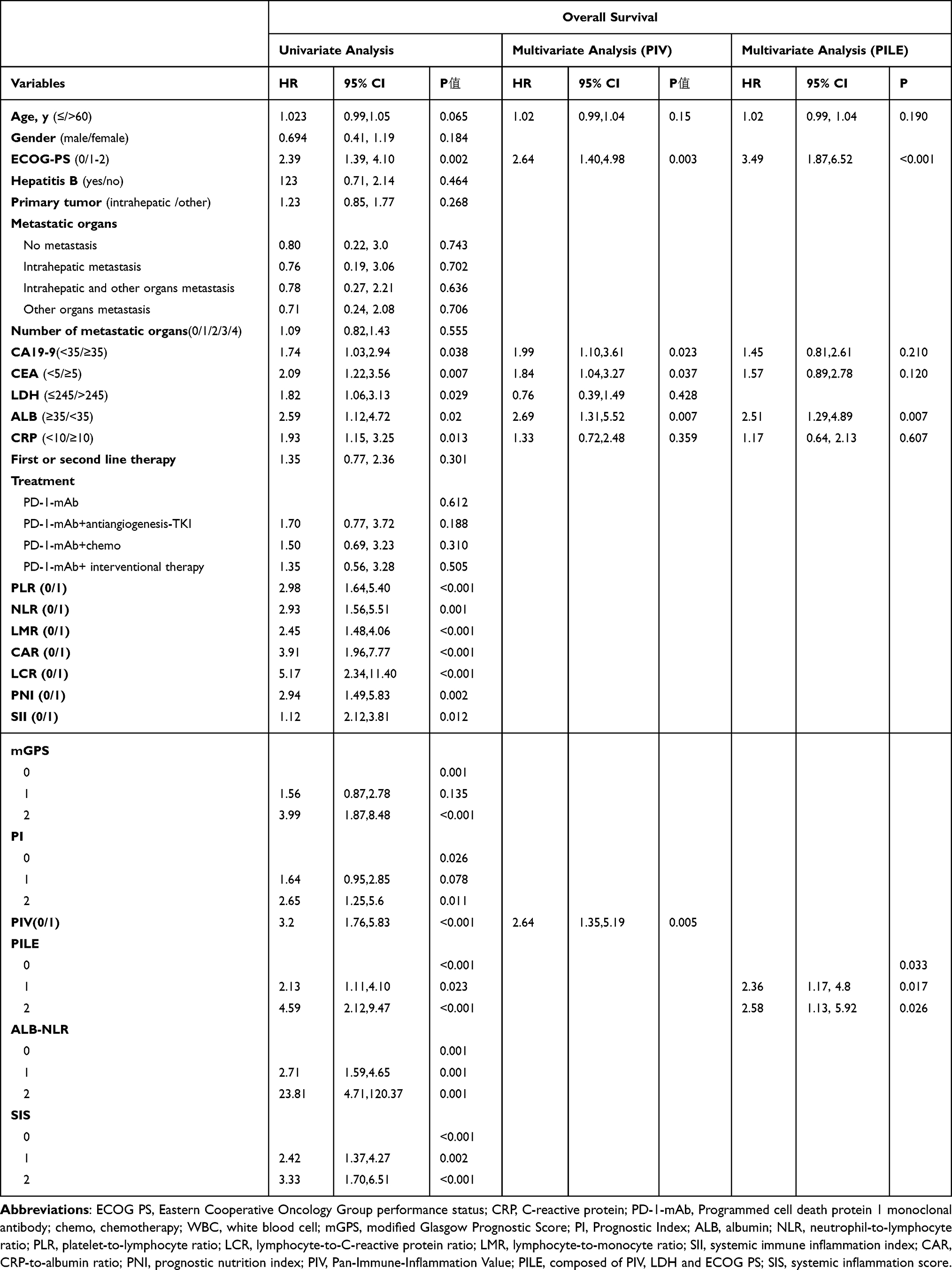 |
Table 3 Univariate and Multivariate Time-Dependent Cox Regression Analyses of the Prognostic Factors for Overall Survival |
 |
Figure 1 Kaplan-Meier curves for overall survival of patients with biliary tract tumors after anti-PD-1 therapy. (A) PLR, (B) NLR, (C) LCR, (D) LMR, (E) CAR, (F) SII, (G) PNI, (H) mGPS, (I) PI, (J) PIV, (K) PILE, (L) SIS, (M) LMR. All indicators of A-K and M were significant influencing factors of OS. The (E) SIS had significant predictive value for OS, but the generation curves of its scores 1 and 2 failed to separate well; F ALB-NLR can distinguish OS better. |
Comparison of the Predictive Value of Inflammation Scores for the Efficacy of Immunotherapy
Time-dependent ROC curves were performed at 6, 12, 18 and 24 months of OS to compare the prognostic value of the 13 inflammation scores (Figure 2). The pan-immune-inflammation value (PIV) and PILE scores were superior to the other scores, especially after the 12-month time point. The dependent area under the curve (AUC) of ROC curves indicated that PIV and PILE scores could better predict survival time after PD-1 regimen for advanced biliary tract tumors. Further, time-dependent AUC plots for consecutive times showed that PIV and PILE scores were consistently higher than other inflammatory scores after the 12-month time point. Even the PILE score was significantly higher than the PIV score after the 12-month time point (Figure 3). The C-index analysis showed that PIV (C-index 0.62, 95% CI: 0.55, 0.68) and PILE (C-index 0.62, 95% CI: 0.55, 0.70) had higher C-index values than the other scoring systems (Table 4). Combined with the time-dependent ROC curves and C-index, it was demonstrated that PILE and PIV scores had a stronger ability to predict survival time after PD-1 regimen treatment for advanced biliary tract tumors than other scoring systems.
 |
Table 4 Concordance Index for the Comparison of Different Inflammatory-Based Scores |
 |
Figure 2 Time-dependent ROC curves for survival prediction of inflammatory scores at 6 (A), 12 (B), 18 (C), and 24(D) months in patients with BTC after anti-PD-1 therapy. (A) No significant distinction among inflammatory index curves. (B) No significant distinction among inflammatory index curves. (C) PIV and PILE curves start to be significantly higher than other inflammatory index curves. (D) PIV, PILE and SII curves were higher than the other inflammatory index curves. |
 |
Figure 3 Time-dependent AUC plot of survival prediction by inflammation score. After the 12-month time point, PILE is above the PIV curve and starts to be significantly higher than the other inflammatory index curves. |
Relationship Between Inflammation Scores and Clinical Characteristics
Table 5 showed the relationships between patients’ clinical characteristics, outcomes, and PIV scores. High PIV scores were associated with higher levels of CRP (p<0.001), CA19-9 (p=0.004), CEA (p=0.006), LDH (p=0.004), and other factors associated with a poor prognosis. Tumor response analysis showed that low PIV group achieved high ORR (p=0.002) DCR (p=0.004). Table 6 showed the relationship between patient clinical characteristics, efficacy, and PILE scores. Higher PIV scores were associated with higher CRP (p<0.001), CA19-9 (p=0.001), CEA (p=0.003), low ALB (p=0.039). Tumor response analysis showed that the low and medium scoring PILE group achieved higher ORR (p=0.005) and DCR (p=0.037). Since LDH and ECOG PS are components of PILE, we will not describe their distribution in the PILE scores groups.
 |
Table 5 Baseline Characteristics of the Patients Grouped by PIV Score |
 |
Table 6 Baseline Characteristics of the Patients Grouped by PILE Score |
Discussion
It is well established that the development of biliary tract cancers is highly correlated with inflammation. PD-L1 expression, immune infiltration, and the exceptionally high mutational load of biliary tract cancers make immune checkpoint inhibitors potentially effective therapeutic strategies.14 Immunotherapy, when combined with chemotherapy, has been recommended as the first-line treatment for advanced biliary tract carcinoma.26,27 However, patients with BTC show extremely variable responses to immunotherapy, underscoring the urgent need for reliable predictors of immunotherapy efficacy.
MMR-deficiency/MSI and TMB-H are likely the most reliable predictive biomarkers for immunotherapy in BTC, although they are rare (4.6% TMB-H; 2.5% MSI/MMRd). Data on the efficacy of immunotherapy in BTC patients with TMB-H or MSI are scarce. Although PD-L1 expression is more frequent (25.6%), its predictive value remains controversial (Frega et al, Cell 2023).6 Studies reporting novel biomarkers for BTC, particularly in patients with mismatch-proficient tumors, are lacking. Currently, there is no universally accepted gold-standard index for predicting the efficacy of BTC, and immunoefficacy prediction remains in the preliminary exploration stage. Our study represents an initial investigation into the predictive capacity of inflammation-based scores for immune efficacy in BTC.
In our study, the PIV and PILE scores outperformed the other inflammation-based scores in predicting OS. To the best of our knowledge, this is the first study to report the predictive value of PIV and PILE scores in patients with BTC after immunotherapy.
PIV has been shown to accurately reflect the systemic immune- and cancer-related inflammatory status in a wide range of solid tumors and is a reliable predictor of clinical outcomes in patients with advanced cancer.28,29 However, the underlying mechanism remains unknown. One possible explanation is that PIV contains all important immune cell components found in peripheral blood. Peripheral blood lymphocytes correlate with the body’s lymphocyte stores and activity, and are an accurate indicator of the patient’s immune status.30,31
The PILE is a three-parameter inflammation score that includes LDH, PIV, and ECOG PS. LDH has been previously identified as an independent prognostic indicator in several tumors, especially non-Hodgkin’s lymphoma, where it is one of the main components of the International Prognostic Index (IPI).32 High LDH levels have been associated with poor first-line chemotherapy efficacy and the overexpression of tumor VEGFA and VEGFR in BTC.33,34
In our study, all inflammation scores were significant predictors of OS after PD-1 treatment for BTC. However, further time-dependent ROC and C-index analyses revealed that PIV (C-index 0.62, 95% CI:0.55–0.68) and PILE (C-index 0.62, 95% CI:0.55–0.70) were the most effective OS predictors. The superiority of PIV and PILE was particularly significant after the 12-month mark. The PILE prediction curve in the continuous time-dependent ROC plot was significantly higher than those of the PIV and other inflammation scores after the 12-month time point. Patients who received effective immunotherapy frequently had prolonged PFS and OS, indicating a trailing effect in the data. Both PILE and PIV had higher predictive abilities after 12 months, which was consistent with the observed trailing effect after PD-1 treatment.
According to Ligorio et al, PIV is a better predictor of overall survival (OS) than other peripheral blood indicators, such as NLR, PLR, and LMR, in patients with HER2-positive metastatic breast cancer treated with trastuzumab plus pertuzumab as first-line therapy.35 Moreover, PIV has been previously described as a novel predictive biomarker in patients with metastatic colorectal cancer receiving first-line therapy, and is superior to other inflammatory markers.36 In 120 patients with advanced cancer of any type who were treated with anti-PD-1 or anti-PD-L1 inhibitors, higher PILE scores were significantly associated with shorter PFS and OS, indicating that PILE is a reliable prognostic indicator for immunotherapy.37 Zeng et al discovered that high PIV and PILE were associated with poor clinical outcomes with anti-PD-1/PD-L1 inhibitors in combination with chemotherapy treatment in a clinical trial of an extensive-stage small cell lung cancer (NCT03041311) dataset and that PIV and PILE may help distinguish patients who do not benefit from anti-PD-1/PD-L1 therapy.38 These findings were validated using real-world datasets. These studies on the predictive values of PILE and PIV for OS support our findings.
In our study, patients with low PIV and PILE scores were more likely to have low CRP levels, which consistently represent a low inflammatory status. Because CRP was not identified as a prognostic factor for OS in the multifactorial Cox analysis, the intergroup distribution of CRP had a minimal impact on the independent predictive power of PIV and PILE. Patients with low PIV and PILE scores were more likely to have low CRP levels, confirming the ability of PIV and PILE scores to reflect systemic inflammatory status. In contrast, the high PIV and PILE score groups demonstrated a stronger association with elevated CEA and CA19-9 levels. Although CEA and CA19-9 have previously been linked to poor clinical outcomes in biliary tract tumors39,40 in multifactorial analysis, CEA, CA19-9, and PIV emerged as independent prognostic factors for OS. Consequently, the impact of the intergroup distribution of CEA and CA19-9 within the PIV was less pronounced in terms of the predictive power of PIV for OS. When CEA and CA19-9 levels were compared with PILE, only PILE remained an independent prognostic factor for OS. Although the influence of the intergroup distribution of CEA and CA19-9 on the prognostic ability of PILE cannot be ruled out, it has significantly less predictive power for OS than the PILE score. Furthermore, the low-PIV score group included more HBV-positive patients. Hepatitis B virus (HBV) infection was reported affect the prognosis of patients with HBV-positive ICC by activating the immune response. To date, the influence of HBV infection on the effectiveness of immunotherapy in BTC remains unclear.41 The HBV positive was not identified as an independent predictor of OS in the univariate analysis in our study. Consequently, its influence on the independent predictive value of PIV for OS was minimal, and its even distribution within the baseline population of the PILE score did not significantly affect our research conclusions.
Owing to the constraints of retrospective data, we obtained PD-L1 and genetic test results from less than 5% of the patients, precluding a direct comparison between peripheral blood immune-inflammatory metrics and the predictive value of PD-L1 and TMB. This is a significant limitation of this study.
Additionally, the study has several other limitations. First, this was a retrospective study conducted on a two-center cohort in China. Despite ensuring strict quality control and between-group balance, unavoidable biases still exist owing to the retrospective nature of the study. We included patients with intrahepatic cholangiocarcinoma, extrahepatic cholangiocarcinoma, or gallbladder carcinoma. In the baseline analysis of the 13 inflammation scores, the primary tumor sites were evenly distributed across the subgroups. However, because of the relatively low incidence of BTC and the retrospective design of the study, we enrolled only 118 eligible patients. Among them, 19 received PD-1 monoclonal antibody (PD-1-mAb) monotherapy, 39 received PD-1-mAb combined with anti-angiogenesis tyrosine kinase inhibitors (PD-1-mAb+antiangiogenesis-TKI), 35 received PD-1-mAb combined with chemotherapy (PD-1-mAb+ chemotherapy), and 25 received PD-1-mAb combined with interventional therapy. The limited sample size restricted our ability to perform subgroup analyses of the inflammatory markers within each treatment regimen. However, as more cases of immunotherapy for biliary tract tumors accumulate, future studies should aim to conduct further subgroup analyses. As Chinese patients with biliary tract tumors have different disease backgrounds than those in the United States, Europe, or Japan, these data cannot be fully extrapolated to other countries and require further validation in external institutions. Second, while we implemented stringent measures to ensure the consistency of each inflammation score, the optimal cutoff values for multiple inflammation scores (including PIV) in this study were derived from the study dataset and were not widely accepted for certification. If necessary, these values should be re-validated and redefined in future studies using authoritative sources. Third, the patients received non-single PD-1 inhibitors throughout the treatment course, which inevitably resulted in bias. Fourth, despite enrolling first- and second-line-treated patients with no contraindications to immunotherapy, chemotherapy, or intervention, the vast majority (116/118) had an ECOG score of 0–1, and there were no cases of obstructive jaundice or biliary tract infection requiring drainage, which largely avoided the impact of biliary tract infection on our data. Hematological parameters, on the other hand, are susceptible to various concomitant medications and pathophysiological states, which will inevitably cause some bias. Finally, the potential regulatory mechanisms of neutrophils, platelets, monocytes, and lymphocytes in peripheral blood during immunotherapy remain unknown and warrant further investigation.
Conclusions
Our findings indicate that the PIV and PILE scores outperform other inflammatory markers as independent prognostic indicators in patients with biliary tract tumors undergoing anti-PD-1 therapy. PIV and PILE have the potential to assist in identifying patients with advanced biliary tract cancer (BTC) who are unlikely to derive benefit from anti-PD-1 therapy. Moreover, this straightforward clinical predictor may aid clinicians in devising optimal anti-PD-1 treatment strategies for BTC patients. Due to the retrospective nature of this analysis, the predictive value of PIV and PILE require validation in further prospective studies.
Acknowledgments
This work was supported by funding from the National Natural Science Foundation of China (82173191).
Author Contributions
All authors contributed significantly to the conception and design, data acquisition, or data analysis and interpretation, participated in the drafting of the article or critically revising it for important intellectual content, agreed to submit to the current journal, gave final approval for the version to be published, and agreed to be accountable for all aspects of the work.
Disclosure
The authors have no actual or potential conflicts of interest related to this manuscript.
References
1. Bertuccio P, Malvezzi M, Carioli G, et al. Reply to: ”Global trends in mortality from intrahepatic and extrahepatic cholangiocarcinoma”. J Hepatol. 2019;71:1262–1263. doi:10.1016/j.jhep.2019.08.033
2. Kelley RK, Ueno M, Yoo C, et al. Pembrolizumab in combination with gemcitabine and cisplatin compared with gemcitabine and cisplatin alone for patients with advanced biliary tract cancer (KEYNOTE-966): a randomised, double-blind, placebo-controlled, phase 3 trial. Lancet. 2023;401:1853–1865. doi:10.1016/S0140-6736(23)00727-4
3. Rimini M, Fornaro L, Lonardi S, et al. Durvalumab plus gemcitabine and cisplatin in advanced biliary tract cancer: an early exploratory analysis of real-world data. Liver Int. 2023;43:1803–1812. doi:10.1111/liv.15641
4. Marabelle A, Fakih M, Lopez J, et al. Association of tumour mutational burden with outcomes in patients with advanced solid tumours treated with pembrolizumab: prospective biomarker analysis of the multicohort, open-label, phase 2 KEYNOTE-158 study. Lancet Oncol. 2020;21:1353–1365. doi:10.1016/S1470-2045(20)30445-9
5. Cristescu R, Mogg R, Ayers M, et al. Pan-tumor genomic biomarkers for PD-1 checkpoint blockade-based immunotherapy. Science. 2018;362: eaar3593. doi:10.1126/science.aar3593
6. Frega G, Cossio FP, Banales JM, et al. Lacking immunotherapy biomarkers for biliary tract cancer: a comprehensive systematic literature review and meta-analysis. Cells-Basel. 2023;12:2098.
7. Marabelle A, Le DT, Ascierto PA, et al. Efficacy of pembrolizumab in patients with noncolorectal high microsatellite instability/mismatch repair-deficient cancer: results from the phase II KEYNOTE-158 study. J Clin Oncol. 2020;38:1–10. doi:10.1200/JCO.19.02105
8. Lemery S, Keegan P, Pazdur R. First FDA approval agnostic of cancer site - when a biomarker defines the indication. New Engl J Med. 2017;377:1409–1412. doi:10.1056/NEJMp1709968
9. Kim RD, Chung V, Alese OB, et al. A phase 2 multi-institutional study of nivolumab for patients with advanced refractory biliary tract cancer. JAMA Oncol. 2020;6:888–894. doi:10.1001/jamaoncol.2020.0930
10. Piha-Paul SA, Oh DY, Ueno M, et al. Efficacy and safety of pembrolizumab for the treatment of advanced biliary cancer: results from the KEYNOTE-158 and KEYNOTE-028 studies. Int J Cancer. 2020;147:2190–2198. doi:10.1002/ijc.33013
11. Yoon SB, Woo SM, Chun JW, et al. The predictive value of PD-L1 expression in response to anti-PD-1/PD-L1 therapy for biliary tract cancer: a systematic review and meta-analysis. Front Immunol. 2024;15:1321813. doi:10.3389/fimmu.2024.1321813
12. Hanahan D, Weinberg RA. Hallmarks of cancer: the next generation. Cell. 2011;144:646–674. doi:10.1016/j.cell.2011.02.013
13. Rosenberg SA. Progress in human tumour immunology and immunotherapy. Nature. 2001;411:380–384. doi:10.1038/35077246
14. Boileve A, Hilmi M, Smolenschi C, Ducreux M, Hollebecque A, Malka D. Immunotherapy in advanced biliary tract cancers. Cancers. 2021;13:1569. doi:10.3390/cancers14010013
15. Dolan RD, Daly L, Sim W, et al. Comparison of the prognostic value of ECOG-PS, mGPS and BMI/WL: implications for a clinically important framework in the assessment and treatment of advanced cancer. Clin Nutr. 2020;39:2889–2895. doi:10.1016/j.clnu.2019.12.024
16. Cupp MA, Cariolou M, Tzoulaki I, Aune D, Evangelou E, Berlanga-Taylor AJ. Neutrophil to lymphocyte ratio and cancer prognosis: an umbrella review of systematic reviews and meta-analyses of observational studies. BMC Med. 2020;18:360. doi:10.1186/s12916-020-01817-1
17. Sun Y, Zhang L. The clinical use of pretreatment NLR, PLR, and LMR in patients with esophageal squamous cell carcinoma: evidence from a meta-analysis. Cancer Manag Res. 2018;10:6167–6179. doi:10.2147/CMAR.S171035
18. Ahn J, Song E, Oh HS, et al. Low lymphocyte-to-monocyte ratios are associated with poor overall survival in anaplastic thyroid carcinoma patients. Thyroid. 2019;29:824–829. doi:10.1089/thy.2018.0684
19. Wang F, He W, Jiang C, et al. Prognostic value of inflammation-based scores in patients receiving radical resection for colorectal cancer. BMC Cancer. 2018;18: 1–10. doi:10.1186/s12885-017-3933-x
20. He K, Si L, Pan X, et al. Preoperative systemic immune-inflammation index (SII) as a superior predictor of long-term survival outcome in patients with stage I-II gastric cancer after radical surgery. Front Oncol. 2022;12:829689. doi:10.3389/fonc.2022.829689
21. Topkan E, Besen AA, Ozdemir Y, et al. Prognostic value of pretreatment systemic immune-inflammation index in glioblastoma multiforme patients undergoing postneurosurgical radiotherapy plus concurrent and adjuvant temozolomide. Mediators Inflamm. 2020;2020:4392189. doi:10.1155/2020/4392189
22. Pan Y, Si H, Jia R, et al. Analysis of a systemic inflammatory biomarker in advanced bile tract carcinoma treated with anti-PD-1 therapy: prognostic and predictive significance of lung immune prognostic index score. J Oncol. 2022;2022:1427779. doi:10.1155/2022/1427779
23. Li J, Wei Q, Wu X, et al. Integrative clinical and molecular analysis of advanced biliary tract cancers on immune checkpoint blockade reveals potential markers of response. Clin Transl Med. 2020;10:e118.
24. Du F, Qiu Z, Ai W, et al. Blood tests predict the therapeutic prognosis of anti-PD-1 in advanced biliary tract cancer. J Leukocyte Biol. 2021;110:327–334. doi:10.1002/JLB.5MA1220-631R
25. Yang Z, Zhang D, Zeng H, et al. Inflammation-based scores predict responses to pd-1 inhibitor treatment in intrahepatic cholangiocarcinoma. J Inflamm Res. 2022;15:5721–5731. doi:10.2147/JIR.S385921
26. Merters J, Lamarca A. Integrating cytotoxic, targeted and immune therapies for cholangiocarcinoma. J Hepatol. 2022;78:652–657. doi:10.1016/j.jhep.2022.11.005
27. Oh DY, Lee KH, Lee DW, et al. Gemcitabine and cisplatin plus durvalumab with or without tremelimumab in chemotherapy-naive patients with advanced biliary tract cancer: an open-label, single-centre, phase 2 study. Lancet Gastroenterol. 2022;7:522–532.
28. Corti F, Lonardi S, Intini R, et al. The pan-immune-inflammation value in microsatellite instability-high metastatic colorectal cancer patients treated with immune checkpoint inhibitors. EUR J CANCER. 2021;150:155–167. doi:10.1016/j.ejca.2021.03.043
29. Guven DC, Sahin TK, Erul E, Kilickap S, Gambichler T, Aksoy S. The association between the pan-immune-inflammation value and cancer prognosis: a systematic review and meta-analysis. Cancers. 2022;14. doi:10.3390/cancers15010014
30. Minnie SA, Waltner OG, Ensbey KS, et al. Depletion of exhausted alloreactive T cells enables targeting of stem-like memory T cells to generate tumor-specific immunity. Sci Immunol. 2022;7:eabo3420.
31. Xu X, Ye L, Zhang Q, et al. Group-2 innate lymphoid cells promote HCC progression through CXCL2-neutrophil-induced immunosuppression. Hepatology. 2021;74:2526–2543. doi:10.1002/hep.31855
32. Chen H, Zhong Q, Zhou Y, et al. Enhancement of the international prognostic index with beta2-microglobulin, platelet count and red blood cell distribution width: a new prognostic model for diffuse large B-cell lymphoma in the rituximab era. BMC Cancer. 2022;22:583. doi:10.1186/s12885-022-09693-z
33. Azuma M, Shi M, Danenberg KD, et al. Serum lactate dehydrogenase levels and glycolysis significantly correlate with tumor VEGFA and VEGFR expression in metastatic CRC patients. Pharmacogenomics. 2007;8:1705–1713. doi:10.2217/14622416.8.12.1705
34. Faloppi L, Del PM, Casadei GA, et al. The correlation between LDH serum levels and clinical outcome in advanced biliary tract cancer patients treated with first line chemotherapy. Sci Rep-UK. 2016;6:24136. doi:10.1038/srep24136
35. Ligorio F, Fuca G, Zattarin E, et al. The pan-immune-inflammation-value predicts the survival of patients with human epidermal growth factor receptor 2 (HER2)-positive advanced breast cancer treated with first-line taxane-trastuzumab-pertuzumab. Cancers. 2021;13:1964.
36. Fuca G, Guarini V, Antoniotti C, et al. The pan-immune-inflammation value is a new prognostic biomarker in metastatic colorectal cancer: results from a pooled-analysis of the Valentino and TRIBE first-line trials. Br J Cancer. 2020;123:403–409. doi:10.1038/s41416-020-0894-7
37. Guven DC, Yildirim HC, Bilgin E, et al. PILE: a candidate prognostic score in cancer patients treated with immunotherapy. Clin Transl Oncol. 2021;23:1630–1636. doi:10.1007/s12094-021-02560-6
38. Zeng R, Liu F, Fang C, et al. PIV and PILE score at baseline predict clinical outcome of anti-PD-1/PD-L1 inhibitor combined with chemotherapy in extensive-stage small cell lung cancer patients. Front Immunol. 2021;12:724443.
39. Loosen SH, Roderburg C, Kauertz KL, et al. Elevated levels of circulating osteopontin are associated with a poor survival after resection of cholangiocarcinoma. J Hepatol. 2017;67:749–757. doi:10.1016/j.jhep.2017.06.020
40. Takahara N, Nakai Y, Isayama H, et al. CA19-9 kinetics during systemic chemotherapy in patients with advanced or recurrent biliary tract cancer. Cancer Chemoth Pharm. 2017;80:1105–1112. doi:10.1007/s00280-017-3456-9
41. Li Z, Gao Q, Wu Y, et al. HBV infection effects prognosis and activates the immune response in intrahepatic cholangiocarcinoma. Hepatol Commun. 2024;8:e0360.
 © 2024 The Author(s). This work is published and licensed by Dove Medical Press Limited. The
full terms of this license are available at https://www.dovepress.com/terms
and incorporate the Creative Commons Attribution
- Non Commercial (unported, 3.0) License.
By accessing the work you hereby accept the Terms. Non-commercial uses of the work are permitted
without any further permission from Dove Medical Press Limited, provided the work is properly
attributed. For permission for commercial use of this work, please see paragraphs 4.2 and 5 of our Terms.
© 2024 The Author(s). This work is published and licensed by Dove Medical Press Limited. The
full terms of this license are available at https://www.dovepress.com/terms
and incorporate the Creative Commons Attribution
- Non Commercial (unported, 3.0) License.
By accessing the work you hereby accept the Terms. Non-commercial uses of the work are permitted
without any further permission from Dove Medical Press Limited, provided the work is properly
attributed. For permission for commercial use of this work, please see paragraphs 4.2 and 5 of our Terms.