
Back to Journals » Drug Design, Development and Therapy » Volume 19
Application of Antimicrobial Peptides in Wound Dressings
Authors Zhu A, Chen B, Ma J, Wang J, Tang R, Liu L, Sun W, Zheng X, Pan G 
Received 28 May 2025
Accepted for publication 2 September 2025
Published 19 September 2025 Volume 2025:19 Pages 8523—8539
DOI https://doi.org/10.2147/DDDT.S543233
Checked for plagiarism Yes
Review by Single anonymous peer review
Peer reviewer comments 3
Editor who approved publication: Professor Tamer Ibrahim
Aoxun Zhu,1,2,* Baiqi Chen,3,* Jing Ma,4 Jiajia Wang,1,2 Rongfang Tang,1,2 Liangeng Liu,1,2 Weixin Sun,1,2 Xingzhong Zheng,1,2 Guangtao Pan1,2,5
1Yancheng Hospital of Traditional Chinese Medicine Affiliated to Nanjing University of Chinese Medicine, Yancheng, Jiangsu, 224000, People’s Republic of China; 2Yancheng Hospital of Traditional Chinese Medicine, Yancheng, Jiangsu, 224000, People’s Republic of China; 3Yancheng NO.1 People’s Hospital, Yancheng, Jiangsu, 224000, People’s Republic of China; 4Xiangshui County Hospital of Traditional Chinese Medicine, Yancheng, Jiangsu, 224000, People’s Republic of China; 5School of Chinese Medicine, Macau University of Science and Technology, Cotai, Macau, People’s Republic of China
*These authors contributed equally to this work
Correspondence: Guangtao Pan, Yancheng Hospital of Traditional Chinese Medicine Affiliated to Nanjing University of Chinese Medicine (Yancheng Hospital of Traditional Chinese Medicine), Yancheng, Jiangsu, 224000, People’s Republic of China, Email [email protected] Xingzhong Zheng, Yancheng Hospital of Traditional Chinese Medicine Affiliated to Nanjing University of Chinese Medicine (Yancheng Hospital of Traditional Chinese Medicine), Yancheng, Jiangsu, 224000, People’s Republic of China, Email [email protected]
Background: Growing antibiotic misuse and the rise of antimicrobial resistance have driven interest in antimicrobial peptides (AMPs) as therapeutic agents for wound dressings and clinical wound management. AMPs are short, cationic peptides with broad‑spectrum activity and diverse mechanisms of action that confer a low propensity for resistance development.
Methods: We performed a focused literature synthesis to review AMP classification, structural features, antimicrobial mechanisms, and strategies for integrating AMPs into wound dressings. We emphasize materials and delivery approaches reported for hydrogels, electrospun fibers, films, scaffolds, and sponges, and we summarize advances in hybrid systems that combine AMPs with functional materials.
Results: AMP‑loaded dressings promote infection control and tissue repair by maintaining a favorable wound microenvironment, enabling controlled peptide release, reducing biofilms, and stimulating cell proliferation and angiogenesis. Hybrid platforms—polysaccharide and stimuli‑responsive hydrogels, metal‑nanoparticle composites, exosome carriers, and cryogels—improve peptide stability and bioavailability while introducing functionalities such as real‑time bacterial sensing, antioxidant activity, and electrical conductivity for electrostimulation. In chronic wounds and burns, AMP‑based dressings show promise for anti‑biofilm activity, immunomodulation, enhanced re‑epithelialization, and reduced risk of resistance compared with conventional antibiotics.
Conclusion: We identify key challenges and propose future directions: rational design of tailored AMPs, smart controlled‑release carriers, nanotechnology‑enabled formulations, and strategies to accelerate clinical translation. Advances in these areas are expected to expedite the clinical adoption of AMP‑based wound therapies, offering safer, more effective, and personalized treatment options.
Keywords: antibiotics, antimicrobial peptides, wound healing, bacterial infection, drug resistance, wound dressings, antimicrobial materials
Introduction
Skin is the body’s largest organ, composed of the epidermis, dermis, and subcutaneous tissues, and serves as a primary barrier against environmental insults. Wound healing restores tissue integrity and barrier function through coordinated stages—hemostasis, inflammation, proliferation, and remodeling—supporting skin homeostasis.1 The skin microbiome contributes to repair, in part via host- and microbiome-derived antimicrobial peptides (AMPs) that shape microbial balance and host defense.2 Antibiotic-resistant infections in chronic wounds increasingly impede repair, raise complication rates, and create a major public health burden worldwide.3 As conventional antibiotics lose efficacy, there is an urgent need for new antimicrobial strategies suitable for wound-site delivery.4 Infection, dysregulated inflammation, and biofilms can disrupt healing, leading to delayed recovery, higher complication rates, and increased healthcare costs.5
AMPs—short, often cationic and amphipathic molecules of the innate immune system—have emerged as promising candidates because of broad-spectrum activity, rapid killing kinetics, and a low tendency to induce resistance. Beyond direct microbicidal effects, many AMPs modulate inflammation, promote cell migration and proliferation, and stimulate angiogenesis, all of which can accelerate tissue repair. Accordingly, a growing number of studies have integrated AMPs into wound-dressing platforms (hydrogels, electrospun fibers, films, scaffolds, sponges) to enable local delivery and multifunctional therapy.6 Notably, recent in vitro and in vivo studies demonstrate the efficacy of AMP-loaded nanofiber and hydrogel platforms in eradicating biofilms and promoting re-epithelialization, which highlight AMP-nanomaterial hybrids and translational challenges7,8 Successful translation requires careful peptide selection, compatible dressing materials, controlled release profiles, thorough biocompatibility testing, and standardized wound assessment protocols. Importantly, many earlier studies — including foundational reports on host antimicrobial peptides and peptide antibiotics (Steiner 1981; Ganz et al 1985; Hancock & Chapple 1999) — also revealed recurrent translational vulnerabilities that persist in contemporary literature: linear L-amino-acid peptides are rapidly degraded in protease-rich wound fluids, many reports rely on small-animal or single-strain assays that poorly model human chronic wounds and biofilms, and study endpoints and biofilm challenge protocols are highly heterogeneous. To address these limitations we suggest integrating standardized in vitro mechanistic assays (membrane versus intracellular action, protease stability), well-defined multi-species biofilm challenges, and higher-fidelity preclinical models (diabetic/ischemic and large-animal wounds) early in development to improve reproducibility and reduce bias. Integrating AMPs with advanced biomaterials can still combine infection control with regenerative support, but doing so requires attention to these historical weaknesses and adoption of harmonized experimental pipelines linking bench mechanistic data to clinically predictive models.
This review synthesizes current knowledge on AMP classification, mechanisms of action, and strategies for their incorporation into diverse dressing platforms, highlights multifunctional hybrid systems, and outlines remaining challenges and future directions to accelerate clinical translation. We conducted a focused literature search in PubMed, Web of Science and Scopus for articles published from 2000 through 2024 using combinations of keywords including “antimicrobial peptide”, “AMP”, “wound dressing”, “hydrogel”, “electrospun”, “nanoparticle”, and “wound healing”. We prioritized primary studies describing AMP integration with wound‑dressing materials and in vitro or in vivo evaluations, together with recent relevant reviews. Non‑English articles and studies not related to wound applications were excluded.
Definition and Classification of AMPs
Definition
AMPs 7 are a class of short peptides with broad-spectrum antimicrobial activity, composed of oligopeptides made up of amino acid residues (up to 100 in length) arranged in either linear or cyclic forms. They are typically composed of L-amino acids and exhibit antimicrobial activity through secondary structures, such as α-helices, β-sheets, or a combination of both.9 Due to their unique multifaceted mechanisms of action, AMPs have attracted increasing attention as a potential novel strategy for treating chronic wound infections. These peptides are inherently cationic10 and are small molecules produced by the innate immune system of organisms, forming an integral part of the biological defense system.9 These biomolecules are cationic, hydrophobic, or amphipathic, and exhibit varying biological activities against Gram-positive and Gram-negative bacteria, viruses, fungi, protozoa, and even tumors.11 When humans encounter potentially harmful pathogens through contact, inhalation, or ingestion, one defense strategy is producing AMPs.12 Unlike the adaptive immune response activated by T cells and B cells to combat specific antigens after pathogen invasion,13 endogenous peptides (expressed or induced) provide a rapid and effective method for defending against pathogens.14
Classification
AMPs are commonly classified by origin, structure, and amino‑acid composition. By origin, AMPs derive from plants (eg, thionins, plant defensins), animals (insects, amphibians and vertebrates — eg, cecropins, magainins, defensins, cathelicidins such as human LL‑37) or microorganisms (bacteriocins and related peptides). Microbial AMPs are particularly diverse and may be ribosomally or non‑ribosomally synthesized; this diversity and the relative ease of microbial production make them attractive for scalable manufacturing and translational applications.15,16 Microbial AMPs (bacteriocins and related peptides) are especially diverse and may be ribosomally or non-ribosomally synthesized; they include bacterial, fungal and actinobacterial peptides with high potential for biotechnological production. Structurally, AMPs are grouped into canonical classes—α-helical,17,18 β-sheet stabilized (often with disulfide bonds),19 looped/cyclic peptides,20,21 and extended or proline/glycine-rich peptides—each class conferring distinct membrane-interaction modes. Classification by amino-acid composition identifies proline-rich, histidine-rich, tryptophan-rich or cationic-rich peptides; composition influences cellular uptake, intracellular targets, and spectrum of activity. For example, proline-rich peptides often translocate into the bacterial cytoplasm to inhibit protein synthesis or chaperone function, whereas amphipathic α-helical peptides commonly destabilize microbial membranes. Because many AMPs also modulate inflammation, angiogenesis and cell migration, their combined antimicrobial and pro-regenerative properties make them especially attractive for wound-dressing applications. Over 100 human AMPs have been described, including α- and β-defensins,22,23 cathelicidins (such as LL-37),24 and histatins (such as Histatin-1, Histatin-5), many of which display both antimicrobial activity and wound-modulatory effects.
Antimicrobial Mechanisms of AMPs
AMPs act via diverse, often complementary mechanisms that can be broadly grouped into membrane-targeting processes and intracellular mechanisms; understanding these modes of action is essential for rational peptide design and formulation for wound dressings (Figure 1). In membrane‑targeting mechanisms, AMPs interact with microbial membranes to cause destabilization, permeabilization and leakage of cellular contents that can lead to rapid bacterial death; the molecular pathway to membrane failure depends on peptide sequence, concentration and membrane composition.19 Biophysical descriptions of membrane action have crystallized into three canonical models — barrel‑stave pores, toroidal pores, and carpet‑like disruption — each describing distinct peptide–lipid arrangements: (a) barrel-stave — amphipathic helices insert and assemble into peptide-lined channels; (b) toroidal pore — peptides induce strong bilayer curvature so pores are lined by both peptide and lipid headgroups; (c) carpet — peptides coat the membrane surface and, at high coverage, cause micellization or membrane fragmentation rather than discrete pore formation.20 Which physical mechanism predominates in a given setting depends on peptide sequence and structure, local concentration, and the lipid composition of the target membrane — factors that also determine bacterial versus host-cell selectivity and thus safety for wound applications.
 |
Figure 1 Two core mechanisms of action of antimicrobial peptides:membrane disruption and non-membrane disruption mechanisms. Mechanisms of membrane disruption include the formation of (A) the barrel-stave model, (B) the toroidal-pore model and (C) the carpet model. Non-membrane disruption mechanisms include (D) blocking RNA synthesis, (E) targeting mitochondria to inhibit cellular respiration and induce reactive oxygen species (ROS) formation, (F) disrupting mitochondrial membrane integrity and (G) inhibiting protein synthesis. Drawn using Figdraw (www.home-for-researchers.com). |
Beyond membrane permeabilization, many AMPs also act via intracellular or cell-wall mechanisms. Representative non-membrane actions include: (i) inhibition of cell-wall synthesis — eg, lantibiotics such as nisin bind lipid II and block peptidoglycan assembly while also forming pores in Gram-positive membranes; (ii) inhibition of protein synthesis — proline-rich peptides (PRPs) such as Bac7 and oncocin are internalized via bacterial uptake systems (for example SbmA) and interact with ribosomes or translation factors to block protein synthesis; (iii) nucleic-acid targeting — tryptophan-rich peptides such as indolicidin can bind DNA/RNA and disrupt nucleic-acid functions; and (iv) induction of oxidative stress or metabolic perturbation — certain peptides increase microbial ROS or disturb energy metabolism, which contributes to killing and may affect fungal/eukaryotic pathogens at higher doses.22 The multiplicity of mechanisms — including combinations of membrane and intracellular actions — reduces single-target vulnerability and is one reason AMPs often show a lower propensity to select for classical resistance mechanisms compared with many single-site antibiotics.
Importantly for wound healing, many AMPs have secondary functions beyond direct microbicidal action. Experimental studies increasingly show that AMPs can modulate inflammation, recruit or polarize immune cells, stimulate angiogenesis, and promote keratinocyte and fibroblast migration — effects that support tissue repair as well as infection control22,25 Thus, AMPs are attractive candidates for multifunctional dressings that simultaneously reduce bioburden and actively promote tissue regeneration. At the molecular level AMPs can down-regulate excessive pro-inflammatory cytokines, up-regulate reparative mediators, act as chemoattractants and modulate macrophage polarization — net effects that can limit tissue damage while supporting clearance and repair.26 Collectively, these antimicrobial and immunomodulatory properties position AMPs as promising agents for dressings that both control infection and actively promote wound healing and barrier restoration.27 The diversity of AMP mechanisms and bioactivities creates opportunities for rational design of tailored peptides and multifunctional formulations optimized for specific wound microenvironments and pathogens. Design objectives include increased pathogen selectivity, improved bacterial versus host selectivity, optimized protease resistance and controlled release; AMPs can also be used as antibiotic adjuvants to enhance activity or mitigate resistance.24 However, practical translation is hindered by vulnerabilities including peptide instability in protease-rich wound fluids, delivery and controlled-release challenges, potential cytotoxicity, manufacturing and scale-up cost barriers, and limited clinical data—issues discussed elsewhere in this paper.18 This review summarizes recent advances in AMP-based wound dressings, synthesizes mechanistic and translational evidence, highlights current limitations, and outlines actionable research priorities to accelerate clinical translation. With coordinated advances in peptide engineering, delivery platforms, standardized preclinical models and manufacturing strategies, AMP-based composite dressings have strong potential to become important clinical tools for managing infected and hard-to-heal wounds.
Application Strategies of AMPs in Wound Dressings
Several advanced wound dressings, such as hydrogels,28 nanofibers,29 films,30 skin scaffolds,31 and sponges,32 not only support wound healing by maintaining low oxygen tension at the wound site and activating factors such as hypoxia-inducible factor-1, but also release AMPs when combined with them (Table 1). These dressings can diffuse, swell, and undergo chemical degradation to release AMPs. Providing therapeutic delivery systems that address infection, inflammation, and other wound-related issues has been a critical solution for enhancing wound care.33 To guide material selection we also compared the principal wound-dressing platforms (hydrogels, electrospun fibers, films, skin scaffolds, and sponges) across key performance dimensions including antimicrobial control, protection from proteolysis and controlled release, mechanical/handling properties, anti-biofilm efficacy, translational maturity and typical side effects (Table 2). In brief: hydrogels typically provide excellent hydration, local sustained release and co-delivery of soluble factors but can show limited mechanical strength and require design for protease protection; electrospun fibers mimic the extracellular matrix and afford high surface area for peptide loading and gradient release but require careful control to avoid burst release and cytotoxicity; films and coatings are simple to apply and can deliver surface-immobilized AMPs with low systemic exposure but are less able to release soluble growth factors; skin scaffolds and sponges provide three-dimensional structure and hemostatic function but vary widely in porosity, degradation and manufacturing complexity.
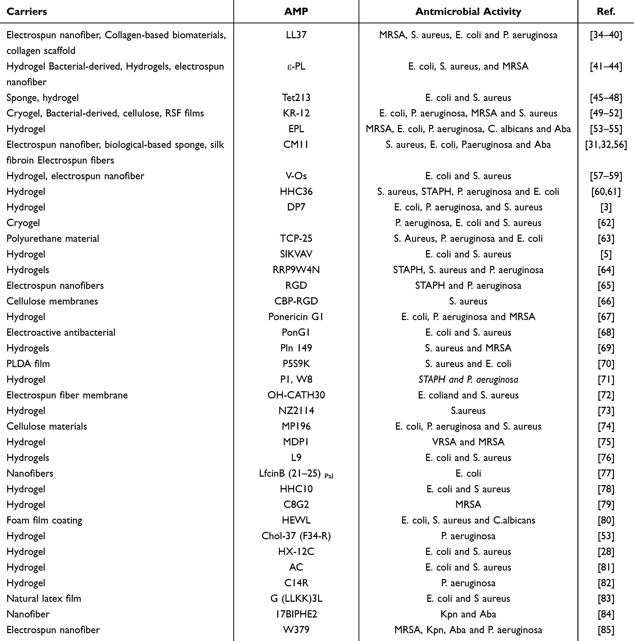 |
Table 1 Carrier Platforms and Representative AMPs |
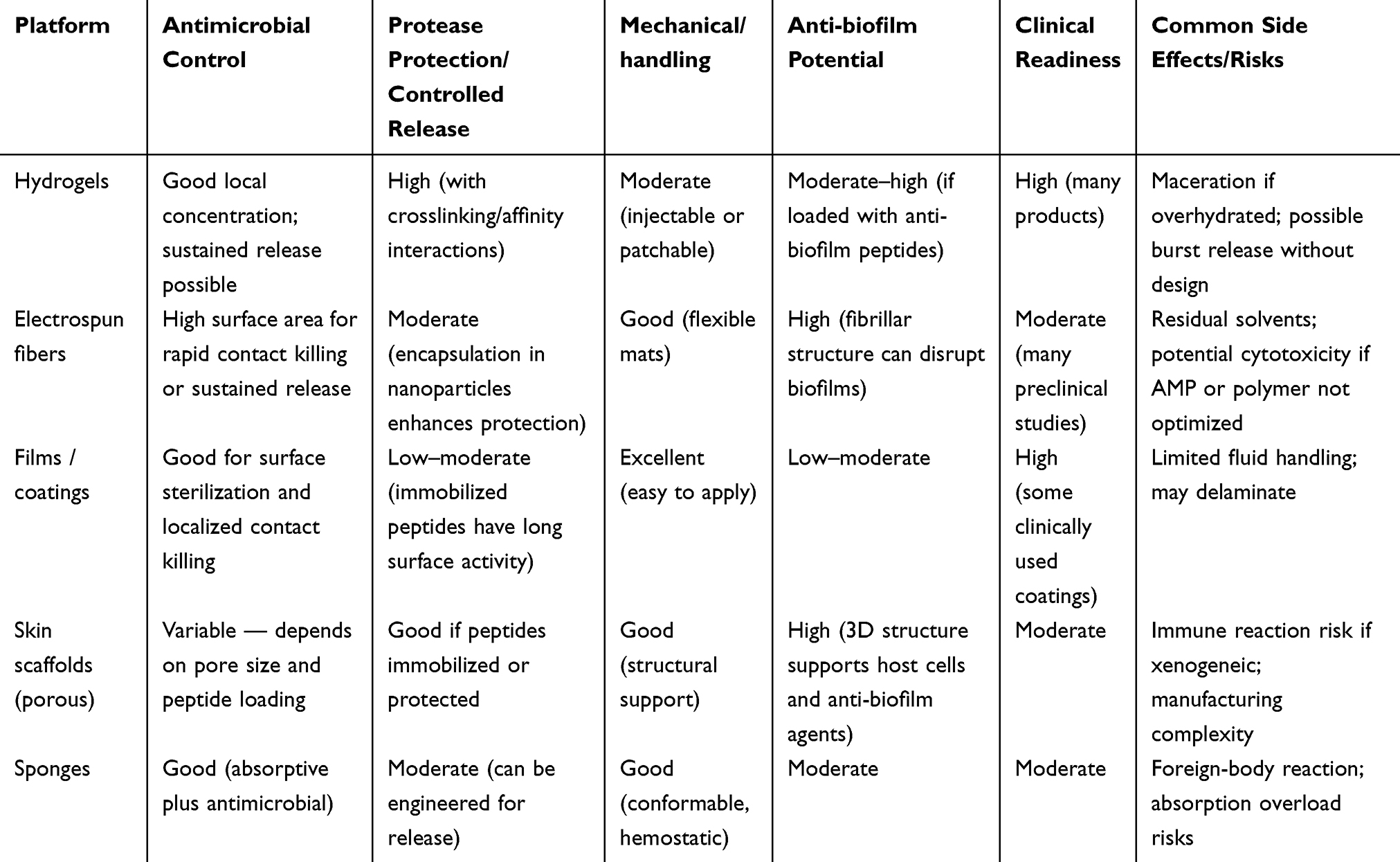 |
Table 2 Comparative Performance of Dressing Platforms |
Hydrogel Wound Dressings
Hydrogels are widely used AMP carriers because their hydrated 3D polymer networks mimic soft tissue, maintain a moist wound milieu, and can be engineered for tunable, sustained or stimulus-responsive release. Natural (hyaluronic acid, alginate, chitosan) and synthetic (PEG, polyacrylamide, PNIPAM) hydrogels can physically entrap or chemically conjugate AMPs, reduce direct protease exposure, slow peptide wash-out, and provide an anti-fouling microenvironment that preserves activity in wound fluid. Tunable crosslink density and mesh size control diffusion-driven release, while affinity interactions (electrostatic binding between cationic AMPs and anionic polysaccharides or specific peptide-binding domains) can prolong residence time and maintain sustained local antimicrobial pressure. Hydrogel matrices readily enable co-delivery of growth factors, metal ions, ROS scavengers or cell-derived exosomes, allowing combination antimicrobial and pro-regenerative activities in a single dressing.53,86
Integration of AMPs with Polysaccharide-Based Hydrogels
Polysaccharide-based hydrogels, such as hyaluronic acid (HA) and sodium alginate, have become invaluable matrix materials in the field of wound care due to their excellent moisturizing ability and injectability. When these hydrogels are combined with AMPs, the resulting dressings not only demonstrate broad-spectrum antimicrobial efficacy but also exhibit exceptional biocompatibility. They efficiently eliminate bacteria, stimulate angiogenesis, and promote cell proliferation, significantly accelerating the wound healing process.87
Through carefully designed strategies, several composite dressings have been successfully developed. For example, Zefeng Lin’s research team88 ingeniously anchored the AMP AMP-Tet213 within a composite matrix of alginate, HA, and collagen. The resulting ALG/HA/COL-AMP dressing not only exhibited excellent physical properties and biodegradability but also displayed potent antimicrobial activity against various pathogens. Furthermore, it effectively promoted the active proliferation of fibroblasts and accelerated wound closure. Another team led by Mahsa Ahmadi89 employed an innovative approach by encapsulating the AMP LL37 within microsphere carriers, which were then embedded in an activated carbon-chitosan hydrogel. This design not only protected LL37 but also enabled targeted action against bacterial toxins, offering a new avenue for the treatment of chronic wound infections. Additionally, the clever combination of HA with the AMP Plantaricin 149 led to the formation of an injectable and adhesive antimicrobial hydrogel (HAD@AMP). This hydrogel demonstrated excellent antimicrobial efficacy, tissue compatibility, antibiotic resistance, and the ability to inhibit bacterial biofilm formation. Moreover, it could regulate inflammatory responses and promote angiogenesis, providing a novel therapeutic perspective for the rapid recovery of infectious wounds.
Furthermore, Shi et al53 successfully developed a sodium alginate-based hydrogel dressing loaded with the novel AMP Chol-37 (F34-R). This dressing not only exhibited excellent cross-sectional structure and good water absorption and swelling properties but also demonstrated significant antimicrobial effects. It could create an impenetrable barrier between the wound and the external environment, effectively preventing bacterial invasion and showing great potential as a novel dressing for the treatment of infectious wounds. In summary, the combination of polysaccharide-based hydrogels and AMPs provides a new pathway for the development of efficient, biocompatible wound dressings. These dressings show great potential in promoting wound healing and preventing infection, offering novel strategies for skin healing.
Integration of AMPs with Smart Responsive Hydrogels
Smart responsive hydrogels have demonstrated significant potential in wound healing by precisely controlling the release of AMPs, thereby enhancing therapeutic efficiency. These hydrogels are capable of sensing changes in the wound environment, such as light, pH, and temperature, and adjusting their release mechanisms accordingly. pH-sensitive hydrogels, for example, release AMPs in response to the pH changes at the wound site.90,91 This responsive design allows the hydrogel to effectively combat infections, especially in cases where the wound’s pH is imbalanced.92 For instance, Li et al79 developed an in situ formed hydrogel by conjugating oxidized β-D-glucan with the AMP C(8)G(2) through a Schiff base reaction. This hydrogel is easy to prepare and has a positive impact on the entire healing process of bacterial-infected wounds, exhibiting significant antimicrobial activity and biocompatibility. The pH-sensitive reversible imine bond enables the hydrogel to self-heal and sustains the release of AMPs, thereby enhancing their bioavailability while reducing toxicity. Photoreponsive hydrogels, which combine AMPs with photosensitizers, enable controlled release under light exposure. This strategy not only improves the precision of the treatment but also reduces unnecessary drug waste and side effects. For example, Fan et al78 proposed a novel antimicrobial hydrogel by covalently coupling photosensitizers with AMPs. This hydrogel showed remarkable antimicrobial effects and good efficacy against drug-resistant bacteria due to the increased treatment precision. Another innovative strategy involves combining thermoresponsive hydrogels with CuS nanoparticles and lysozyme-loaded AMPs. By utilizing photothermal regulation to control the release of AMPs, this hydrogel can effectively combat chronic wound infections.93 In summary, smart responsive hydrogels have broad application prospects in the field of wound treatment. With continued optimization of design and fabrication processes, these hydrogels are expected to provide more efficient, precise, and safe solutions for wound healing.
Combination of AMPs and Metal Nanomaterials
Nanomaterials have emerged as a research frontier in antimicrobial materials due to their remarkable antibacterial properties and their ability to significantly enhance the mechanical strength and stability of wound dressings.94 By strategically integrating AMPs with these high-performance metallic nanomaterials, researchers are working to develop innovative metallic nanohydrogel dressings that exhibit synergistically enhanced antimicrobial effects and excellent mechanical properties. These dressings enable precise control over the release of both AMPs and metal nanoparticles, ensuring long-lasting antimicrobial efficacy while accelerating the wound healing process.95
Specifically, Xianhao Su93 successfully developed an innovative thermosensitive hydrogel composite material based on poly-N-isopropylacrylamide (PNIPAM) and polyacrylamide (PAM). This hydrogel incorporates CuS nanoparticles (CuS/PP) and one-dimensional lysozyme nanofibers loaded with the AMP melittin (M). Through photothermal regulation, this hydrogel allows for flexible control of melittin release, providing a novel strategy for the treatment of chronic wound infections.
Another notable study by Xinrui Li6 designed a dynamic, multifunctional HA-based hydrogel dressing that mimics the extracellular matrix (ECM) and integrates antimicrobial, immunomodulatory, and angiogenic functionalities for the treatment of infected skin wounds. The dynamic network structure of this hydrogel is constructed through reversible metal-ligand coordination between thiol groups and bioactive metal ions, with antimicrobial silver ions and immunomodulatory zinc ions being ingeniously incorporated into thiolated HA and angiogenic peptides. This dressing not only exhibits excellent bioactivity for infected wounds but also possesses self-healing and injectable properties, offering enhanced flexibility and potential for wound care applications.
In summary, the combination of AMPs and metal-based antimicrobial nanomaterials has opened a new avenue for the development of next-generation multifunctional, high-performance wound dressings. These innovations hold significant promise for advancing wound treatment strategies and are expected to play a critical role in future wound care applications.
Combination of AMPs and Exosomes
Exosomes,96 as nanoscale carriers naturally secreted by cells, exhibit excellent biocompatibility and efficient delivery capabilities, demonstrating immense potential in biomedical applications. Recent studies have explored a cutting-edge approach: the integration of AMPs with exosomes derived from mesenchymal stem cells (MSCs) to develop composite hydrogel systems that offer long-term storage stability, rapid re-dissolution properties, and high antimicrobial activity.96 This innovative strategy not only deepens our understanding of the delivery mechanisms of bioactive molecules but also provides new solutions for tissue repair and regeneration.97
The composite hydrogel system exhibits multiple biological effects, effectively regulating the activity of fibroblasts, vascular endothelial cells, and macrophages. Additionally, it demonstrates outstanding performance in inhibiting fibrosis mediated by myofibroblasts, which is crucial for promoting healthy tissue regeneration and reducing scar formation. These findings offer novel perspectives for addressing skin injuries, wound healing, and other tissue repair needs.98
Of particular interest, YuLing Yang3 proposed an innovative preparation strategy involving the construction of a macroporous hydrogel (HD-DP7/Exo) containing human umbilical cord mesenchymal stem cell-derived exosomes (HucMSC-Exos) using rapid freeze-drying technology. This hydrogel was further enhanced with an AMP coating on its surface. This method not only achieves stable encapsulation of exosomes within the HA-based macroporous hydrogel but also imparts the hydrogel with long-term storage and transport advantages, as well as the capability for instant re-dissolution for therapeutic use.
Comparative experiments demonstrated3 that hydrogel systems loaded with HucMSC-Exos exhibited similar positive effects in regulating cellular behavior, inhibiting fibrosis, and promoting tissue regeneration compared to hydrogels directly loaded with HucMSCs. These findings further validate the unique therapeutic value of HucMSC-Exos as extracellular communication mediators and highlight their enhanced therapeutic potential when combined with AMPs within hydrogel systems.
In summary, the integration of AMPs and HucMSC-derived exosomes has not only paved the way for efficient bioactive molecule delivery but has also brought revolutionary progress to tissue engineering and regenerative medicine. This approach holds tremendous promise for promoting healthy tissue recovery and reducing complications, with broad applications in future therapeutic scenarios.
Combination of AMPs and Cryogels
Cryogels, as emerging star materials in the biomedical field, are characterized by highly tunable porous structures and outstanding water absorption properties conferred by low-temperature gelation techniques. These features make cryogels ideal carriers for bioactive molecules, ensuring stable release and precise delivery within the body. Their excellent biocompatibility and biodegradability further guarantee their safety in clinical applications.99
Wang et al62 designed an innovative macroporous hydrogel (DA7CG@C system) that integrates AMPs and stem cells. This hydrogel, constructed through the polymerization of an HA backbone with dopamine, combines the AMP DP7 with placental mesenchymal stem cells (PMSCs), achieving comprehensive wound care from inflammation suppression and tissue regeneration to scar-free healing. The system significantly optimizes the wound healing process.
Similarly, Menglong Liu et al49 developed a multifunctional cryogel based on HA, tannic acid, and the AMP KR-12 (HA/TA2/KR2). This cryogel stands out for its shape-memory properties, strong absorption capacity, hemostatic effect, and antimicrobial activities. The intelligent release mechanism of KR-12 effectively eradicates various pathogens, while TA synergistically eliminates reactive oxygen species and regulates immune responses, thereby accelerating wound healing and demonstrating excellent in vivo performance.
In conclusion, cryogels, through meticulous design, achieve efficient delivery of bioactive molecules and precise regulation of cellular behavior while promoting healthy tissue regeneration. These advancements represent a revolutionary breakthrough and innovation in the biomedical field.
Electrospun Fiber (ESF) Wound Dressings
ESFs mats provide extracellular-matrix-like architecture that promotes cell attachment and ingrowth while offering very high surface area for AMP loading and contact killing.100 The fibrous geometry can physically block bacterial penetration and limit biofilm maturation, and fiber porosity supports oxygen and gas exchange essential for healing. However, the same high surface exposure that promotes antimicrobial contact can also accelerate peptide loss and degrade peptide stability unless peptides are encapsulated or immobilized; moreover, residual solvents, polymer choice and high local AMP concentration can contribute to cytotoxicity or local irritation. Common mitigation strategies that have been validated in preclinical studies include core–shell or multilayer fiber architectures, nanoparticle loading and post-spinning crosslinking to reduce burst release, and graded release designs to preserve antimicrobial activity while minimizing host toxicity. When properly designed, ESFs are promising for both acute and chronic wounds, but translation requires explicit attention to solvent removal, peptide stability assays in wound-like fluids, and standardized cytotoxicity testing.
The flexibility of nanofiber fabrication is attributed to its compatibility with a wide variety of polymeric materials and the ability to incorporate bioactive components. This enables the creation of an efficient and precise drug and antimicrobial agent delivery system.29 For instance, the integration of carboxymethyl chitosan nanoparticles with the AMP OH-CATH30 imparts antimicrobial efficacy and wound-healing potential to PVA/CS nanofibers.72 Similarly, recombinant LL37 peptide and VEGF encapsulated in chitosan nanoparticles and fabricated through a multilayered electrospinning process result in wound dressings with superior biophysical properties and notable efficacy against drug-resistant pathogens like MRSA.34 Polycaprolactone (PCL) fibers, another highlight, exhibit strong antibacterial activity against Staphylococcus aureus and Pseudomonas aeruginosa through the physical adsorption of nisin.101 Additionally, the synergistic strategy of combining cationic bio-based polymers with AMPs offers a novel approach for developing multifunctional ESFs, which demonstrate significant potential in the biomedical field, particularly in wound dressings.102
Of particular interest, the incorporation of insulin-like growth factor 1 (IGF1) and AMP Os into PLGA electrospun scaffolds via an innovative small pentapeptide tagging technology fused with 3,4-dihydroxyphenylalanine (DA) yields fibers that mimic the porous structure of natural ECM. These scaffolds provide an optimal microenvironment for cell growth and achieve controlled release of growth factors and AMPs, significantly enhancing wound healing and tissue regeneration.57
In summary, the exploration and application of ESFs and their composites in wound healing are rapidly advancing, showcasing their immense potential. With continuous optimization of material design and fabrication processes, ESFs are poised to offer more efficient, safer, and patient-centered solutions for wound care, heralding a new era in wound healing treatments.
Other Types of Dressings
At the forefront of research on AMP-based wound dressings, innovative strategies continue to emerge, optimizing wound management approaches. Jiamei Zhang et al successfully incorporated the AMP KR-12 into regenerated silk fibroin films. This novel design not only enables the sustained release of the AMP but also imparts the dressing with multiple functionalities, including antimicrobial activity, enhanced cell adhesion, and macrophage activity regulation. In vivo experiments have demonstrated the significant wound healing effects of this dressing, offering a breakthrough in the field of wound treatment.
Another study, conducted by Mehrdad Moosazadeh Moghaddam et al,31 focused on the innovative application of amniotic skin scaffolds. Through enhanced processing of silk fibroin and coating with the AMP CM11, the team developed a dressing with superior antimicrobial performance and excellent biocompatibility. This amniotic skin scaffold exhibited remarkable efficacy in promoting wound closure, providing a novel solution for the treatment of complex wounds.
Hatef Ghasemi Hamidabadi et al32 extracted bio-based sponges from human placental tissue and skillfully loaded them with AMPs to create a skin substitute with high antimicrobial activity. This innovative dressing is currently advancing to preclinical research stages and holds the potential to bring revolutionary changes to future wound care.
Additionally, Wang et al83 developed a glycerol-AMP natural latex film with significant antimicrobial activity and excellent biocompatibility. This dressing effectively promotes the healing of infected wounds, providing new insights and approaches for wound management and expanding the application scope of AMP-based antimicrobial dressings.
In summary, these pioneering studies collectively advance the development of AMP-based antimicrobial dressings, offering unprecedented hope for wound treatment. As research deepens and technologies continue to innovate, it is anticipated that more efficient, safe, and convenient AMP-based antimicrobial dressings will emerge, providing more comprehensive and effective solutions for patient wound care.
Multifunctional Wound Dressings
In recent years, AMP-based multifunctional wound dressings have emerged in the field of wound management, offering comprehensive and efficient solutions by integrating cutting-edge technologies. The core advantage of these dressings lies in their multifunctionality, which includes real-time bacterial monitoring, promotion of angiogenesis and tissue regeneration, antioxidation, and electrostimulation-assisted healing.
First, the real-time bacterial monitoring capability allows dressings to respond rapidly to wound infections.45 By incorporating built-in sensors or indicators, healthcare providers can promptly assess bacterial loads in wounds and adjust treatment strategies accordingly, effectively controlling infections.90 For instance, certain studies46 synthesized composite dressings combining AIEgens mesoporous bioactive glass and AMPs, enabling real-time bacterial detection, broad-spectrum antimicrobial activity, and tissue regeneration, thereby accelerating the healing of chronic wounds.
Second, these dressings are highly effective in promoting angiogenesis and tissue regeneration. By releasing AMPs and other bioactive molecules, they stimulate cell proliferation and migration, accelerate the formation of new blood vessels to supply oxygen and nutrients, and facilitate tissue repair and regeneration.70 For example, some multifunctional hydrogels achieve sequential two-stage release via near-infrared stimulation, enabling precise wound treatment while promoting angiogenesis and collagen deposition.28
Third, these dressings exhibit outstanding antioxidant properties, scavenging free radicals and mitigating oxidative stress-induced cellular damage to create a favorable environment for wound healing. For instance, Dong Yang et al67 developed multifunctional dynamic boronic acid-crosslinked hydrogels through specific chemical reactions. These hydrogels not only exhibit rapid gelation properties but also carry cationic AMPs, achieving both antimicrobial and antioxidative effects.
Finally, electrostimulation assistance represents another innovative feature of these dressings.68 Mild electric currents can further activate cellular activities, promoting cell proliferation and differentiation to accelerate wound healing. For example, a research team led by Dawei Ren58 developed multifunctional conductive gelatin methacrylate hydrogels. By using superior conductive materials as electrodes and incorporating modified AMPs to inhibit bacterial infections, this dressing accelerates skin damage repair through external electrical stimulation, enhancing cell survival rates and upregulating tissue repair-related gene expression.
In conclusion, AMP-based multifunctional wound dressings demonstrate immense potential and broad application prospects in wound management due to their outstanding multifunctionality. With ongoing advancements in technology and deeper clinical applications, these dressings are poised to become a new frontier in wound treatment, offering hope to more patients. Moving forward, we anticipate the emergence of even more innovative AMP-based multifunctional wound dressings, injecting new vitality into wound management.
Clinical Applications and Prospects of AMP-Based Wound Dressings
Innovations in Chronic Wound Treatment
Chronic wounds,103 such as diabetic foot ulcers and wounds induced by venous or arterial diseases, are often challenging to treat due to persistent infections and impaired blood circulation. In addressing these complexities, AMPs have demonstrated their unique therapeutic value. AMPs are highly effective against both Gram-negative bacteria (eg, Pseudomonas aeruginosa) and Gram-positive bacteria (eg, Staphylococcus aureus), which are major culprits in chronic wound infections.35 By inhibiting bacterial communication, reducing biofilm formation, and immobilizing bacterial cells to prevent proliferation, AMPs play a critical role in infection control.104 For instance, methacrylated chitosan (MECs) hydrogels coated with melittin-derived peptide 1 (MDP1)75 utilize an “explosive release” strategy to rapidly eliminate planktonic bacteria while exhibiting anti-biofilm activity. Moreover, AMPs are not limited to their antimicrobial function; they also modulate host immune responses, promote cell migration and proliferation, and thereby accelerate wound healing.105 A prominent example is human β-defensin-3 (HBD-3), which activates the FGFR/JAK2/STAT3 pathway critical for healing, enhances angiogenesis, and promotes fibroblast migration, significantly expediting wound repair.106 Most notably, unlike traditional antibiotics, AMPs’ unique bactericidal mechanisms make them less likely to induce resistance. This characteristic is particularly valuable for chronic wounds, which are often associated with multidrug-resistant bacteria.107 For example, AMP-based wound dressings have demonstrated superior antimicrobial efficacy against methicillin-resistant Staphylococcus aureus (MRSA),64 multidrug-resistant Pseudomonas aeruginosa (MDR-PA),108 and extensively drug-resistant Acinetobacter baumannii.
Comprehensive Care for Burn Wounds
Burn wounds are highly susceptible to bacterial infections due to the compromised skin barrier, and their healing process is often complex and variable.56 The application of AMPs in burn wound treatment has shown remarkable advantages.109 In preventing infection, AMPs rapidly inhibit bacterial growth on the surface of burn wounds, effectively reducing the risk of infection. This is critical for preventing wound deterioration and protecting the wound bed from secondary injury.86 AMPs also promote the epithelialization process in burn wounds by enhancing epithelial cell migration and coverage over the wound bed, forming a new skin barrier. This not only accelerates wound healing but also minimizes scar formation.62 Furthermore, AMPs enhance the expression of vascular endothelial growth factor (VEGF), epidermal growth factor (EGF), and angiogenic markers such as CD31, thereby expediting the wound healing process.73 In terms of immune modulation, AMPs inhibit the expression of pro-inflammatory cytokines, including interleukin-6 (IL-6), IL-1β, and tumor necrosis factor-α (TNF-α), while upregulating anti-inflammatory mediators such as transforming growth factor-β1 (TGF-β1) and platelet endothelial cell adhesion molecule-1 (CD31). These effects not only alleviate patient discomfort but also provide robust support for successful wound healing.110
Challenges and Limitations
Despite substantial preclinical promise, AMP-based wound dressings face multiple translational challenges — peptide instability and proteolytic degradation, delivery and controlled-release limitations, scale-up and manufacturing costs, cytotoxicity and off-target effects, regulatory barriers, imperfect animal models, and limited clinical data — that must be addressed to enable clinical adoption. A primary limitation of many natural AMPs is poor stability in protease-rich wound environments: host and bacterial proteases rapidly degrade linear, L-amino-acid peptides, reducing local bioavailability and efficacy.9 Additionally, achieving therapeutic peptide concentrations at the wound site for the required duration is challenging: simple topical application often produces rapid wash-out, burst release or inactivation, limiting bactericidal exposure. Controlled-release matrices, particulate carriers and biological carriers have shown promise to sustain and localize AMP delivery while protecting peptides from proteolysis and dilution.9 Moreover, cost and manufacturability remain significant barriers: chemically synthesized peptides, particularly long or extensively modified sequences, are expensive to produce at scale, and formulation stability further complicates product development. Although several AMP candidates have advanced to clinical testing, the number of well-controlled human trials of AMP-based wound dressings remains small, and some clinical programs have stalled because of limited efficacy, toxicity or commercial decisions. To accelerate translation, investigators should from the outset integrate stability-enhancing chemistries, protective delivery platforms, rigorous safety and cytotoxicity testing, manufacturability assessments and cost analyses, improved and standardized animal models, and early regulatory engagement into their development pipelines. Such an integrated approach will better position AMP-based dressings for robust clinical testing and, ultimately, routine use in complex wound care.22,111
Future Research Directions
Ongoing advances in AMP discovery, peptide engineering and delivery technologies are expanding the translational potential of AMP‑based wound dressings for chronic wounds and burns. Key technical and translational areas likely to deliver near-term impact include: First, rational peptide engineering — using computational design, high-throughput screening, genetic/biotechnological production and chemical strategies — should produce AMPs with enhanced potency, selectivity and protease resistance.112 Designed peptides should be matched to wound microenvironments or dominant pathogens to maximize efficacy while minimizing off-target cytotoxicity.113 Second, validated delivery platforms (responsive hydrogels, electrospun matrices, polymeric nanoparticles, liposomes and exosome-based vesicles) must be optimized and validated to protect peptides from proteolysis and to provide controlled, site-matched release.114 These carriers can shield peptides from enzymatic degradation, provide sustained or on‑demand release, and localize antimicrobial activity to the wound bed—maintaining local antimicrobial pressure while reducing systemic exposure.65 Engineering multifunctional dressings that combine antimicrobial action with anti-inflammatory, pro-angiogenic and matrix-remodeling activities (via co-delivery or multifunctional materials) is an important translational objective.115 More broadly, advanced chemistries and formulation strategies will expand design options to precisely control peptide presentation, stability and release kinetics appropriate to wound type and healing stage.116 Beyond chronic and acute infected wounds, these approaches could be adapted for surgical prophylaxis, implant coatings and other dermatological indications.84,89,117 Finally, adoption of standardized, higher-fidelity preclinical models (diabetic/ischemic and large-animal wounds, standardized biofilm challenges, bioluminescent and imaging readouts) will improve quantitative, longitudinal assessment of AMP activity and kinetics and better predict clinical performance.96,118 With coordinated progress in peptide engineering, delivery platforms, standardized preclinical models, manufacturing scale-up and regulatory alignment, AMP-based dressings may become integral components of next-generation wound care.
Conclusion
Ongoing advances in AMP discovery, peptide engineering and delivery technologies are expanding the translational potential of AMP-based wound dressings for chronic wounds, burns and other indications. AMPs combine broad-spectrum antimicrobial activity with immunomodulatory and pro-regenerative effects, making them attractive candidates for multifunctional dressings that address infection and support tissue repair. However, realizing clinical impact requires parallel progress in peptide optimization, protective and responsive delivery systems, robust preclinical models, manufacturability and regulatory pathways.
Funding
This study was supported by the Project of Yancheng Health Commission (No. YK2023072).
Disclosure
The authors declare no conflict of interest. Aoxun Zhu and Baiqi Chen contributed to the work equally and should be regarded as co-first authors.
References
1. Takeo M, Lee W, Ito M. Wound healing and skin regeneration. Cold Spring Harb Perspect Med. 2015;5(1):a023267. doi:10.1101/cshperspect.a023267
2. Rodrigues M, Kosaric N, Bonham CA, Gurtner GC. Wound healing: a cellular perspective. Physiol Rev. 2019;99(1):665–706. doi:10.1152/physrev.00067.2017
3. Yang Y, Zhang J, Wu S, et al. Exosome/antimicrobial peptide laden hydrogel wound dressings promote scarless wound healing through miR-21-5p-mediated multiple functions. Biomaterials. 2024;308:122558. doi:10.1016/j.biomaterials.2024.122558
4. Sultana A, Luo H, Ramakrishna S. Antimicrobial peptides and their applications in biomedical sector. Antibiotics. 2021;10(9):1094. doi:10.3390/antibiotics10091094
5. Jin J, Sun C, Xu K, Sun X, Cao L, Liu L. Multifunctional self-healing peptide hydrogel for wound healing. Int J Biol Macromol. 2024;261(Pt 1):129734. doi:10.1016/j.ijbiomac.2024.129734
6. Li X, Chen X, Guan L, et al. Bioactive metal ion-coordinated dynamic hydrogel with antibacterial, immunomodulatory, and angiogenic activities for infected wound repair. ACS Appl Mater Interfaces. 2024;16(25):32104–32117. doi:10.1021/acsami.4c05967
7. Sacks D, Baxter B, Campbell BCV, et al. Multisociety consensus quality improvement revised consensus statement for endovascular therapy of acute ischemic stroke. Int J Stroke. 2018;13(6):612–632. doi:10.1177/1747493018778713
8. Rai A, Ferrão R, Marta D, et al. Antimicrobial peptide-tether dressing able to enhance wound healing by tissue contact. ACS Appl Mater Interfaces. 2022;14(21):24213–24228. doi:10.1021/acsami.2c06601
9. Hancock RE, Sahl HG. Antimicrobial and host-defense peptides as new anti-infective therapeutic strategies. Nat Biotechnol. 2006;24(12):1551–1557. doi:10.1038/nbt1267
10. Sánchez-Vásquez L, Silva-Sanchez J, Jiménez-Vargas JM, et al. Enhanced antimicrobial activity of novel synthetic peptides derived from vejovine and hadrurin. Biochim Biophys Acta. 2013;1830(6):3427–3436. doi:10.1016/j.bbagen.2013.01.028
11. Miao F, Li Y, Tai Z, et al. Antimicrobial peptides: the promising therapeutics for cutaneous wound healing. Macromol Biosci. 2021;21(10):e2100103. doi:10.1002/mabi.202100103
12. Mickels N, McManus C, Massaro J, et al. Clinical and microbial evaluation of a histatin-containing mouthrinse in humans with experimental gingivitis. J Clin Periodontol. 2001;28(5):404–410. doi:10.1034/j.1600-051x.2001.028005404.x
13. Mahlapuu M, Sidorowicz A, Mikosinski J, et al. Evaluation of LL-37 in healing of hard-to-heal venous leg ulcers: a multicentric prospective randomized placebo-controlled clinical trial. Wound Repair Regen. 2021;29(6):938–950. doi:10.1111/wrr.12977
14. Butler MS, Cooper MA. Antibiotics in the clinical pipeline in 2011. J Antibiot. 2011;64(6):413–425. doi:10.1038/ja.2011.44
15. Steiner H, Hultmark D, Engström A, Bennich H, Boman HG. Sequence and specificity of two antibacterial proteins involved in insect immunity. Nature. 1981;292(5820):246–248. doi:10.1038/292246a0
16. Ganz T, Selsted ME, Szklarek D, et al. Defensins. Natural peptide antibiotics of human neutrophils. J Clin Invest. 1985;76(4):1427–1435. doi:10.1172/jci112120
17. Bui Thi Phuong H, Doan Ngan H, Le Huy B, Vu Dinh H, Luong Xuan H. The amphipathic design in helical antimicrobial peptides. ChemMedChem. 2024;19(7):e202300480. doi:10.1002/cmdc.202300480
18. Zhu X, Dong N, Wang Z, et al. Design of imperfectly amphipathic α-helical antimicrobial peptides with enhanced cell selectivity. Acta Biomater. 2014;10(1):244–257. doi:10.1016/j.actbio.2013.08.043
19. Ringstad L, Kacprzyk L, Schmidtchen A, Malmsten M. Effects of topology, length, and charge on the activity of a kininogen-derived peptide on lipid membranes and bacteria. Biochim Biophys Acta. 2007;1768(3):715–727. doi:10.1016/j.bbamem.2006.11.016
20. Hooper DC, Wolfson JS. Mode of action of the quinolone antimicrobial agents: review of recent information. Rev Infect Dis. 1989;11 Suppl 5(Supplement_5):S902–11. doi:10.1093/clinids/11.supplement_5.s902
21. Ramamurthy R, Mehta CH, Nayak UY. Structurally nanoengineered antimicrobial peptide polymers: design, synthesis and biomedical applications. World J Microbiol Biotechnol. 2021;37(8):139. doi:10.1007/s11274-021-03109-z
22. Mahlapuu M, Håkansson J, Ringstad L, Björn C. Antimicrobial peptides: an emerging category of therapeutic agents. Front Cell Infect Microbiol. 2016;6:194. doi:10.3389/fcimb.2016.00194
23. Lei J, Sun L, Huang S, et al. The antimicrobial peptides and their potential clinical applications. Am J Transl Res. 2019;11(7):3919–3931.
24. Bahar AA, Ren D. Antimicrobial peptides. Pharmaceuticals. 2013;6(12):1543–1575. doi:10.3390/ph6121543
25. W G. Human antimicrobial peptides and proteins. Discover Immunity. 2014;545–594. doi:10.3390/ph7050545
26. de la Fuente-Núñez C, Reffuveille F, Fernández L, Hancock RE. Bacterial biofilm development as a multicellular adaptation: antibiotic resistance and new therapeutic strategies. Curr Opin Microbiol. 2013;16(5):580–589. doi:10.1016/j.mib.2013.06.013
27. Mangoni ML, McDermott AM, Zasloff M. Antimicrobial peptides and wound healing: biological and therapeutic considerations. Exp Dermatol. 2016;25(3):167–173. doi:10.1111/exd.12929
28. Chen H, Song Y, Peng Y, et al. Hydrogel complex containing the antimicrobial peptide HX-12C accelerates healing of infected wounds. Macromol Biosci. 2023;23(4):e2200514. doi:10.1002/mabi.202200514
29. Bombin ADJ, Dunne N, McCarthy HO. Delivery of a peptide/microRNA blend via electrospun antimicrobial nanofibres for wound repair. Acta Biomater. 2023;155:304–322. doi:10.1016/j.actbio.2022.10.059
30. Choudhary P, Ramalingam B, Das SK. Fabrication of chitosan-reinforced multifunctional graphene nanocomposite as antibacterial scaffolds for hemorrhage control and wound-healing application. ACS Biomater Sci Eng. 2020;6(10):5911–5929. doi:10.1021/acsbiomaterials.0c00923
31. Moosazadeh Moghaddam M, Farhadie B, Mirnejad R, Kooshki H. Evaluation of an antibacterial peptide-loaded amniotic membrane/silk fibroin electrospun nanofiber in wound healing. Int Wound J. 2023;20(9):3443–3456. doi:10.1111/iwj.14215
32. Ghasemi Hamidabadi H, Alizadeh S, Mahboobi L, et al. Antimicrobial peptide-loaded decellularized placental sponge as an excellent antibacterial skin substitute against XDR clinical isolates. Amino Acids. 2023;55(8):955–967. doi:10.1007/s00726-023-03277-2
33. Zhang Y, Wang J, Cui Z, et al. Preparation of antibacterial hydrogel from poly(aspartic hydrazide) and quaternized N-[3-(dimethylamino) propyl] methylacrylamide copolymer with antioxidant and hemostatic effects for wound repairing. Colloids Surf B Biointerfaces. 2024;238:113881. doi:10.1016/j.colsurfb.2024.113881
34. Fahimirad S, Khaki M, Ghaznavi-Rad E, Abtahi H. Investigation of a novel bilayered PCL/PVA electrospun nanofiber incorporated Chitosan-LL37 and Chitosan-VEGF nanoparticles as an advanced antibacterial cell growth-promoting wound dressing. Int J Pharm. 2024;661:124341. doi:10.1016/j.ijpharm.2024.124341
35. Hu W, Chen Z, Chen X, et al. Double-network cellulose-based hybrid hydrogels with favourable biocompatibility and antibacterial activity for wound healing. Carbohydr Polym. 2023;319:121193. doi:10.1016/j.carbpol.2023.121193
36. Lim BY, Azmi F, Ng SF. LL37 microspheres loaded on activated carbon-chitosan hydrogel: anti-bacterial and anti-toxin wound dressing for chronic wound infections. AAPS Pharm Sci Tech. 2024;25(5):110. doi:10.1208/s12249-024-02826-6
37. Wei Z, Rolle MW, Camesano TA. Characterization of LL37 binding to collagen through peptide modification with a collagen-binding domain. ACS Omega. 2023;8(38):35370–35381. doi:10.1021/acsomega.3c05328
38. Jelodari S, Daemi H, Mohammadi P, et al. Assessment of the efficacy of an LL-37-encapsulated keratin hydrogel for the treatment of full-thickness wounds. ACS Appl Bio Mater. 2023;6(6):2122–2136. doi:10.1021/acsabm.2c01068
39. Lozeau LD, Grosha J, Smith IM, Stewart EJ, Camesano TA, Rolle MW. Alginate affects bioactivity of chimeric collagen-binding LL37 antimicrobial peptides adsorbed to collagen-alginate wound dressings. ACS Biomater Sci Eng. 2020;6(6):3398–3410. doi:10.1021/acsbiomaterials.0c00227
40. Yang X, Guo JL, Han J, et al. Chitosan hydrogel encapsulated with LL-37 peptide promotes deep tissue injury healing in a mouse model. Mil Med Res. 2020;7(1):20. doi:10.1186/s40779-020-00249-5
41. Chilunga FP, Henneman P, Requena-Méndez A, et al. Hyperuricaemia and its association with 10-year risk of cardiovascular disease among migrant and non-migrant African populations: the RODAM study. Trop Med Int Health. 2020;25(4):496–505. doi:10.1111/tmi.13362
42. Yi X, He J, Wei X, Li H, Liu X, Cheng F. A polyphenol and ε-polylysine functionalized bacterial cellulose/PVA multifunctional hydrogel for wound healing. Int J Biol Macromol. 2023;247:125663. doi:10.1016/j.ijbiomac.2023.125663
43. Yang Q, Xie Z, Hu J, Liu Y. Hyaluronic acid nanofiber mats loaded with antimicrobial peptide towards wound dressing applications. Mater Sci Eng C Mater Biol Appl. 2021;128:112319. doi:10.1016/j.msec.2021.112319
44. Tian MP, Zhang AD, Yao YX, Chen XG, Liu Y. Mussel-inspired adhesive and polypeptide-based antibacterial thermo-sensitive hydroxybutyl chitosan hydrogel as BMSCs 3D culture matrix for wound healing. Carbohydr Polym. 2021;261:117878. doi:10.1016/j.carbpol.2021.117878
45. Chen Y, Qian H, Peng D, et al. Antimicrobial peptide-modified AIE visual composite wound dressing for promoting rapid healing of infected wounds. Front Bioeng Biotechnol. 2023;11:1338172. doi:10.3389/fbioe.2023.1338172
46. Lin Y, Tan Y, Deng R, et al. Antibacterial antimicrobial peptide grafted HA/SF/Alg wound dressing containing aiegens for infected wound treating. ACS Omega. 2024;9(22):23499–23511. doi:10.1021/acsomega.4c00649
47. Jin Y, Yang Y, Duan W, Qu X, Wu J. Synergistic and on-demand release of Ag-AMPs loaded on porous silicon nanocarriers for antibacteria and wound healing. ACS Appl Mater Interfaces. 2021;13(14):16127–16141. doi:10.1021/acsami.1c02161
48. Xuan C, Hao L, Liu X, et al. Wet-adhesive, haemostatic and antimicrobial bilayered composite nanosheets for sealing and healing soft-tissue bleeding wounds. Biomaterials. 2020;252:120018. doi:10.1016/j.biomaterials.2020.120018
49. Liu M, Ding R, Li Z, et al. Hyaluronidase-responsive bactericidal cryogel for promoting healing of infected wounds: inflammatory attenuation, ROS scavenging, and immune regulation. Adv Sci. 2024;11(17):e2306602. doi:10.1002/advs.202306602
50. van Zyl EM, Coburn JM. Functionalization of bacterial cellulose with the antimicrobial peptide KR-12 via chimerical cellulose-binding peptides. Int J Mol Sci. 2024;25(3):1462. doi:10.3390/ijms25031462
51. Zhang J, Wang L, Xu C, et al. Transparent silk fibroin film-facilitated infected-wound healing through antibacterial, improved fibroblast adhesion and immune modulation. J Mater Chem B. 2024;12(2):475–488. doi:10.1039/d3tb02146g
52. Liu M, Liu T, Zhang X, et al. Fabrication of KR-12 peptide-containing hyaluronic acid immobilized fibrous eggshell membrane effectively kills multi-drug-resistant bacteria, promotes angiogenesis and accelerates re-epithelialization. Int J Nanomed. 2019;14:3345–3360. doi:10.2147/ijn.S199618
53. Shi S, Dong H, Chen X, et al. Sustained release of alginate hydrogel containing antimicrobial peptide Chol-37(F34-R) in vitro and its effect on wound healing in murine model of Pseudomonas aeruginosa infection. J Vet Sci. 2023;24(3):e44. doi:10.4142/jvs.22319
54. Gao L, Chen J, Feng W, et al. A multifunctional shape-adaptive and biodegradable hydrogel with hemorrhage control and broad-spectrum antimicrobial activity for wound healing. Biomater Sci. 2020;8(24):6930–6945. doi:10.1039/d0bm00800a
55. Rezaei N, Hamidabadi HG, Khosravimelal S, et al. Antimicrobial peptides-loaded smart chitosan hydrogel: release behavior and antibacterial potential against antibiotic resistant clinical isolates. Int J Biol Macromol. 2020;164:855–862. doi:10.1016/j.ijbiomac.2020.07.011
56. Khosravimelal S, Chizari M, Farhadihosseinabadi B, Moosazadeh Moghaddam M, Gholipourmalekabadi M. Fabrication and characterization of an antibacterial chitosan/silk fibroin electrospun nanofiber loaded with a cationic peptide for wound-dressing application. J Mater Sci Mater Med. 2021;32(9):114. doi:10.1007/s10856-021-06542-6
57. Jiao J, Peng C, Li C, Qi Z, Zhan J, Pan S. Dual bio-active factors with adhesion function modified electrospun fibrous scaffold for skin wound and infections therapeutics. Sci Rep. 2021;11(1):457. doi:10.1038/s41598-020-80269-2
58. Ren D, Zhang Y, Du B, Wang L, Gong M, Zhu W. An antibacterial, conductive nanocomposite hydrogel coupled with electrical stimulation for accelerated wound healing. Int J Nanomed. 2024;19:4495–4513. doi:10.2147/ijn.S460700
59. Liang C, Wang H, Lin Z, Zhang C, Liu G, Hu Y. Augmented wound healing potential of photosensitive GelMA hydrogel incorporating antimicrobial peptides and MXene nanoparticles. Front Bioeng Biotechnol. 2023;11:1310349. doi:10.3389/fbioe.2023.1310349
60. Huang X, Xu L, Yu X, et al. Near-infrared light-responsive multifunctional hydrogel releasing peptide-functionalized gold nanorods sequentially for diabetic wound healing. J Colloid Interface Sci. 2023;639:369–384. doi:10.1016/j.jcis.2023.02.078
61. Cheng H, Shi Z, Yue K, et al. Sprayable hydrogel dressing accelerates wound healing with combined reactive oxygen species-scavenging and antibacterial abilities. Acta Biomater. 2021;124:219–232. doi:10.1016/j.actbio.2021.02.002
62. Wang S, Wu S, Yang Y, et al. Versatile hydrogel dressings that dynamically regulate the healing of infected deep burn wounds. Adv Healthc Mater. 2023;12(30):e2301224. doi:10.1002/adhm.202301224
63. Strömdahl AC, Ignatowicz L, Petruk G, et al. Peptide-coated polyurethane material reduces wound infection and inflammation. Acta Biomater. 2021;128:314–331. doi:10.1016/j.actbio.2021.04.045
64. Atefyekta S, Blomstrand E, Rajasekharan AK, et al. Antimicrobial peptide-functionalized mesoporous hydrogels. ACS Biomater Sci Eng. 2021;7(4):1693–1702. doi:10.1021/acsbiomaterials.1c00029
65. Varshosaz J, Arabloo K, Sarrami N, et al. RGD peptide grafted polybutylene adipate-co-terephthalate/gelatin electrospun nanofibers loaded with a matrix metalloproteinase inhibitor drug for alleviating of wounds: an in vitro/in vivo study. Drug Dev Ind Pharm. 2020;46(3):484–497. doi:10.1080/03639045.2020.1730397
66. Weishaupt R, Zünd JN, Heuberger L, et al. Antibacterial, cytocompatible, sustainably sourced: cellulose membranes with bifunctional peptides for advanced wound dressings. Adv Healthc Mater. 2020;9(7):e1901850. doi:10.1002/adhm.201901850
67. Yang D, Shou Z, Xie X, et al. Gelatin-based dynamic response antioxidant, anti-inflammatory multifunctional hydrogel for enhanced diabetic wound repair. Int J Biol Macromol. 2024;260(Pt 2):129453. doi:10.1016/j.ijbiomac.2024.129453
68. Zang XL, Gao F, Zhang Z, Shen LH, Pan YH. Synergistic effects of electroactive antibacterial material and electrical stimulation in enhancing skin tissue regeneration: a next-generation dermal wound dressing. Skin Res Technol. 2023;29(11):e13465. doi:10.1111/srt.13465
69. Lin X, Fu T, Lei Y, et al. An injectable and light curable hyaluronic acid composite gel with anti-biofilm, anti-inflammatory and pro-healing characteristics for accelerating infected wound healing. Int J Biol Macromol. 2023;253(Pt 5):127190. doi:10.1016/j.ijbiomac.2023.127190
70. Hou L, Wang W, Wang MK, Song XS. Acceleration of healing in full-thickness wound by chitosan-binding bfgf and antimicrobial peptide modification chitosan membrane. Front Bioeng Biotechnol. 2022;10:878588. doi:10.3389/fbioe.2022.878588
71. Boden A, Dart A, Liao TY, et al. Enhancing the activity of surface immobilized antimicrobial peptides using thiol-mediated tethering to Poly(ethylene glycol). Macromol Biosci. 2023;23(9):e2200411. doi:10.1002/mabi.202200411
72. Zou P, Lee WH, Gao Z, et al. Wound dressing from polyvinyl alcohol/chitosan electrospun fiber membrane loaded with OH-CATH30 nanoparticles. Carbohydr Polym. 2020;232:115786. doi:10.1016/j.carbpol.2019.115786
73. Huang Y, Yang N, Teng D, et al. Antibacterial peptide NZ2114-loaded hydrogel accelerates Staphylococcus aureus-infected wound healing. Appl Microbiol Biotechnol. 2022;106(9–10):3639–3656. doi:10.1007/s00253-022-11943-w
74. Barbosa M, Simões H, Pinto SN, Macedo AS, Fonte P, Prazeres DMF. Fusions of a carbohydrate binding module with the small cationic hexapeptide RWRWRW confer antimicrobial properties to cellulose-based materials. Acta Biomater. 2022;143:216–232. doi:10.1016/j.actbio.2022.02.042
75. Ekhtiari-Sadegh S, Samani S, Barneh F, Dashtbin S, Shokrgozar MA, Pooshang Bagheri K. Rapid eradication of vancomycin and methicillin-resistant Staphylococcus aureus by MDP1 antimicrobial peptide coated on photocrosslinkable chitosan hydrogel: in vitro antibacterial and in silico molecular docking studies. Front Bioeng Biotechnol. 2024;12:1385001. doi:10.3389/fbioe.2024.1385001
76. D’Souza A, Yoon JH, Beaman H, et al. Nine-residue peptide self-assembles in the presence of silver to produce a self-healing, cytocompatible, antimicrobial hydrogel. ACS Appl Mater Interfaces. 2020;12(14):17091–17099. doi:10.1021/acsami.0c01154
77. Román JT, Fuenmayor CA, Zuluaga Dominguez CM, et al. Pullulan nanofibers containing the antimicrobial palindromic peptide LfcinB (21-25)(Pal) obtained via electrospinning. RSC Adv. 2019;9(35):20432–20438. doi:10.1039/c9ra03643a
78. Fan D, Xie R, Liu X, et al. A peptide-based pH-sensitive antibacterial hydrogel for healing drug-resistant biofilm-infected diabetic wounds. J Mater Chem B. 2024;12(22):5525–5534. doi:10.1039/d4tb00594e
79. Li L, Wang Y, Huang Z, et al. An additive-free multifunctional β-glucan-peptide hydrogel participates in the whole process of bacterial-infected wound healing. J Control Release. 2023;362:577–590. doi:10.1016/j.jconrel.2023.09.010
80. Kummer N, Huguenin-Elie L, Zeller A, et al. 2D foam film coating of antimicrobial lysozyme amyloid fibrils onto cellulose nanopapers. Nanoscale Adv. 2023;5(19):5276–5285. doi:10.1039/d3na00370a
81. Wang X, Zhao D, Li Y, et al. Collagen hydrogel with multiple antimicrobial mechanisms as anti-bacterial wound dressing. Int J Biol Macromol. 2023;232:123413. doi:10.1016/j.ijbiomac.2023.123413
82. Kraemer M, Bellion M, Kissmann AK, et al. Aptamers as novel binding molecules on an antimicrobial peptide-armored composite hydrogel wound dressing for specific removal and efficient eradication of pseudomonas aeruginosa. Int J Mol Sci. 2023;24(5):4800. doi:10.3390/ijms24054800
83. Wang R, Li R, Li F, Zheng P, Wang Z, Qian S. Glycerol and antimicrobial peptide-modified natural latex for bacteriostasis of skin wounds. ACS Omega. 2023;8(1):1505–1513. doi:10.1021/acsomega.2c07008
84. Su Y, Wang H, Mishra B, et al. Nanofiber dressings topically delivering molecularly engineered human cathelicidin peptides for the treatment of biofilms in chronic wounds. Mol Pharm. 2019;16(5):2011–2020. doi:10.1021/acs.molpharmaceut.8b01345
85. Su Y, Mainardi VL, Wang H, et al. Dissolvable microneedles coupled with nanofiber dressings eradicate biofilms via effectively delivering a database-designed antimicrobial peptide. ACS Nano. 2020;14(9):11775–11786. doi:10.1021/acsnano.0c04527
86. Qianqian O, Songzhi K, Yongmei H, et al. Preparation of nano-hydroxyapatite/chitosan/tilapia skin peptides hydrogels and its burn wound treatment. Int J Biol Macromol. 2021;181:369–377. doi:10.1016/j.ijbiomac.2021.03.085
87. Gribova V, Boulmedais F, Dupret-Bories A, et al. Polyanionic hydrogels as reservoirs for polycationic antibiotic substitutes providing prolonged antibacterial activity. ACS Appl Mater Interfaces. 2020;12(17):19258–19267. doi:10.1021/acsami.9b23140
88. Lin Z, Wu T, Wang W, et al. Biofunctions of antimicrobial peptide-conjugated alginate/hyaluronic acid/collagen wound dressings promote wound healing of a mixed-bacteria-infected wound. Int J Biol Macromol. 2019;140:330–342. doi:10.1016/j.ijbiomac.2019.08.087
89. Ahmadi M, Sabzini M, Rastgordani S, Farazin A. Optimizing wound healing: examining the influence of biopolymers through a comprehensive review of nanohydrogel-embedded nanoparticles in advancing regenerative medicine. Int J Low Extrem Wounds. 2024;15347346241244890. doi:10.1177/15347346241244890
90. Ran P, Xia T, Zheng H, et al. Light-triggered theranostic hydrogels for real-time imaging and on-demand photodynamic therapy of skin abscesses. Acta Biomater. 2023;155:292–303. doi:10.1016/j.actbio.2022.11.039
91. Biscari G, Pitarresi G, Fiorica C, et al. Near-infrared light-responsive and antibacterial injectable hydrogels with antioxidant activity based on a dopamine-functionalized gellan gum for wound healing. Int J Pharm. 2022;627:122257. doi:10.1016/j.ijpharm.2022.122257
92. Koohzad F, Asoodeh A. Cross-linked electrospun pH-sensitive nanofibers adsorbed with Temporin-Ra for promoting wound healing. ACS Appl Mater Interfaces. 2023;15(12):15172–15184. doi:10.1021/acsami.2c23268
93. Su X, Feng Y, Shi H, et al. A hydrogel dressing with tunable critical temperature and photothermal modulating melittin release for multiply antibacterial treatment. Int J Biol Macromol. 2023;239:124272. doi:10.1016/j.ijbiomac.2023.124272
94. Liu W, Yu Y, Cheng W, et al. D-A structured high-performance photothermal/photodynamic thionin-synthetic melanin nanoparticles for rapid bactericidal and wound healing effects. Adv Healthc Mater. 2023;12(21):e2203303. doi:10.1002/adhm.202203303
95. Barrett JP, Raby E, Wood F, et al. An in vitro study into the antimicrobial and cytotoxic effect of Acticoat™ dressings supplemented with chlorhexidine. Burns. 2022;48(4):941–951. doi:10.1016/j.burns.2021.09.019
96. Shiekh PA, Singh A, Kumar A. Exosome laden oxygen releasing antioxidant and antibacterial cryogel wound dressing OxOBand alleviate diabetic and infectious wound healing. Biomaterials. 2020;249:120020. doi:10.1016/j.biomaterials.2020.120020
97. Qiao Z, Wang X, Zhao H, et al. The effectiveness of cell-derived exosome therapy for diabetic wound: a systematic review and meta-analysis. Ageing Res Rev. 2023;85:101858. doi:10.1016/j.arr.2023.101858
98. Rousselle P, Braye F, Dayan G. Re-epithelialization of adult skin wounds: cellular mechanisms and therapeutic strategies. Adv Drug Deliv Rev. 2019;146:344–365. doi:10.1016/j.addr.2018.06.019
99. Hou S, Liu Y, Feng F, Zhou J, Feng X, Fan Y. Polysaccharide-peptide cryogels for multidrug-resistant-bacteria infected wound healing and hemostasis. Adv Healthc Mater. 2020;9(3):e1901041. doi:10.1002/adhm.201901041
100. Sousa MGC, Rezende TMB, Franco OL. Nanofibers as drug-delivery systems for antimicrobial peptides. Drug Discov Today. 2021;26(8):2064–2074. doi:10.1016/j.drudis.2021.03.008
101. Dart A, Sarviya N, Babaie A, et al. Highly active nisin coated polycaprolactone electrospun fibers against both Staphylococcus aureus and Pseudomonas aeruginosa. Biomater Adv. 2023;154:213641. doi:10.1016/j.bioadv.2023.213641
102. Chiloeches A, Zágora J, Plachá D, et al. Synergistic combination of antimicrobial peptides and cationic polyitaconates in multifunctional PLA fibers. ACS Appl Bio Mater. 2023;6(11):4805–4813. doi:10.1021/acsabm.3c00576
103. Lee DU, Kim SC, Choi DY, Jung WK, Moon MJ. Basic amino acid-mediated cationic amphiphilic surfaces for antimicrobial pH monitoring sensor with wound healing effects. Biomater Res. 2023;27(1):14. doi:10.1186/s40824-023-00355-0
104. Moon AY, Bailey EJ, Polanco JA, Kurata WE, Pierce LM. Antibacterial efficacy of a chitosan-based hydrogel modified with epsilon-Poly-l-Lysine against pseudomonas aeruginosa in a murine-infected burn wound model. Mil Med. 2023;188(Suppl 6):52–60. doi:10.1093/milmed/usad013
105. Ding X, Tang Q, Xu Z, et al. Challenges and innovations in treating chronic and acute wound infections: from basic science to clinical practice. Burns Trauma. 2022;10:tkac014. doi:10.1093/burnst/tkac014
106. Dong Z, Zhang X, Zhang Q, et al. Anticancer mechanisms and potential anticancer applications of antimicrobial peptides and their nano agents. Int J Nanomed. 2024;19:1017–1039. doi:10.2147/ijn.S445333
107. Grebnev GA, Bolekhan VN, Golota AS, et al. Modern aspects of combat maxillofacial trauma (review of foreign scientific-medical publications during 2012-2013). Voen Med Zh. 2013;334(4):51–53.
108. Alizadeh S, Farshi P, Farahmandian N, et al. Synergetic dual antibiotics-loaded chitosan/poly (vinyl alcohol) nanofibers with sustained antibacterial delivery for treatment of XDR bacteria-infected wounds. Int J Biol Macromol. 2023;229:22–34. doi:10.1016/j.ijbiomac.2022.11.288
109. Li W, Yu Q, Yao H, et al. Superhydrophobic hierarchical fiber/bead composite membranes for efficient treatment of burns. Acta Biomater. 2019;92:60–70. doi:10.1016/j.actbio.2019.05.025
110. Zheng Y, Yuan W, Liu H, Huang S, Bian L, Guo R. Injectable supramolecular gelatin hydrogel loading of resveratrol and histatin-1 for burn wound therapy. Biomater Sci. 2020;8(17):4810–4820. doi:10.1039/d0bm00391c
111. Craik DJ, Fairlie DP, Liras S, Price D. The future of peptide-based drugs. Chem Biol Drug Des. 2013;81(1):136–147. doi:10.1111/cbdd.12055
112. Manoharan C, Thomas DS, Yashwant RS, et al. Bioengineered and functionalized silk proteins accelerate wound healing in rat and human dermal fibroblasts. Integr Biol. 2022;14(7):151–161. doi:10.1093/intbio/zyac014
113. Huang HN, Chan YL, Wu CJ, Chen JY. Tilapia piscidin 4 (TP4) stimulates cell proliferation and wound closure in MRSA-infected wounds in mice. Mar Drugs. 2015;13(5):2813–2833. doi:10.3390/md13052813
114. Andreu V, Mendoza G, Arruebo M, Irusta S. Smart dressings based on nanostructured fibers containing natural origin antimicrobial, anti-inflammatory, and regenerative compounds. Materials. 2015;8(8):5154–5193. doi:10.3390/ma8085154
115. Obuobi S, Tay HK, Tram NDT, et al. Facile and efficient encapsulation of antimicrobial peptides via crosslinked DNA nanostructures and their application in wound therapy. J Control Release. 2019;313:120–130. doi:10.1016/j.jconrel.2019.10.013
116. Mofazzal Jahromi MA, Sahandi zangabad P, Moosavi Basri SM, et al. Nanomedicine and advanced technologies for burns: preventing infection and facilitating wound healing. Adv Drug Deliv Rev. 2018;123:33–64. doi:10.1016/j.addr.2017.08.001
117. Dart A, Bhave M, Kingshott P. Antimicrobial peptide-based electrospun fibers for wound healing applications. Macromol Biosci. 2019;19(9):e1800488. doi:10.1002/mabi.201800488
118. Badal D, Dayal D, Singh G, Sachdeva N. Role of DNA-LL37 complexes in the activation of plasmacytoid dendritic cells and monocytes in subjects with type 1 diabetes. Sci Rep. 2020;10(1):8896. doi:10.1038/s41598-020-65851-y
 © 2025 The Author(s). This work is published and licensed by Dove Medical Press Limited. The
full terms of this license are available at https://www.dovepress.com/terms
and incorporate the Creative Commons Attribution
- Non Commercial (unported, 4.0) License.
By accessing the work you hereby accept the Terms. Non-commercial uses of the work are permitted
without any further permission from Dove Medical Press Limited, provided the work is properly
attributed. For permission for commercial use of this work, please see paragraphs 4.2 and 5 of our Terms.
© 2025 The Author(s). This work is published and licensed by Dove Medical Press Limited. The
full terms of this license are available at https://www.dovepress.com/terms
and incorporate the Creative Commons Attribution
- Non Commercial (unported, 4.0) License.
By accessing the work you hereby accept the Terms. Non-commercial uses of the work are permitted
without any further permission from Dove Medical Press Limited, provided the work is properly
attributed. For permission for commercial use of this work, please see paragraphs 4.2 and 5 of our Terms.
Recommended articles
Antimicrobial Peptides Against Antimicrobial-Resistant Bacteria: Focus on Machine Learning
Tahmasebi H, Arabestani MR
Infection and Drug Resistance 2026, 19:602699
Published Date: 23 June 2026