Back to Journals » International Journal of Nanomedicine » Volume 21
Antimicrobial Hydrogel for Diabetic Wound Treatment
Authors Huang H, Zhang P, Li S, Song Q, Hu Z, Zhou C, Yang L ![]()
Received 4 October 2025
Accepted for publication 17 March 2026
Published 23 March 2026 Volume 2026:21 572181
DOI https://doi.org/10.2147/IJN.S572181
Checked for plagiarism Yes
Review by Single anonymous peer review
Peer reviewer comments 3
Editor who approved publication: Dr Kamakhya Prakash Misra
Huan Huang,1– 3,* Puhua Zhang,3,4,* Songjie Li,5 Qiuhe Song,1,3 Zhongcheng Hu,1 Chunxia Zhou,1,3 Lei Yang2
1Department of Dermatology, Affiliated Hospital of Jiujiang University, Jiujiang, 332000, People’s Republic of China; 2Zhejiang Provincial Key Laboratory of Silk and Silk Protein New Materials, Zhejiang Academy of Agricultural Sciences, Hangzhou, 310021, People’s Republic of China; 3Graduate Department, Gannan Medical University, Ganzhou, 341000, People’s Republic of China; 4Department of Cardiology, Jiujiang University Affiliated Hospital, Jiujiang, 332000, People’s Republic of China; 5Department of Plastic and Reconstructive Surgery, Xijing Hospital, Fourth Military Medical University, Xi’an, 710032, People’s Republic of China
*These authors contributed equally to this work
Correspondence: Lei Yang, Chunxia Zhou, Email [email protected]; [email protected]
Abstract: Diabetic wound infections pose a significant clinical challenge. Chronic wounds associated with diabetes mellitus exhibit delayed healing and, if not managed appropriately, are highly susceptible to infections that may lead to life-threatening complications in severe cases. Antimicrobial hydrogels have become promising materials for diabetic wound management due to their moisture retention, biocompatibility, biodegradability, and intrinsic antimicrobial properties. As wound dressings, they offer advantages such as wound exudate absorption, controlled drug 2release, and minimized toxic side effects. By incorporating antimicrobial components (e.g. chitosan (CS), polyethylenimine (PEI)) or serving as carriers for antimicrobial agents (e.g. antibiotics, antimicrobial peptides (AMPs), inorganic metal materials, and carbon nanomaterials (CNMs)), antimicrobial hydrogels effectively inhibit microbial growth. With ongoing research, the development of intelligent, multifunctional, and highly responsive antimicrobial hydrogels continues to advance, offering more precise and rapid therapeutic solutions for diabetic wounds. This review first examines the impact of infection on diabetic wound healing, followed by an overview of the definition and classification of hydrogels. Subsequently, various antimicrobial hydrogels and their mechanisms of action are summarized, along with an exploration of their fabrication methods. By analyzing recent advancements in antimicrobial hydrogel research, this review aims to provide insights into future research directions and potential clinical applications for diabetic wound management.
Keywords: hydrogel, antimicrobial hydrogel, diabetic wound, wound healing
Introduction
Diabetic wounds are characterized by difficult healing and troublesome treatment.1 Current first-line treatments for diabetic wounds include wound care, debridement of necrotic tissue, offloading of pressure from the affected area, glycemic control, antibiotic therapy, and improved circulation.2 While these treatments address some clinical challenges, high recurrence rates remain a persistent issue, largely due to bacterial infections. Studies have shown that diabetic wound infections are characterized by a high bacterial load, with many of these microorganisms exhibiting resistance to conventional antibiotics.3,4 Furthermore, biofilm formation on the wound surface significantly impairs antimicrobial drug penetration, thereby reducing treatment efficacy.5,6 In addition to infection-related challenges, conventional wound dressings such as gauze suffer from several limitations, including a lack of antimicrobial properties, frequent replacement requirements, incomplete wound coverage, and restrictions on joint mobility. Antimicrobial hydrogels offer a promising alternative to conventional dressings due to their flexibility, biocompatibility, and ability to incorporate various antimicrobial efficacy.6,7
Hydrogels are highly versatile materials with applications across multiple industries, including agriculture, cosmetics, and biomedicine. In the medical field, they are widely utilized for controlled drug release, wound dressings, tissue engineering scaffolds, and enzyme immobilization.8–12 Wound healing is generally divided into four phases, including hemostasis, inflammation, proliferation, and remodeling. Hydrogels can promote wound healing by regulating these four phases.13–16 As a subclass of hydrogels, antimicrobial hydrogels exhibit unique advantages, such as the ability to absorb wound exudates, regulate drug release, minimize cytotoxicity, and prevent secondary wound damage upon removal. The antimicrobial properties of these hydrogels can be achieved through the integration of intrinsically antimicrobial materials (eg., chitosan (CS), polyethylenimine (PEI)) or through the incorporation of antimicrobial agents (eg., antibiotics, antimicrobial peptides (AMPs), inorganic metal nanoparticles (NPs), and carbon-based nanomaterials). Furthermore, smart antimicrobial hydrogels have been designed to be responsive to external stimuli such as light, temperature, redox conditions, pH, and enzyme activity, triggering the release of antimicrobial agents or exposing antimicrobial functional groups for enhanced therapeutic action. These dynamic properties enable sustained and controlled antimicrobial release, mitigating the risks associated with antibiotic overuse and resistance development. Composite hydrogels that synergize the above antimicrobial mechanisms and new antimicrobial hydrogels that integrate antimicrobial and wound-healing-promoting drugs in the same hydrogel are also emerging. In addition, the development of a specialized response to diabetic wounds to specifically monitor the antimicrobial of the wound and real-time antimicrobial also began to gradually research and achieved some results, through the antimicrobial hydrogel is expected toprovide diabetic wounds with a more effective, accurate and rapid treatment options. This review begins with an introduction to the fundamental concepts of hydrogels, including their definition, classification, and key properties. It then transitions to a discussion of antimicrobial hydrogels, outlining their various types and mechanisms of action. Additionally, the article summarizes commonly used fabrication methods for antimicrobial hydrogels. Finally, the current applications of antimicrobial hydrogels in diabetic wound treatment are reviewed, along with a discussion of existing challenges and future research directions. Compared with previous reviews, this paper establishes a coherent logical framework that spans from material design to clinical application.17
Hydrogel
Definition
A hydrogel is a 3D polymeric network capable of absorbing and retaining large amounts of water while maintaining its structural integrity without dissolving.18 The water absorption capacity of hydrogels is strongly linked to their degree of cross-linking; higher cross-linking density results in reduced water absorption attributable to the constrained movement of polymer chains. Hydrogels exist in a unique semi-solid state, exhibiting properties of both solids and liquids. While solids retain their shape and volume under specific conditions, hydrogels allow solute diffusion and controlled permeability, similar to liquids.8,19 Over the past few years, hydrogels have garnered significant attention as promising materials for wound healing applications due to their high water content and solid-like mechanical properties. Their ability to mimic the extracellular matrix makes them suitable for tissue engineering and drug delivery. Currently, antimicrobial hydrogels represent a rapidly advancing area of research in biomedicine, with numerous innovative formulations being developed. These hydrogels offer advantages such as high solubility, enhanced oxygen permeability, superior biocompatibility, efficient drug loading and release, and structural versatility, making them highly effective for infection control and wound management.20
Classification
Hydrogels can be classified into synthetic polymer hydrogels and natural polymer hydrogels based on the composition of their precursor materials. Synthetic polymer hydrogels are typically synthesized from polymers such as polymethyl methacrylate, polyethylene glycol, PVA, polyvinylpyrrolidone, poly(lactic-co-glycolic acid) (PLGA), and polyurethane. These materials provide excellent mechanical strength and stability but often lack biodegradability. In contrast, natural polymer hydrogels are primarily derived from polysaccharides (eg., hyaluronic acid (HA), alginate (Alg), CS) and proteins (eg., albumin, collagen, elastin). Unlike synthetic polymers, natural polymer-based hydrogels exhibit intrinsic biodegradability, superior biocompatibility, and bioactivity, enhancing their suitability for biomedical applications.21–23 However, their mechanical properties, particularly tensile strength and rigidity, are generally weaker compared to synthetic hydrogels. To address these limitations, chemical modifications can be introduced to natural polymers to enhance their mechanical strength and bioactivity by introducing cross-linking sites suitable for structural modifications. On the other hand, synthetic polymer networks can be constructed through copolymerization of polymer backbones with cross-linking monomers or by reacting synthetic macromolecules with cross-linking agents. A promising approach involves the hybridization of natural and synthetic polymers, resulting in natural-synthetic hybrid hydrogels that integrate the biodegradability and bioactivity of natural materials with the mechanical robustness and tunability of synthetic polymers. These hybrid hydrogels offer enhanced functionality, rendering them particularly advantageous for advanced biomedical applications.24
Characterization
Degradability
Traditional wound dressings, while providing cleanliness and breathability, are non-degradable, often leading to strong adhesion to the wound bed and secondary tissue damage upon removal. In contrast, hydrogels offer the advantage of biodegradability, enabling gradual decomposition within the tissue environment, thereby minimizing adhesion-related complications and promoting wound healing.25–27 The degradability of hydrogels is primarily governed by the breakdown of the polymer backbone or the cleavage of cross-linked chemical bonds. These processes can be influenced by mechanical stress, pH fluctuations, and immune responses, with the resulting degradation products being metabolized or excreted by the body.26 Furthermore, conventional dressings lack controlled drug release mechanisms, increasing the risk of overdosing or suboptimal therapeutic effects. In contrast, the slow degradation of hydrogels facilitates sustained drug release, maintaining a stable drug concentration over time, thereby enhancing therapeutic efficacy.28 Recent research has highlighted the potential of antimicrobial hydrogels for precise drug delivery and enhanced wound healing. Lan et al29 developed an antimicrobial hydrogel foam dressing designed for the controlled release of gallium maltolate (GaM), an antimicrobial agent known for its broad-spectrum antimicrobial activity, low toxicity, reduced bacterial resistance, and efficacy in controlling persistent infections (Figure 1A). The dressing contains GaM-loaded microspheres, and the release kinetics can be regulated by adjusting microsphere size and drug-loading capacity, effectively reducing the healing time of infected wounds. Correspondingly, Tharindu et al30 engineered a force-triggered self-destructing hydrogel, incorporating nuclease-doped, DNA cross-linked polymer networks. Under an externally applied Pico-Newtonian force, enzyme cleavage sites within the DNA cross-links become selectively exposed, triggering rapid polymer degradation and targeted drug release. These innovative hydrogels hold significant promise for the treatment of diseases associated with abnormally high cellular traction forces or elevated shear stress conditions.
 |
Figure 1 Properties of hydrogels. (A) (i) PEGDA hydrogel foam and its encapsulated microspheres. (ii) SEM images of GaM microspheres with different GaM loading ratios29 Licensed under CC BY 4.0. (B) (i) The shrinkage image of the hydrogel at 37°C.(ii) HE staining of the wound in the hydrogel group.The black arrow indicates the regenerated hair follicle. (iii) SEM detection of bacterial adhesion to hydrogels31 Copyright 2022, American Chemical Society. (C) (i) Fluorescence imaging showed that 1.0% FHPCH was almost completely degraded within 48 hours. (ii) Pictures of two hydrogels under different temperatures and light conditions32 Copyright 2022, Elsevier Ltd. |
Mechanical Properties
Compared to traditional wound dressings, hydrogels exhibit superior mechanical properties due to their ability to undergo physical and chemical cross-linking, which enhances their adhesion and elasticity. These properties enable hydrogels to conform closely to tissues, including highly mobile areas such as joints, without excessive tightening. This ensures unrestricted blood circulation, prevents excessive compression, and provides sufficient mechanical strength to resist rupture.33 Recent advancements have further enhanced the mechanical adaptability of hydrogels. Cai et al31 constructed a special hydrogel system comprising two different temperature-responsive antimicrobial hydrogels, an outer layer responsive to ambient temperature and an inner layer responsive to body temperature (Figure 1B).The inner layer shrinks more rapidly, whereas the outer layer exhibits minimal shrinking during application. This differential contraction facilitates wound closure and allows the structure to endure mechanical deformation. In addition, the hydrogel demonstrates excellent shape recovery following cyclic stretching, strong skin adhesion, and intrinsic antimicrobial activity. Additionally, it promotes wound healing, making it particularly advantageous for dynamic wound management.
Monitorability
The intrinsic transparency of hydrogels allows for direct visualization of wound healing progress without requiring the removal of the dressing, thereby minimizing the risk of secondary injuries.34 Moreover, some advanced hydrogel dressings are capable of real-time infection monitoring by incorporating responsive materials that react to bacterial presence, metabolic byproducts, or environmental changes. These materials enable dynamic detection mechanisms, such as color changes or fluorescence shifts, facilitating timely medical interventions.35 Recent studies have demonstrated innovative monitoring-integrated hydrogel systems. Huang et al32 introduced an aggregation-induced emission fluorogenic covalently incorporated into hydroxypropyl chitin (HPCH), yielding thermosensitive fluorescent hydroxypropyl chitin (FHPCH) (Figure 1C).Under UV irradiation, FHPCH hydrogels exhibit strong yellow-green fluorescence, which can dynamically respond to temperature fluctuations in a reversible manner. Notably, the fluorescence intensity decay correlates with hydrogel weight loss, enabling real-time tracking of hydrogel degradation. Since bacterial infections often induce local temperature variations, infection progression can be monitored by analyzing fluorescence intensity changes. Additionally, You et al36 developed a hydrogel-based skin sensor utilizing amphiphilic carboxymethylcellulose (CMC) to continuously monitor pH and temperature at the wound site. In animal experiments, relative resistance signals demonstrated that the sensor dressing exhibited high sensitivity to temperature and pH fluctuations, with in vitro results aligning with in vitro observations. Such a hydrogel-based sensing platform offers great potential for real-time clinical management of infected or chronic wounds. Beyond passive monitoring, some hydrogels integrate bacterial identification and sterilization, functioning as closed-loop, on-demand drug delivery systems. Pan et al37 reported a hydrogel-based RF H2S sensor, constructed by incorporating silver nanoparticles-chlorhexidine (AG-AgNPs-CHL hydrogel) with a split-ring resonator. When pathogenic bacteria generate H2S, AgNPs selectively bind to sulfide, triggering a wireless sensing signal transmission to a mobile interface. In response, a broad-spectrum antimicrobial agent, chlorhexidine, is released, effectively sterilizing the wound site.
Scalability
Unlike traditional dressings, which typically serve a single function, hydrogels act as versatile platforms capable of incorporating various bioactive agents, thereby extending their functionality beyond simple wound coverage.21 By loading hydrogels with therapeutic compounds, such as small-molecule drugs (eg., antibiotics, antitumor agents) or large biomolecules (eg., proteins, peptides, stem cells, and growth factors), hydrogels can enhance drug stability, optimize therapeutic efficacy, and reduce drug overuse. For instance, Hu et al38 developed a HA-based hydrogel embedded with polymeric nanoparticles (NPs) targeting human chondroitin sulfate proteoglycans and interleukin-15. When implanted into the tumor cavities of mice following subcutaneous melanoma resection, this hydrogel exhibited a sustained drug release profile, significantly reducing drug consumption while effectively preventing tumor recurrence, outperforming traditional treatment methods.
In the context of large-molecule drug delivery, hydrogels provide enhanced stability and protection against premature degradation.39 Peptides, which consist of natural amino acid derivatives linked via amide bonds, exhibit a stable secondary structure akin to natural proteins, enabling them to accelerate wound healing and tissue regeneration.40 Xi et al41 designed a dual-loaded thermosensitive HPCH hydrogel system, which facilitated stem cell recruitment and chondrogenic differentiation by gradually releasing stromal-derived factor-1α-like peptide (SDFP) and karyopherin (KGN). In animal experiments, this hydrogel effectively repaired articular cartilage defects, demonstrating its potential in orthopedic applications. Beyond drug delivery, hydrogels serve as cell carriers, maintaining cell viability and supporting stem cell-based regenerative therapies. Stem cells possess multilineage differentiation potential, making them promising for peripheral nerve repair. Xu et al42 designed a biodegradable multifunctional hydrogel for stem cell encapsulation, which significantly upregulated gene expression associated with mesenchymal stem cells (MSCs) and Schwann-like cell differentiation. This hydrogel enhanced myelin sheath formation and axonal regeneration, providing a powerful tool for neural tissue repair. Hydrogels also serve as scaffolds for growth factor delivery, promoting tissue and vascular regeneration. Growth factors play significant roles in regulating cell proliferation, differentiation, and apoptosis regulation, making them widely used in wound healing and regenerative medicine.43 Niu et al44 incorporated growth factors into a hydrogel, which subsequently promoted both bone and angiogenesis. Additionally, hydrogels can be functionalized with magnetic NPs, enabling applications in magnetic targeting therapy and magnetic resonance imaging. The magnetic NP-loaded hydrogel functions as a magnetic scaffold, capable of stimulating cell proliferation and differentiation to enhance angiogenesis. Under a fluctuating magnetic field, it can activate both the hydrogel matrix and embedded cells, while under a static magnetic field, it provides structural support with tunable magnetic properties. Hong et al45 developed a magnetic fibronectin nanofibrous hydrogel for the targeted delivery of iron oxide magnetic NPs, demonstrating peripheral nerve regeneration with efficacy comparable to autografts. In the field of neuroscience, magnetic scaffolds and magnetic stimulation are increasingly recognized for their potential to enhance neurite outgrowth and neural regeneration.
Classification of Antimicrobial Hydrogels and Their Antimicrobial Mechanisms
The antimicrobial efficacy of antimicrobial hydrogels primarily depends on either the intrinsic antimicrobial properties of their base materials or the incorporation of antimicrobial agents. Based on their mechanisms of action, antimicrobial hydrogels can be categorized into intrinsic antimicrobial hydrogels, antimicrobial agent-releasing hydrogels, and responsive antimicrobial hydrogels. Intrinsic antimicrobial hydrogels exert their antimicrobial effects through the inherent antimicrobial properties of their base materials. Antimicrobial agent-releasing hydrogels function by gradually releasing encapsulated antimicrobial agents to achieve sustained antimicrobial activity. Responsive antimicrobial hydrogels are formulated to be responsive to particular environmental stimuli (eg., light exposure, temperature variations, redox conditions, pH shifts, and enzymatic activity), triggering the release of antimicrobial agents or inducing structural changes that expose antimicrobial functional groups, thereby enhancing their antimicrobial efficacy (Table 1).
 |
Table 1 Comparison of Various Types of Antimicrobial Hydrogels |
Intrinsic Antimicrobial Hydrogels
Intrinsic antimicrobial hydrogels are formulated from materials that inherently possess antimicrobial properties.58 These hydrogels exhibit antimicrobial properties without requiring additional antimicrobial agents or antibiotics.33 The synthesized material can be classified according to its origin as hydrogels derived from natural antimicrobial materials and those derived from synthetic materials.
Antimicrobial Hydrogels Derived From Natural Materials
Common natural antimicrobial hydrogels include CS and its derivatives, HA, natural polysaccharides, and turmeric extracts.59 The antimicrobial mechanisms of hydrogels derived from natural and synthetic materials differ significantly. For example, the antimicrobial activity of CS and its derivatives primarily arises from the interaction between cationic groups on the polymer chain and the negatively charged bacterial surface, which disrupts cell membrane integrity, ultimately leading to bacterial death. Additionally, positively charged CS binds to microbial DNA, inhibiting replication and transcription, thereby exerting bactericidal effects.60 Based on this principle, Cao et al61 developed a CS sponge composed of CS and quaternized chitosan (QCS), demonstrating strong antimicrobial properties that protect wounds from bacterial invasion (Figure 2A).Simultaneously, the porous structure of the QCS/CS sponge facilitates rapid absorption of tissue fluids, reducing skin hydration and promoting wound healing.
 |
Figure 2 A variety of different hydrogels. (A) (i) Preparation of QCS/CS hydrogel (ii) Pictures of QCS/CS sponges prepared from different CS. (iii) The antibacterial performance of QCS/CS4 sponge61 Copyright 2024, Elsevier Ltd. (B) (i) The anti-inflammatory, anti-infective and wound healing- promoting effects of multifunctional hydrogel. (ii) 3D CLSM images of S. aureus Xen36 biofilms after different treatments. Live bacteria are indicated by green fluorescence, while dead bacteria are indicated by red fluorescence. G4 is a G-quadruplex hydrogel without Mn, NPs and bFGF. (iii) Optical images of the wound regions in mice with different treatments at various time points62 Copyright 2024, Elsevier B.V. (C) (i) SEM images of the different hydrogels. (ii) Images of the hydrogel during stretching, bending, and compression63 Copyright 2024, Elsevier B.V. |
Antimicrobial Hydrogels Derived From Synthetic Materials
Synthetic material-based hydrogels typically comprise PEI, polyacrylic acid, polyacrylamide (PAM), and their derivatives In contrast, synthetic antimicrobial materials, such as PEI, exhibit antimicrobial activity through structural domains that bind to bacteria, leading to bacterial lysis and death62 (Figure 2B). Tikhomirov et al64 synthesized a nanocomposite hydrogel composed of cellulose nanofibrils (CNF) and branched low molecular weight polyethylenimine (CNF-PEI 800) via a catalytic reaction. The incorporated PEI conferred intrinsic antimicrobial properties to the hydrogel. Furthermore, Chun et al65 reported a hydrogel consisting of a crosslinked network of poly(ethylene glycol) dimethacrylate and an overhanging PEI star copolymer. Upon bacterial contact, the hydrogel degrades, releasing PEI at the infection site, effectively eliminating bacteria remaining through direct contact. Additionally, the hydrogel facilitates the removal of bacterial biofilms. Animal studies demonstrated that this hydrogel achieves a >99.9% reduction in biofilms of methicillin-resistant Staphylococcus aureus. However, single-component antimicrobial materials often fail to meet all application requirements, necessitating the combination of multiple antimicrobial agents. Zhang et al63 developed hydrogels with interpenetrating polymer networks composed of ferrocene-grafted polyethylenimine (PEI-Fc), sodium alginate (SA), and poly(vinyl alcohol) (PVA) (Figure 2C). These hydrogels not only exhibited enhanced antimicrobial efficacy but also demonstrated improved mechanical properties through various non-covalent interactions.
Antimicrobial Agent-Releasing Hydrogels
Antimicrobial agent-releasing hydrogels are designed to incorporate antimicrobial agents within the hydrogel matrix, enabling their controlled release through the hydrogel’s unique structural properties to achieve sustained antimicrobial effects. Compared to the conventional clinical application of antimicrobial agents, such as direct topical administration, loading antimicrobial agents onto hydrogels not only enhances antimicrobial efficacy but also reduces the risk of drug overuse. Common antimicrobial agents integrated into hydrogels include antibiotics, AMPs, various inorganic metal-antimicrobial materials, and carbon nanomaterials (CNMs).
Hydrogels Loaded with Antibiotics
With the increasing complexity of wound infections and the emergence of drug-resistant bacteria, hydrogels with enhanced antimicrobial properties are needed. While antibiotics remain widely used in clinical antimicrobial treatments, systemic administration poses risks such as antibiotic overuse and adverse side effects. Antimicrobial agent-releasing hydrogels provide a potential remedy for these challenges. By incorporating various antimicrobial agents into hydrogels through different loading techniques, controlled and sustained drug release can be achieved, leveraging the advantages of hydrogels such as biodegradability, biocompatibility, and scalability. This method not only improves antimicrobial effectiveness but also reduces side effects.17,66
Commonly incorporated antibiotics in hydrogels include quinolones, beta-lactams, macrolides, aminoglycosides, and tetracyclines. Quinolone antibiotics exhibit broad-spectrum antimicrobial activity by inhibiting bacterial DNA replication through interactions with DNA gyrase. Representative quinolones include ciprofloxacin (CIP), ofloxacin, and difloxacin. Jin et al67 demonstrated that incorporating ofloxacin into hydrogels enhanced antimicrobial activity and promoted wound healing. Similarly, Fahimeh et al68 developed a CS hydrogel containing zinc tetraamino phthalocyanine (ZnTAPc) and difloxacin, enabling controlled difloxacin release. This formulation not only expanded the antimicrobial spectrum but also reduced drug dosage and mitigated side effects. Furthermore, Krishna et al69 synthesized a nanocomposite hydrogel based on carbon quantum dots (CQD), incorporating polyacrylamide/dextran (PAM/Dex/CQD) via in situ polymerization of acrylamide and loading CIP as an antimicrobial agent. This hydrogel system facilitated the controlled and sustained release of CIP, improved its solubility and bioavailability, and reduced the risk of drug-related complications.
The antimicrobial mechanism of β-lactam antibiotics primarily involves inhibiting bacterial cell wall synthesis, leading to cell expansion and lysis. Common β-lactam antibiotics include cefazolin, nafcillin and carbapenems.70 Akmal et al71 developed a hydrogel microneedle system incorporating cefazolin, enabling transdermal drug delivery by penetrating the stratum corneum in a minimally invasive manner. This system offers a promising strategy for treating deep tissue infections, such as infectious arthritis, osteomyelitis, and cellulitis. In animal studies, the hydrogel demonstrated effective drug delivery while mitigating the risk of antibiotic overuse associated with systemic administration. Gabriela et al72 designed a photocrosslinkable nanocomposite hydrogel incorporating nafcillin, using tris (ethylene glycol) divinyl ether (TEGDVE) as a crosslinking agent. This nanocomposite hydrogel enables targeted nafcillin release and exhibits strong antimicrobial activity against Gram-positive bacteria. Experiment results confirmed its high bactericidal efficacy against S. aureus. Angeliki et al73 constructed glutathione-conjugated polyethylene glycol hydrogels loaded with meropenem. In vitro porcine skin assays demonstrated strong antimicrobial activity against Pseudomonas aeruginosa, highlighting the hydrogel’s enhanced targeting capability and convenient application.
The antimicrobial mechanism of macrolide antibiotics involves inhibiting RNA-dependent protein synthesis by reversibly binding to the 50S ribosomal subunit of bacterial cells. Common macrolides include erythromycin (EM), roxithromycin, azithromycin, and clarithromycin (Cla).74,75 Zheng et al76 encapsulated EM into bifunctional polyhedral oligomeric sesquisiloxanes (BPOSS) within a poly(PEG/PPG ethyl carbamate) (BPEP) hydrogel matrix. This hydrogel exhibits excellent self-assembly properties and gelation at the target site. Additionally, it maintains excellent cytocompatibility and enhances the water solubility of EM. The antimicrobial efficacy of the EM-loaded NPs against S. aureus was confirmed via the zone of inhibition assay. Moreover, in vitro and in vivo studies demonstrated the hydroge’s excellent biocompatibility, bioavailability, and biosafety. He et al77 developed a hydrogel with Cla. This hydrogel enables controlled Cla release under specific conditions, thereby enhancing its antimicrobial efficacy through a synergistic antimicrobial mechanism. Given its potent antimicrobial performance, this hydrogel shows significant potential for future wound dressing applications.
The antimicrobial mechanism of aminoglycoside antibiotics primarily relies on their amino and hydroxyl groups (−OH), which interact with the A-site of the 16S ribosomal RNA (rRNA) in bacterial cells, ultimately inhibiting protein synthesis. Common aminoglycosides include gentamicin, tobramycin, and amikacin.78,79 Stalin et al80 developed gentamicin-Alg hydrogels, in which gentamicin sulfate molecules were covalently attached to the Alg backbone through amide bond formation. The antimicrobial effect was most pronounced at a gentamicin-to-Alg molar ratio of 2:1. Xu et al81 fabricated a highly stretchable, adhesive, transparent, and antimicrobial hydrogel by copolymerizing THMA and AA via free radical polymerization, using CS and tobramycin as dynamic physical cross-linking agents. Tobramycin not only provided antimicrobial effects but also integrated into the hydrogel network through electrostatic interactions between the carboxyl groups of the copolymer and the amino groups of tobramycin. The hydrogel’s enhanced mechanical strength and controlled drug release make it a promising candidate for treating diabetic wound infections. Hui et al82 constructed a hydrogel by combining amikacin and dynamic carboxymethyl chitosan (CMCS) as antimicrobial components, with dialdehyde PEG as a cross-linking agent via Schiff base reaction (Figure 3A). The hydrogel undergoes degradation and releases amikacin under slightly alkaline conditions. In vitro experiments demonstrated its strong antimicrobial efficacy against S. aureus and P. aeruginosa. Additionally, the presence of imine bonds in the hydrogel imparts self-repairing properties, enhancing its durability and potential for long-term applications.
 |
Figure 3 Antibacterial hydrogels loaded with antibiotics. (A) (i) Schematic illustration of favorable characteristics of Gel, such as self-healing, swelling and moisture. (ii) Demonstration of the compressive and tensile properties of hydrogels. (iii) Schematic diagram of the gel’s functions of infection prevention, antibacterial and cytocompatibility. (iv) The Self-healing of Gel.The two red boxes indicate the process in which the blank gel and the red - labeled gel merge with each other, resulting in a blurred interface.(v) Flowchart of the antibacterial and wound healing promotion of hydrogels83 Copyright 2024, Elsevier B.V. (B) (i) The working principle of the asymmetric microfluidic/CS device. (ii) Images of PDMAEMA hydrogel at different pH values. (iii) The picture of the fabricated asymmetric microfluidic/CS device. The three red lines labeled in the figure represent, from top to bottom, the chitosan membrane, the drug reservoir, and the grid plate. (iv)SEM images of CS membranes and osteoblasts attached to the surface of CS after 5 days of culture84 Licensed under CC BY 4.0. |
Tetracycline antibiotics exert their antimicrobial effects by inhibiting bacterial protein synthesis. Commonly used tetracyclines include tetracycline, doxycycline, and minocycline.83,85 Chen et al86 developed a composite hydrogel by incorporating CMCS and calcium pre-crosslinked oxidized gelling gel (OGG) via Schiff base reaction, with tetracycline encapsulated alongside composite hydroxyapatite (HAp). This hydrogel exhibited controlled drug release properties and showed antimicrobial activity against S. aureus and Escherichia coli. Hu et al87 constructed HG/Doxy hydrogels by cross-linking dynamic borate bonds between HA and hyaluronic gelatin (HG) while loading doxycycline. Animal studies confirmed the hydrogel’s biocompatibility and strong antimicrobial efficacy against both Gram-positive and Gram-negative bacteria. Chen et al88 developed a pH-sensitive PDMAEMA hydrogel that undergoes volumetric changes and releases minocycline at pH 5 and 6 (Figure 3B).This controlled release mechanism effectively inhibited the growth of S. aureus and Streptococcus pyogenes.
Hydrogels Loaded with AMPs
AMPs are short amino acid sequences with antimicrobial activity that induce minimal bacterial resistance and effectively disrupt bacterial biofilms, making them a promising alternative to antibiotics in addressing antibiotic resistance. Their primary antimicrobial mechanism mainly involves disrupting the cell membrane of bacteria, leading to cell lysis, while also inhibiting normal reproduction. However, the clinical application of AMPs is limited due to their susceptibility to enzymatic hydrolysis, short duration of action, and potential cytotoxic effects. The incorporation of AMPs into hydrogels offers a promising strategy to overcome these problems, and hydrogel-based AMP delivery systems are constantly being developed. Common AMPs include tensin and defensin.84,89,90 Zhao et al91 developed Npx-FFEY/KN-17 hydrogels based on the structural modification of Aspergillus B-derived peptide KN-17. This hydrogel promoted vascular development, stimulated odontogenic differentiation of hDPSC, and effectively inhibited the growth and biofilm formation of Enterococcus faecalis. Sang et al92 developed an injectable hydrogel capable of precise and controlled release of AMPs, specifically triggered only by the presence of both matrix metalloproteinase (MMP) and reactive oxygen species (ROS) associated with bacterial infection. This system exhibited reduced cytotoxicity and prolonged bioactivity compared to natural AMPs, establishing it as a promising candidate for the treatment of chronic wounds, particularly in diabetic patients. Hu et al93 constructed a photopolymerization composite hydrogel (CMSA/Pep-B) using human defensin 1-derived Pep-B and CMCS. UV-induced photopolymerization formed a stable hydrogel matrix, enabling sustained AMP release over an extended period. In animal studies, it demonstrated strong antimicrobial efficacy, reduced the inflammatory response, and restored the osteogenic differentiation of stem cells under the inflammatory state. With the continuous discovery of novel AMPs, their antimicrobial efficacy has significantly improved. Luo et al94 reported a newly identified AMP, SR25, which kills both Gram-negative and Gram-positive bacteria through a unique dual-targeting mechanism without detectable resistance. In vitro experiments confirmed its potent antimicrobial activity against mixed infections of E. coli and methicillin-resistant S. aureus (MRSA). In recent years, various hydrogel formulations incorporating different AMPs have been developed, a selection of which is summarized in (Table 2).
 |
Table 2 Hydrogels Loaded with Various AMPs |
Hydrogels Loaded with Inorganic Metal Antimicrobial Materials
The widespread issue of antibiotic resistance and the adverse consequences associated with their long-term, high-dose usage have prompted the exploration of alternative antimicrobial strategies. In this context, inorganic metal-based materials been recognized as potential candidates owing to their broad-spectrum antimicrobial activity and reduced likelihood of inducing resistance.103,104 These materials primarily include inorganic metal ions and metal oxides.
Inorganic metal ions exhibit strong and are widely used in the field of antimicrobials, primarily by disrupting bacterial cell membranes and interfering with bacterial protein synthesis. Common inorganic metal ions used in antimicrobial applications include Ag+, Cu2+, Zn2+, and Au+. However, these metal ions, classified as heavy metals, can be cytotoxic, posing risks to normal cells during long-term application.105 Therefore, in order to achieve antimicrobial efficacy while minimizing cytotoxicity to healthy tissues, incorporating metal ions into hydrogels has emerged as a promising strategy.106–108 Li et al109 developed a dual-network (DN) hydrogel. This DN hydrogel exhibited excellent mechanical properties due to its interpenetrating or semi-permeable polymer network structure. Moreover, AgNPs within the hydrogel provided broad-spectrum antimicrobial activity while maintaining low cytotoxicity. The hydrogel system also facilitated the regulated release of Ag QDs, further enhancing its antimicrobial effectiveness while reducing potential toxicity. The hydrogel demonstrated strong antimicrobial activity against Gram-negative E. coli and Gram-positive S. aureus, along with effective anti-biofilm properties and long-lasting antimicrobial effect. Meng et al110 incorporated CS-AuNPs into a hydrogel dressing, and the resulting hydrogel showed good antimicrobial activity and a dose-dependent (Figure 4A). Among the tested formulations, Gel/CS-Au25, which contained 25% CS-AuNPs, demonstrated the strongest antimicrobial efficacy and optimal wound healing properties. Li et al111 designed a composite hydrogel GSC/PBE@Lut, by ligand cross-linking Cu2+-Alg with a dual network hydrogel GSC. This hydrogel exhibited a pH-responsive release mechanism, enabling the rapid release of Cu2+ in the acidic microenvironment induced by bacterial infections during the acute phase, exerting antimicrobial effects. Huo et al112 developed PADM@CZ, which allowed for the controlled slow release of Zn2+, thereby minimizing NP-induced cytotoxicity while enhancing wound healing (Figure 4B). The hydrogel exhibits excellent antimicrobial and immunomodulatory properties. In a mouse model involving excisional skin infections, PADM@CZ hydrogel treatment led to a substantial reduction in bacterial survival, with nearly complete bacterial eradication observed by day 10.
 |
Figure 4 Antibacterial hydrogels incorporating sterile metal materials. (A) (i) Flowchart for synthesis of gel/CS-AuNPs and animal experiments. (ii) H&E- and MT staining of wound sections from TIIDM rats in control and Gel/CS-Au25 groups (Red arrow shows wound length. Black dashed box shows enlarged region. Green dashed curve shows dermal tissue boundary at wound site. Yellow arrow shows granulation tissue thickness. Red dashed lines show granulation tissue margins. Yellow dashed lines show epidermal boundary. Green arrow shows neovascularization. Black arrow shows skin appendages). (iii) The antibacterial performance of Gel /CS-Au25111 Licensed under CC BY-NC 4.0. (B) (i) SEM images of PADM@CZ hydrogels at different concentrations. (ii) MRSA morphology was examined through representative TEM images after employing different treatments (The red dashed box represents the damaged cells, and the red arrow indicates the damaged bacterial cell membrane). (iii) Superimposed images of wound healing under different treatment methods, day 0 (blue), day 4 (Orange), day 7 (purple), day 10 (yellow), and day 14 (green)113 Licensed under CC BY 4.0. |
Similar to inorganic metal ions, metal oxides and their NPs also exhibit strong antimicrobial effects. Their antimicrobial mechanisms include disruption of bacterial cell membranes, generation of ROS, and electrostatic interactions with bacterial cells.113 Common metal oxide NPs used for antimicrobial applications include TiO2, MgO, CaO and ZnO. Shang et al114 developed a bioactive composite hydrogel composed of exosome-embedded CMCS, chitosan nanoparticles (CS-NPs), bioactive glass and TiO2 NPs. The hydrogel exhibited excellent antimicrobial activity against both Gram-negative and Gram-positive bacteria. Dong et al115 synthesized DOCMCHM hydrogels containing MgO, which not only exerted antimicrobial effects and reduced inflammation but also promoted angiogenesis and bone regeneration (Figure 5A). In vitro experiments showed that the hydrogel effectively killed over 80% of S. aureus and E. coli. Control experiments further demonstrated that the DOCMCHM hydrogel loaded with MgO was more effective than the unloaded hydrogel in facilitating cranial bone healing in mice. H Mohamed et al116 fabricated a polymeric hydrogel matrix composed of κ-carrageenan and SA containing CaO nanocomposites. CaO exhibits antimicrobial properties by dissociating and lysing bacteria while simultaneously releasing Ca2+ to promote platelet aggregation, thereby initiating the coagulation cascade reaction. This property makes it particularly suitable for application in acute wound hemostasis. ZnO exerts its antimicrobial effects by directly interacting with bacterial cells, disrupting their membranes, and interfering with their physiological processes. Based on this principle, Feng et al117 designed a bioinspired hydrogel by co-modifying bionic nano-enzymes and zinc oxide microspheres based on rhubarbate (Figure 5B). In vitro experiments demonstrated potent bactericidal activity against both E. coli and S. aureus, with antimicrobial efficacy rates of 99.64% and 99.99%, respectively. Given its multifunctional properties, this hydrogel shows significant potential for the treatment of chronic and refractory wounds.
 |
Figure 5 Antibacterial hydrogel loaded with metal oxides. (A) (i)The self-healing performance of the hydrogels. (ii) The antimicrobial property of DOCMCHM hydrogel. (iii) Masson staining images of the new bone tissue in the defect area, 6 weeks after the operation. (iv) Micro-CT images of new bone formation116 Copyright 2024, American Chemical Society. (B) (i) Schematic diagram of smart hydrogel promoting L929 activity while inhibiting macrophage differentiation. (ii) Sol-gel state diagram. (iii) Morphology of bacteria after different treatments118 Copyright 2024, American Chemical Society. |
Hydrogels Loaded with CNMs
CNMs are a class of natural or synthetic materials composed mainly of carbon, usually ranging in size from 1 nm and 1 μm. These materials exhibit excellent antimicrobial properties and can function as antimicrobial agents while simultaneously reducing bacterial drug resistance due to their unique interactions with bacteria and distinctive photophysical properties. Common ones are graphene, fullerenes, carbon nanotubes (CNTs), and CQD.118–120
Graphene has extensive utilization owing to its exceptional physicochemical properties, and its diverse antimicrobial mechanisms make it a promising candidate for addressing antibiotic resistance. Among graphene-based materials, graphene derivatives, such as graphene oxide (GO), reduced graphene oxide (rGO), have garnered significant attention for their antimicrobial properties. The primary antimicrobial mechanisms of these derivatives include: (1) direct puncture of bacterial cell membranes, (2) isolation of bacteria and blocking of nutrient sources, (3) oxidative stress, (4) disruption of lipid molecules, (5) interference with electron transfer mechanisms, and (6) photothermal effects.121–123 Clinically, graphene and its derivatives are often integrated with hydrogels to enhance their antimicrobial efficacy. Mai et al124 developed a novel antimicrobial hydrogel by incorporating GO into a hydrogel matrix consisting of tartaric acid gum (TG) and PVA. The antimicrobial activity was evaluated using the agar disk diffusion method. The hydrogel exhibited relative cell viability ranging from 76% to 99%, indicating no significant cytotoxicity. Yan et al125 developed an SF/Ag@rGO hydrogel, which combines the synergistic antimicrobial effects of AgNPs and rGO. In addition to the inherent antimicrobial properties of its components, the hydrogel further enhances antimicrobial ability through a photothermal effect under the irradiation of near-infrared (NIR) light. In vitro and in vivo experiments demonstrated that the SF/Ag@rGO hydrogel exhibits strong photothermal antimicrobial ability while also promoting wound healing through angiogenesis, collagen production, alleviating inflammation, and cell proliferation. These properties make SF/Ag@rGO hydrogel dressings a promising candidate for clinical applications in the treatment of full-layer bacterial-stained wounds.
Fullerene, as antimicrobial agents, exert their effects through multiple mechanisms. They can directly interact with bacterial cells, physically damaging the bacterial membrane. Additionally, through the lipid peroxidation mechanism, the hydrophobic surface of fullerenes interacts with bacterial membrane lipids, embedding within the membrane and triggering lipid peroxidation, ultimately compromising membrane integrity. Most notably, fullerenes can function as photosensitizers, generating ROS or singlet oxygen under specific wavelength irradiation. These reactive species exhibit strong oxidative properties, effectively eliminating bacteria.126–129 Chen et al130 incorporated fullerene nanocomposites (C60@PDA) into GelMA hydrogels to fabricate a hybridized hydrogel. The antimicrobial activity of the hydrogel was evaluated by in vitro surface antimicrobial assays, which demonstrated a significant increase in antimicrobial activity against E. coli and S. aureus. Additionally, the hydrogel exhibited wound healing-promoting properties. In vivo animal studies revealed that the hydrogel-treated group showed complete wound closure by day 14, whereas the control group still exhibited crusted blood clots. Zhang et al128 reported the development of a hybrid supramolecular hydrogel prepared by the modulated self-assembly of fullerene peptides. This hydrogel relies on non-covalent interactions between peptides and fullerenes to enable targeted and sustained photodynamic antimicrobial therapy. Both in vitro and in vivo antimicrobial findings indicated that the peptide-fullerene hydrogel effectively inhibited MRSA while promoting wound healing.
CNTs are multifunctional materials with excellent tensile strength, electrical, thermal stability, and antimicrobial properties.131,132 Their antimicrobial mechanism primarily involves physical sterilization and triggering of oxidative stress, thereby destroying the cell membrane of bacteria. CNTs are categorized into single-walled CNTs (typical diameter of 0.4–2 nm) and multi-walled CNTs (typical diameter of 10–100 nm). Despite their antimicrobial properties, CNTs tend to aggregate, which can lead to cytotoxicity when directly interacting with normal cells, thereby limiting their biomedical applications. However, functionalizing CNTs and incorporating them into hydrogels can address this issue.133 Jayachandran et al134 developed CS-CNT hydrogels and examined the dispersion of CNT particles within the hydrogel matrix using optical microscopy, confirming uniform CNT distribution in the CS matrix. In vitro experiments showed that the hydrogel exhibited strong antimicrobial activity against S. aureus, E. coli and Candida tropicalis. Pooyan et al135 developed a thermosensitive HA-based hydrogel incorporating biosynthesized AgNP-modified multi-walled carbon nanotubes (Ag/MWCNTs) (Figure 6A). In addition to its inherent antimicrobial properties, the hydrogel also leveraged photodynamic therapy (PDT) by utilizing the thermal conductivity of CNTs as an additional antimicrobial mechanism. Monica et al136 reported the development of a novel hydrogel by incorporating GO and single-walled carbon nanotubes (SWNT) into a hydrogel. Under NIR laser irradiation, the SWNT-based hydrogel reached a temperature of 52°C, whereas the GO-based hydrogel exhibited a maximum temperature of 34°C. The synergistic effects of GO and SWNTs significantly enhanced antimicrobial activity. In vitro antimicrobial assays demonstrated that S. aureus cell counts were reduced by more than 65% without NIR irradiation, and by up to 86% under NIR irradiation.
 |
Figure 6 Hydrogel loaded with CNMs. (A) (i) The preparation process of tea extract.(ii) TEM images of free AgNPs and AgNPs-modified MWCNTs. (iii) Flowchart for preparing MWCNT nanocomposites modified by AgNPs136 Licensed under CC BY 4.0. (B) (i) H&E staining of wound skin tissue after GA dressings treatment (left) and GA/WS-CQDs dressings treatment (right). (ii) SEM images of the GA-gel (right) and GA/WS-CQDs-gel (left)137 Licensed under CC BY-NC 4.0. (C) (i) Fluorescence of hydrogels. (ii) Antibacterial activity of hydrogels (The area where the two yellow circles do not overlap is the bacteriostatic zone)138 Licensed under CC BY 4.0. |
CQDs are a class of carbon-based nanomaterials with an emerging particle size below 10 nm. They are highly distributed in water, exhibit low toxicity, maintain excellent optical stability, and possess hydrophilic groups on the surface, making them highly biocompatible.139 The antimicrobial mechanism of CQD is multifaceted, with its primary mode of action involving the production of ROS. Additionally, CQD can exert direct bactericidal effects through contact with bacterial membranes and can bind to microbial DNA, thereby regulating gene expression and leading to cell death.140 Fu et al141 developed a hydrogel incorporating glycyrrhizic acid (GA) and tryptophan-sorbitol carbon quantum dots (WS-CQDs), which demonstrated excellent anti-MRSA efficacy in in vitro assays (Figure 6B). In addition, the GA/WS-CQDs hydrogel exhibited sustained drug release, with over 90% cumulative release of WS-CQDs over a 60 h period. Furthermore, in vivo studies revealed that the hydrogel significantly reduced the expression of inflammatory factors (TNF-α, IL-1β, and IL-6), effectively promoting the healing of MRSA-infected wounds while exhibiting minimal systemic toxicity. Although CQDs exhibit low toxicity, their biocompatibility can be further enhanced by doping nitrogen into their lattice structure. Hanan et al137 developed CQDs@Chs hydrogels and NCQDs@Chs hydrogels by impregnating CS with CQDs and nitrogen-containing carbon quantum dots (NCQDs), respectively (Figure 6C). Comparative antimicrobial analysis demonstrated that NCQDs@Chs hydrogels showed stronger antimicrobial properties, with enhanced efficacy against S. aureus and E. coli. Additionally, NCQDs@Chs hydrogels exhibited fluorescence intensity.
Responsive Antimicrobial Hydrogels
Responsive antimicrobial hydrogels are a class of smart hydrogels capable of altering their properties in response to external environmental stimuli, thereby enhancing antimicrobial efficacy by the exposure of active functional groups or the controlled release of embedded antimicrobial agents. Compared to traditional antimicrobial materials (eg., traditional cotton-woven materials, foam dressings, etc)., these hydrogels not only amplify antimicrobial activity but also reduce the incidence of multiple infections and antibiotic resistance. Based on their response mechanisms, environment-responsive antimicrobial hydrogels can be categorized into several types: light-responsive antimicrobial hydrogels that respond to specific light stimuli, pH-responsive antimicrobial hydrogels that ARE triggered by bacterial-induced microenvironmental pH changes, and temperature-responsive antimicrobial hydrogels that respond to fluctuations in environmental temperature, enzyme-responsive antimicrobial hydrogels that are activated by specific bacterial-secreted enzymes, and redox-responsive antimicrobial hydrogels that react to changes in the redox state of the environment.138,142
Light-Responsive Antimicrobial Hydrogels
Light-responsive antimicrobial hydrogels are a kind of intelligent hydrogel that react to specific light stimulation, including ultraviolet (UV) light, NIR light, and visible light. The irradiation dosage can also be accurately controlled by controlling the light intensity and exposure duration, thereby regulating the extent of the hydrogel’s response. The antimicrobial mechanisms of these hydrogels primarily involve two pathways: 1) the controlled release of functional substances or nanomaterials with antimicrobial activity encapsulated within the hydrogel upon light exposure, and 2) the absorption of light energy, leading to the production of ROS or localized heat generation, both of which effectively eliminate bacteria within a relatively short period. Light-responsive antimicrobial hydrogels are mainly classified into photothermal antimicrobial hydrogels and photodynamic antimicrobial hydrogels.142–144
Photothermal antimicrobial hydrogels incorporate photothermal agents (eg., inorganic metal ions, carbon-based nanomaterials) that, upon exposure to specific light wavelengths, generate localized heat, thereby achieving sterilization through Photothermal therapy (PTT).145,146 Based on this principle, Wang et al147 utilized MnO2 nanosheets as a photothermal agent to construct a BMH hydrogel, which exhibited high photothermal conversion efficiency under NIR light irradiation. Although localized heat generated by photothermal agents upon light stimulation is sufficient to eradicate bacteria, excessive temperature elevation may cause damage to surrounding tissue. To this end, Huang et al148 developed a NIR light-responsive nanocarrier system by incorporating β-cyclodextrin (βCD)-functionalized GO. Upon irradiation with 808 nm NIR light, GO exhibited photothermal conversion and triggered the NO release, where the synergistic interaction between photothermal and NO-based mechanisms not only reduced potential thermal damage to surrounding tissues but also significantly enhanced antimicrobial efficacy. Not coincidentally, Chen et al149 developed a nanofiber membrane/thermally responsive hydrogel composite (Figure 7A). This hydrogel utilizes Prussian blue nanocubes (PB NCs) as a photothermal agent. Under NIR laser irradiation, this hydrogel undergoes a rapid phase transition upon reaching a critical temperature, generating a light-scattering center, which effectively restricts the over-penetration of the NIR laser, thereby preventing overheating at the wound site.
 |
Figure 7 Photoresponsive antibacterial hydrogel. (A) (i) After laser irradiation, the hydrogel layer gradually becomes opaque due to the phase transition process induced by the excellent photothermal performance of PB NCs@PLA nanofibrous membrane. (ii) PB NCs@PLA/P (NIAM-AM) (+) hydrogel promotes wound healing. (iii) Thermoresponsive P(NIPAM-AM) could effectively regulated the photothermal temperature. (iv) SEM images of S. aureus and E. coli with different treatments150 Copyright 2024, Elsevier Inc. (B) (i) Preparation and Mechanism of Action of Au25Capt18 hydrogel. (ii) SEM images of carrageenan hydrogel and the Au25Capt18 hydrogel containing 25, 50 μg/mL of Au25Capt18. Scale bar, 200 μm.(iii) Histological images of wound tissues on 7 day of treatment. Scale bar, 100 μm151 Copyright 2023, Elsevier B.V. |
Photodynamic antimicrobial hydrogels with photosensitizers that, upon exposure to light of specific wavelengths, generate cytotoxic ROS to eliminate bacteria, a process known as PDT.150,152 Sonam et al153 developed a heterogeneous hydrogel using protoporphyrin IX (PpIX) as a photosensitizer. Upon direct irradiation with visible light, PpIX generated ROS, enabling sustained antimicrobial activity. In vitro experiments showed that this hydrogel effectively released ROS over time and exhibited an excellent bactericidal effect against Gram-positive (S. aureus). To achieve both antimicrobial activity and enhanced wound healing, Zhao et al154 fabricated a composite hydrogel incorporating curcumin (Cur), a natural photosensitizer. Under blue light irradiation, the hydrogel exhibited rapid and effective bactericidal effects against MRSA and broad-spectrum β-lactamase E. coli. In addition, Cur can effectively inhibit the expression of pro-inflammatory cytokines in skin tissue-forming cells, greatly accelerating wound healing. Although PDT therapy offers advantages such as non-invasiveness, strong antimicrobial efficacy, and a low resistance rate, it must rely on an external light source, which limits its application scenarios. Therefore, in order to solve this shortcoming, emerging technologies have been developed that utilize in situ self-luminescence.In situ autoluminescence, such as bioluminescence, electrochemiluminescence (ECL), and chemiluminescence (CL), as an internal excitation source to drive the PDT.155–157 Zhang et al158 developed a hydrogel incorporating Nano-assemblies of electroluminescent (EL) materials and photosensitizers. The system was designed based on the principle that electrical energy released from an integrated flexible power source induces EL molecules to emit fluorescence, which in turn activates the photosensitizer to generate singlet oxygen (1 O2). In vitro experiments demonstrated that, after being energized for 10 minutes under dark conditions, the hydrogel exhibited potent ROS-induced bactericidal effects, achieving a >99.9% inactivation rate against drug-resistant bacteria. PTT and PDT, two common methods of phototherapy, can also be combined to achieve synergistic antimicrobial effects. Zhang et al159 reported a hydrogel incorporating an atomically precise captopril-capped Au nanocluster (Au25Capt18), embedded within a natural polysaccharide-based keratan gum hydrogel (Figure 7B). As a dual-function phototherapeutic agent, Au25Capt18 exhibited both PTT and PDT effects under a single NIR light irradiation, enabling simultaneous thermal generation and singlet oxygen. In vitro experiments demonstrated the antimicrobial activity of Au25Capt18 hydrogel against both Gram-positive S. aureus and Gram-negative E. coli.
pH-Responsive Antimicrobial Hydrogels
pH-responsive antimicrobial hydrogels are designed to release antimicrobial agents in response to pH changes in infected wounds. Bacterial infections typically alter the wound microenvironment, leading to pH fluctuations. These hydrogels leverage such changes to trigger the controlled release of antimicrobial substances, effectively inhibiting bacterial proliferation. The antimicrobial mechanism primarily depends on the type of loaded antimicrobial agent, utilizing their specific bactericidal mechanisms to achieve antimicrobial efficacy.151,160,161 Wang et al162 developed a hydrogel in which a cationic short peptide (Nap-FFKKK) was co-assembled with Cur, enabling controlled release at pH 5.5 (Figure 8A). This system allowed Cur to exert its antimicrobial effects synergistically with the lysine-rich cationic peptide. In vitro antimicrobial assays demonstrated that the co-assembled system significantly reduced the minimum inhibitory concentration of Cur against MRSA by 10-fold. Additionally, in vivo experiments using a mouse model of MRSA-infected wounds showed enhanced wound healing. For chronic wounds, where bacterial metabolism typically results in an alkaline pH, María et al163 designed a keratin-based hydrogel incorporating ZnO nanoparticles (nZnO). This hydrogel swelled under alkaline conditions, increasing its pore size and facilitating the controlled release of nZnO. The released NPs exhibited bactericidal effects through ROS generation and direct interaction with bacterial cell membranes, effectively inhibiting bacterial growth. To improve the sensitivity of pH-responsive antimicrobial hydrogels while maintaining long-term efficacy and minimizing the risk of resistance or adverse effects, Hanif et al164 developed a multi-stimuli-responsive hydrogel with high sensitivity to both pH and temperature fluctuations in the wound microenvironment. The hydrogel was loaded with AgNPs, which exhibited an accelerated release at an alkaline pH (>7.4) or elevated wound temperatures >38°C. In vivo antimicrobial efficacy showed that this system achieved over 95% bacterial clearance in S. aureus-infected wounds. Given the complex and dynamic pathological microenvironment of diabetic wounds, Feng et al165 designed a composite hydrogel consisting of an Alg hydrogel combined with a biodegradable copper hydrogen phosphate (CuP) nano-enzyme (Figure 8B). Under an alkaline pH environment, the nano-enzyme catalyzed the generation of dissolved oxygen while facilitating the controlled release of Cu2+, which promoted angiogenesis and accelerated wound healing. In response to bacterial infection, the hydrogel can generate toxic hydroxyl radicals (·OH) with Cu2+ to effectively kill bacteria and disrupt biofilms. Additionally, the nano-enzymes exhibited strong NIR photothermal conversion capability, enabling mild photothermal effects under NIR irradiation. This process amplified the catalytic activity of Cu2+ and enhanced its bioactivity, thereby promoting anti-infection and diabetic wound healing.
 |
Figure 8 PH-responsive antibacterial hydrogel. (A) (i) The wound healed significantly after treatment with hydrogel (The group of Nap-pep@Cur(S3)). (ii) On day 9, H&E staining, Masson trichrome staining, and immunohistochemical staining results of myeloperoxidase (MPO) in the Nap-pep@Cur (S3) hydrogel (scale bar = 200 μm), with red boxes indicating the absence of significant inflammatory cells. (iii) SEM images of PBS, Cur, Nap-pep, and Nap-pep@Cur (S3) incubated with MRSA (the red arrows indicated the changes of the MRSA surface). (iv) photographs of bacterial colonies of MRSA after 6 h incubation with PBS, Cur, Nap-pep, and Nap-pep@Cur163 Copyright 2024, American Chemical Society. (B) (i) Preparation of pH-Switchable CuP Nanozyme Hydrogel (The red box indicates the preparation of a complete Alg/Cup hydrogel). (ii) In vitro tube formation performance of HUVECs for 6h. (iii) The photo of CuP MF and CuP NS. (iv) Under NIR light irradiation, Alg/CuP hydrogel exhibited better photothermal properties166 Copyright 2024, American Chemical Society. |
Temperature-Responsive Antimicrobial Hydrogels
Temperature-responsive antimicrobial hydrogels are smart hydrogel materials capable of sensing changes in external temperature, triggering alterations in their physical properties (eg., sol-gel phase transition, contraction or expansion of lattice structure) or chemical conformation (eg., arrangement of polymer chain segments) to exert antimicrobial effects.166–168 Their antimicrobial mechanisms mainly involve the controlled release of antimicrobial agents, physical encapsulation or extrusion of bacteria, and direct bactericidal action through intrinsic functional groups.169 Zheng et al170 develop catechol-modified quaternized chitosan (QCS-C) (Figure 9A). This modification not only preserved the antimicrobial activity of CS but also reduced the sol-gel transition temperature of the hydrogel, thereby improving gelation efficiency and enhancing both adhesion and mechanical properties. In vivo experiments using a partial tear wound model in mice demonstrated the hydrogel’s strong antimicrobial efficacy and its ability to accelerate wound healing. In order to enable selective antimicrobial activity for diverse applications, Hai et al171 synthesized P(NIPAM-co-MQ) hydrogels by simple radical random copolymerization of two monomers, 5-(2-methacryloyloxyethoxymethyl)-8-quinolinol (MQ) and NIPAM. AgNPs were bonded to it to constitute a novel hydrogel. By varying the molar ratios of AgNO3 and MQ monomer units and conducting in vitro antimicrobial tests at 37 °C, the hydrogel demonstrated tunable antimicrobial activity. Specifically, an increase in the number of MQ moles enhanced its antimicrobial activity against E. coli. In order to minimize the risk of secondary injury in diabetic wounds while promoting wound healing, Huang et al172 developed a temperature-responsive self-shrinking nanofiber/hydrogel (TSNH) composite dressing (Figure 9B). This dressing featured a dual-layer structure, consisting of a biaxially oriented PLATMC nanofiber substrate and a hydrogel functional layer loaded with AMP@CS NPs. At 37°C, PLATMC exhibited a contractile response, generating mechanical force that facilitates wound edge contraction and promotes wound closure. Simultaneously, the hydrogel was loaded with Epi-1, which provided potent antimicrobial activity while supporting tissue regeneration. Additionally, temperature-sensitive antimicrobial hydrogels derived from natural bioactive substances have recently attracted considerable attention in the treatment of diabetic wounds. Rahmi et al173 developed a novel antimicrobial hydrogel incorporating peppermint leaf extract that degrades at 37°C, enabling the release of the extract. Animal experiment results demonstrated that this hydrogel effectively promoted wound healing.
 |
Figure 9 Temperature-responsive antibacterial hydrogel. (A) (i) Thermo-sensitive injectable PLEL-nBG-QCS-C composite hydrogel can respond to the body temperature and form in situ gel.l.(ii) Fluorescent images of HUVECs cell migration after scratch (scale bar = 300 μm).(iii) PLEL-nBG-QCS-C1 hydrogel could firmly adhere on the wound in mice skin treated by stretching and inversion after in situ gelation.171 Copyright 2020, Elsevier Ltd. (B) (i) The preparation process of TSNH composite dressings. (ii) SEM and TEM images of CS-Epi-1 NPs. (iii) Physical image of “plaster type” composite dressing173 Copyright 2024, Published by Elsevier Ltd. |
Enzyme-Responsive Antimicrobial Hydrogels
During bacterial infections, pathogens secrete enzymes (eg., proteases, lipases, phosphatases, phosphatases, etc.) to degrade macromolecules in the surrounding environment and acquire nutrients. Enzyme-responsive antimicrobial hydrogels were developed based on this property of bacteria and contain structural units that recognize and respond to specific enzymes. Upon bacterial infection, the presence of these enzymes catalyzes the cleavage or formation of specific chemical bonds within the hydrogel, thereby modifying its physical and chemical properties and triggering the controlled release of antimicrobial agents The main antimicrobial principles of these hydrogels involve either the release of antimicrobial agents or direct physical restriction of bacteria proliferation.174–177 Based on this principle, Guo et al178 developed a nanocomposite hydrogel by covalently linking DNA templated silver nanoclusters (Ag NCs) to a polymer network, using gelatin and poly(ethylene glycol) as the main components, with 4-arm (poly(ethylene glycol)-thiol serving as a cross-linking agent (Figure 10A). This hydrogel was designed to respond to both Proteases and Dnase, leading to the controlled release of Ag+, which exerted antimicrobial effects. In vitro experiments demonstrated that the hydrogel exhibited potent bactericidal activity against S. aureus and E. coli, two major pathogens commonly associated with clinical wound infections. Li et al179 developed a guanidinium-rich lipopeptide-functionalized solvatochromic liquid crystal hydrogel, designed based on the principle that a large amount of phospholipase is secreted during bacterial infections. Upon exposure to phospholipase-containing environments, the hydrogel undergoes degradation, triggering the release of the N-terminal palmitoylated arginine-rich tetrapeptide (C16R4) that mimics the N-terminal palmitoylated tetrapeptide (C16R4) of Host defense peptides/peptidomimetics (HDP). This mimic (C16R4) exhibits high efficiency bacterial capture and elimination. In vitro experiments showed that the hydrogel achieved bactericidal rates of 98.8% against MRSA and 94.9% of P. aeruginosa. Valeria et al180 developed a peptide-rich hydrogel that responds to elastase secreted by pathogens during infection, thereby exerting a targeted antimicrobial effect. Given the need for comprehensive strategies that simultaneously enhance antimicrobial efficacy, alleviate inflammation, and accelerate angiogenesis for chronic wound healing,181 Yang et al182 designed a composite hydrogel (Figure 10B). This hydrogel responded to lipase secretion by releasing Cu2+ through a cascade reaction, thereby exerting strong antimicrobial effects. Additionally, it effectively eliminated MRSA biofilms, captured bacteria efficiently, and promoted neovascularization, facilitating wound healing.
 |
Figure 10 Enzyme-responsive antibacterial hydrogel. (A) (i) Bacterial-responsive degradation and Ag release of the hydrogel in the wound site. (ii) Hydrogels have a good antibacterial effect. (iii) SEM images of E. coli and S. aureus coincubated with hydrogel. (iv) Pictures of protease-catalyzed degradation of hydrogels179 Copyright 2024, Elsevier B.V. (B) (i) Schematic illustration of the synthesis of the MSCO nanozyme. (ii) The macroscopic wound closure pictures. (iii)TEM images of (B) MoS2 nanosheets, CuO2 NPs and MSCO Nanozyme183 Copyright 2024, The Author(s). Published by Oxford University Press on behalf of China Science Publishing & Media Ltd. |
Redox-Responsive Antimicrobial Hydrogel
During bacterial infections, the local wound microenvironment undergoes significant redox alterations (eg., bacterial metabolism generates ROS at the infection site, whereas some anaerobic bacteria produce reducing substances), thereby impeding wound healing.183–185 Redox-responsive antimicrobial hydrogels, developed based on this special wound environment, respond to these alterations in the redox environment by breaking or reorganizing their internal chemical bonds, achieving bacterial eradication either through physical encapsulation of bacteria or the controlled release of antimicrobial substances.186–190 Wang et al191 designed a redox-responsive composite hydrogel by incorporating CS microspheres into CMC hydrogels using the disulfide cross-linking agent cystamine dihydrochloride (CYS). In a redox environment, the disulfide bonds in the hydrogel underwent cleavage, enabling the rapid release of CS microspheres encapsulated with the antimicrobial agent tetracycline hydrochloride. Mariam et al192 developed a redox-responsive degradable hydrogel loaded with an AMP (vancomycin, in which disulfide bonds within the polymer backbone responded to the redox environment (caused by glutathione and ROS), leading to hydrogel degradation and the subsequent release of vancomycin for antimicrobial purposes. Diabetic wounds are often characterized by a highly oxidative environment due to the excessive production of ROS, not only from bacterial metabolism but also from immune cells (eg., neutrophils, macrophages) generated by inflammation. This oxidative stress further complicates the wound healing process.193 Wang et al194 developed Redox-type antimicrobial hydrogels by combining ε-polylysine (EPL)-coated MnO nanosheets (EM) with insulin-containing hydrogels, aiming to achieve both antimicrobial properties and wound healing. EM can catalyze the decomposition of ROS (HO) into O, thereby reducing oxidative stress and alleviating hypoxia within the wound microenvironment. Meanwhile, EPL exerted antimicrobial effects by disrupting bacterial cell membranes and interfering with bacterial metabolism. In a continuous wound healing assay, wounds treated with this hydrogel showed significant healing progress, with only 11.70% of the wound area remaining unhealed by day 14.
Preparation of Antimicrobial Hydrogel
A substantial portion of the antimicrobial efficacy of antimicrobial hydrogels arises from the incorporated antimicrobial agents. The controlled and sustained release of these agents is essential for effective infection management, as it maintains therapeutic concentrations while minimizing the risk of excessive drug exposure. Consequently, the uniform distribution and stable immobilization of antimicrobial agents within the hydrogel matrix are critical determinants of their practical performance. Common preparation strategies for antimicrobial hydrogels include physical blending, in situ polymerization, immersion, chemical bonding, template-based fabrication, and physical adsorption.
To examine the morphology and evaluate the properties of the prepared antibacterial hydrogels, comprehensive physicochemical characterization techniques are required. These methods mainly involve analyzing the microstructure and pore architecture through scanning electron microscopy (SEM) and transmission electron microscopy (TEM), assessing viscoelastic behavior and mechanical strength using rheological and mechanical testing systems, determining physical stability and exudate management capacity by measuring swelling ratio, porosity, and in vitro degradation rate, identifying chemical structure and functional groups via Fourier transform infrared spectroscopy (FTIR), and evaluating surface wettability and charge properties through water contact angle and zeta potential measurements.
Physical Blending Method
Physical blending entails thoroughly mixing the antimicrobial agent with the hydrogel matrix in solution, followed by cross-linking to obtain the antimicrobial hydrogel. Owing to its operational simplicity and cost-effectiveness, this approach is widely adopted. It preserves the intrinsic biocompatibility and drug release characteristics of the hydrogel while enhancing adsorption capacity and mechanical performance.195,196 Wang et al197 prepared a composite hydrogel film by first modifying HA with succinylated pullulan to obtain HA-st-Pu and subsequently blending it with CS. After freeze-drying, a CS/HA-st-Pu film was formed. SEM revealed a 3D porous architecture, and the surface pores of the CS/HA-st-Pu film were notably larger than those observed in the HA-st-Pu film alone.
In situ Polymerization Method
In situ polymerization is another widely applied method, in which the antimicrobial agent is combined with hydrogel monomers and initiators, followed by polymerization under controlled conditions to construct the network structure. This approach facilitates the homogeneous incorporation of the antimicrobial agent within the hydrogel matrix.198 Its advantages include procedural simplicity, reduced solvent consumption, and the preservation of effective antimicrobial performance. Ji et al199 prepared a PAM/SA/Ag hydrogel using acrylamide, SA, AgNO3, Cu(NO3)2, and (NH4)2S2O8 as precursors. The components were dissolved in deionized water and reacted at room temperature. In vitro assays demonstrated complete bactericidal activity against S. aureus, while MTT analysis indicated negligible cytotoxicity.
Immersion Method
The immersion method is widely recognized for its operational simplicity and scalability. In this approach, the hydrogel is first prepared and subsequently immersed in a solution containing the antimicrobial agent, allowing the agent to diffuse into the network and achieve uniform distribution.200 Cheng et al201 fabricated AGAR@PVA-TA hydrogels using tannic acid (TA) as the antimicrobial component and agar and polyvinyl alcohol (PVA) as the matrix materials. Agar was initially dissolved in deionized water under heating, followed by the addition of PVA to obtain a homogeneous solution. After cooling, agar-PVA hydrogels were formed and subjected to freeze–thaw cycles to produce AGAR@PVA hydrogels. These hydrogels were then immersed in a TA solution to yield AGAR@PVA-TA hydrogels. FTIR revealed a characteristic benzene ring stretching vibration at approximately 1540 cm−1, confirming successful TA incorporation. Drug release analysis using an Agilent 8453 UV spectrophotometer demonstrated sustained TA release, with the highest release rate observed at 6 h.
Chemical Bonding Method
Chemical bonding refers to the formation of covalent linkages between the antimicrobial agent and the hydrogel matrix, thereby incorporating the agent into the network structure. This strategy enhances structural stability, prolongs antimicrobial activity, and strengthens the interaction between the drug and the matrix.58,202 Furthermore, partial chemical cross-linking can impart additional functionalities, including self-healing capacity, antioxidant activity, and tissue adhesion, thereby broadening potential applications. Lu et al203 prepared a Cn-Nm hydrogel by cross-linking CS dissolved in an alkaline solution through Schiff base reactions between aldehyde and amino groups. Macromolecular aldehyde-tetra-armed poly(ethylene glycol) (4r-PEG-CHO) served as the cross-linking agent, while amino-tetra-armed poly(ethylene glycol) (4r-PEG-NH2) was introduced as an additive. SEM of freeze-dried samples revealed a uniform and continuous network structure.
Template Method
The template method employs the antimicrobial agent as a structural template, around which polymerization occurs, thereby encapsulating the agent within the hydrogel matrix.204 This strategy enables precise regulation of the network architecture, which can improve mechanical properties and antimicrobial performance while enhancing overall functionality. Long et al205 fabricated antimicrobial and antioxidant scaffolds with tailored microstructures through an emulsion templating approach. Dialdehyde starch (DS) was first utilized as both a reducing and stabilizing agent for the preparation of AgNPs. Cur was subsequently introduced and confined within the DS cavities to form a hybrid antimicrobial system. Gelatin microspheres with a controlled size distribution were then produced via a water-in-oil (W/O) emulsion and cross-linked with genipin. After immersion in the hybrid antimicrobial solution and freeze-drying, gelatin-based scaffolds with defined porous architectures were obtained. SEM revealed a porosity exceeding 74%. Swelling and in vitro degradation assessments indicated a swelling ratio above 640% and a degradation duration longer than 18 days.
Physical Adsorption Method
The physical adsorption method is based on intermolecular interactions, such as electrostatic attraction and van der Waals forces, to immobilize antimicrobial agents on the hydrogel surface or within its internal pore structure.206 This approach features rapid loading, low energy requirements, and preservation of the biological activity of the antimicrobial agents. Kristine et al207 prepared a HA/T-PL hydrogel through electrostatic cross-linking. SEM of freeze-dried samples revealed a 3D interconnected network with relatively uniform pore distribution. In vitro assays demonstrated pronounced antimicrobial activity, achieving more than 99.999% inhibition of Gram-negative E. coli.
 |
Table 3 Antimicrobial Hydrogel for Diabetic Wounds |
Application of Antimicrobial Hydrogel in the Treatment of Diabetic Wounds
Antimicrobial Hydrogels with a Single Antimicrobial Mechanism
Antimicrobial hydrogels play a strong role in the treatment of diabetic wounds. (Table 3) provides a partial summary of relevant studies. CS as a widely used natural antimicrobial material, has demonstrated significant potential in diabetic wound healing. Xu et al212 developed a non-crosslinked CS hydrogel (1%), which exhibited strong antimicrobial activity against E. coli, S. aureus, and MRSA, while also promoting diabetic wound healing. Similarly, Wang et al213 designed hydrogels based on PVA and CS, which also showed strong antimicrobial capacity. AMPs are known for their broad-spectrum antimicrobial activity, low resistance development, and ability to disrupt bacterial biofilms. Fan et al214 constructed a novel antimicrobial hydrogel, COA-T3, incorporating the AMP HHC10. In vitro and in vivo experiments have shown that COA-T3 exhibited potent antimicrobial efficacy against drug-resistant bacteria while promoting the healing of diabetic foot ulcers by creating a wound environment favorable for diabetic wound recovery. The advent of metallic nanomaterials has further enabled drug delivery by enabling controlled release mechanisms, reducing side effects on healthy tissues while enhancing antimicrobial efficacy. Zhou et al215 developed a NanoAg@QAC hydrogel incorporating nanosilver, which demonstrated effective antimicrobial activity in treating infections caused by S. aureus and P. aeruginosa in vivo. Zhu et al216 constructed a smart hydrogel dressing composed of TMB/Fe2+/PF127/GOx where oxidized TMB (3,3’,5,5’-tetramethylbenzidine) showed strong absorption in the NIR region, enabling efficient photothermal conversion for PTT. In vitro experiments demonstrated that this hydrogel achieved nearly 100% bacterial eradication against S. aureus and E. coli. Pan et al217 developed a novel antimicrobial hydrogel using tetrakis(4-carboxyphenyl)porphyrin (mPL-TCPP) as a photosensitizer. Upon light irradiation, mPL-TCPP produced ROS exhibiting strong antimicrobial efficacy while simultaneously reducing inflammation, as confirmed by in vitro antimicrobial experiments. Liu et al218 designed a Cs/small molecule compound (Cs-cpd.2) antimicrobial dressing, in which cpd.2 acts as a photosensitizer. Under visible light irradiation, the hydrogel exhibited significant bactericidal activity against S. aureus, demonstrating its potential for application in antimicrobial wound dressings.
Antimicrobial Hydrogels with Multiple Antimicrobial Mechanisms
In order to enhance antimicrobial efficacy and expand the antimicrobial spectrum, constructing antimicrobial hydrogels with multiple antimicrobial mechanisms has emerged as an effective strategy. Mónica et al219 developed a novel hydrogel based on ferulic isocyanate-dendrophosphorylated PVP-crosslinked CS loaded with CIP. In vitro experiments demonstrated that the CIP-loaded hydrogel effectively inhibited the growth of Gram-positive and Gram-negative bacteria. Bu et al220 fabricated QNGH (Quaternized N-Halogenated Amine Grafted GO Hydrogel) using quaternized N-halogenated amine-modified GO and PVA. In this system, GO exerts a physical antimicrobial effect, contributing to the mitigation of bacterial drug resistance. Building on the photothermal properties of GO, Zhang et al221 incorporated GO-BPEI (branched PEI grafted GO) into a novel hydrogel to develop a multifunctional antimicrobial system. In addition to its physical antimicrobial properties, this hydrogel demonstrated the ability to accelerate the proliferation of NIH-3T3 cells under NIR irradiation, thereby promoting wound healing. Yang et al222 developed a nanocomposite hydrogel (M/P-SNO/G, which exhibited PTT and NO synergistic antimicrobial effects under 660 nm laser irradiation. This hydrogel effectively inhibited bacterial proliferation while promoting angiogenesis. In vitro experiments demonstrated strong bactericidal and anti-biofilm capabilities. In a full-thickness skin defect model in diabetic mice, M/P-SNO/G significantly enhanced bacterial clearance and angiogenesis, significantly accelerating wound healing. Liu et al223 designed a photothermally synergized CS-based temperature-sensitive hydrogel (h-EGF-CS/β-GP-MPDA@Cip), which exhibited rapid phase transition and controlled drug release under NIR light irradiation. The NIR was utilized as a “trigger switch” to enable on-demand drug release and enhance antimicrobial therapy through photothermal effects, thereby improving treatment precision and efficacy. Li et al224 developed a glucose and pH-responsive hydrogel tailored to the high-glucose and low-pH microenvironment of diabetic wounds. Upon exposure to relevant stimuli, the hydrogel facilitated the controlled release of AMPs, ensuring targeted antimicrobial effects while addressing the specific wound conditions associated with diabetes.
Multifunctional Antimicrobial Hydrogel
The healing of diabetic wounds is affected by multiple factors, and beyond antimicrobial activity, microenvironmental regulation is critical. Multi-functional antimicrobial hydrogels not only inhibit or eliminate bacterial infections but also integrate additional therapeutic functions such as wound healing promotion and hemostasis. Chen et al225 developed a multifunctional antimicrobial hydrogel consisting of PVA, N1-(4-boronbenzyl)-N3-(4-boronphenyl)-N1,N1,N3,N3-teramethylpropane-1,3-diamine (TSPBA) and bi-drug-loaded gelatinized methacryloyl (GM) microgel. The GM microgels were loaded with sodium clathrate (SF) and nanoliposomes (LP) containing metformin hydrochloride (MH). In this system, SF provided antimicrobial activity, MH exerted a hypoglycemic effect, and TSPBA formed dynamic phenylboronate bonds capable of scavenging excess reactive ROS. In vivo animal experiments have shown that the hydrogel effectively promotes diabetic wound healing. He et al226 designed a PAG-CuS hydrogel through the copolymerization of acrylic acid (AA), GelMA, and copper sulfide NPs coated with sodium thiooctanoate (LAS) (CuS@LAS). This hydrogel exerted antimicrobial activity through a photothermal effect while releasing Cu2+, which played a role in promoting neovascularization and scavenging free radicals. The synergistic effects of these mechanisms contributed to an enhanced wound healing process.
Antimicrobial Hydrogel for Monitoring and Therapeutic Integration
The development of integrated antimicrobial hydrogels for monitoring and treating diabetic wounds addresses the complexity and variability of wound healing, which requires timely detection and intervention. The hyperglycemic microenvironment and bacterial infections are critical clinical challenges that necessitate real-time monitoring and targeted therapy. Based on this principle, Yang et al227 designed a hydrogel incorporating glucose oxidase (GOx) and gold nanoclusters (AuNCs). This hydrogel exhibited fluorescence intensity changes in response to glucose concentration. As glucose concentration increases, the fluorescence intensity of AuNCs gradually decreases. By collecting the color changes at different stages of the hydrogel via a smartphone and converting the data to RGB values through calibrated linear equations, the glucose concentration at the wound site could be accurately assessed, allowing real-time adjustments to the treatment plan. Yang et al228 developed TA/QCMCS/OSA@CQD hydrogels incorporating CQDs with stable photoluminescence and excellent pH response, along with TA and quaternized CMCS, both of which exhibit antimicrobial activity. This hydrogel system enabled real-time monitoring of wound healing by capturing image signals from the hydrogel while also providing pH measurements of diabetic wounds. In order to enhance the precision and speed of microenvironmental monitoring and treatment, Lei et al229 constructed a nanocomposite DN hydrogel system designed to detect pH changes using bromothymol blue, while also monitoring exudate volume and wound temperature variations through borax. The second cross-linking layer facilitated in situ curing, providing sealing and hemostatic functions. Additionally, borax and TA within the nanogel acted as antimicrobial agents. This multifunctional system enabled early diagnosis, real-time monitoring, and active regulation of drug delivery, allowing for timely and comprehensive treatment of diabetic wounds.
Summary and Future Directions
Diabetic wounds are characterized by difficulty in healing, prolonged duration of disease, and a high susceptibility to infection, etc., necessitating advanced wound dressings for effective treatment. Antimicrobial hydrogel offers excellent biocompatibility, moisture retention and biodegradability which enable the controlled release of antimicrobial substances to combat bacterial infections and protect wound sites. Based on their antimicrobial mechanisms, antimicrobial hydrogels can be classified into three major types: intrinsic antimicrobial hydrogels, antimicrobial agent-releasing hydrogels, and responsive antimicrobial hydrogels. Intrinsic antimicrobial hydrogels exert antimicrobial effects through their synthetic raw materials, antimicrobial agent-releasing hydrogels rely on the controlled release of loaded antimicrobial agents, while responsive antimicrobial hydrogels intelligently release antimicrobial substances in response to specific bacterial-induced environmental changes. To broaden the antimicrobial spectrum and enhance efficacy, combining different antimicrobial mechanisms to develop composite hydrogels is an effective approach. Overall, antimicrobial hydrogels provide a valuable tool for the treatment of diabetic wounds and hold significant potential for improving patient prognosis and quality of life. It is hoped that through summarizing the latest research achievements of antibacterial hydrogels in recent years, the understanding of antibacterial hydrogels in the field of diabetic wound treatment can be enhanced.
Although antimicrobial hydrogels hold great potential for diabetic wound treatment, several challenges remain. Firstly, current antibacterial hydrogels available for clinical application often exhibit restricted functionality. Despite the development of various antibacterial systems based on diverse mechanisms in recent years, the majority remain confined to preclinical animal studies. Secondly, the multifactorial and complex nature of diabetic wounds means that no single hydrogel formulation can address all pathological challenges, resulting in elevated costs and limited scalability. Thridly, concerns regarding biosafety and in vivo biocompatibility of newly developed antibacterial hydrogels further constrain their clinical implementation. Therefore, distinct categories of antibacterial hydrogels necessitate tailored research strategies to overcome these barriers. Natural antimicrobial hydrogels demonstrate satisfactory antibacterial activity and possess certain mechanical advantages; however, their clinical utility is restricted by a relatively narrow antibacterial spectrum and moderate biocompatibility. Antimicrobial agent-releasing hydrogels are more extensively applied, with several products already commercialized, and they generally provide a broader antibacterial range compared with natural systems. However, their application is constrained by issues including antibiotic resistance, the limited stability and potential toxicity of AMPs, and the dose-dependent cytotoxicity associated with inorganic metal ions, necessitating further evaluation of long-term biocompatibility. Although CNMs exhibit strong antibacterial performance, concerns remain regarding possible immune responses, cytotoxic effects, and high production costs. In contrast, responsive antimicrobial hydrogels offer the capacity to address complex infected wounds through environmental responsiveness, enabling more precise infection management while minimizing unnecessary drug release and related side effects. Future investigations should emphasize the identification of additional biomarkers or physicochemical cues to enhance therapeutic specificity and accuracy.
In summary, while antimicrobial hydrogels have made remarkable progress in the treatment of diabetic wounds, multiple issues have yet to be resolved. Future research on antimicrobial hydrogels for diabetic wound treatment should focus on integrating emerging technologies such as artificial intelligence (AI), machine learning algorithms, and 3D printing to enable intelligent, precise, and personalized treatment approaches. 3D printing enables the fabrication of scaffolds tailored to individual wound morphology, ensuring improved conformity and overcoming the limitations of incomplete coverage associated with conventional dressings. For instance, the antibacterial hydrogel developed by Reshma et al through 3D printing demonstrated close wound adherence and a marked reduction in scar formation. These engineered scaffolds address key pathological features of diabetic wounds, including infection control, delayed healing, and biofilm formation, through synergistic functional design. With continued advances in AI, data-driven antimicrobial hydrogel design is emerging as a promising direction. Such approaches may shorten development cycles, reduce experimental costs, and improve performance matching, thereby facilitating individualized treatment planning. Jiang and his team developed an AI-guided design platform (AMP-hydrogel-Designer) that utilizes generative design and multi-objective constrained optimization to generate a novel thiol-containing highly efficient antimicrobial peptide (AMP). This platform integrates with hydrogel functionality, providing an innovative solution for the precise treatment of drug-resistant bacterial infections. Despite encouraging outcomes, significant barriers to clinical translation persist, including ethical considerations, challenges in technology transfer, and high production costs. Consequently, sustained efforts are required to refine drug delivery systems and expand clinical validation to support the safe and effective implementation of novel therapeutic strategies. These developments are expected to enhance treatment efficacy, lower healthcare burdens, and provide more efficient and patient-centered solutions for diabetic wound care.
Data Sharing Statement
No datasets were generated or analysed during the current study.
Funding
This work was supported by the National Natural Science Foundation of China (reference 82260147 and 82460170 to PZ), the National Natural Science Foundation of China (reference 82360626 to CZ), Natural Science Foundation of Jiangxi Province (reference 20224BAB206030 to Dr. PZ), Natural Science Foundation of Jiangxi Province (reference 20242BAB20370 to CZ), Science and Technology Research Project of Jiangxi Provincial Department of Education (reference GJJ2201920 to PZ), Science and Technology Research Project of Jiangxi Provincial Department of Education (reference GJJ2201941 to CZ).
Disclosure
The authors report no conflicts of interest in this work.
References
1. Monaghan MG, Borah R, Thomsen C, Browne S. Thou shall not heal: overcoming the non-healing behaviour of diabetic foot ulcers by engineering the inflammatory microenvironment. Adv Drug Deliv Rev. 2023;203:115120. doi:10.1016/j.addr.2023.115120
2. Abel ED, Gloyn AL, Evans-Molina C, et al. Diabetes mellitus-Progress and opportunities in the evolving epidemic. Cell. 2024;187(15):3789–37. doi:10.1016/j.cell.2024.06.029
3. Armstrong DG, Tan TW, Boulton AJM, Bus SA. Diabetic Foot Ulcers: a Review. JAMA. 2023;330(1):62–75. doi:10.1001/jama.2023.10578
4. Wang G, Lin Z, Li Y, et al. Colonizing microbiota is associated with clinical outcomes in diabetic wound healing. Adv Drug Deliv Rev. 2023;194:114727. doi:10.1016/j.addr.2023.114727
5. Dwivedi J, Sachan P, Wal P, Wal A, Rai AK. Current State and Future Perspective of Diabetic Wound Healing Treatment: present Evidence from Clinical Trials. Curr Diabetes Rev. 2024;20(5):e280823220405. doi:10.2174/1573399820666230828091708
6. Xie J, Liu G, Chen R, et al. NIR-activated electrospun nanodetonator dressing enhances infected diabetic wound healing with combined photothermal and nitric oxide-based gas therapy. J Nanobiotechnol. 2024;22(1):232. doi:10.1186/s12951-024-02474-9
7. Turzańska K, Adesanya O, Rajagopal A, Pryce MT, Fitzgerald Hughes D. Improving the Management and Treatment of Diabetic Foot Infection: challenges and Research Opportunities. Int J Mol Sci. 2023;24(4):3913. doi:10.3390/ijms24043913
8. Cao H, Duan L, Zhang Y, Cao J, Zhang K. Current hydrogel advances in physicochemical and biological response-driven biomedical application diversity. Signal Transduct Target Ther. 2021;6(1):426. doi:10.1038/s41392-021-00830-x
9. Yang L, Li W, Zhao Y, Shang L. Magnetic Polysaccharide Mesenchymal Stem Cells Exosomes Delivery Microcarriers for Synergistic Therapy of Osteoarthritis. ACS Nano. 2024. doi:10.1021/acsnano.4c01406
10. Yang L, Li W, Zhao Y, Wang Y, Shang L. Stem cell recruitment polypeptide hydrogel microcarriers with exosome delivery for osteoarthritis treatment. J Nanobiotechnol. 2024;22(1):512. doi:10.1186/s12951-024-02765-1
11. Liu J, Zhang Z, Lin X, et al. Magnesium metal-organic framework microneedles loaded with curcumin for accelerating oral ulcer healing. J Nanobiotechnol. 2024;22(1):594. doi:10.1186/s12951-024-02873-y
12. Feng J, Wang F, Shao Y, Jin A, Lei L. Engineered protein-based materials for tissue repair: a review. Int J Biol Macromol. 2025;303:140674. doi:10.1016/j.ijbiomac.2025.140674
13. Gong H, Yang L, Li Y, et al. Metal-polyphenol nanocomposite hybrid hydrogel: a multifunctional platform for treating diabetic foot ulcers through metabolic microenvironment reprogramming. Biomaterials. 2025;322:123414. doi:10.1016/j.biomaterials.2025.123414
14. Jose J, Pai AR, Gopakumar DA, et al. Novel 3D porous aerogels engineered at nano scale from cellulose nano fibers and curcumin: an effective treatment for chronic wounds. Carbohydr Polym. 2022;287:119338. doi:10.1016/j.carbpol.2022.119338
15. Long L-Y, Hu C, Liu W, et al. Microfibrillated cellulose-enhanced carboxymethyl chitosan/oxidized starch sponge for chronic diabetic wound repair. Biomater Adv. 2022;135:112669. doi:10.1016/j.msec.2022.112669
16. Zhang W, Liu W, Long L, et al. Responsive multifunctional hydrogels emulating the chronic wounds healing cascade for skin repair. J Control Release. 2023;354:821–834. doi:10.1016/j.jconrel.2023.01.049
17. Li Y, Han Y, Li H, et al. Antimicrobial Hydrogels: potential Materials for Medical Application. Small. 2024;20(5):e2304047. doi:10.1002/smll.202304047
18. Zhang X, Qin M, Xu M, et al. The fabrication of antibacterial hydrogels for wound healing. Eur Polym J. 2021;146:110268. doi:10.1016/j.eurpolymj.2021.110268
19. Caliari SR, Burdick JA. A practical guide to hydrogels for cell culture. Nat Methods. 2016;13(5):405–414. doi:10.1038/nmeth.3839
20. Xu W, Dong S, Han Y, Li S, Liu Y. Hydrogels as Antibacterial Biomaterials. Curr Pharm Des. 2018;24(8):843–854. doi:10.2174/1381612824666180213122953
21. Zöller K, To D, Bernkop-Schnürch A. Biomedical applications of functional hydrogels: innovative developments, relevant clinical trials and advanced products. Biomaterials. 2025;312:122718. doi:10.1016/j.biomaterials.2024.122718
22. Ju Y, Xue H, Yang P, et al. Nanosphere-reinforced polysaccharide self-healing hydrogels for infected wound healing. Int J Biol Macromol. 2024;264:130593. doi:10.1016/j.ijbiomac.2024.130593
23. Su C, Lin D, Huang X, et al. Developing hydrogels for gene therapy and tissue engineering. J Nanobiotechnol. 2024;22(1):182. doi:10.1186/s12951-024-02462-z
24. Zhao X, Chen X, Yuk H, et al. Soft Materials by Design: unconventional Polymer Networks Give Extreme Properties. Chem Rev. 2021;121(8):4309–4372. doi:10.1021/acs.chemrev.0c01088
25. Liu J, Jiang W, Xu Q, Zheng Y. Progress in Antibacterial Hydrogel Dressing. Gels. 2022;8(8):503. doi:10.3390/gels8080503
26. Freedman BR, Uzun O, Luna NMM, et al. Degradable and Removable Tough Adhesive Hydrogels. Adv Mater. 2021;33(17):e2008553. doi:10.1002/adma.202008553
27. Chen H, Yang P, Xue P, et al. Food-Derived Tripeptide-Copper Self-Healing Hydrogel for Infected Wound Healing. Biomater Res. 2025;29:0139. doi:10.34133/bmr.0139
28. Graça MFP, Miguel SP, Cabral CSD, Correia IJ. Hyaluronic acid-Based wound dressings: a review. Carbohydr Polym. 2020;241:116364. doi:10.1016/j.carbpol.2020.116364
29. Lan Z, Guo L, Fletcher A, et al. Antimicrobial hydrogel foam dressing with controlled release of gallium maltolate for infection control in chronic wounds. Bioact Mater. 2024;42:433–448. doi:10.1016/j.bioactmat.2024.08.044
30. Rajasooriya T, Ogasawara H, Dong Y, Mancuso JN, Salaita K. Force-Triggered Self-Destructive Hydrogels. Adv Mater. 2023;35(52):e2305544. doi:10.1002/adma.202305544
31. Cai C, Zhu H, Chen Y, et al. Mechanoactive Nanocomposite Hydrogel to Accelerate Wound Repair in Movable Parts. ACS Nano. 2022;16(12):20044–20056. doi:10.1021/acsnano.2c07483
32. Huang L, Zhan H, Jiang X. Visualization of degradation of injectable thermosensitive hydroxypropyl chitin modified by aggregation-induced emission. Carbohydr Polym. 2022;293:119739. doi:10.1016/j.carbpol.2022.119739
33. Zhu J, Cheng H, Zhang Z, et al. Antibacterial Hydrogels for Wound Dressing Applications: current Status, Progress, Challenges, and Trends. Gels. 2024;10(8):495. doi:10.3390/gels10080495
34. Lei H, Zhao J, Ma X, Li H, Fan D. Antibacterial Dual Network Hydrogels for Sensing and Human Health Monitoring. Adv Healthc Mater. 2021;10(21):e2101089. doi:10.1002/adhm.202101089
35. Sun X, Ding C, Qin M, Li J. Hydrogel-Based Biosensors for Bacterial Infections. Small. 2024;20(11):e2306960. doi:10.1002/smll.202306960
36. Long Y, Bai M, Liu X, et al. A zwitterionic cellulose-based skin sensor for the real-time monitoring and antibacterial sensing wound dressing. Carbohydr Polym. 2022;297:119974. doi:10.1016/j.carbpol.2022.119974
37. Pan J, Li X, Sun R, et al. Hydrogel-based radio frequency H(2)S sensor for in situ periodontitis monitoring and antibacterial treatment. Biosens Bioelectron. 2024;259:116404. doi:10.1016/j.bios.2024.116404
38. Hu Q, Li H, Archibong E, et al. Inhibition of post-surgery tumour recurrence via a hydrogel releasing CAR-T cells and anti-PDL1-conjugated platelets. Nat Biomed Eng. 2021;5(9):1038–1047. doi:10.1038/s41551-021-00712-1
39. Muir VG, Burdick JA. Chemically Modified Biopolymers for the Formation of Biomedical Hydrogels. Chem Rev. 2021;121(18):10908–10949. doi:10.1021/acs.chemrev.0c00923
40. Yang J, Wang Z, Liang X, Wang W, Wang S. Multifunctional polypeptide-based hydrogel bio-adhesives with pro-healing activities and their working principles. Adv Colloid Interface Sci. 2024;327:103155. doi:10.1016/j.cis.2024.103155
41. Yuan X, Wan J, Yang Y, et al. Thermosensitive hydrogel for cartilage regeneration via synergistic delivery of SDF-1α like polypeptides and kartogenin. Carbohydr Polym. 2023;304:120492. doi:10.1016/j.carbpol.2022.120492
42. Xu C, Wu P, Yang K, et al. Multifunctional Biodegradable Conductive Hydrogel Regulating Microenvironment for Stem Cell Therapy Enhances the Nerve Tissue Repair. Small. 2024;20(23):e2309793. doi:10.1002/smll.202309793
43. Shan BH, Wu FG. Hydrogel-Based Growth Factor Delivery Platforms: strategies and Recent Advances. Adv Mater. 2024;36(5):e2210707. doi:10.1002/adma.202210707
44. Niu X, Xiao S, Huang R, et al. ZIF-8-modified hydrogel sequentially delivers angiogenic and osteogenic growth factors to accelerate vascularized bone regeneration. J Control Release. 2024;374:154–170. doi:10.1016/j.jconrel.2024.08.011
45. Hong J, Wu D, Wang H, et al. Magnetic fibrin nanofiber hydrogel delivering iron oxide magnetic nanoparticles promotes peripheral nerve regeneration. Regen Biomater. 2024;11:rbae075. doi:10.1093/rb/rbae075
46. Tan M, Zeng J, Zhang F-Z, et al. Double-Layer Hydrogel with Glucose-Activated Two-Stage ROS Regulating Properties for Programmed Diabetic Wound Healing. ACS Appl Mater Interfaces. 2023;15(44):50809–50820. doi:10.1021/acsami.3c10607
47. Sun X, Wang P, Tang L, et al. Multifunctional Hydrogel Containing Oxygen Vacancy-Rich WOx for Synergistic Photocatalytic O2 Production and Photothermal Therapy Promoting Bacteria-Infected Diabetic Wound Healing. Adv Funct Mater. 2024;34(52):2411117. doi:10.1002/adfm.202411117
48. Yang X, Jiang X, Ning X, et al. Skin-inspired conductive hydrogels with antibacterial and pro-angiogenic abilities customized for accelerating diabetic wound healing. Sci China Mater. 2025;68(4):1271–1284. doi:10.1007/s40843-024-3262-y
49. Chen R, Hao Y, Francesco S, Mao X, Huang WC. A chitosan-based antibacterial hydrogel with injectable and self-healing capabilities. Mar Life Sci Technol. 2024;6(1):115–125. doi:10.1007/s42995-023-00211-z
50. Liu Y, Yang X, Wu K, et al. Skin-Inspired and Self-Regulated Hydrophobic Hydrogel for Diabetic Wound Therapy. Adv. Mater. 2025;37(16):2414989. doi:10.1002/adma.202414989
51. Bei Z, Ye L, Tong Q, et al. Thermostimulated shrinking and adhesive hydrogel dressing for treating chronic diabetic wounds. Cell Rep Phy Sci. 2024;5(11):102289. doi:10.1016/j.xcrp.2024.102289
52. Chen X, Zhou C, Wang J, et al. Improving the Hemocompatibility of Antimicrobial Peptidomimetics through Amphiphilicity Masking Using a Secondary Amphiphilic Polymer. Adv Healthc Mater. 2022;11(15):e2200546. doi:10.1002/adhm.202200546
53. Kumari A, Singh B. Functionalization of sterculia gum for making platform hydrogels via network formation for use in drug delivery. Int J Biol Macromol. 2024;264:130814. doi:10.1016/j.ijbiomac.2024.130814
54. Ye Y-T, Xiao X, Xia H-Y, et al. Photo-controlled spatiotemporal sequential release of MXenes/NO gas from bilayer (clean-cure) hydrogel promotes healing of MRSA biofilm-infected diabetic ulcer wounds. J Control Release. 2026;391:114611. doi:10.1016/j.jconrel.2026.114611
55. Ding X, Bai M, Yang J, et al. Metal-carbon dual network reinforced bismuth-based chitosan hydrogels for improved photoresponsiveness, antibacterial activity, and wound healing. Int J Biol Macromol. 2025;319:145376. doi:10.1016/j.ijbiomac.2025.145376
56. Ji H, Li H, Wang J, et al. Multifunctional hydrogels-based dressings for diabetic chronic wounds: mechanisms and clinical translation. Int J Biol Macromol. 2026;336:149373. doi:10.1016/j.ijbiomac.2025.149373
57. Jia B, Li G, Cao E, et al. Recent progress of antibacterial hydrogels in wound dressings. Mater Today Bio. 2023;19:100582. doi:10.1016/j.mtbio.2023.100582
58. Li S, Dong S, Xu W, et al. Antibacterial Hydrogels. Adv Sci. 2018;5(5):1700527. doi:10.1002/advs.201700527
59. Tang Y, Xu H, Wang X, et al. Advances in preparation and application of antibacterial hydrogels. J Nanobiotechnol. 2023;21(1):300. doi:10.1186/s12951-023-02025-8
60. Kazemi Shariat Panahi H, Dehhaghi M, Amiri H, et al. Current and emerging applications of saccharide-modified chitosan: a critical review. Biotechnol Adv. 2023;66:108172. doi:10.1016/j.biotechadv.2023.108172
61. Cao S, Ji P, Hao L, et al. Self-assembling chitosan based injectable and expandable sponge with antimicrobial property for hemostasis and wound healing. Carbohydr Polym. 2025;347:122699. doi:10.1016/j.carbpol.2024.122699
62. Wang Y, Zhao X, Zhou X, et al. A supramolecular hydrogel dressing with antibacterial, immunoregulation, and pro-regeneration ability for biofilm-associated wound healing. J Control Release. 2024;368:740–755. doi:10.1016/j.jconrel.2024.03.024
63. Zhang H, Hu H, Li Y, Wang J, Ma L. A ferrocene-based hydrogel as flexible electrochemical biosensor for oxidative stress detection and antioxidation treatment. Biosens Bioelectron. 2024;248:115997. doi:10.1016/j.bios.2023.115997
64. Tikhomirov E, Franconetti A, Johansson M, et al. A Simple and Cost-Effective FeCl3-Catalyzed Functionalization of Cellulose Nanofibrils: toward Adhesive Nanocomposite Materials for Medical Implants. ACS Appl Mater Interfaces. 2024;16(23):30385–30395. doi:10.1021/acsami.4c04351
65. Yeo CK, Vikhe YS, Li P, et al. Hydrogel Effects Rapid Biofilm Debridement with ex situ Contact-Kill to Eliminate Multidrug Resistant Bacteria in vivo. ACS Appl Mater Interfaces. 2018;10(24):20356–20367. doi:10.1021/acsami.8b06262
66. Zheng BD, Ye J, Yang YC, Huang YY, Xiao MT. Self-healing polysaccharide-based injectable hydrogels with antibacterial activity for wound healing. Carbohydr Polym. 2022;275:118770. doi:10.1016/j.carbpol.2021.118770
67. Jin Y, Shang Y, Wu C, et al. Conformal immunomodulatory hydrogels for the treatment of otitis media. J Nanobiotechnol. 2024;22(1):619. doi:10.1186/s12951-024-02908-4
68. Bayat F, Karimi AR, Adimi T. Design of nanostructure chitosan hydrogels for carrying zinc phthalocyanine as a photosensitizer and difloxacin as an antibacterial agent. Int J Biol Macromol. 2020;159:598–606. doi:10.1016/j.ijbiomac.2020.05.086
69. Sahu KM, Biswal A, Manisha U, Swain SK. Synthesis and drug release kinetics of ciprofloxacin from polyacrylamide/dextran/carbon quantum dots (PAM/Dex/CQD) hydrogels. Int J Biol Macromol. 2024;269:132132. doi:10.1016/j.ijbiomac.2024.132132
70. Vollmer W, Blanot D, de Pedro MA. Peptidoglycan structure and architecture. FEMS Microbiol Rev. 2008;32(2):149–167. doi:10.1111/j.1574-6976.2007.00094.x
71. Sabri AHB, Anjani QK, Utomo E, Ripolin A, Donnelly RF. Development and characterization of a dry reservoir-hydrogel-forming microneedles composite for minimally invasive delivery of cefazolin. Int J Pharm. 2022;617:121593. doi:10.1016/j.ijpharm.2022.121593
72. Toader G, Podaru I, Rusen E, et al. Nafcillin-Loaded Photocrosslinkable Nanocomposite Hydrogels for Biomedical Applications. Pharmaceutics. 2023;15(6):1588. doi:10.3390/pharmaceutics15061588
73. Andrianopoulou A, Sokolowski K, Wenzler E, Bulman ZP, Gemeinhart RA. Assessment of antibiotic release and antibacterial efficacy from pendant glutathione hydrogels using ex vivo porcine skin. J Control Release. 2024;365:936–949. doi:10.1016/j.jconrel.2023.12.008
74. Alzolibani AA, Zedan K. Macrolides in chronic inflammatory skin disorders. Mediators Inflamm. 2012;2012:159354. doi:10.1155/2012/159354
75. Friedlander AL, Albert RK. Chronic macrolide therapy in inflammatory airways diseases. Chest. 2010;138(5):1202–1212. doi:10.1378/chest.10-0196
76. Zheng L, Chen Y, Han Y, et al. Thermosensitive Polyhedral Oligomeric Silsesquioxane Hybrid Hydrogel Enhances the Antibacterial Efficiency of Erythromycin in Bacterial Keratitis. Biomater Res. 2024;28:0033. doi:10.34133/bmr.0033
77. He L, Li G, Wang H, et al. Rapidly molded sodium alginate/soy protein adhesive hydrogel with 808-nm laser inhibition of infected wounds. Int J Biol Macromol. 2024;279:135471. doi:10.1016/j.ijbiomac.2024.135471
78. Dezanet C, Kempf J, Mingeot-Leclercq MP, Décout JL. Amphiphilic Aminoglycosides as Medicinal Agents. Int J Mol Sci. 2020;21(19):7411. doi:10.3390/ijms21197411
79. Thamban Chandrika N, Garneau-Tsodikova S. Comprehensive review of chemical strategies for the preparation of new aminoglycosides and their biological activities. Chem Soc Rev. 2018;47(4):1189–1249. doi:10.1039/c7cs00407a
80. Kondaveeti S, Bueno PVDA, Carmona-Ribeiro AM, et al. Microbicidal gentamicin-alginate hydrogels. Carbohydr Polym. 2018;186:159–167. doi:10.1016/j.carbpol.2018.01.044
81. Xu A, Zhang N, Su S, et al. A highly stretchable, adhesive, and antibacterial hydrogel with chitosan and tobramycin as dynamic cross-linkers for treating the infected diabetic wound. Carbohydr Polym. 2024;324:121543. doi:10.1016/j.carbpol.2023.121543
82. Hui Z, Pan X, Li Y, et al. Dynamic carboxymethyl chitosan prodrug hydrogel precisely mediates robust therapy on wound infection. Int J Biol Macromol. 2024;260:129529. doi:10.1016/j.ijbiomac.2024.129529
83. Rusu A, Buta EL. The Development of Third-Generation Tetracycline Antibiotics and New Perspectives. Pharmaceutics. 2021;13(12):2085. doi:10.3390/pharmaceutics13122085
84. Ciulla MG, Gelain F. Structure-activity relationships of antibacterial peptides. Microb Biotechnol. 2023;16(4):757–777. doi:10.1111/1751-7915.14213
85. Wang Y, Su J, Zhou Z, et al. Baicalein Resensitizes Multidrug-Resistant Gram-Negative Pathogens to Doxycycline. Microbiol Spectr. 2023;11(3):e0470222. doi:10.1128/spectrum.04702-22
86. Chen M, Tan H, Xu W, et al. A self-healing, magnetic and injectable biopolymer hydrogel generated by dual cross-linking for drug delivery and bone repair. Acta Biomater. 2022;153:159–177. doi:10.1016/j.actbio.2022.09.036
87. Hu Y, Yu B, Jia Y, et al. Hyaluronate- and gelatin-based hydrogels encapsulating doxycycline as a wound dressing for burn injury therapy. Acta Biomater. 2023;164:151–158. doi:10.1016/j.actbio.2023.04.021
88. Chen H. A pH-Responsive Asymmetric Microfluidic/Chitosan Device for Drug Release in Infective Bone Defect Treatment. Int J Mol Sci. 2023;24:4616. doi:10.3390/ijms24054616
89. Copling A, Akantibila M, Kumaresan R, et al. Recent Advances in Antimicrobial Peptide Hydrogels. Int J Mol Sci. 2023;24(8):7563. doi:10.3390/ijms24087563
90. Hou Y, Tan T, Guo Z, et al. Gram-selective antibacterial peptide hydrogels. Biomater Sci. 2022;10(14):3831–3844. doi:10.1039/d2bm00558a
91. Zhao B, Zhang Q, Yang H, et al. Peptide KN-17-Loaded Supramolecular Hydrogel Induces the Regeneration of the Pulp-Dentin Complex. ACS Biomater Sci Eng. 2024;10(4):2523–2533. doi:10.1021/acsbiomaterials.3c01376
92. Jeong SH, Cheong S, Kim TY, Choi H, Hahn SK. Supramolecular Hydrogels for Precisely Controlled Antimicrobial Peptide Delivery for Diabetic Wound Healing. ACS Appl Mater Interfaces. 2023;15(13):16471–16481. doi:10.1021/acsami.3c00191
93. Hu Z, Zhou Y, Wu H, et al. An injectable photopolymerizable chitosan hydrogel doped anti-inflammatory peptide for long-lasting periodontal pocket delivery and periodontitis therapy. Int J Biol Macromol. 2023;252:126060. doi:10.1016/j.ijbiomac.2023.126060
94. Luo X-Y, Hu C-M, Yin Q, et al. Dual-Mechanism Peptide SR25 has Broad Antimicrobial Activity and Potential Application for Healing Bacteria-infected Diabetic Wounds. Adv Sci. 2024;11(30):e2401793. doi:10.1002/advs.202401793
95. Guo Z, Hou Y, Tian Y, et al. Antimicrobial Peptide Hydrogel with pH-Responsive and Controllable Drug Release Properties for the Efficient Treatment of Helicobacter pylori Infection. ACS Appl Mater Interfaces. 2024;16(39):51981–51993. doi:10.1021/acsami.4c09185
96. Zhou J, Wang Z, Yang C, et al. A carrier-free, dual-functional hydrogel constructed of antimicrobial peptide Jelleine-1 and 8Br-cAMP for MRSA infected diabetic wound healing. Acta Biomater. 2022;151:223–234. doi:10.1016/j.actbio.2022.07.066
97. Guo Z, Wang Y, Tan T, et al. Antimicrobial d-Peptide Hydrogels. ACS Biomater Sci Eng. 2021;7(4):1703–1712. doi:10.1021/acsbiomaterials.1c00187
98. Qi C, Sun Q, Xiao D, et al. Tetrahedral framework nucleic acids/hyaluronic acid-methacrylic anhydride hybrid hydrogel with antimicrobial and anti-inflammatory properties for infected wound healing. Int J Oral Sci. 2024;16(1):30. doi:10.1038/s41368-024-00290-3
99. Moorcroft SCT, Roach L, Jayne DG, Ong ZY, Evans SD. Nanoparticle-Loaded Hydrogel for the Light-Activated Release and Photothermal Enhancement of Antimicrobial Peptides. ACS Appl Mater Interfaces. 2020;12(22):24544–24554. doi:10.1021/acsami.9b22587
100. Atefyekta S, Blomstrand E, Rajasekharan AK, et al. Antimicrobial Peptide-Functionalized Mesoporous Hydrogels. ACS Biomater Sci Eng. 2021;7(4):1693–1702. doi:10.1021/acsbiomaterials.1c00029
101. Wang G, Huang Y, Shi Y, et al. The novel cathelicidin-DM antimicrobial peptide conjugated carbomer and thermosensitive chitosan hydrogel speeds up wound-healing in both non-infected and S. aureus-infected wounds. Int J Biol Macromol. 2024;288:138659. doi:10.1016/j.ijbiomac.2024.138659
102. Sun X, et al. Oxygenous and biofilm-targeted nanosonosensitizer anchored with Pt nanozyme and antimicrobial peptide in the gelatin/sodium alginate hydrogel for infected diabetic wound healing. Int J Biol Macromol. 2024;293:139356. doi:10.1016/j.ijbiomac.2024.139356
103. Tian M, Zhou L, Fan C, et al. Bimetal-organic framework/GOx-based hydrogel dressings with antibacterial and inflammatory modulation for wound healing. Acta Biomater. 2023;158:252–265. doi:10.1016/j.actbio.2022.12.049
104. Haidari H, Bright R, Strudwick XL, et al. Multifunctional ultrasmall AgNP hydrogel accelerates healing of S. aureus infected wounds. Acta Biomater. 2021;128:420–434. doi:10.1016/j.actbio.2021.04.007
105. Silver S, Phung T, Silver G. Silver as biocides in burn and wound dressings and bacterial resistance to silver compounds. J Ind Microbiol Biotechnol. 2006;33(7):627–634. doi:10.1007/s10295-006-0139-7
106. Zhang Y, Kang J, Chen X, et al. Ag nanocomposite hydrogels with immune and regenerative microenvironment regulation promote scarless healing of infected wounds. J Nanobiotechnol. 2023;21(1):435. doi:10.1186/s12951-023-02209-2
107. da Silva AB, Rufato KB, de Oliveira AC, et al. Composite materials based on chitosan/gold nanoparticles: from synthesis to biomedical applications. Int J Biol Macromol. 2020;161:977–998. doi:10.1016/j.ijbiomac.2020.06.113
108. Chen S, Tang F, Tang L, Li L. Synthesis of Cu-Nanoparticle Hydrogel with Self-Healing and Photothermal Properties. ACS Appl Mater Interfaces. 2017;9(24):20895–20903. doi:10.1021/acsami.7b04956
109. Li J, Yang H, Cai Y, et al. Ag quantum dots-doped poly (vinyl alcohol)/chitosan hydrogel coatings to prevent catheter-associated urinary tract infections. Int J Biol Macromol. 2024;282:136405. doi:10.1016/j.ijbiomac.2024.136405
110. Meng H, Zhao Y, Cai H, et al. Hydrogels Containing Chitosan-Modified Gold Nanoparticles Show Significant Efficacy in Healing Diabetic Wounds Infected with Antibiotic-Resistant Bacteria. Int J Nanomed. 2024;19:1539–1556. doi:10.2147/ijn.S448282
111. Li Y, Wang Y, Ding Y, et al. A Double Network Composite Hydrogel with Self-Regulating Cu 2+/Luteolin Release and Mechanical Modulation for Enhanced Wound Healing. ACS Nano. 2024;18(26):17251–17266. doi:10.1021/acsnano.4c04816
112. Huo S, Liu S, Liu Q, et al. Copper-Zinc-Doped Bilayer Bioactive Glasses Loaded Hydrogel with Spatiotemporal Immunomodulation Supports MRSA-Infected Wound Healing. Adv Sci. 2024;11(5):e2302674. doi:10.1002/advs.202302674
113. El Fadl FIA, Hegazy DE, Maziad NA, Ghobashy MM. Effect of nano-metal oxides (TiO(2), MgO, CaO, and ZnO) on antibacterial property of (PEO/PEC-co-AAm) hydrogel synthesized by gamma irradiation. Int J Biol Macromol. 2023;250:126248. doi:10.1016/j.ijbiomac.2023.126248
114. Shang S, Zhuang K, Chen J, et al. A bioactive composite hydrogel dressing that promotes healing of both acute and chronic diabetic skin wounds. Bioact Mater. 2024;34:298–310. doi:10.1016/j.bioactmat.2023.12.026
115. Dong Z, Xu J, Lun P, et al. Dynamic Cross-Linking, Self-Healing, Antibacterial Hydrogel for Regenerating Irregular Cranial Bone Defects. ACS Appl Mater Interfaces. 2024;16(30):39035–39050. doi:10.1021/acsami.4c07057
116. Haseef HMA, Dinesh S, Prakash J, et al. Calcium oxide/silica nanocomposite and L. coromandelica bark incorporated κ-carrageenan/sodium alginate hydrogel for rapid hemostasis. Int J Biol Macromol. 2024;254:127951. doi:10.1016/j.ijbiomac.2023.127951
117. Feng S, Peng X, Deng Y, et al. Biomimetic Nanozyme-Decorated Smart Hydrogel for Promoting Chronic Refractory Wound Healing. ACS Appl Mater Interfaces. 2024;16(44):59862–59879. doi:10.1021/acsami.4c13220
118. Liu Y, Liu H, Guo S, et al. A review of carbon nanomaterials/bacterial cellulose composites for nanomedicine applications. Carbohydr Polym. 2024;323:121445. doi:10.1016/j.carbpol.2023.121445
119. Mammari N, Duval RE. Photothermal/Photoacoustic Therapy Combined with Metal-Based Nanomaterials for the Treatment of Microbial Infections. Microorganisms. 2023;11(8):2084. doi:10.3390/microorganisms11082084
120. Sadat Z, Farrokhi-Hajiabad F, Lalebeigi F, et al. A comprehensive review on the applications of carbon-based nanostructures in wound healing: from antibacterial aspects to cell growth stimulation. Biomater Sci. 2022;10(24):6911–6938. doi:10.1039/d2bm01308h
121. Gungordu Er S, Edirisinghe M, Tabish TA. Graphene-Based Nanocomposites as Antibacterial, Antiviral and Antifungal Agents. Adv Healthc Mater. 2023;12(6):e2201523. doi:10.1002/adhm.202201523
122. Kaur H, Garg R, Singh S, et al. Progress and challenges of graphene and its congeners for biomedical applications. J Mol Liq. 2022;368:120703. doi:10.1016/j.molliq.2022.120703
123. Ni F, Chen Y, Wang Z, et al. Graphene derivative based hydrogels in biomedical applications. J Tissue Eng. 2024;15:20417314241282131. doi:10.1177/20417314241282131
124. Elshahawy MF, Mohamed RD, Ali AE, Raafat AI, Ahmed NA. Electron beam irradiation developed cinnamon oil- (polyvinyl alcohol/gum tragacanth)/graphene oxide dressing hydrogels: antimicrobial and healing assessments. Int J Biol Macromol. 2024;277:134384. doi:10.1016/j.ijbiomac.2024.134384
125. Yan S, Xu S, Wang Y, et al. A Hydrogel Dressing Comprised of Silk Fibroin, Ag Nanoparticles, and Reduced Graphene Oxide for NIR Photothermal-Enhanced Antibacterial Efficiency and Skin Regeneration. Adv Healthc Mater. 2024;13(23):e2400884. doi:10.1002/adhm.202400884
126. Rašović I, Piacenti AR, Contera S, Porfyrakis K. Hierarchical Self-Assembly of Water-Soluble Fullerene Derivatives into Supramolecular Hydrogels. Small. 2024;20(42):e2401963. doi:10.1002/smll.202401963
127. Sun J, Jia W, Qi H, et al. An Antioxidative and Active Shrinkage Hydrogel Integratedly Promotes Re-Epithelization and Skin Constriction for Enhancing Wound Closure. Adv Mater. 2024;36(21):e2312440. doi:10.1002/adma.202312440
128. Zhang Y, Zhang H, Zou Q, et al. An injectable dipeptide-fullerene supramolecular hydrogel for photodynamic antibacterial therapy. J Mater Chem B. 2018;6(44):7335–7342. doi:10.1039/c8tb01487f
129. Hou W, Shi G, Wu S, et al. Application of Fullerenes as Photosensitizers for Antimicrobial Photodynamic Inactivation: a Review. Front Microbiol. 2022;13:957698. doi:10.3389/fmicb.2022.957698
130. Chen X, Zhang Y, Yu W, et al. In situ forming ROS-scavenging hybrid hydrogel loaded with polydopamine-modified fullerene nanocomposites for promoting skin wound healing. J Nanobiotechnol. 2023;21(1):129. doi:10.1186/s12951-023-01879-2
131. Raphey VR, Henna TK, Nivitha KP, et al. Advanced biomedical applications of carbon nanotube. Mater Sci Eng C Mater Biol Appl. 2019;100:616–630. doi:10.1016/j.msec.2019.03.043
132. Alavi M, Jabari E, Jabbari E. Functionalized carbon-based nanomaterials and quantum dots with antibacterial activity: a review. Expert Rev Anti Infect Ther. 2021;19(1):35–44. doi:10.1080/14787210.2020.1810569
133. Lanone S, Andujar P, Kermanizadeh A, Boczkowski J. Determinants of carbon nanotube toxicity. Adv Drug Deliv Rev. 2013;65(15):2063–2069. doi:10.1016/j.addr.2013.07.019
134. Venkatesan J, Jayakumar R, Mohandas A, Bhatnagar I, Kim SK. Antimicrobial Activity of Chitosan-Carbon Nanotube Hydrogels. Materials. 2014;7(5):3946–3955. doi:10.3390/ma7053946
135. Makvandi P, Ashrafizadeh M, Ghomi M, et al. Injectable hyaluronic acid-based antibacterial hydrogel adorned with biogenically synthesized AgNPs-decorated multi-walled carbon nanotubes. Prog Biomater. 2021;10(1):77–89. doi:10.1007/s40204-021-00155-6
136. Sardaru MC, Rosca I, Ursu C, et al. Photothermal Hydrogel Composites Featuring G4-Carbon Nanomaterial Networks for Staphylococcus aureus Inhibition. ACS Omega. 2024;9(14):15833–15844. doi:10.1021/acsomega.3c07724
137. Ahmed HB, Emam HE, Shaheen TI. Fluorescent antimicrobial hydrogel based on fluorophore N-doped carbon dots originated from cellulose nanocrystals. Sci Rep. 2024;14(1):29226. doi:10.1038/s41598-024-80222-7
138. Li L, Scheiger JM, Levkin PA. Design and Applications of Photoresponsive Hydrogels. Adv Mater. 2019;31(26):e1807333. doi:10.1002/adma.201807333
139. Cai R, Xiao L, Liu M, Du F, Wang Z. Recent Advances in Functional Carbon Quantum Dots for Antitumour. Int J Nanomed. 2021;16:7195–7229. doi:10.2147/ijn.S334012
140. Chen S, Zeng Q, Tan X, et al. Photodynamic antibacterial chitosan/nitrogen-doped carbon dots composite packaging film for food preservation applications. Carbohydr Polym. 2023;314:120938. doi:10.1016/j.carbpol.2023.120938
141. Fu X, Ni Y, Wang G, et al. Synergistic and Long-Lasting Wound Dressings Promote Multidrug-Resistant Staphylococcus Aureus-Infected Wound Healing. Int J Nanomed. 2023;18:4663–4679. doi:10.2147/ijn.S418671
142. Jia X, Dou Z, Zhang Y, et al. Smart Responsive and Controlled-Release Hydrogels for Chronic Wound Treatment. Pharmaceutics. 2023;15(12):2735. doi:10.3390/pharmaceutics15122735
143. Cao J, Yuan P, Wu B, Liu Y, Hu C. Advances in the Research and Application of Smart-Responsive Hydrogels in Disease Treatment. Gels. 2023;9(8):662. doi:10.3390/gels9080662
144. Rapp TL, DeForest CA. Visible Light-Responsive Dynamic Biomaterials: going Deeper and Triggering More. Adv Healthc Mater. 2020;9(7):e1901553. doi:10.1002/adhm.201901553
145. Maleki A, He J, Bochani S, et al. Multifunctional Photoactive Hydrogels for Wound Healing Acceleration. ACS Nano. 2021;15(12):18895–18930. doi:10.1021/acsnano.1c08334
146. Xu M, Li L, Hu Q. The recent progress in photothermal-triggered bacterial eradication. Biomater Sci. 2021;9(6):1995–2008. doi:10.1039/d0bm02057e
147. Wang S, Zheng H, Zhou L, et al. Injectable redox and light responsive MnO(2) hybrid hydrogel for simultaneous melanoma therapy and multidrug-resistant bacteria-infected wound healing. Biomaterials. 2020;260:120314. doi:10.1016/j.biomaterials.2020.120314
148. Huang S, Liu H, Liao K, et al. Functionalized GO Nanovehicles with Nitric Oxide Release and Photothermal Activity-Based Hydrogels for Bacteria-Infected Wound Healing. ACS Appl Mater Interfaces. 2020;12(26):28952–28964. doi:10.1021/acsami.0c04080
149. Chen Y, Gu B, Hao X, Lu Z, Wang D. Nanofibrous membrane/thermoresponsive hydrogel composites with temperature-controlled capability for enhancing infected wounds healing. J Colloid Interface Sci. 2024;680:172–180. doi:10.1016/j.jcis.2024.10.170
150. Lee KW, Wan Y, Li X, et al. Recent Progress of Alkyl Radicals Generation-Based Agents for Biomedical Applications. Adv Healthc Mater. 2021;10(10):e2100055. doi:10.1002/adhm.202100055
151. Ding H, Tan P, Fu S, et al. Preparation and application of pH-responsive drug delivery systems. J Control Release. 2022;348:206–238. doi:10.1016/j.jconrel.2022.05.056
152. Sowa A, Voskuhl J. Host-guest complexes - Boosting the performance of photosensitizers. Int J Pharm. 2020;586:119595. doi:10.1016/j.ijpharm.2020.119595
153. Kumari S, Rajit Prasad S, Mandal D, Das P. Carbon dot-DNA-protoporphyrin hybrid hydrogel for sustained photoinduced antimicrobial activity. J Colloid Interface Sci. 2019;553:228–238. doi:10.1016/j.jcis.2019.06.034
154. Zhao G, Lu G, Fan H, et al. Herbal Products-Powered Thermosensitive Hydrogel with Phototherapy and Microenvironment Reconstruction for Accelerating Multidrug-Resistant Bacteria-Infected Wound Healing. Adv Healthc Mater. 2024;13(15):e2400049. doi:10.1002/adhm.202400049
155. Yu L, Liu Z, Xu W, et al. Towards overcoming obstacles of type II photodynamic therapy: endogenous production of light, photosensitizer, and oxygen. Acta Pharm Sin B. 2024;14(3):1111–1131. doi:10.1016/j.apsb.2023.11.007
156. Cui X, Li X, Peng C, et al. Beyond External Light: on-Spot Light Generation or Light Delivery for Highly Penetrated Photodynamic Therapy. ACS Nano. 2023;17(21):20776–20803. doi:10.1021/acsnano.3c05619
157. Chen MM, Xu CH, Zhao W, Chen HY, Xu JJ. Single Cell Imaging of Electrochemiluminescence-Driven Photodynamic Therapy. Angew Chem Int Ed Engl. 2022;61(16):e202117401. doi:10.1002/anie.202117401
158. Zhang J, Jia Q, Yue Z, et al. An Electroluminodynamic Flexible Device for Highly Efficient Eradication of Drug-Resistant Bacteria. Adv Mater. 2022;34(17):e2200334. doi:10.1002/adma.202200334
159. Zheng Y, Zhu Y, Dai J, et al. Atomically precise Au nanocluster-embedded carrageenan for single near-infrared light-triggered photothermal and photodynamic antibacterial therapy. Int J Biol Macromol. 2023;230:123452. doi:10.1016/j.ijbiomac.2023.123452
160. Han Z, Yuan M, Liu L, et al. pH-Responsive wound dressings: advances and prospects. Nanoscale Horiz. 2023;8(4):422–440. doi:10.1039/d2nh00574c
161. Bazban-Shotorbani S, Hasani-Sadrabadi MM, Karkhaneh A, et al. Revisiting structure-property relationship of pH-responsive polymers for drug delivery applications. J Control Release. 2017;253:46–63. doi:10.1016/j.jconrel.2017.02.021
162. Wang Y, Shi J, Wang M, et al. pH-Responsive Co-Assembled Peptide Hydrogel to Inhibit Drug-Resistant Bacterial Infection and Promote Wound Healing. ACS Appl Mater Interfaces. 2024;16(15):18400–18410. doi:10.1021/acsami.3c18436
163. Villanueva ME, Cuestas ML, Pérez CJ, Campo Dall Orto V, Copello GJ. Smart release of antimicrobial ZnO nanoplates from a pH-responsive keratin hydrogel. J Colloid Interface Sci. 2019;536:372–380. doi:10.1016/j.jcis.2018.10.067
164. Haidari H, Vasilev K, Cowin AJ, Kopecki Z. Bacteria-Activated Dual pH- and Temperature-Responsive Hydrogel for Targeted Elimination of Infection and Improved Wound Healing. ACS Appl Mater Interfaces. 2022;14(46):51744–51762. doi:10.1021/acsami.2c15659
165. Feng Y, Su L, Zhang Z, et al. pH-Responsive Wound Dressing Based on Biodegradable CuP Nanozymes for Treating Infected and Diabetic Wounds. ACS Appl Mater Interfaces. 2024;16(1):95–110. doi:10.1021/acsami.3c12997
166. Tian MP, Zhang AD, Yao YX, Chen XG, Liu Y. Mussel-inspired adhesive and polypeptide-based antibacterial thermo-sensitive hydroxybutyl chitosan hydrogel as BMSCs 3D culture matrix for wound healing. Carbohydr Polym. 2021;261:117878. doi:10.1016/j.carbpol.2021.117878
167. Ge P, Chang S, Wang T, et al. An antioxidant and antibacterial polydopamine-modified thermo-sensitive hydrogel dressing for Staphylococcus aureus -infected wound healing. Nanoscale. 2023;15(2):644–656. doi:10.1039/d2nr04908b
168. Gao G, Jiang YW, Jia HR, Wu FG. Near-infrared light-controllable on-demand antibiotics release using thermo-sensitive hydrogel-based drug reservoir for combating bacterial infection. Biomaterials. 2019;188:83–95. doi:10.1016/j.biomaterials.2018.09.045
169. Cheng W, Chen Y, Teng L, et al. Antimicrobial colloidal hydrogels assembled by graphene oxide and thermo-sensitive nanogels for cell encapsulation. J Colloid Interface Sci. 2018;513:314–323. doi:10.1016/j.jcis.2017.11.018
170. Zheng Z, Bian S, Li Z, et al. Catechol modified quaternized chitosan enhanced wet adhesive and antibacterial properties of injectable thermo-sensitive hydrogel for wound healing. Carbohydr Polym. 2020;249:116826. doi:10.1016/j.carbpol.2020.116826
171. Ji H, Zhou S, Fu Y, et al. Size-controllable preparation and antibacterial mechanism of thermo-responsive copolymer-stabilized silver nanoparticles with high antimicrobial activity. Mater Sci Eng C Mater Biol Appl. 2020;110:110735. doi:10.1016/j.msec.2020.110735
172. Huang Y, Song M, Li X, et al. Temperature-responsive self-contraction nanofiber/hydrogel composite dressing facilitates the healing of diabetic-infected wounds. Mater Today Bio. 2024;28:101214. doi:10.1016/j.mtbio.2024.101214
173. Rahmi NS, Maharani MP, Irianto K, et al. Green Extraction of Peppermint as an Antibacterial Agent in Thermosensitive Hydrogel for Diabetic Wound Healing. ChemistrySelect. 2025;10(4):e202405765. doi:10.1002/slct.202405765
174. Zhang Y, Wu BM. Current Advances in Stimuli-Responsive Hydrogels as Smart Drug Delivery Carriers. Gels. 2023;9(10):838. doi:10.3390/gels9100838
175. Toda H, Yamamoto M, Uyama H, Tabata Y. Fabrication of hydrogels with elasticity changed by alkaline phosphatase for stem cell culture. Acta Biomater. 2016;29:215–227. doi:10.1016/j.actbio.2015.10.036
176. Anjum F, Lienemann PS, Metzger S, et al. Enzyme responsive GAG-based natural-synthetic hybrid hydrogel for tunable growth factor delivery and stem cell differentiation. Biomaterials. 2016;87:104–117. doi:10.1016/j.biomaterials.2016.01.050
177. Sobczak M. Enzyme-Responsive Hydrogels as Potential Drug Delivery Systems-State of Knowledge and Future Prospects. Int J Mol Sci. 2022;23(8):4421. doi:10.3390/ijms23084421
178. Guo S, Zhang Q, Li X, et al. Bacterial-responsive biodegradable silver nanoclusters composite hydrogel for infected wound therapy. Colloids Surf B Biointerfaces. 2025;245:114213. doi:10.1016/j.colsurfb.2024.114213
179. Li F, Lin L, Chi J, et al. Guanidinium-rich lipopeptide functionalized bacteria-absorbing sponge as an effective trap-and-kill system for the elimination of focal bacterial infection. Acta Biomater. 2022;148:106–118. doi:10.1016/j.actbio.2022.05.052
180. Castelletto V, Edwards-Gayle CJC, Hamley IW, et al. Peptide-Stabilized Emulsions and Gels from an Arginine-Rich Surfactant-like Peptide with Antimicrobial Activity. ACS Appl Mater Interfaces. 2019;11(10):9893–9903. doi:10.1021/acsami.9b00581
181. Yu L, Hu P, Chen Y. Gas-Generating Nanoplatforms: material Chemistry, Multifunctionality, and Gas Therapy. Adv Mater. 2018;30(49):e1801964. doi:10.1002/adma.201801964
182. Yang Y, Wang J, Huang S, et al. Bacteria-responsive programmed self-activating antibacterial hydrogel to remodel regeneration microenvironment for infected wound healing. Natl Sci Rev. 2024;11(4):nwae044. doi:10.1093/nsr/nwae044
183. Zhang M, Song CC, Du FS, Li ZC. Supersensitive Oxidation-Responsive Biodegradable PEG Hydrogels for Glucose-Triggered Insulin Delivery. ACS Appl Mater Interfaces. 2017;9(31):25905–25914. doi:10.1021/acsami.7b08372
184. Xu Y, Lu G, Chen M, et al. Redox and pH dual-responsive injectable hyaluronan hydrogels with shape-recovery and self-healing properties for protein and cell delivery. Carbohydr Polym. 2020;250:116979. doi:10.1016/j.carbpol.2020.116979
185. Xu P, Wang L, Zhang X, Yan J, Liu W. High-Performance Smart Hydrogels with Redox-Responsive Properties Inspired by Scallop Byssus. ACS Appl Mater Interfaces. 2022;14(1):214–224. doi:10.1021/acsami.1c18610
186. Jain M, Trapani G, Trappmann B, Ravoo BJ. Stiffness Modulation and Pulsatile Release in Dual Responsive Hydrogels. Angew Chem Int Ed Engl. 2024;63(22):e202403760. doi:10.1002/anie.202403760
187. Cheng Q, Hao A, Xing P. Stimulus-responsive luminescent hydrogels: design and applications. Adv Colloid Interface Sci. 2020;286:102301. doi:10.1016/j.cis.2020.102301
188. Ma X, Sekhar KPC, Zhang P, Cui J. Advances in stimuli-responsive injectable hydrogels for biomedical applications. Biomater Sci. 2024;12(21):5468–5480. doi:10.1039/d4bm00956h
189. Salahuddin A, Ashraf A, Ahmad K, Hou H. Recent advances in chitosan-based smart hydrogel for drug delivery systems. Int J Biol Macromol. 2024;13:5803. doi:10.1016/j.ijbiomac.2024.135803
190. Terriac L, Helesbeux -J-J, Maugars Y, et al. Boronate Ester Hydrogels for Biomedical Applications: challenges and Opportunities. Chem Mater. 2024;36(14):6674–6695. doi:10.1021/acs.chemmater.4c00507
191. Wang F, Zhang Q, Li X, et al. Redox-responsive blend hydrogel films based on carboxymethyl cellulose/chitosan microspheres as dual delivery carrier. Int J Biol Macromol. 2019;134:413–421. doi:10.1016/j.ijbiomac.2019.05.049
192. Cherri M, Stergiou PS, Ahmadian Z, et al. Redox-Responsive Hydrogels Loaded with an Antibacterial Peptide as Controlled Drug Delivery for Healing Infectious Wounds. Adv Healthc Mater. 2024;13(27):e2401289. doi:10.1002/adhm.202401289
193. Lu B, Han X, Zou D, et al. Catechol-chitosan/polyacrylamide hydrogel wound dressing for regulating local inflammation. Mater Today Bio. 2022;16:100392. doi:10.1016/j.mtbio.2022.100392
194. Wang S, Zheng H, Zhou L, et al. Nanoenzyme-Reinforced Injectable Hydrogel for Healing Diabetic Wounds Infected with Multidrug Resistant Bacteria. Nano Lett. 2020;20(7):5149–5158. doi:10.1021/acs.nanolett.0c01371
195. Li H, Williams GR, Wu J, et al. Thermosensitive nanofibers loaded with ciprofloxacin as antibacterial wound dressing materials. Int J Pharm. 2017;517(1–2):135–147. doi:10.1016/j.ijpharm.2016.12.008
196. Radoor S, Karayil J, Jayakumar A, et al. Recent advances in cellulose- and alginate-based hydrogels for water and wastewater treatment: a review. Carbohydr Polym. 2024;323:121339. doi:10.1016/j.carbpol.2023.121339
197. Wang Z, Li K, Xu Q, et al. Preparation and evaluation of chitosan- and hyaluronic acid-grafted pullulan succinate films for skin wound healing. Int J Biol Macromol. 2022;223:1432–1442. doi:10.1016/j.ijbiomac.2022.11.100
198. Li L, Wang Y, Huang Z, et al. An additive-free multifunctional β-glucan-peptide hydrogel participates in the whole process of bacterial-infected wound healing. J Control Release. 2023;362:577–590. doi:10.1016/j.jconrel.2023.09.010
199. Ji H, Song X, Cheng H, et al. Biocompatible In Situ Polymerization of Multipurpose Polyacrylamide-Based Hydrogels on Skin via Silver Ion Catalyzation. ACS Appl Mater Interfaces. 2020;12(28):31079–31089. doi:10.1021/acsami.0c02495
200. Huang M, Lou Z, Zhao W, et al. Immersion grinding and in-situ polymerization synthesis of poly(ionic liquid)s incorporation into MOF composites as radioactive TcO(4)(-) scavenger. J Hazard Mater. 2022;422:126871. doi:10.1016/j.jhazmat.2021.126871
201. Cheng C, Peng X, Xi L, et al. An agar-polyvinyl alcohol hydrogel loaded with tannic acid with efficient hemostatic and antibacterial capacity for wound dressing. Food Funct. 2022;13(18):9622–9634. doi:10.1039/d2fo02251f
202. Guo B, Liang Y, Dong R. Physical dynamic double-network hydrogels as dressings to facilitate tissue repair. Nat Protoc. 2023;18(11):3322–3354. doi:10.1038/s41596-023-00878-9
203. Lu J, Chen Y, Ding M, et al. A 4arm-PEG macromolecule crosslinked chitosan hydrogels as antibacterial wound dressing. Carbohydr Polym. 2022;277:118871. doi:10.1016/j.carbpol.2021.118871
204. Zhang W, Wang Y, Wu D. Mapping hierarchical networks of poly(vinyl alcohol)/cellulose nanofiber composite hydrogels via viscoelastic probes. Carbohydr Polym. 2022;288:119372. doi:10.1016/j.carbpol.2022.119372
205. Long T, Xu T, Li R, et al. Emulsion template fabricated gelatin-based scaffold functionalized by dialdehyde starch complex with antibacterial antioxidant properties for accelerated wound healing. Int J Biol Macromol. 2024;254:127918. doi:10.1016/j.ijbiomac.2023.127918
206. Lamch Ł, Pucek A, Kulbacka J, et al. Recent progress in the engineering of multifunctional colloidal nanoparticles for enhanced photodynamic therapy and bioimaging. Adv Colloid Interface Sci. 2018;261:62–81. doi:10.1016/j.cis.2018.09.002
207. Salma-Ancane K, Sceglovs A, Tracuma E, et al. Effect of crosslinking strategy on the biological, antibacterial and physicochemical performance of hyaluronic acid and ɛ-polylysine based hydrogels. Int J Biol Macromol. 2022;208:995–1008. doi:10.1016/j.ijbiomac.2022.03.207
208. Chen Y, Gu B, Hao X, Lu Z, Wang D. Nanofibrous membrane/thermoresponsive hydrogel composites with temperature-controlled capability for enhancing infected wounds healing. J Colloid Interface Sci. 2025;680:172–180. doi:10.1016/j.jcis.2024.10.170
209. An R, Shi C, Tang Y, et al. Chitosan/rutin multifunctional hydrogel with tunable adhesion, anti-inflammatory and antibacterial properties for skin wound healing. Carbohydr Polym. 2024;343:122492. doi:10.1016/j.carbpol.2024.122492
210. Chen Y, Younis MR, He G, et al. Oxidative Stimuli-Responsive “Pollen-Like” Exosomes from Silver Nanoflowers Remodeling Diabetic Wound Microenvironment for Accelerating Wound Healing. Adv Healthc Mater. 2023;12(23):e2300456. doi:10.1002/adhm.202300456
211. Jiang S, Xie D, Hu Z, et al. Enhanced diabetic wound healing with injectable hydrogel containing self-assembling nanozymes. J Control Release. 2024;372:265–280. doi:10.1016/j.jconrel.2024.06.040
212. Xu J, Lin Y, Wang Y, et al. Multifunctional Regeneration Silicon-Loaded Chitosan Hydrogels for MRSA-Infected Diabetic Wound Healing. Adv Healthc Mater. 2024;13(10):e2303501. doi:10.1002/adhm.202303501
213. Wang M, Deng Y, Huang C, et al. A chitosan-based hydrogel loaded with fenofibrate for diabetic wound healing. Biomater Sci. 2024;12(18):4682–4694. doi:10.1039/d4bm00499j
214. Fan D, Xie R, Liu X, et al. A peptide-based pH-sensitive antibacterial hydrogel for healing drug-resistant biofilm-infected diabetic wounds. J Mater Chem B. 2024;12(22):5525–5534. doi:10.1039/d4tb00594e
215. Zhou Y, Huang H, Yuan Q, et al. Hydrogel dressing composed of nanoAg@QAC promotes the healing of bacterial infected diabetic wounds. Biomater Adv. 2025;169:214143. doi:10.1016/j.bioadv.2024.214143
216. Zhu W, Liu Y-Q, Liu P, et al. Blood-Glucose-Depleting Hydrogel Dressing as an Activatable Photothermal/Chemodynamic Antibacterial Agent for Healing Diabetic Wounds. ACS Appl Mater Interfaces. 2023;15(20):24162–24174. doi:10.1021/acsami.3c03786
217. Ran P, Xia T, Zheng H, et al. Light-triggered theranostic hydrogels for real-time imaging and on-demand photodynamic therapy of skin abscesses. Acta Biomater. 2023;155:292–303. doi:10.1016/j.actbio.2022.11.039
218. Liu ZJ, Lin S-C, Lee P-Y, et al. Dual-acting antibacterial porous chitosan film embedded with a photosensitizer. Sci Technol Adv Mater. 2020;21(1):562–572. doi:10.1080/14686996.2020.1795431
219. García MC, Aldana AA, Tártara LI, et al. Bioadhesive and biocompatible films as wound dressing materials based on a novel dendronized chitosan loaded with ciprofloxacin. Carbohydr Polym. 2017;175:75–86. doi:10.1016/j.carbpol.2017.07.053
220. Bu D, Liu X, Cao Y, et al. Preparation of quaternized N-halamine modified graphene oxide based antibacterial hydrogel and wound healing of bacterial infection. Colloids Surf B Biointerfaces. 2023;229:113451. doi:10.1016/j.colsurfb.2023.113451
221. Zhang X, Tan B, Wu Y, et al. An injectable, self-healing carboxymethylated chitosan hydrogel with mild photothermal stimulation for wound healing. Carbohydr Polym. 2022;293:119722. doi:10.1016/j.carbpol.2022.119722
222. Yang H. Corrole-based photothermal nanocomposite hydrogel with nitric oxide release for diabetic wound healing. Acta Biomater. 2024;2024:1. doi:10.1016/j.actbio.2024.12.020
223. Liu S, Liu Z, Wu M, et al. NIR as a “trigger switch” for rapid phase change, on-demand release, and photothermal synergistic antibacterial treatment with chitosan-based temperature-sensitive hydrogel. Int J Biol Macromol. 2021;191:344–358. doi:10.1016/j.ijbiomac.2021.09.093
224. Li Z, Chen L, Yang S, et al. Glucose and pH dual-responsive hydrogels with antibacterial, reactive oxygen species scavenging, and angiogenesis properties for promoting the healing of infected diabetic foot ulcers. Acta Biomater. 2024;190:205–218. doi:10.1016/j.actbio.2024.10.020
225. Chen R, Wang P, Xie J, et al. A multifunctional injectable, self-healing, and adhesive hydrogel-based wound dressing stimulated diabetic wound healing with combined reactive oxygen species scavenging, hyperglycemia reducing, and bacteria-killing abilities. J Nanobiotechnol. 2024;22(1):444. doi:10.1186/s12951-024-02687-y
226. He D, Liao C, Li P, Liao X, Zhang S. Multifunctional photothermally responsive hydrogel as an effective whole-process management platform to accelerate chronic diabetic wound healing. Acta Biomater. 2024;174:153–162. doi:10.1016/j.actbio.2023.11.043
227. Yang Z, Zhang J, Wang C, et al. A glucose responsive multifunctional hydrogel with antibacterial properties and real-time monitoring for diabetic wound treatment. Biomater Sci. 2024;13(1):275–286. doi:10.1039/d4bm01097c
228. Yang Z, Wang C, Zhang Z, et al. A pH responsive tannic acid/quaternized carboxymethyl chitosan/oxidized sodium alginate hydrogels for accelerated diabetic wound healing and real-time monitoring. Int J Biol Macromol. 2024;264:130741. doi:10.1016/j.ijbiomac.2024.130741
229. Lei H, Yu X, Fan D. Nanocomposite Hydrogel for Real-Time Wound Status Monitoring and Comprehensive Treatment. Adv Sci. 2024;11(42):e2405924. doi:10.1002/advs.202405924
 © 2026 The Author(s). This work is published and licensed by Dove Medical Press Limited. The
full terms of this license are available at https://www.dovepress.com/terms
and incorporate the Creative Commons Attribution
- Non Commercial (unported, 4.0) License.
By accessing the work you hereby accept the Terms. Non-commercial uses of the work are permitted
without any further permission from Dove Medical Press Limited, provided the work is properly
attributed. For permission for commercial use of this work, please see paragraphs 4.2 and 5 of our Terms.
© 2026 The Author(s). This work is published and licensed by Dove Medical Press Limited. The
full terms of this license are available at https://www.dovepress.com/terms
and incorporate the Creative Commons Attribution
- Non Commercial (unported, 4.0) License.
By accessing the work you hereby accept the Terms. Non-commercial uses of the work are permitted
without any further permission from Dove Medical Press Limited, provided the work is properly
attributed. For permission for commercial use of this work, please see paragraphs 4.2 and 5 of our Terms.
Recommended articles
Photobiomodulation at 830 nm Stimulates Migration, Survival and Proliferation of Fibroblast Cells

Oyebode OA, Houreld NN
Diabetes, Metabolic Syndrome and Obesity 2022, 15:2885-2900
Published Date: 21 September 2022
A Moisture-Balancing Hydropolymer Gel Dressing with a Tissue Boost Effect - Experimental and Clinical Evidence
Wiegand C, Wesenberg U, Heggemann J
Chronic Wound Care Management and Research 2023, 10:11-21
Published Date: 5 October 2023
Design Strategies and Application Potential of Multifunctional Hydrogels for Promoting Angiogenesis

Wang M, Chen J, Luo Y, Feng M, Yang Q, Tang Y, Tang Z, Xiao W, Zheng Y, Li L
International Journal of Nanomedicine 2024, 19:12719-12742
Published Date: 27 November 2024
Alginate-Based Hydrogels with Amniotic Membrane Stem Cells for Wound Dressing Application
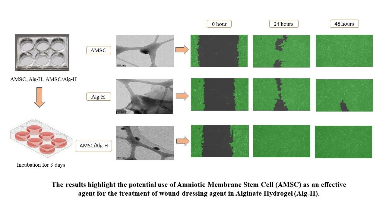
Fitriani N, Wilar G, Narsa AC, Elamin KM, Wathoni N
Stem Cells and Cloning: Advances and Applications 2025, 18:1-13
Published Date: 10 January 2025
Gallic Acid Nanocrystal Hydrogel: A Novel Strategy for Promoting Wound Healing and Inhibiting Scar Formation

Yan M, Huang S, Li X, Wang Y, Zhong S, Ban J, Zhang S
International Journal of Nanomedicine 2025, 20:4607-4626
Published Date: 11 April 2025