
Back to Journals » Drug Design, Development and Therapy » Volume 20
Advances and Therapeutic Potential of Anthraquinone Compounds in Neurodegenerative Diseases: A Comprehensive Review
Authors Liu Z  , Zhang H , Wan B, Yin S, Yue R
Received 10 November 2025
Accepted for publication 14 January 2026
Published 22 January 2026 Volume 2026:20 580330
DOI https://doi.org/10.2147/DDDT.S580330
Checked for plagiarism Yes
Review by Single anonymous peer review
Peer reviewer comments 2
Editor who approved publication: Professor Anastasios Lymperopoulos
Zheng Liu,1 Huize Zhang,2 Bin Wan,1 Sili Yin,3 Rensong Yue1
1Department of Endocrinology, Hospital of Chengdu University of Traditional Chinese Medicine, Chengdu, Sichuan, People’s Republic of China; 2School of Basic Medicine, Chengdu University of Traditional Chinese Medicine, Chengdu, Sichuan, People’s Republic of China; 3Department of Gynecology, Leshan City Central Maternal and Child Health Hospital, Leshan, Sichuan, People’s Republic of China
Correspondence: Rensong Yue, Department of Endocrinology, Hospital of Chengdu University of Traditional Chinese Medicine, No. 39-41 Shierqiao Avenue, Chengdu, Sichuan, People’s Republic of China, Tel +86-28-18384203191, Email [email protected]
Background: Rhubarb, traditionally used in China for neurological disorders, has recently attracted considerable scientific attention for its neuroprotective and cerebrovascular benefits. The main therapeutic components of rhubarb are anthraquinones, including emodin, aloe-emodin, chrysophanol, rhein, and physcion. Accumulating experimental evidence indicates that anthraquinones are of importance in neurodegenerative diseases (NDDs), such as Alzheimer’s disease, Parkinson’s disease, Huntington’s disease, and multiple sclerosis. However, as a promising candidate for drug development, the mechanisms by which anthraquinones treat NDDs have not been systematically reviewed. Therefore, this article outlines the anti-neurodegenerative effects of anthraquinones, focusing on their molecular mechanisms.
Objective: This article reviews recent research progress of anthraquinones in NDDs, focusing on their potential targets and pathways to provide new ideas for the intervention and treatment of NDDs.
Methods: A comprehensive search of PubMed, Web of Science, and Google Scholar was conducted for articles on the intervention of anthraquinones in NDDs in the past 20 years. The collected information was then summarized and analyzed.
Results: Anthraquinones ameliorate NDDs through multiple mechanisms. They exhibit antioxidant and anti-inflammatory effects, protect mitochondria, and regulate microglial polarization. Furthermore, anthraquinones inhibit pyroptosis, apoptosis, tau phosphorylation, Aβ/α-synuclein aggregation, and acetylcholinesterase activity, while restoring metal homeostasis, activating estrogen receptors, modulating gut microbiota, increasing BDNF levels, and preserving blood-brain barrier permeability. More notably, these compounds play a neuroprotective role by mediating multiple signaling pathways and targets, including Nrf2, ERK1/2, PI3K/mTOR, ROS/TXNIP, SIRT1/PCG-1α, NLRP3, PI3K/Akt, MAPK, TLR4-NFκB, CaM/CaMKIV, and Ca2+/EGFR/PLCγ.
Conclusion: The pleiotropic actions of anthraquinones highlight their potential as therapeutic candidates for NDDs, yet clinical validation remains essential. Future studies should emphasize rigorously designed clinical trials and optimized brain-targeted delivery platforms. This review consolidates current evidence to support their translational development.
Keywords: anthraquinones, neurodegenerative diseases, neurodegeneration, neuroprotection, cognition
Introduction
The worldwide socioeconomic burden of neurodegenerative diseases (NDDs), such as Alzheimer’s disease (AD), Parkinson’s disease (PD), and amyotrophic lateral sclerosis (ALS), is growing significantly as life expectancy rises.1 Due to its irreversibility and lack of effective treatment, NDDs are becoming highly prevalent.2 Numerous NDDs advance to dementia,3 which is anticipated to impact around 150 million individuals worldwide by 2050, incurring an economic burden of $10 trillion and incalculable costs to patients and their families.4,5
Despite the wide variability in the pathological presentations of NDDs, they exhibit comparable pathological characteristics, including the progressive deterioration of neuronal structure and function, along with analogous clinical symptoms, such as cognitive, motor, and behavioral deficits.6–8 NDDs are intricately linked to cerebrovascular conditions, including cerebral ischemia-reperfusion injury (CIRI), traumatic brain injury, intracerebral hemorrhage (ICH), and ischemic stroke (IS), as well as neurological disorders such as epilepsy (EPI). Clinical and pathological evidence suggests that they share pathogenic mechanisms, including neuroinflammation, oxidative stress (OS), pyroptosis, and mitochondrial dysfunction.9–13 Moreover, converging evidence indicates that vascular dysfunction is a key pathological component of NDDs. Vascular alterations occur in the majority of NDDs, particularly in capillaries. Recent investigations have demonstrated that these modifications induce vascular dysfunction and facilitate the course of NDDs.14–17 In addition, there is a close clinical correlation among these diseases. Patients with Vascular Cognitive Impairment Disorder (VCID) frequently exhibit episodic memory impairments, challenges in word retrieval, temporal disorientation, and mild executive dysfunctions. Consequently, distinguishing AD from VCID can be challenging, as they are often indistinguishable during clinical evaluation.18 Cerebral ischemia can induce the aggregation of proteins linked to neurodegenerative diseases.19 Stroke-induced secondary neurodegeneration shares a number of striking similarities to other NDDs, such as AD, especially with the substantial accumulation of the neurotoxic protein amyloid-β (Aβ).20 There is growing evidence indicating the presence of clinical and sub-clinical EPI in AD, Lewy body dementia, PD, and other less prevalent NDDs. For instance, Epidemiological studies have shown that unprovoked EPI occurs in 10–22% of patients with AD. Furthermore, biomarkers related to NDDs have been detected in the cerebrospinal fluid or blood of patients with EPI, such as Aβ, Tau, and α-synuclein (α-syn).21,22 Some studies have grouped these diseases with typical NDDs.23,24 Therefore, these diseases were adopted for our discussion.
In the fight against NDDs, extensive research has sought to elucidate mechanisms and potential therapeutic targets.8 However, current therapeutic approaches offer no permanent solution and only provide symptomatic relief.6 Natural products, particularly those derived from medicinal plants, have been utilized for centuries in disease therapy, supported by their inherent multi-component, multi-target, and multi-pathway synergistic mechanisms.25 Their intrinsic ability to cross the blood-brain barrier (BBB) and act on intracellular targets has made small-molecule compounds highly attractive for neurotherapeutic development.26
The Biological Activity and Mechanism of Anthraquinones
Anthraquinones, primarily derived from rhubarb (Rheum officinale Baill., Rheum tanguticum Maxim. ex Balf., Rheum palmatum L)., comprise emodin (EM), aloe-emodin (AE), chrysophanol (CHR), rhein (RHE), and physcion (PHY). Chemically, these compounds share a planar anthraquinone core with diverse substituent patterns. CHR is 1,8-dihydroxy-3-methylanthraquinone,27 EM is 1,3,8-trihydroxy-6-methylanthraquinone,28 and RHE is 4,5-dihydroxyanthraquinone-2-carboxylic acid.29 AE and PHY share a similar skeleton, with variations in hydroxyl, methyl, and carboxyl groups,30 as shown in Figure 1. Their structural arrangement facilitates reversible oxidation-reduction processes.31
 |
Figure 1 The chemical structure of anthraquinones. |
Functionally, these anthraquinones provide diverse neuroprotection via antioxidant and mitochondrial-supporting mechanisms, particularly by activating the nuclear factor erythroid-2-related factor 2 (Nrf2) and sirtuin1/peroxisome proliferator-activated receptor-γ coactivator-1α (SIRT1/PGC-1α) pathways. They effectively modulate neuroinflammation by inhibiting NLRP3 inflammasome activation, suppressing pro-inflammatory cytokines, and regulating microglial polarization towards an anti-inflammatory M2 phenotype. Additionally, anthraquinones mitigate neuronal loss by modulating intricate signaling pathways that regulate cell death processes, including apoptosis, pyroptosis, and ferroptosis. By suppressing Aβ aggregation and tau hyperphosphorylation, these compounds directly engage the central molecular pathology of AD. Beyond AD, they also exert neuroprotective effects in PD and Huntington’s disease (HD) models, attenuating motor deficits and proteinopathy. The therapeutic effects and molecular mechanisms of anthraquinones on NDDs have been summarized in Tables 1, 2 and 3.
 |
Table 1 Summary of Preclinical Information on the Therapeutic Effects and Molecular Mechanisms of Emodin on NDDs |
 |
Table 2 Summary of Preclinical Information on the Therapeutic Effects and Molecular Mechanisms of CHR on NDDs |
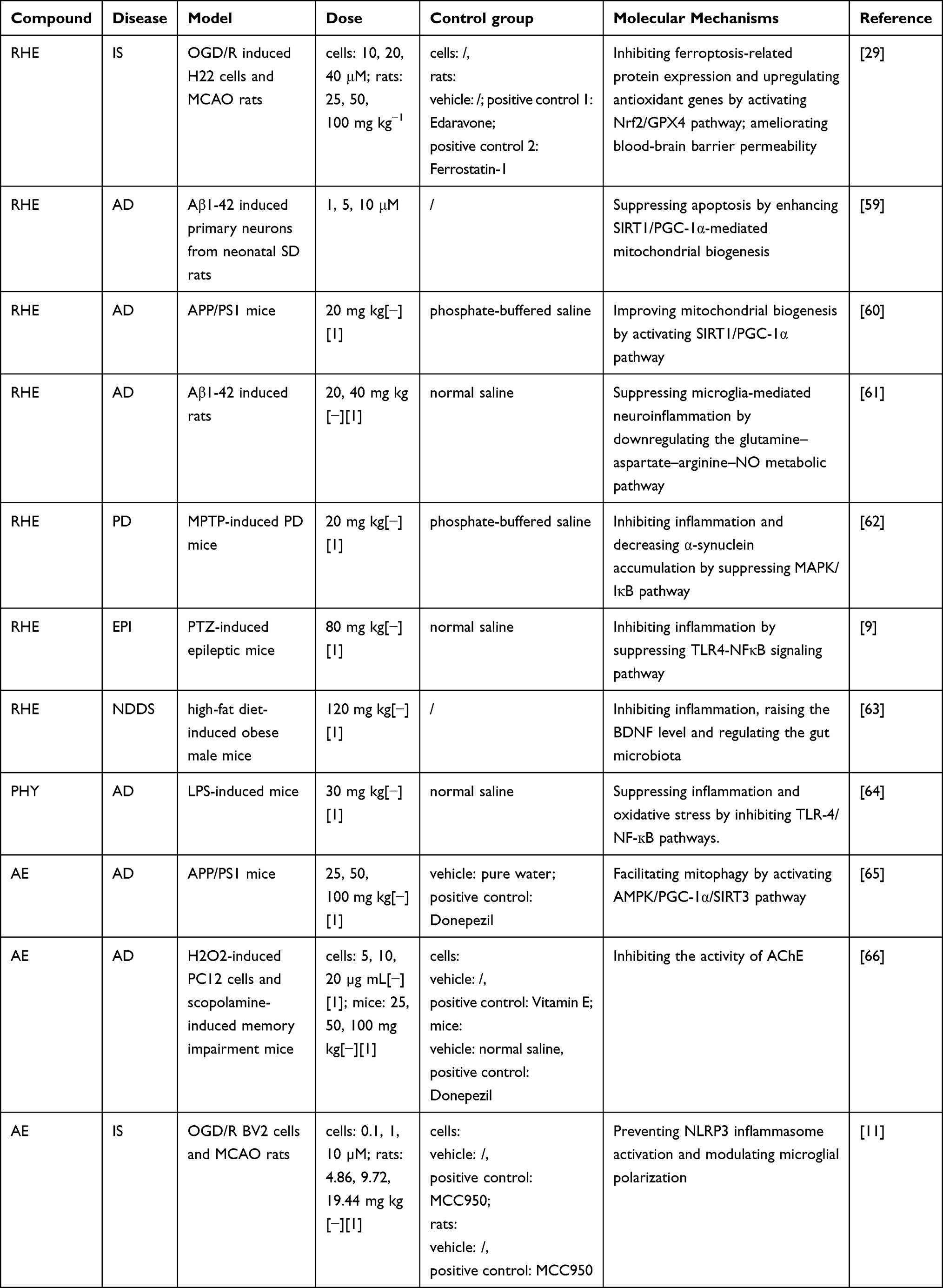 |
Table 3 Summary of Preclinical Information on the Therapeutic Effects and Molecular Mechanisms of RHE, PHY, and AE on NDDs |
Nevertheless, no summary of contemporary pharmacological research in this area focuses on action targets and pathways. Consequently, emerging evidence has delineated the molecular targets and signaling pathways by which anthraquinones modulate NDDs pathology. The pathway regulation related to AD is plotted in Figure 2, whereas Figures 3 and 4 present the corresponding pathways for PD and IS, respectively. A comprehensive review of the included literature aims to provide valuable references for future research and accelerate the clinical application of anthraquinones.
 |
Figure 2 The molecular mechanism of anthraquinone-based treatment for AD. Anthraquinones exert anti-AD effects primarily by regulating OS and mitochondrial homeostasis, suppressing neuroinflammation, and inhibiting programmed cell death. Specifically, these pathways involve activation of antioxidant signaling and mitochondrial biogenesis, leading to reduced ROS and restored mitochondrial function; inhibition of inflammasome-related inflammatory signaling and microglial activation, resulting in decreased pro-inflammatory cytokine production; and modulation of apoptosis and ferroptosis through the regulation of caspase activation and LPO. In addition, pathways associated with Aβ and Tau pathology, neurotrophic support, calcium signaling, and metal homeostasis are also involved. Collectively, these multitarget actions converge to mitigate AD-associated neurodegeneration. |
 |
Figure 3 The molecular mechanism of anthraquinone-based treatment for PD. Anthraquinones exert anti-PD effects primarily by modulating redox balance, inflammatory signaling, and mitochondrial integrity. Regulation of antioxidant responses limits excessive ROS accumulation and mitigates OS, while coordinated modulation of MAPK- and PI3K-associated signaling dampens inflammatory cascades and reduces pro-inflammatory cytokine release. In parallel, restoration of mitochondrial function and energy metabolism contributes to the restraint of apoptotic and ferroptotic cell death. Additional regulatory processes, including α-syn aggregation, metal ion homeostasis, and neurotrophic factor support, further participate in the overall neuroprotective outcome in PD. |
 |
Figure 4 The molecular mechanism of anthraquinone-based treatment for IS. Anthraquinones exert anti-IS effects primarily by suppressing inflammation, OS, and programmed cell death. ERK1/2, Nrf2, and NLRP3 signaling pathways were involved. Meanwhile, promotion of the BBB function contributes to overall neuroprotection in IS. |
Molecular Mechanisms of Neuroprotection by Anthraquinones
Anti-Oxidative Stress
Due to its high lipid content and substantial oxygen demand, the brain is particularly susceptible to OS.67 Excessive generation of reactive oxygen species (ROS) leads to OS, a central pathological hallmark of NDDs that drives neuronal dysfunction and cell death.68–70 Most NDDs, including AD, PD, HD, and ALS, are characterized by elevated ROS levels and compromised antioxidant defense mechanisms.68
Nrf2
Nuclear factor erythroid 2-related factor 2 (Nrf2) serves as the master regulator of the cellular antioxidant defense system, orchestrating redox homeostasis and protecting against oxidative injury.71–73 Nrf2 regulates the expression of numerous antioxidant enzymes, including heme oxygenase-1 (HO-1), glutathione reductase, superoxide dismutase-1 (SOD-1), and catalase (CAT), all of which can scavenge endogenous ROS.74,75 Under resting conditions, Nrf2 is primarily localized in the cytoplasm, where it interacts with Kelch-like ECH-associated protein 1.76 During the occurrence and development of OS, Nrf2 translocates to the nucleus and binds to antioxidant response elements (ARE), triggering the activation of several downstream proteins and detoxifying enzymes. This process regulates the expression of many antioxidant genes and maintains a proper redox balance.77–79
CHR
CHR activated the Nrf2/HO-1 pathway in the hippocampus and cortex of PTZ-induced epileptic mice, decreasing OS markers (malondialdehyde [MDA] and nitric oxide [NO]) and increasing antioxidant enzymes (glutathione S-transferase and CAT). Molecular docking confirmed that CHR directly interacted with Nrf2.45
Emodin
In PC12 cells exposed to Aβ25-35, the translocation of Nrf2 from the cytoplasm to the nucleus was inhibited, whereas emodin reversed this effect. Emodin treatment groups at varying doses significantly reduced OS and enhanced the expression levels of cystine/glutamate antiporter and glutathione peroxidase-4 (GPX4).32 Similarly, emodin upregulated Nrf2 and its downstream effectors (HO-1, SOD-1, and CAT) to reduce ROS and preserved mitochondrial membrane potential (MMP) in AD models.28
RHE
According to Cai et al, RHE treatment significantly increased glutathione (GSH) levels and GPX4 expression in the brain, while markedly reducing MDA content compared with the middle cerebral artery occlusion (MCAO) group, suggesting a reduction in OS. Additionally, total Nrf2 expression increased distinctly in the H-RHE group, and the protein bands in the RHE-treated group showed higher intensity than those in the MCAO group. Evaluations of GPX4 and nuclear Nrf2 yielded similar findings.29
PHY
Ahmad et al confirmed that PHY elevated Nrf2 and HO-1 expression, and reversed the effects of LPS-induced OS. The concentration of GSH in the cortex and hippocampus decreased after LPS treatment but was significantly increased following PHY treatment, while MDA levels were markedly reduced.64
ERK1/2
Extracellular signal-regulated protein kinases 1/2 (ERK1/2) belong to the mitogen-activated protein kinase (MAPK) family and play crucial roles in neuronal differentiation, survival, structural plasticity, long-term potentiation, and memory formation.80 Transient ERK activation promotes neuronal survival,81 whereas sustained ERK hyperphosphorylation drives premature senescence, apoptosis, and dysregulated autophagy.82 An abnormal accumulation of activated ERK1/2 in neurons has been documented in AD brains.83 In vivo and in vitro, phosphorylated ERK expression was markedly elevated in the PD model group.84
SH-SY5Y cells have been utilized in numerous models of NDDs, including PD and AD.85,86 Chen et al exposed SH-SY5Y 26 cells to zinc sulfate and different concentrations of emodin, followed by assessment of ERK1/2 expression, OS, and mitochondrial function. Emodin pretreatment rectified zinc-induced synaptic dysfunction, diminished OS by inhibiting phospho-ERK1/2 signaling, and curtailed mitochondrial dissipation.38 In the rat MCAO model, emodin reduced infarct volume and cell death, and additionally restored PC12 cells’ viability while decreasing ROS generation and glutamate release under ischemic and hypoxic conditions, possibly via activation of the ERK1/2 signaling pathway.39
PI3K/mTOR
The phosphatidylinositol 3-kinase (PI3K)/mammalian target of rapamycin (mTOR) signaling pathway is a critical regulator of cellular survival, proliferation, and metabolism.87 Growth hormone, oxygen, amino acid availability, and cellular energy status all influence mTORC1 activity.88
PI3K/AKT/mTOR
PI3K phosphorylates protein kinase B (AKT), triggering downstream effects that stimulate mTOR. AKT is essential for neuronal proliferation and synapse formation.89 Numerous NDDs, such as AD,90 multiple sclerosis (MS),91 HD,92 and PD,93 have been linked to the pathophysiology of the PI3K/AKT/mTOR signaling pathway.
In ICH-associated neurological defects in rats, CHR was neuroprotective by reversing neurobehavioral changes and dramatically lowering the increased levels of PI3K, AKT, and mTOR proteins. The treatment doses of CHR at 10 mg/kg and 20 mg/kg resulted in significant increases in GSH, CAT, SOD, and GPX levels compared with the vehicle and sham control groups.46
PI3K/mTOR/GSK3β
Glycogen synthase kinase-3 (GSK-3) is an evolutionarily conserved serine/threonine kinase prevalent in mammalian eukaryotic cells.41 GSK3β, a GSK-3 isoform, plays a critical role in neuronal cell death, and its pharmacological inhibition has been shown to reduce α-syn accumulation in PD.94
In H2O2-induced SH-SY5Y cells, p-PI3K, p-mTOR, and p-GSK3β expression were significantly elevated, whereas emodin significantly reversed the above expression. Emodin could prevent the activation of the PI3K/mTOR/GSK3β pathway induced by H2O2, thereby exerting neuroprotective effects.41
ROS/TXNIP
Thioredoxin-interacting protein (TXNIP), also known as thioredoxin (TRX) binding protein-2,95 possesses an α-arrestin domain that engages with and diminishes the activity of cytosolic TRX and mitochondrial TRX (activated form), hence regulating cellular redox signaling.96 TRX1 and TRX2 modulate ROS levels in the cytoplasm and mitochondria, respectively. However, TXNIP inhibits TRX’s antioxidant function. Consequently, TXNIP acts as a pro-oxidant, augmenting ROS production and leading to OS.95
In the d-galactose- and Aβ25-35-induced AD model, CHR significantly improved the survival rate and attenuated OS. Furthermore, CHR markedly decreased MDA and lactate dehydrogenase (LDH) levels, concomitantly enhancing the activities of T-SOD, CAT, and GSH. Mechanistically, CHR simultaneously downregulated TXNIP at the protein and mRNA levels and upregulated TRX expression, likely via modulation of the ROS/TXNIP signaling pathway.47
Mitochondrial Protection
Within the central nervous system (CNS), mitochondria are essential for multiple cellular processes, including neurotransmitter metabolism, ROS generation, calcium homeostasis, and regulation of cell death.97 Mitochondria are the principal source of intracellular ROS and serve as critical targets of Aβ toxicity during AD pathogenesis.98 Mitochondrial impairment is characterized by diminished metabolic activity, elevated ROS generation, membrane permeability transition, and the release of mitochondrial proteins into the cytoplasm.74 Anthraquinones exert mitochondrial protection via multiple pathways.
Promotion of Mitochondrial Biogenesis
Mitochondrial biogenesis is the process by which new mitochondria are generated from pre-existing mitochondria.99 Mitochondrial dysfunction is considered a pivotal factor in the pathogenesis of various neurological illnesses, and adaptive mitochondrial biogenesis has been observed in the nervous system.100 Moreover, mitochondrial biogenesis, an intrinsic antioxidant defense mechanism, is actively upregulated in pathological conditions to counteract intracellular OS.101,102
SIRT1/PCG-1α
SIRT1 is an NAD+-dependent protein deacetylase that belongs to the SIRT family and plays a vital role in aging, cell death, mitochondrial biogenesis, inflammation, and energy regulation by deacetylating histone and a variety of non-histone proteins.103–105 SIRT1 deacetylates and activates the mitochondrial regulator PGC-1α.59 Activated PGC-1α targets and enhances the expression of downstream mitochondrial transcription factor A (TFAM). TFAM stimulates mitochondrial DNA transcription and replication, producing new, healthy mitochondria.60,106,107
Yin et al demonstrated that RHE alleviated mitochondrial OS in Aβ oligomer-induced primary neurons by enhancing SIRT1/PGC-1α-mediated mitochondrial biogenesis.59 In the Aβ precursor protein/presenilin 1 (APP/PS1) transgenic mouse model of AD, RHE considerably improved cognitive and memory deficits. SIRT1, PGC-1α, and nuclear respiratory factor 1 (NRF1) expression were significantly lower in the APP/PS1 group than in the WT (age-matched wild-type mice with the same background) group, indicating compromised mitochondrial biogenesis. However, RHE therapy significantly raised SIRT1, PGC-1α, and NRF1 expression. These results suggested that RHE enhanced SIRT1/PGC-1α-mediated mitochondrial biogenesis.60 Moreover, RHE-loaded nanoparticles (K8@Fe−Rh/Pda NPs) activated SIRT1/PGC-1α pathway, thereby correcting mitochondrial dynamics and increasing cytochrome c oxidase activity in Aβ42 oligomer-burdened neurons, helping repair damage triggered by Aβ aggregation.108
Facilitation of Mitophagy
Cellular mitophagy refers to the selective encapsulation and degradation of damaged mitochondria by cells via an autophagic process, thereby preserving mitochondrial integrity.109 Mitophagy plays a vital part in energy availability, neuronal survival, mitochondrial and metabolic balance, and general health.110 OS, nutritional deficiencies, and aging can cause mitochondrial depolarization, leading to mitochondrial dysfunction and disruption of cellular homeostasis, which are closely associated with NDDs.65,111–113 Four primary steps make up mitophagy: 1) Damaged mitochondria lose their membrane potential and depolarize, 2) Autophagosomes wrap mitochondria to produce mitochondrial autophagosomes, 3) Mitochondrial autophagosomes merge with lysosomes, and 4) Lysosomes break down mitochondrial contents.114 Several studies have shown that AD patients’ brains exhibit aberrant mitophagy, which impairs the normal clearance of damaged mitochondria.109
In the APP/PS1 double transgenic mouse model, AE reduced hippocampal neuronal damage and ameliorated cognitive dysfunction. AE treatment markedly decreased sequestosome 1 (p62), Beclin-1, microtubule-associated protein 1 light chain 3 beta (LC3B), PGC-1α, p-AMPK, and SIRT3, suggesting that AE induced mitophagy via the AMPK/PGC-1α/SIRT3 pathway.65
Maintenance of Mitochondrial Integrity
Structure determines function. Maintaining mitochondrial structure integrity is another crucial way to exert mitochondrial function.
CHR
In Aβ25-35-induced PC12 cells, transmission electron microscopy revealed that the model group showed substantial mitochondrial morphological damage, including reduced cristae and fractured mitochondrial membranes. The CHR-treated model group displayed markedly improved mitochondrial morphology, as evidenced by restoration of mitochondrial cristae and double-membrane structures.49
Emodin
Xia et al demonstrated that emodin reversed MMP impairment in Aβ25-35-treated PC12 cells.32 Ubiquinol-cytochrome c reductase core protein 1 (UQCRC1), a crucial part of mitochondrial complex III, sustains bioenergetic homeostasis via mitochondrial preservation and apoptotic control. In the MPP+-induced PD model, emodin could enhance UQCRC1 expression, stabilize mitochondrial function, and prevent MMP collapse.40
Anti-Inflammatory Effects
Neuroinflammation, a core CNS response to acute injury and chronic NDDs, plays a central role in neurodegeneration and disease progression.115,116 Anthraquinones exert anti-neuroinflammatory effects mainly through the following pathways.
Regulation of Inflammatory Signaling Pathways
NLRP3 Inflammasome
The NLRP3 inflammasome is a complex that orchestrates the innate immune response by detecting diverse damage and pathogen-associated molecular patterns.117 Three major components of the NLRP3 inflammasome are NLRP3, the adaptor protein apoptosis-associated speck-like protein containing a caspase activation and recruitment domain (ASC), and pro-caspase-1.118 Upon receiving a danger signal, NLRP3 activates ASC, which attracts pro-caspase-1 and cleaves it into active caspase-1, which facilitates the maturation of pro-interleukin-1β (IL-1β) and induces inflammation.119 Enhanced activation of NLRP3 has been associated with various chronic conditions, including neurodegenerative disorders.120
Inhibition of Inflammation
In Aβ25-35-induced PC12 cells, the expression of key inflammatory signaling molecules, including Toll-like receptor-4 (TLR4), phosphorylated nuclear factor kappa-B (p-NF-κB), NLRP3, as well as the proinflammatory cytokines IL-1β and tumor necrosis factor-alpha (TNF-α), were markedly increased. Emodin effectively attenuated signaling protein levels and cytokine levels. These findings provide compelling evidence that emodin downregulated the TLR4/p-NF-κB/NLRP3 pathway and exerted anti-inflammatory effects in AD.32 Experimental autoimmune encephalomyelitis (EAE) is presently the predominant animal model for MS.121 In this model, microglia and the NLRP3 inflammasome intensified neuroinflammation, which was crucial for the onset of MS. Emodin suppressed NLRP3 inflammasome signaling by upregulating SIRT1 and PGC-1α, thereby attenuating demyelination and neuroinflammatory responses and ultimately ameliorating behavioral deficits and neuropathological alterations.42
Zhang et al reported that cerebral ischemia/reperfusion (I/R) induced brain tissue injury and activated the NLRP3 inflammasome. CHR could prevent NLRP3 activation and provide neural protection against IS.52 In the Aβ25-35-induced AD model, CHR markedly enhanced learning and memory deficits, mitigated hippocampus neuronal injury, and substantially decreased the protein and mRNA expression levels of NLRP3, IL-1β, and IL-18, potentially through the ROS/TXNIP/NLRP3 signaling pathway.47
AE exerted neuroprotective effects in brain I/R models by preventing NLRP3 inflammasome activation, thereby regulating microglial polarization and pyroptosis.11
In another study on quinones as preventive agents in AD, Chen et al reported that quinones in Traditional Chinese Medicine (TCM) and Chinese Medicinal Formulas were promising prophylactic agents due to their ability to regulate the NLRP3 pathway. Furthermore, anthraquinones exhibited the most potent effect among quinones, especially AE, CHR, and RHE.115
Inhibition of Pyroptosis
Pyroptosis, an inflammatory form of programmed cell death that occurs in both microglia and neurons, is initiated by various inflammasomes (such as NLRP3 inflammasome) and executed by caspases and the gasdermin (GSDM) family.122,123 Activated caspase-1 cleaves gasdermin D (GSDMD) to generate the N-terminal fragment (GSDMD-NT), which oligomerizes at the plasma membrane to form pores, leading to cell swelling, membrane rupture, and the subsequent release of intracellular contents, thereby inducing cell death.124,125 A variety of NDDs, including AD, PD, ALS, and MS, are linked to pyroptosis.125–129
In the MCAO/R models, CHR suppressed GSDMD transcription by activating the Nrf2/ARE signaling pathway, consequently diminishing pyroptosis in hippocampal neurons.54 In a similar study, Xia et al observed that CHR postconditioning protected against CIRI by inhibiting NLRP3-mediated pyroptosis.55
Cui et al demonstrated that emodin significantly reduced pyroptosis in EAE rats by reducing the molecular level of the NLRP3 but had no noticeable effect in healthy rats, providing a solid experimental foundation for emodin’s therapeutic application in the clinical treatment of MS patients.42 Besides, Jiang et al found that emodin mitigated LPS/ATP-induced pyroptosis in BV2 cells by inhibiting NLRP3 inflammasome activity and the cleavage of the pyroptotic effector protein GSDMD, thereby exerting neuroprotective properties.36
PI3K/Akt
PI3K modulates numerous critical processes in the inflammatory response to infection and injury. By attracting and activating innate immune cells, such as macrophages, the PI3K/Akt axis can modulate inflammatory responses.130 Previous studies have shown that targeting the PI3K/AKT signaling pathway and its downstream mediators exerts neuroprotective effects by reducing neuronal inflammation.131,132
PI3K/Akt/mTOR
Integrative metabolomics and network pharmacology analyses were conducted to elucidate the metabolic mechanisms and therapeutic potential of Didang decoction (DDD, a traditional TCM decoction) in CIRI, revealing that emodin is a major bioactive constituent and AKT1 is a core molecular target. Molecular docking further predicted a stable interaction between emodin and AKT1, suggesting a direct regulatory effect on the pathway. DDD treatment significantly enhanced phosphorylation of PI3K, Akt, and mTOR. PI3K/Akt/mTOR activation was implicated in suppressing neuroinflammation by phosphorylating NF-κB, downregulating NLRP3, and lowering the systemic production of pro-inflammatory cytokines.133
A recent study demonstrated that CHR ameliorated neurobehavioral deficits by downregulating the expression of PI3K, AKT, and mTOR proteins. Additionally, CHR significantly inhibited the production of inflammatory cytokines, including IL-1β, IL-6, and TNF-α. The findings suggested that in rats with ICH-induced neurological dysfunction, CHR might be a promising therapeutic strategy for repairing neuronal damage driven by upregulation of the PI3K/AKT/mTOR signaling pathway.46
Myd88/PI3K/Akt/ NF-κB
Myeloid differentiation factor 88 (MyD88) is a key adapter protein in innate immunity, plays a critical role in TLR-mediated signaling transduction and downstream signaling cascades, including PI3K/Akt.43,134 Upon phosphorylation, the PI3K/Akt pathway activates NF-κB, thereby initiating an inflammatory cascade.135
In EAE mice, emodin reduced IκB (NF-κB inhibitor) kinase activation and the consequent IκB degradation by suppressing MyD88 and preventing PI3K and Akt phosphorylation. Consequently, NF-κB nuclear translocation was reduced, thereby suppressing the production of proinflammatory cytokines and preventing microglial overactivation.43
MAPK
MAPK signaling is a recognized prototypical pro-inflammatory signaling pathway. The MAPK family encompasses ERK1/2, JNK, and p38 signaling pathways.136 Upon activation of the MAPK cascade, IκB undergoes ubiquitination and proteasome-mediated degradation, thereby releasing NF-κB and initiating an inflammatory cascade.137–139 Several studies have confirmed that inhibition of MAPK signaling pathways can reduce neuroinflammation and ameliorate the neurodegenerative process.140,141
In MPTP-induced PD mice, RHE improved motor symptoms and reduced dopaminergic neuron damage by suppressing activation of the MAPK/IκB signaling pathway and reducing levels of pro-inflammatory cytokines such as IL-1β, IL-6, and TNF-α.62
TLR4-NFκB
The NF-κB family of transcription factors is crucial for regulating various biological processes, including inflammation, immune responses, cell survival, and cellular differentiation.142 TLRs are crucial membrane proteins involved in signal transduction and the innate immune system. The quintessential route involves the interaction of lipopolysaccharide with TLR4 on microglial surfaces, thereby activating multiple signaling transduction pathways, including PI3K/AKT, MAPK, and mTOR, ultimately leading to the activation of NF-κB.139,143
RHE
In PTZ-induced epileptic mice, RHE exerted anticonvulsant and neuroprotective effects by inhibiting TLR4-NFκB signaling and reducing the secretion of inflammatory cytokines, including TNF-α, IL-6, IL-1β, and IL-18.9
PHY
Liu et al reported that PHY treatment significantly reduced the expression of NF-κB, TNF-α, and IL-1β in both the cortex and hippocampus in an LPS-induced gliosis model. Moreover, PHY could reduce the relative protein abundance of TLR4 and the responsiveness of glial cells in the mouse brain. These results might provide a modern theoretical framework for clinical research that examines the pathogenic mechanisms and treatment modalities for AD and neuroinflammation.64
CHR
The fundamental processes of ethanol-induced neurodegeneration are intricate and involve several cellular functions, including inflammation, OS, and apoptosis.56 It has been demonstrated that ethanol exposure activates the NF-κB pathway in several brain regions, particularly the cerebral cortex and hippocampus.144 In ethanol-induced neurodegeneration mice, administration of CHR decreased the expression of TLR4, NFκB, IL-1β, TNF-α, and Caspase-3 in the cortex and hippocampal regions.56 In addition, CHR ameliorated memory loss and reduced histopathological changes in neurons in stress-intensified PTZ-induced EPI in mice. Meanwhile, CHR significantly reduced the mRNA expression levels of TLR-4, NF-κB, TNF-α, IL-1β, and Caspase-3.45
Suppression of Pro-Inflammatory Cytokines
Suppression of pro-inflammatory cytokines is another critical anti-inflammatory mechanism. It has been suggested that the basal inflammatory state and aging play a significant role in the rising prevalence of chronic illnesses, such as metabolic, neurodegenerative, and cardiovascular disorders.145 Proinflammatory cytokines, such as IL-1β, IL-6, IL-12, IL-18, and TNF, together with anti-inflammatory cytokines including IL-4, IL-10, IL-13, and IL-19, regulate immune responses through interactions with immune cells.146,147 Relevant studies showed that patients with AD or PD have higher levels of these pro-inflammatory cytokines in their serum, cerebrospinal fluid (CSF), and post-mortem brain tissue.148–150
Emodin
Zheng et al investigated the effects of emodin in EAE models. Treatment with emodin significantly downregulated the mRNA expression of IL-6 and IL-17A, indicating that emodin alleviated inflammatory injury by suppressing the transcription of proinflammatory cytokines.43 An additional study concluded that emodin reduced levels of IL-18, IL-1β, and TNF-α in LPS/ATP-induced BV2 cells.36
RHE
An analogous study observed that in the MPTP-induced PD model, RHE decreased levels of pro-inflammatory cytokines (IL-1β, IL-6, and TNF-α) and microglial M1 polarization markers. Consequently, PD movement abnormalities improved while neurodegeneration and α-syn deposition decreased.62
CHR
In diabetic encephalopathy (DE), sustained hyperglycemia and advanced glycation end products induce chronic neuroinflammation, thereby compromising neuronal function and synaptic plasticity. These changes were accompanied by elevated levels of inflammatory cytokines, ultimately leading to cognitive deficits.151–153 In the streptozotocin (STZ)-induced DE model, the levels of pro-inflammatory cytokines (IL-1β, IL-4, IL-6, TNF-α) in the hippocampus were markedly increased. However, CHR alleviated learning and memory impairments by preventing the overexpression of pro-inflammatory cytokines in the hippocampal regions of DE mice.57
Modulation of Microglial Polarization
Microglia, the indigenous macrophages of the CNS, employ their own receptor repertoire to surveil the brain’s surroundings dynamically.154 In a homeostatic brain, microglia perform functions in synapse pruning, damage repair, maintenance of homeostasis, phagocytosis, support of other glial cells, and intercellular communication.155 Microglia are generally classified into two phenotypes based on their activation state. Upon M1 activation, microglia secrete pro-inflammatory chemotactic molecules and cytokines that have neurotoxic effects.156 Whereas M2 microglia release anti-inflammatory cytokines and brain-derived neurotrophic factors (BDNF), which perform neuroprotective benefits by inhibiting inflammatory responses and promoting regeneration and repair.157 Aberrant activation of microglia and the overproduction of proinflammatory mediators are pivotal contributors to the pathogenesis of NDDs, as well as to neuronal injury secondary to traumatic brain injury and stroke.158,159
CHR
By arresting microglia toward a pro-inflammatory phenotype and downregulating pro-inflammatory cytokine expression, CHR demonstrated neuroprotection and improved the brain plasticity milieu of stroke rats in the chronic phase.13 In LPS-induced BV2 cells, treatment with CHR reduced the release of proinflammatory mediators, including inducible nitric oxide synthase, prostaglandin E2, TNF-α, IL-6, and IL-1β. Furthermore, CHR exerted its anti-inflammatory effects by suppressing the activation of p38, ERK1/2, and JNK signaling pathways in microglia.58
Emodin
Iba-1 and CD68 indicate the extent of microglial aggregation and the level of M1 phenotypic activation, respectively.160 In the EAE models, emodin therapy mitigated Iba-1 and CD68 levels.42 In a similar study, emodin reduced the expression of phosphorylated PI3K, Akt, NF-κB, and MyD88, as well as the levels of markers CD86 and CD206 in microglia.43 These findings demonstrated the neuroprotective and anti-inflammatory effects of emodin mediated by regulating microglial polarization.
RHE
Chi et al demonstrated that activated microglia drive neuroinflammation by regulating NO production through the glutamine-aspartate-arginine pathway. Additionally, RHE suppressed microglia-mediated neuroinflammation by suppressing NO production in microglia.61
AE
In the CIRI model, AE decreased microglial NLRP3 inflammasome activation, induced a M1 to M2 phenotypic change, and inhibited microglial pyroptosis, thus performing a neuroprotective effect.11
Inhibition of Apoptosis
Apoptosis is a form of programmed cell death in which BCL-2-associated X protein (Bax), Bcl-2, and Caspase-3 play crucial regulatory roles.161 Several investigations have demonstrated that increased neuronal death is a factor behind NDDs.162,163
CHR
Apoptotic and necrotic signaling pathways are activated as a result of nitrosative/oxidative stress in cerebral I/R animals. CHR inhibited nitrosative/oxidative stress by reducing 3-NT levels, inhibiting caspase-3 activation, and suppressing NOx generation, thereby preventing NO-mediated neuronal cell death.53 CHR reduced endoplasmic reticulum stress-mediated apoptosis in AD mice by downregulating endoplasmic reticulum stress markers (GRP78, CHOP) and apoptotic caspases (3, 9, 12), an effect mediated through inhibition of the PERK signaling pathway.48
Emodin
Emodin, another prominent anthraquinone, reduced glutamate release via presynaptic adenosine A1 receptors, thereby mitigating excitotoxicity-induced apoptosis.164 In Aβ1-42-induced U251 cells, emodin activated the Nrf2 signaling pathway, leading to upregulation of Bcl-2, downregulation of Bax, and suppression of caspase-3, −8, and −9 activation, ultimately reducing apoptosis.28 Moreover, emodin regulated the PKM2/Nrf2/ARE pathway in OS-induced PC12 cells, in which PKM2 dissociation promoted Nrf2 activation, leading to upregulation of antioxidant genes (Gclc, Gclm) and restoration of GSH levels, thereby diminishing caspase-3 activation.34 Emodin additionally upregulated activin A through the activin A/Smads pathway to downregulate caspase-3 in neurons deprived of oxygen and glucose,35 and regulated multiple survival pathways, including PI3K/Akt, AMPK/GSK-3β, and PI3K/mTOR/GSK3β.41,165,166
RHE
RHE protected Aβ1-42-oligomer-burdened neurons from apoptosis by activating the SIRT1/PGC-1α pathway, which enhanced mitochondrial biogenesis, suppressed cytochrome c release, and inhibited caspase-3 activation.59
Inhibition of Amyloid-Beta Aggregation and Tau Pathology
Characterized by behavioral impairment and cognitive dysfunction,167 AD is the most common NDD. Aβ peptides aggregate to form senile plaques, whereas hyperphosphorylated tau assembles into helical filaments. Together, these structures form neurofibrillary tangles (NFTs), which are key clinical features of AD and contribute to chronic inflammation, synapse loss, and neuronal death.168–170 Furthermore, tau was identified as the secondary agent in mixed pathology forms of PD,171 MS,172 and HD.173
Inhibition of Aβ Aggregation
Emodin
A vital factor in the pathophysiology of AD is the impairment of APP metabolism. By controlling the breakdown of amyloid linked to AD, the protein kinase C (PKC) pathway exhibits neuroprotective benefits. After 4 hours of emodin treatment, cell membrane PKC activity increased by almost 52%.33 As reported by Wang et al, cognitive impairment in AD transgenic mice was reduced by 60–70%, and Aβ deposition levels decreased by 50–70% when a high dose of emodin was administered to 8-month-old B6C3-Tg mice for two months.26
CHR
BACE1 (beta-site APP cleaving enzyme 1) is a type I transmembrane protein. In AD, APP is sequentially cleaved by BACE1 and γ-secretase, leading to the production and release of the Aβ peptide in the brain.174 Razieh Amini et al evaluated the inhibitory effects of 79 plant-derived compounds on BACE1 through virtual screening and molecular docking. Among them, CHR showed nanomolar-range BACE1 inhibitory activity and demonstrated stability in molecular dynamics simulations, suggesting its therapeutic potential against AD and PD.175
AE
By reducing beta-sheet-rich aggregates, encouraging alpha-helical structures, and lowering oligomeric formation, AE suppressed hemoglobin aggregation—a mechanism related to amyloid-like misfolding, and might provide a therapeutic basis for diseases caused by protein aggregation, such as AD.176
Inhibition of Tau Phosphorylation
CaM/CaMKIV
In AD, Aβ accumulation could oversaturate neurons with calcium, trigger the calmodulin (CaM)/calcium/calmodulin-dependent protein kinase IV (CaMKIV) signaling cascade, and subsequently hyperphosphorylate tau.50,177
Ye et al conducted a series of studies demonstrating the neuroprotective effects of CHR in AD. CHR prevented ultrastructural alterations in hippocampal CA1 neurons, alleviated hippocampal neuronal damage, and decreased the levels of CaM, p-CaMKIV, and p-tau in the hippocampus of AD rats. These findings provide compelling evidence that CHR exerts neuroprotective effects by downregulating the CaM/CaMKIV signaling pathway and reducing p-tau expression.50 The same results were observed in the cell model.51
Ca2+/EGFR-PLCγ
Cellular calcium ion (Ca2+) homeostasis is essential for regulating neuronal physiology, encompassing development and differentiation, synaptic plasticity, and cognitive functions such as learning and memory.27 Dysregulation of Ca2⁺ homeostasis occurs in AD, suggesting its involvement in the onset of this disease.178 Elevated intracellular Ca2⁺ promotes tau phosphorylation,179 while the epidermal growth factor receptor (EGFR)/phospholipase C gamma (PLCγ)/CaMK pathway facilitates the Ca2+ release.180
Compared with Aβ25-35-treated PC12 cells, cells in the CHR group exhibited reduced cellular damage and tighter intercellular junctions. Moreover, treatment with CHR markedly decreased tau mRNA and phosphorylated tau protein levels, as well as mRNA and protein levels of EGFR, PLCγ, and inositol 1,4,5-trisphosphate receptor (a Ca2+ channel protein). The results indicated that CHR exerted neuroprotective effects by alleviating calcium overload, preventing Ca2⁺ from binding to CaM, and attenuating tau hyperphosphorylation, likely through inhibition of the EGFR-PLCγ signaling pathway.27
Previous studies had shown that emodin simultaneously suppressed Aβ accumulation and tau phosphorylation. In the APP/PS1 mice, emodin improved AD-like behavior, and emodin-treated animals, especially those given a dose of 20 mg/kg, showed reduced expression levels of Aβ1-42 and p-tau when compared to untreated APP/PS1 mice.28 Hyperhomocysteinemia (HHcy) is an autonomous risk factor for dementia.181 HHcy-induced dementia rats were characterized by excessive Aβ production, tau hyperphosphorylation, and depletion of neuronal and synaptic proteins. Administration of emodin effectively reversed these pathological changes by reducing BACE1 levels and increasing protein phosphatase 2A activity.182
Inhibition of Acetylcholinesterase Activity
Acetylcholine (ACh) is a neurotransmitter in the nervous system that controls how cholinergic neurons work physically and behaviorally. It was regulated and degraded by acetylcholinesterase (AChE) in the CNS.183,184 AChE facilitates the aggregation of Aβ into oligomers and fibrils in the brain, enhancing their neurotoxicity and thereby promoting neuroinflammation, OS, and synaptic dysfunction.185,186 To date, one of the primary therapeutic medications used to treat AD is acetylcholinesterase inhibitors (AChEIs).
AE
In a scopolamine-induced amnesia animal model, AE significantly ameliorated cognitive deficits (>70%). In vitro, AE demonstrated dose-dependent inhibition of AChE activity.66
Emodin
A recent study reported that emodin directly inhibited AChE activity and exerted neuroprotective effects against Aβ25-35-induced cytotoxicity in PC12 cells.32 Systems pharmacology combined with molecular simulation studies revealed that emodin exhibited high-affinity binding to AChE, resulting in potent enzyme inhibition. It occupied the active site via hydrogen bonds and hydrophobic interactions, thereby preventing ACh degradation and enhancing cholinergic nerve transmission. Molecular dynamics simulations further verified the structural stability of the emodin-AChE complex. Its mechanism of action is similar to that of the clinical drug Donepezil, suggesting that emodin could serve as a potential natural AChE inhibitor.187 Polygoni Multiflori Radix Praeparata combined with Acori Tatarinowii Rhizoma is widely employed in TCM for the prevention and treatment of AD. Xie et al reported that the combination decoction improved cognitive performance and significantly decreased AChE expression, an effect likely mediated by bioactive compounds, including emodin,188 but further investigation is warranted.
Focusing on the investigation of new AChEIs, Duran assessed the effects of selected quinones on AChE in vitro by comparing them with the reference drug Tacrine. The findings indicated that anthraquinones could associate with either the unbound enzyme or the enzyme-substrate complex, consequently resulting in enzyme inhibition. It provides a new basis for exploring alternative inhibitors of traditional AChEIs.183
Regulation of the Metallic Equilibrium State
A variety of physiological processes in the CNS, such as energy metabolism, DNA replication, protein synthesis, and myelin and neurotransmitter synthesis, all rely critically on metal ions. Dysregulation of metal ions leads to neuronal death via OS, ferroptosis, copper excision, cellular senescence, or neuroinflammation.189 Several amyloidogenic proteins associated with NDDs, such as prion protein, α-syn, and Aβ, can bind metal ions and influence metal homeostasis.190 Therefore, agents capable of normalizing metal ion homeostasis may represent potential therapeutic strategies for NDDs.
Ferroptosis
Ferroptosis, a regulated form of cell death driven by iron overload, inactivation of GPX4, and subsequent lipid peroxidation (LPO),191,192 has been widely reported in the pathologies of AD and PD.193 Meanwhile, a variety of neurological disorders have been linked to iron buildup in defective brain regions.194–198
Emodin
Fe2⁺ accumulation induced cytotoxicity in Aβ25-35-incubated PC12 cells, while emodin significantly inhibited Fe2⁺ accumulation and conferred cellular protection by upregulating GPX4 via Nrf2 to suppress LPO.32 A study of the mechanisms by which emodin attenuated dopaminergic neuron loss revealed that emodin reduced erastin-induced ferroptotic death by upregulating UQCRC1 and preventing MPP+-induced ferroptotic cytotoxicity. This provided a clinical potential for emodin’s intervention in ferroptosis-related neurodegeneration.40
CHR
In Aβ25-35-treated rat and PC12 cells, CHR attenuated ferroptosis by upregulating GPX4 expression, GPX activity, and GSH levels, while concomitantly decreasing ROS and LPO accumulation.49
RHE
Moreover, in CIRI models, Liu et al reported that RHE suppressed the expression of ferroptosis-related proteins both in vitro and in vivo, through a mechanism involving the Nrf2/SLC7A11/GPX4 pathway.29
Inhibition of Zinc-Induced Neurotoxicity
Bush et al proposed the “Metal Hypothesis of Alzheimer’s Disease”, which pointed out that zinc and copper are critical for Aβ aggregation in AD brain.199,200 In vitro, higher exogenously applied zinc concentrations (>300 μM) can cause neuronal loss.201 Meanwhile, Noxious stimuli that raise intracellular zinc ions can trigger widespread neuronal death.202 Moreover, some studies have confirmed that aberrant zinc release from metallothionein (MT)-3 may result in neuronal loss in the thalamus and the hippocampal CA1 area.203
The entry of Zn2⁺ into neurons is considered a critical step in Zn2⁺-induced neurotoxicity.204 Emodin prevented zinc entry into neuronal cells, thereby reducing intracellular zinc levels and subsequent ATP and NAD⁺ depletion. This process suppressed ROS accumulation and endoplasmic reticulum stress, inactivated the AMPK/ACC signaling pathway, and ultimately protected SH-SY5Y neuroblastoma cells from apoptosis.37
Prolongation of Metallothionein Induction
MT, an endogenous metal-detoxifying protein, regulates metal ion homeostasis by forming metal-sulfhydryl bonds, thereby ensuring proper metal distribution and exerting neuroprotective effects.205
By prolonging MT release, emodin was effective against Aβ and α-syn toxicity in aged transgenic Caenorhabditis elegans, demonstrating a promising lead in AD and PD research.44
Activation of the Estrogen Receptor
Estrogen, the female sex hormone, is attributed with significant neuroprotective qualities.206 An increased incidence of AD in women during and after menopause has been linked to a drop in estrogen levels.207 Several studies have reported that women exhibit more pronounced Aβ-related neuropathology and cognitive decline than men.208,209 The estrogen receptor (ER) is a significant class of ligand-activated transcription factors that play vital functions in modulating the effects of estrogens within a cellular context, including ERα and ERβ.207,210 ERs in the brain regulate neuronal differentiation, proliferation, neuroinflammation, cholesterol metabolism, synaptic plasticity, and behavior.206 In AD, ER has been reported to co-localize with NFTs,211 inhibit tau phosphorylation,212 and serve as an early biomarker in prodromal AD.213
Systemic pharmacology and molecular modeling studies showed that emodin could bind to and activate ERα, with binding stability comparable to that of estradiol. Through ERα, emodin regulated key signaling pathways, including MAPK, PI3K/AKT, and EGFR. It enhanced synaptic plasticity, promoted neuronal survival, and inhibited inflammatory responses and OS, thereby exerting a neuroprotective effect. This mechanism provided a molecular basis for the application of emodin in AD, especially in female patients with estrogen deficiency.187
Regulation of the Gut Microbiota
Throughout human history, microbes have exerted a pivotal influence on human health and survival. Growing evidences indicate that the gut microbiome and the CNS communicate bidirectionally, forming the so-called “microbiota-gut-brain axis”.214,215 Diverse microorganisms in the gastrointestinal tract can significantly influence the pathophysiology of CNS diseases through neurological, endocrine, and immune pathways associated with the microbiota-gut-brain axis, particularly in PD and AD.216–218
Wang et al demonstrated that chronic RHE treatment improved recognition memory deficits in high-fat (HF) diet-induced obese male mice. Moreover, RHE suppressed the elevation of plasma LPS levels and the accumulation of proinflammatory macrophages in the colon. Additionally, it altered the gut microbiota by increasing the abundance of Bifidobacterium and Lactobacillus species, while decreasing that of the Bacteroides-Prevotella group and Desulfovibrio species. Therefore, as a preventive strategy against chronic HF diet-induced cognitive decline, microbiome dysregulation, and neuroinflammation, supplementation with RHE-enriched foods or herbal medicines may be beneficial.63 Nonetheless, given the limited available evidence, this process requires further investigation.
Upregulation of BDNF Levels
BDNF, a pivotal neurotrophic factor in the CNS, plays a crucial role in neuronal differentiation, maturation, and survival.219,220 In addition, neurotrophins and their receptors, particularly BDNF, are thought to be molecular mediators of functional and morphological synaptic plasticity since they are expressed in brain regions with high levels of plasticity, such as the cerebral cortex and hippocampus.221 Furthermore, reduced BDNF levels have been observed in the development of NDDs, including AD,222 PD,223 MS,224 and HD.225
In the HF diet-induced obesity model, long-term RHE treatment improved recognition memory by raising BDNF in the perirhinal cortex.63
Inhibition of α-Syn
The overexpression and abnormal aggregation of α-syn are key contributors to PD pathogenesis.226–228 Mechanistic studies show that chronic exposure to preformed α-syn fibrils or α-syn overexpression induces dopaminergic neuron dysfunction and subsequent cell death.229,230
RHE
In a mouse model of PD, RHE ameliorated motor deficits and reduced dopaminergic neuronal damage. Furthermore, RHE reduced the production of α-syn by suppressing neuroinflammation mediated by MAPK/IκB.62 However, the specific mechanism by which RHE decreased α-syn accumulation still needs to be explored.
Emodin
Garcia’s work assessed the stability of primary metabolites in the roots of R. palmatum against key protein targets relevant to PD, as well as their absorption, distribution, metabolism, excretion, and toxicity properties. The results indicated that emodin exhibited notable inhibitory potential against α-syn aggregation.231
Amelioration of Blood-Brain Barrier Permeability in NDDs and Related Cerebrovascular Diseases
BBB dysfunction, characterized by increased permeability, allows the infiltration of toxins and immune cells into the CNS.232 BBB disruption contributes to the pathology of various neurological disorders, including NDDs, stroke, and EPI.233
CHR
Zhang et al assessed BBB permeability using the Evans blue assay to explore the protective effects of CHR against CIRI. Animals from the MCAO and vehicle groups exhibited marked BBB breakdown, while treatment with CHR-H or CHR-M significantly reduced Evans blue extravasation.52
RHE
In a rat model of CIRI, RHE significantly reduced cerebral infarction volume and BBB disruption, and exhibited a dose-dependent neuroprotective effect.29
Complementary Effects and Multi-Target Therapeutic Strategies
Combination therapies based on anthraquinones and other bioactive compounds have shown considerable potential to ameliorate the multifactorial pathogenesis of NDDs by collectively modulating multiple targets. For instance, hybrid molecules integrating anthraquinone scaffolds with other pharmacophores further extend multi-target capabilities: the RHE-based hybrid RHE-HUP (an inhibitor of AChE) simultaneously inhibited AChE and BACE1, reduced APP levels, increased ADAM10 (non-amyloidogenic pathway) and insulin-degrading enzyme (Aβ clearance), and suppressed protein tyrosine phosphatase 1B to alleviate neuroinflammation, ultimately restoring dendritic spines and cognitive function in APP/PS1 mice.234 Similarly, in I/R injury, the anthraquinone emodin combined with ginsenoside Rb1 exerted multi-pathway neuroprotection by simultaneously downregulating Connexin 43 (gap junction communication) and Aquaporin 4 (cerebral edema), resulting in a reduced infarction area, improved BBB integrity, and recovery of neurological function compared to monotherapies, exemplifying TCM “component compatibility” for multi-pathway regulation.12
In addition, combinations of multiple anthraquinones have also demonstrated considerable potential. An optimized formulation of rhubarb aglycones (including AE, RHE, emodin, CHR, and PHY) regulated metabolic disturbances (lipid, energy, and amino acid metabolism) in I/R rats, collectively reducing cerebral infarction and neuronal apoptosis through coordinated modulation of plasma/urine metabolites such as lactate, taurine, and phosphocreatine.235 In the MCAO model, RHE, emodin, and kaempferol, the principal blood-entering constituents of the TCM formula DDD, exerted dual regulatory effects on metabolism and programmed cell death by modulating multiple pathways and processes, including PI3K/Akt signaling, autophagy, and glycerophospholipid metabolism.133
Innovative delivery systems enhance additively effects: self-assembled carrier-free nanoassemblies of berberin and RHE with 3D porous structures exerted superior cholinesterase inhibition, Aβ aggregation regulation, and ROS elimination compared to single components or physical mixtures. Moreover, the resultant BER-RHE complex exhibited Fickian diffusion-controlled sustained-release properties, synergistic biological activities, and mild neurotoxicity.236 Similarly, nanoplatforms like K8@Fe-Rh/Pda NPs (RHE + polydopamine) simultaneously targeted Aβ aggregation (via polydopamine) and mitochondrial OS (via RHE-mediated SIRT1/PGC-1α activation), reducing neuronal apoptosis and improving cognition in APP/PS1 mice through BBB-penetrating and Aβ-targeted delivery.108
Additionally, polyherbal formulations such as BHD (which contains emodin) exhibited multi-target effects via estrogen signaling (ESR1) and cholinergic (AChE inhibition) mechanisms, potentially complementing anthraquinones as adjuvants to standard therapies.187 Anthraquinone-based multi-target strategies also extended to PD: emodin inhibited ferroptosis via dual mechanisms (iron chelation and mitochondrial UQCRC1 activation), stabilizing electron transport and restoring redox homeostasis in PD models, highlighting coordinated modulation of iron-dependent cell death and mitochondrial function.40
These examples collectively demonstrated that anthraquinone-based combination therapies addressed the complexity of NDDs by simultaneously modulating overlapping pathological cascades, thereby offering superior efficacy compared to single-target approaches.
Challenges
Despite encouraging preclinical results, current studies on anthraquinone compounds for NDDs face several limitations and research gaps, such as efficacy and safety in clinical trials. For instance, emodin lacks human clinical data regarding its potential to ameliorate AD phenotypes. Clinical trials are therefore needed to assess its benefits in AD and mild cognitive impairment, as well as to thoroughly evaluate its toxic effects at therapeutic doses in humans. This is crucial because higher doses (500–1500 mg/kg body weight) in animal studies have been associated with toxicity, including inflammation and hepatotoxicity.237,238 Notably, the clinical applicability of emodin remains constrained by its toxicity, and the absence of randomized controlled trials in humans assessing pure emodin prevents accurate determination of its tolerable dosage. Machine-learning-based modeling of serum alanine aminotransferase levels indicated that emodin may transition from hepatoprotective to hepatotoxic effects at doses above 45.74 mg/kg/day or with treatment durations exceeding 30.41 days.239 The dose-dependent neuroprotective effects of RHE observed in experimental models need to be translated into human studies, with clinical trials required to confirm both efficacy and safety in IS.29
Addressing bioavailability issues remains a major challenge for anthraquinones due to their poor intestinal absorption, extensive first-pass metabolism, and low cell membrane permeability.240,241 A comprehensive pharmacokinetic analysis by Zhou et al demonstrated that emodin exhibits low absolute bioavailability (~3.2%) in rats, with approximately 56% of the administered dose excreted unchanged in feces.242 Glucuronidation metabolism seems to be a primary factor contributing to the exceedingly low oral bioavailability of emodin.243 Piperine markedly increased the in vivo bioavailability of emodin by inhibiting glucuronidation; nevertheless, the underlying mechanisms of this enhancement remain to be fully elucidated.244 Nano-formulations represent a promising research direction because they can improve pharmacokinetics, enhance tissue distribution, and increase bioavailability by reducing particle size and improving absorption and solubility relative to traditional formulations.245 Fang et al conceived and fabricated AE@ZIF-8 NPs, a novel class of biomineralized nanocarrier, which improved AE’s penetration through the BBB and reduced the toxicity.246
Gender-related limitations and potential endocrine disruption risks represent another challenge in the clinical application of anthraquinones. Firstly, sex-dependent differences in pharmacokinetics may arise from variations in physiological and biochemical factors, including body weight, plasma volume, gastric emptying time, plasma protein concentrations, and drug transporter activity.247 In the subchronic toxicity study of mice, female mice exhibited faster emodin metabolism and higher levels of glucuronidated metabolites than male mice.248 The US National Toxicology Program demonstrated that emodin exposure led to heightened occurrences of renal tubule pigmentation in both male and female mice, as well as increased instances of nephropathy in female mice.249 Moreover, higher hepatic accumulation of emodin had been observed in female rats, which is associated with increased hepatotoxicity and may be attributed to sex-related differences in hepatic distribution.250 Secondly, certain anthraquinones demonstrated weak phytoestrogenic activity, raising concerns regarding potential endocrine disruption. In vitro studies have demonstrated that emodin and AE suppress breast cancer cell proliferation by inhibiting ERα activation, downregulating ERα protein expression, and exerting anti-estrogenic effects.251 These findings suggest that low-dose exposure may exert estrogen-like effects and disrupt endocrine homeostasis, whereas prolonged or high-dose exposure could raise safety concerns for hormone-sensitive individuals, particularly women of reproductive age. In summary, the clinical utility of anthraquinones necessitates careful balancing of therapeutic benefits against potential risks, underscoring the need for additional epidemiological and clinical investigations in humans.
Conclusions
The multi-target and multi-pathway nature of anthraquinones’ intervention in NDDs stems from their intricate mechanisms. Pharmacological evidence from diverse in vitro and in vivo disease models demonstrates improvements in cognitive and motor functions, reductions in pathological hallmarks, and modulation of multiple molecular targets. Synergistic strategies combining anthraquinones with other bioactive compounds or advanced drug-delivery systems are considered promising approaches for enhancing therapeutic efficacy and targeting multiple pathological pathways. Nevertheless, challenges remain in clinical validation, understanding detailed mechanisms, optimizing bioavailability, and addressing safety concerns. In the future, researchers may focus on the specific efficacy and safety of anthraquinones in NDDs, whether as standalone supplements or components of complex plant extracts.
Abbreviations
NDDs, neurodegenerative diseases; AD, Alzheimer’s disease; PD, Parkinson’s disease; HD, Huntington’s disease; MS, multiple sclerosis; ALS, amyotrophic lateral sclerosis; DE, diabetic encephalopathy; ICH, intracerebral hemorrhage; OS, oxidative stress; ROS, reactive oxygen species; VCID, Vascular Cognitive Impairment Disorder; Nrf2, nuclear factor erythroid-2-related factor 2; SIRT, sirtuin; PGC-1α, peroxisome proliferator-activated receptor-γ coactivator-1α; HO-1, heme oxygenase-1; CAT, catalase; ARE, antioxidant response elements; SOD, superoxide dismutase; NO, nitric oxide; MDA, malondialdehyde; GSH, glutathione; GPX, glutathione peroxidase; MCAO, middle cerebral artery occlusion; ERK1/2, extracellular signal-regulated protein kinase 1/2; MAPK, mitogen-activated protein kinase; PI3K, phosphatidylinositol 3-kinase; mTOR, mammalian target of rapamycin; AKT, protein kinase B; GSK-3, glycogen synthase kinase-3; TXNIP, thioredoxin-interacting protein; TRX, thioredoxin; CNS, central nervous system; TFAM, mitochondrial transcription factor A; APP/PS1, amyloid precursor protein/presenilin 1; MMP, mitochondrial membrane potential; UQCRC1, ubiquinol-cytochrome c reductase core protein 1; MyD88, myeloid differentiation factor 88; ASC, apoptosis-associated speck-like protein; TLR, Toll-like receptor; IL, interleukin; NF-κB, nuclear factor kappa-B; TNF-α, tumor necrosis factor-alpha; AChE, acetylcholinesterase; EAE, experimental autoimmune encephalomyelitis; CIRI, cerebral ischemia-reperfusion injury; IκB, inhibitor of NF-κB; STZ, streptozotocin; BDNF, brain-derived neurotrophic factors; LDH, lactate dehydrogenase; I/R, ischemia/reperfusion; Bax, BCL-2-associated X protein; GSDM, Gasdermin; NFTs, neurofibrillary tangles; PKC, protein kinase C; BACE1, beta-site amyloid precursor protein cleaving enzyme 1; CaM, calmodulin; CaMK, calcium/calmodulin-dependent protein kinase; EGFR, epidermal growth factor receptor; PLCγ, phospholipase C gamma; HHcy, hyperhomocysteinemia; Ach, acetylcholine; AChE, acetylcholinesterase; AChEIs, acetylcholinesterase inhibitors; MT, metallothionein; ER, estrogen receptor; HF, high-fat; α-syn, α-synuclein; BBB, blood-brain barrier; CSF, cerebrospinal fluid; TCM, Traditional Chinese Medicine; IS, ischemic stroke; VCI, vascular cognitive impairment; VD, vascular dementia; LPO, lipid peroxidation; NRF1, nuclear respiratory factor 1; EPI, epilepsy; PHY, physcion; CHR, chrysophanol; AE, aloe-emodin; RHE, rhein; EM, emodin.
Acknowledgments
We would like to acknowledge the reviewers for their helpful comments on this paper.
Author Contributions
All authors made a significant contribution to the work reported, whether that is in the conception, study design, execution, acquisition of data, analysis and interpretation, or in all these areas; took part in drafting, revising or critically reviewing the article; gave final approval of the version to be published; have agreed on the journal to which the article has been submitted; and agree to be accountable for all aspects of the work.
Funding
This study was supported by “Joint Innovation Fund of Health Commission of Chengdu and Chengdu University of Traditional Chinese Medicine” (grant number WXLH202402006).
Disclosure
The authors report no conflicts of interest in this work.
References
1. Cova I, Markova A, Campini I, Grande G, Mariani C, Pomati S. Worldwide trends in the prevalence of dementia. J Neurol Sci. 2017;379:259–31. doi:10.1016/j.jns.2017.06.030
2. Di Paolo M, Papi L, Gori F, Turillazzi E. Natural Products in Neurodegenerative Diseases: a Great Promise but an Ethical Challenge. Int J Mol Sci. 2019;20(20):5170. doi:10.3390/ijms20205170
3. Temple S. Advancing cell therapy for neurodegenerative diseases. Cell Stem Cell. 2023;30(5):512–529. doi:10.1016/j.stem.2023.03.017
4. GBD. Dementia forecasting collaborators. Estimation of the global prevalence of dementia in 2019 and forecasted prevalence in 2050: an analysis for the global burden of disease study 2019. Lancet Public Health. 2019;7(2):e105–e125. doi:10.1016/s2468-2667(21)00249-8
5. Nandi A, Counts N, Chen S, et al. Global and regional projections of the economic burden of Alzheimer’s disease and related dementias from 2019 to 2050: a value of statistical life approach. EClinicalMedicine. 2022;51:101580. doi:10.1016/j.eclinm.2022.101580
6. Kademani A, Avraam C, Montenegro D, et al. Exploring the Emerging Role of Stem Cell Therapy in Neurodegenerative Diseases and Spinal Cord Injury: a Narrative Review. Cureus. 2025;17(8):e89629. doi:10.7759/cureus.89629
7. Shi M, Chu F, Zhu J. Stem cells therapy in neurodegenerative and neuroimmune diseases: current status of treatments and future prospects. Pharmacol Res. 2025;221:107960. doi:10.1016/j.phrs.2025.107960
8. Rahman MH, Bajgai J, Fadriquela A, et al. Therapeutic Potential of Natural Products in Treating Neurodegenerative Disorders and Their Future Prospects and Challenges. Molecules. 2021;26(17):5327. doi:10.3390/molecules26175327
9. Yu L, Yang J, Yu W, Cao J, Li X. Rhein attenuates PTZ‑induced epilepsy and exerts neuroprotective activity via inhibition of the TLR4-NFκB signaling pathway. Neurosci Lett. 2021;758:136002. doi:10.1016/j.neulet.2021.136002
10. Bi F, Ma H, Ji C, Chang C, Liu W, Xie K. Rhein Protects Against Neurological Deficits After Traumatic Brain Injury in Mice via Inhibiting Neuronal Pyroptosis. Front Pharmacol. 2020;11:564367. doi:10.3389/fphar.2020.564367
11. Li X, Yao M, Li L, et al. Aloe-emodin alleviates cerebral ischemia-reperfusion injury by regulating microglial polarization and pyroptosis through inhibition of NLRP3 inflammasome activation. Phytomedicine. 2024;129:155578. doi:10.1016/j.phymed.2024.155578
12. Zhao X, Zhou H, Pan Z, Guo J, Yang J, Wan H. Ginsenoside Rd and chrysophanol: modulating the serine-glycine-one-carbon pathway to enhance neuroprotection in intracerebral hemorrhage. Bioorg Chem. 2025;160:108493. doi:10.1016/j.bioorg.2025.108493
13. Liu X, Zhang X, Chen J, et al. Chrysophanol facilitates long-term neurological recovery through limiting microglia-mediated neuroinflammation after ischemic stroke in mice. Int Immunopharmacol. 2022;112:109220. doi:10.1016/j.intimp.2022.109220
14. Nielsen RB, Egefjord L, Angleys H, et al. Capillary dysfunction is associated with symptom severity and neurodegeneration in Alzheimer’s disease. Alzheimers Dement. 2017;13(10):1143–1153. doi:10.1016/j.jalz.2017.02.007
15. Erdener ŞE, Dalkara T. Small Vessels Are a Big Problem in Neurodegeneration and Neuroprotection. Front Neurol. 2019;10:889. doi:10.3389/fneur.2019.00889
16. Korte N, Nortley R, Attwell D. Cerebral blood flow decrease as an early pathological mechanism in Alzheimer’s disease. Acta Neuropathol. 2020;140(6):793–810. doi:10.1007/s00401-020-02215-w
17. Fang YC, Hsieh YC, Hu CJ, Tu YK. Endothelial Dysfunction in Neurodegenerative Diseases. Int J Mol Sci. 2023;24(3):2909. doi:10.3390/ijms24032909
18. Braaten AJ, Parsons TD, McCue R, Sellers A, Burns WJ. Neurocognitive differential diagnosis of dementing diseases: alzheimer’s Dementia, Vascular Dementia, Frontotemporal Dementia, and Major Depressive Disorder. Int J Neurosci. 2006;116(11):1271–1293. doi:10.1080/00207450600920928
19. Kahl A, Blanco I, Jackman K, et al. Cerebral ischemia induces the aggregation of proteins linked to neurodegenerative diseases. Sci Rep. 2018;8(1):2701. doi:10.1038/s41598-018-21063-z
20. Ong LK, Walker F, Nilsson PM Is stroke a neurodegenerative condition? A critical review of secondary neurodegeneration and amyloid-beta accumulation after stroke. AIMS Med Sci. 2017;4(1):1–16. doi:10.3934/medsci.2017.1.1
21. Friedman D, Honig LS, Scarmeas N. Seizures and epilepsy in Alzheimer’s disease. CNS Neurosci Ther. 2012;18(4):285–294. doi:10.1111/j.1755-5949.2011.00251.x
22. Negi D, Granak S, Shorter S, O’Leary VB, Rektor I, Ovsepian SV. Molecular Biomarkers of Neuronal Injury in Epilepsy Shared with Neurodegenerative Diseases. Neurotherapeutics. 2023;20(3):767–778. doi:10.1007/s13311-023-01355-7
23. Guo D, Liu Z, Zhou J, Ke C, Li D. Significance of Programmed Cell Death Pathways in Neurodegenerative Diseases. Int J Mol Sci. 2024;25(18):9947. doi:10.3390/ijms25189947
24. Rauf A, Badoni H, Abu-Izneid T, et al. Neuroinflammatory Markers: key Indicators in the Pathology of Neurodegenerative Diseases. Molecules. 2022;27(10):3194. doi:10.3390/molecules27103194
25. Seo EJ, Fischer N, Efferth T. Phytochemicals as inhibitors of NF-κB for treatment of Alzheimer’s disease. Pharmacol Res. 2018;129:262–273. doi:10.1016/j.phrs.2017.11.030
26. Wang L, Liu S, Xu J, et al. Emodin inhibits aggregation of amyloid-β peptide 1-42 and improves cognitive deficits in Alzheimer’s disease transgenic mice. J Neurochem. 2021;157(6):1992–2007. doi:10.1111/jnc.15156
27. Huang W, Chen Q, Zhou P, Ye S, Fang Z. Neuroprotective effect of chrysophanol in Alzheimer disease via modulating the Ca(2+)/EGFR-PLCγ pathway. Neurosci Lett. 2024;824:137684. doi:10.1016/j.neulet.2024.137684
28. Li Z, Bi H, Jiang H, et al. Neuroprotective effect of emodin against Alzheimer’s disease via Nrf2 signaling in U251 cells and APP/PS1 mice. Mol Med Rep. 2021;23(2):11747. doi:10.3892/mmr.2020.11747
29. Liu H, Zhang TA, Zhang WY, Huang SR, Hu Y, Sun J. Rhein attenuates cerebral ischemia-reperfusion injury via inhibition of ferroptosis through NRF2/SLC7A11/GPX4 pathway. Exp Neurol. 2023;369:114541. doi:10.1016/j.expneurol.2023.114541
30. Li X, Chu S, Liu Y, Chen N. Neuroprotective Effects of Anthraquinones from Rhubarb in Central Nervous System Diseases. Evid Based Complement Alternat Med. 2019;2019:3790728. doi:10.1155/2019/3790728
31. Mitra S, Anjum J, Muni M, et al. Exploring the journey of emodin as a potential neuroprotective agent: novel therapeutic insights with molecular mechanism of action. Biomed Pharmacother. 2022;149:112877. doi:10.1016/j.biopha.2022.112877
32. Xia N, Xu L, Xu D, et al. Neuroprotective effect of emodin on Aβ(25-35)-induced cytotoxicity in PC12 cells involves Nrf2/GPX4 and TLR4/p-NF-κB/NLRP3 pathways. Brain Res. 2024;1841:149019. doi:10.1016/j.brainres.2024.149019
33. Du C, Shi L, Wang M, et al. Emodin attenuates Alzheimer’s disease by activating the protein kinase C signaling pathway. Cell Mol Biol. 2019;65(5):32–37.
34. Ding Z, Da HH, Osama A, Xi J, Hou Y, Fang J. Emodin ameliorates antioxidant capacity and exerts neuroprotective effect via PKM2-mediated Nrf2 transactivation. Food Chem Toxicol. 2022;160:112790. doi:10.1016/j.fct.2021.112790
35. Guo H, Shen X, Xu Y, Yuan J, Zhao D, Hu W. Emodin prevents hypoxic-ischemic neuronal injury: involvement of the activin A pathway. Neural Regen Res. 2013;8(15):1360–1367. doi:10.3969/j.issn.1673-5374.2013.15.002
36. Jiang W, Liu Z, Wu S, et al. Neuroprotection of Emodin by Inhibition of Microglial NLRP3 Inflammasome-Mediated Pyroptosis. J Integr Neurosci. 2023;22(2):48. doi:10.31083/j.jin2202048
37. Liu W, Fan Z, Gao F, et al. Emodin inhibits zinc-induced neurotoxicity in neuroblastoma SH-SY5Y cells. Biosci Rep. 2019;39(5):2378. doi:10.1042/bsr20182378
38. Chen Q, Lai C, Chen F, et al. Emodin Protects SH-SY5Y Cells Against Zinc-Induced Synaptic Impairment and Oxidative Stress Through the ERK1/2 Pathway. Front Pharmacol. 2022;13:821521. doi:10.3389/fphar.2022.821521
39. Leung SW, Lai JH, Wu JC, et al. Neuroprotective Effects of Emodin against Ischemia/Reperfusion Injury through Activating ERK-1/2 Signaling Pathway. Int J Mol Sci. 2020;21(8):2899. doi:10.3390/ijms21082899
40. Yusun A, Wan HM, Chen HX, Sun M, Zhang CN, Ding XD. Emodin ameliorates dopaminergic neuron loss in the MPP(+) induced Parkinson’s disease model: significant inhibition of ferroptosis by activating UQCRC1 protein. Nat Prod Res. 2025;1–12. doi:10.1080/14786419.2025.2502155
41. Li R, Liu W, Ou L, et al. Emodin Alleviates Hydrogen Peroxide-Induced Inflammation and Oxidative Stress via Mitochondrial Dysfunction by Inhibiting the PI3K/mTOR/GSK3β Pathway in Neuroblastoma SH-SY5Y Cells. Biomed Res Int. 2020;2020:1562915. doi:10.1155/2020/1562915
42. Cui YR, Bu ZQ, Yu HY, Yan LL, Feng J. Emodin attenuates inflammation and demyelination in experimental autoimmune encephalomyelitis. Neural Regen Res. 2023;18(7):1535–1541. doi:10.4103/1673-5374.358612
43. Zheng K, Lv B, Wu L, et al. Protecting effect of emodin in experimental autoimmune encephalomyelitis mice by inhibiting microglia activation and inflammation via Myd88/PI3K/Akt/NF-κB signalling pathway. Bioengineered. 2022;13(4):9322–9344. doi:10.1080/21655979.2022.2052671
44. Pretsch D, Rollinger JM, Schmid A, et al. Prolongation of metallothionein induction combats Aß and α-synuclein toxicity in aged transgenic Caenorhabditis elegans. Sci Rep. 2020;10(1):11707. doi:10.1038/s41598-020-68561-7
45. Khan JZ, Zainab SR, Rehman MU, et al. Chrysophanol attenuates cognitive impairment, neuroinflammation, and oxidative stress by TLR4/NFκB-Nrf2/HO-1 and BDNF/VEGF signaling in stress-intensified PTZ induced epilepsy in mice. Front Pharmacol. 2024;15:1446304. doi:10.3389/fphar.2024.1446304
46. Jadaun KS, Mehan S, Sharma A, Siddiqui EM, Kumar S, Alsuhaymi N. Neuroprotective Effect of Chrysophanol as a PI3K/AKT/mTOR Signaling Inhibitor in an Experimental Model of Autologous Blood-induced Intracerebral Hemorrhage. Curr Med Sci. 2022;2022:2. doi:10.1007/s11596-022-2522-7
47. Zhang M, Ding ZX, Huang W, et al. Correction to: chrysophanol exerts a protective effect against Aβ(25‑35)‑induced Alzheimer’s disease model through regulating the ROS/TXNIP/NLRP3 pathway. Inflammopharmacology. 2024;32(2):1655–1656. doi:10.1007/s10787-023-01411-w
48. Li X, Cheng Y, Qin Y, et al. Chrysophanol exerts neuroprotective effects via interfering with endoplasmic reticulum stress apoptotic pathways in cell and animal models of Alzheimer’s disease. J Pharm Pharmacol. 2022;74(1):32–40. doi:10.1093/jpp/rgab148
49. Luo J, Lu Q, Sun B, et al. Chrysophanol improves memory impairment and cell injury by reducing the level of ferroptosis in Aβ(25-35) treated rat and PC12 cells. 3 Biotech. 2023;13(11):348. doi:10.1007/s13205-023-03769-8
50. Ye T, Li X, Zhou P, et al. Chrysophanol improves memory ability of d-galactose and Aβ(25-35) treated rat correlating with inhibiting tau hyperphosphorylation and the CaM-CaMKIV signal pathway in hippocampus. 3 Biotech. 2020;10(3):111. doi:10.1007/s13205-020-2103-z
51. Ye T, Gao HW, Xuan WT, et al. The Regulating Mechanism of Chrysophanol on Protein Level of CaM-CaMKIV to Protect PC12 Cells Against Aβ(25-35)-Induced Damage. Drug Des Devel Ther. 2020;14:2715–2723. doi:10.2147/dddt.S245128
52. Zhang N, Zhang X, Liu X, et al. Chrysophanol inhibits NALP3 inflammasome activation and ameliorates cerebral ischemia/reperfusion in mice. Mediators Inflamm. 2014;2014:370530. doi:10.1155/2014/370530
53. Zhao Y, Huang Y, Fang Y, et al. Chrysophanol attenuates nitrosative/oxidative stress injury in a mouse model of focal cerebral ischemia/reperfusion. J Pharmacol Sci. 2018;138(1):16–22. doi:10.1016/j.jphs.2018.08.002
54. Zeng F, Liang Y, Wei Y, et al. Chrysophanol inhibits ischemia/reperfusion-induced neuronal pyroptosis by regulating NRF2/ARE/GSDMD signaling pathway. Tissue Cell. 2025;97:103082. doi:10.1016/j.tice.2025.103082
55. Xia P, Marjan M, Liu Z, et al. Chrysophanol postconditioning attenuated cerebral ischemia-reperfusion injury induced NLRP3-related pyroptosis in a TRAF6-dependent manner. Exp Neurol. 2022;357:114197. doi:10.1016/j.expneurol.2022.114197
56. Khan JZ, Zainab SR, Alattar A, Alshaman R, Shah FA, Tipu MK. Chrysophanol Attenuates Cognitive Impairment, Neuroinflammation, and Oxidative Stress by TLR4/ NFκB-Nrf2/HO-1 Signaling in Ethanol-Induced Neurodegeneration. Neurochem Res. 2025;50(4):255. doi:10.1007/s11064-025-04486-9
57. Chu X, Zhou S, Sun R, et al. Chrysophanol Relieves Cognition Deficits and Neuronal Loss Through Inhibition of Inflammation in Diabetic Mice. Neurochem Res. 2018;43(4):972–983. doi:10.1007/s11064-018-2503-1
58. Lin F, Zhang C, Chen X, et al. Chrysophanol affords neuroprotection against microglial activation and free radical-mediated oxidative damage in BV2 murine microglia. Int J Clin Exp Med. 2015;8(3):3447–3455.
59. Yin Z, Geng X, Zhang Z, Wang Y, Gao X. Rhein Relieves Oxidative Stress in an Aβ(1-42) Oligomer-Burdened Neuron Model by Activating the SIRT1/PGC-1α-Regulated Mitochondrial Biogenesis. Front Pharmacol. 2021;12:746711. doi:10.3389/fphar.2021.746711
60. Yin Z, Gao D, Du K, et al. Rhein Ameliorates Cognitive Impairment in an APP/PS1 Transgenic Mouse Model of Alzheimer’s Disease by Relieving Oxidative Stress through Activating the SIRT1/PGC-1α Pathway. Oxid Med Cell Longev. 2022;2022:2524832. doi:10.1155/2022/2524832
61. Chi B, Zhang Z, Zhang Z, et al. Rhein Inhibits Microglia-Mediated Neuroinflammation and Neuronal Damage of Alzheimer’s Disease via Regulating the Glutamine-Aspartate-Arginine-NO Metabolic Pathway. Int J Mol Sci. 2025;26(13):6404. doi:10.3390/ijms26136404
62. Qin X, Wang S, Huang J, et al. Rhein alleviates MPTP-induced Parkinson’s disease by suppressing neuroinflammation via MAPK/IκB pathway. Front Neurosci. 2024;18:1396345. doi:10.3389/fnins.2024.1396345
63. Wang S, Huang XF, Zhang P, et al. Chronic rhein treatment improves recognition memory in high-fat diet-induced obese male mice. J Nutr Biochem. 2016;36:42–50. doi:10.1016/j.jnutbio.2016.07.008
64. Ahmad S, Choe K, Badshah H, et al. Physcion Mitigates LPS-Induced Neuroinflammation, Oxidative Stress, and Memory Impairments via TLR-4/NF-кB Signaling in Adult Mice. Pharmaceuticals. 2024;17(9):1199. doi:10.3390/ph17091199
65. Wang Y, Ge Y, Hua S, Shen C, Cai B, Zhao H. Aloe-Emodin Improves Mitophagy in Alzheimer’s Disease via Activating the AMPK/PGC-1α/SIRT3 Signaling Pathway. CNS Neurosci Ther. 2025;31(3):e70346. doi:10.1111/cns.70346
66. Tao L, Xie J, Wang Y, et al. Protective effects of aloe-emodin on scopolamine-induced memory impairment in mice and H2O2-induced cytotoxicity in PC12 cells. Bioorg Med Chem Lett. 2014;24(23):5385–5389. doi:10.1016/j.bmcl.2014.10.049
67. Singh A, Kukreti R, Saso L, Kukreti S. Oxidative Stress: a Key Modulator in Neurodegenerative Diseases. Molecules. 2019;24(8):1583. doi:10.3390/molecules24081583
68. Domanskyi A, Parlato R. Oxidative Stress in Neurodegenerative Diseases. Antioxidants. 2022;11(3):504. doi:10.3390/antiox11030504
69. Lin MT, Beal MF. Mitochondrial dysfunction and oxidative stress in neurodegenerative diseases. Nature. 2006;443(7113):787–795. doi:10.1038/nature05292
70. Teleanu DM, Niculescu AG, Lungu II, et al. An Overview of Oxidative Stress, Neuroinflammation, and Neurodegenerative Diseases. Int J Mol Sci. 2022;23(11):5938. doi:10.3390/ijms23115938
71. Murakami S, Kusano Y, Okazaki K, Akaike T, Motohashi H. NRF2 signalling in cytoprotection and metabolism. Br J Pharmacol. 2023;2023:16246. doi:10.1111/bph.16246
72. Shaw P, Chattopadhyay A. Nrf2-ARE signaling in cellular protection: mechanism of action and the regulatory mechanisms. J Cell Physiol. 2020;235(4):3119–3130. doi:10.1002/jcp.29219
73. Yu C, Xiao JH. The Keap1-Nrf2 System: a Mediator between Oxidative Stress and Aging. Oxid Med Cell Longev. 2021;2021:6635460. doi:10.1155/2021/6635460
74. Chen QM. Nrf2 for protection against oxidant generation and mitochondrial damage in cardiac injury. Free Radic Biol Med. 2022;179:133–143. doi:10.1016/j.freeradbiomed.2021.12.001
75. Ma Q, Xiong Y, Jie Z, et al. Damascenone inhibits osteoclastogenesis by epigenetically modulating Nrf2-mediated ROS scavenge and counteracts OVX-induced osteoporosis. Phytomedicine. 2024;135:156205. doi:10.1016/j.phymed.2024.156205
76. Zgorzynska E, Dziedzic B, Walczewska A. An Overview of the Nrf2/ARE Pathway and Its Role in Neurodegenerative Diseases. Int J Mol Sci. 2021;22(17):9592. doi:10.3390/ijms22179592
77. Kaur SJ, McKeown SR, Rashid S. Mutant SOD1 mediated pathogenesis of Amyotrophic Lateral Sclerosis. Gene. 2016;577(2):109–118. doi:10.1016/j.gene.2015.11.049
78. Kang MI, Kobayashi A, Wakabayashi N, Kim SG, Yamamoto M. Scaffolding of Keap1 to the actin cytoskeleton controls the function of Nrf2 as key regulator of cytoprotective Phase 2 genes. Proc Natl Acad Sci U S A. 2004;101(7):2046–2051. doi:10.1073/pnas.0308347100
79. Zhao CY, Wang XL, Peng Y. Role of Nrf2 in neurodegenerative diseases and recent progress of its activators. Yao Xue Xue Bao. 2015;50(4):375–384.
80. Schafe GE, Atkins CM, Swank MW, Bauer EP, Sweatt JD, LeDoux JE. Activation of ERK/MAP kinase in the amygdala is required for memory consolidation of pavlovian fear conditioning. J Neurosci. 2000;20(21):8177–8187. doi:10.1523/jneurosci.20-21-08177.2000
81. Lin E, Cavanaugh JE, Leak RK, Perez RG, Zigmond MJ. Rapid activation of ERK by 6-hydroxydopamine promotes survival of dopaminergic cells. J Neurosci Res. 2008;86(1):108–117. doi:10.1002/jnr.21478
82. Cagnol S, Chambard JC. ERK and cell death: mechanisms of ERK-induced cell death--apoptosis, autophagy and senescence. Febs J. 2010;277(1):2–21. doi:10.1111/j.1742-4658.2009.07366.x
83. Pei JJ, Gong CX, An WL, et al. Okadaic-acid-induced inhibition of protein phosphatase 2A produces activation of mitogen-activated protein kinases ERK1/2, MEK1/2, and p70 S6, similar to that in Alzheimer’s disease. Am J Pathol. 2003;163(3):845–858. doi:10.1016/s0002-9440(10)63445-1
84. Ye J, Sun X, Jiang Q, et al. Umbilical cord blood-derived exosomes attenuate dopaminergic neuron damage of Parkinson’s disease mouse model. J Nanobiotechnol. 2024;22(1):567. doi:10.1186/s12951-024-02773-1
85. Xicoy H, Wieringa B, Martens GJ. The SH-SY5Y cell line in Parkinson’s disease research: a systematic review. Mol Neurodegener. 2017;12(1):10. doi:10.1186/s13024-017-0149-0
86. Kunzler A, Kolling EA, Da silva Jr JD, et al. Retinol (Vitamin A) Increases α-Synuclein, β-Amyloid Peptide, Tau Phosphorylation and RAGE Content in Human SH-SY5Y Neuronal Cell Line. Neurochem Res. 2017;42(10):2788–2797. doi:10.1007/s11064-017-2292-y
87. Mendoza MC, Er EE, Blenis J. The Ras-ERK and PI3K-mTOR pathways: cross-talk and compensation. Trends Biochem Sci. 2011;36(6):320–328. doi:10.1016/j.tibs.2011.03.006
88. Hennig M, Fiedler S, Jux C, Thierfelder L, Drenckhahn JD. Prenatal Mechanistic Target of Rapamycin Complex 1 (m TORC1) Inhibition by Rapamycin Treatment of Pregnant Mice Causes Intrauterine Growth Restriction and Alters Postnatal Cardiac Growth, Morphology, and Function. J Am Heart Assoc. 2017;6(8):e5506. doi:10.1161/jaha.117.005506
89. Jaworski J, Spangler S, Seeburg DP, Hoogenraad CC, Sheng M. Control of dendritic arborization by the phosphoinositide-3’-kinase-Akt-mammalian target of rapamycin pathway. J Neurosci. 2005;25(49):11300–11312. doi:10.1523/jneurosci.2270-05.2005
90. Hodges SL, Reynolds CD, Smith GD, Jefferson TS, Nolan SO, Lugo JN. Molecular interplay between hyperactive mammalian target of rapamycin signaling and Alzheimer’s disease neuropathology in the NS-Pten knockout mouse model. Neuroreport. 2018;29(13):1109–1113. doi:10.1097/wnr.0000000000001081
91. Giacoppo S, Pollastro F, Grassi G, Bramanti P, Mazzon E. Target regulation of PI3K/Akt/mTOR pathway by cannabidiol in treatment of experimental multiple sclerosis. Fitoterapia. 2017;116:77–84. doi:10.1016/j.fitote.2016.11.010
92. Abd-Elrahman KS, Ferguson SSG. Modulation of mTOR and CREB pathways following mGluR5 blockade contribute to improved Huntington’s pathology in zQ175 mice. Mol Brain. 2019;12(1):35. doi:10.1186/s13041-019-0456-1
93. Chen Y, Zheng X, Wang Y, Song J. Effect of PI3K/Akt/mTOR signaling pathway on JNK3 in Parkinsonian rats. Exp Ther Med. 2019;17(3):1771–1775. doi:10.3892/etm.2018.7120
94. Golpich M, Amini E, Hemmati F, et al. Glycogen synthase kinase-3 beta (GSK-3β) signaling: implications for Parkinson’s disease. Pharmacol Res. 2015;97:16–26. doi:10.1016/j.phrs.2015.03.010
95. Choi EH, Park SJ. TXNIP: a key protein in the cellular stress response pathway and a potential therapeutic target. Exp Mol Med. 2023;55(7):1348–1356. doi:10.1038/s12276-023-01019-8
96. Tsubaki H, Tooyama I, Walker DG. Thioredoxin-Interacting Protein (TXNIP) with Focus on Brain and Neurodegenerative Diseases. Int J Mol Sci. 2020;21(24):9357. doi:10.3390/ijms21249357
97. Lin F, Luo SQ. Mitochondria in neurodegenerative diseases. CNS Neurosci Ther. 2019;25(7):813–815. doi:10.1111/cns.13183
98. Nesi G, Sestito S, Digiacomo M, Rapposelli S. Oxidative Stress, Mitochondrial Abnormalities and Proteins Deposition: multitarget Approaches in Alzheimer’s Disease. Curr Top Med Chem. 2017;17(27):3062–3079. doi:10.2174/1568026617666170607114232
99. Li PA, Hou X, Hao S. Mitochondrial biogenesis in neurodegeneration. J Neurosci Res. 2017;95(10):2025–2029. doi:10.1002/jnr.24042
100. Zhang Q, Wu Y, Zhang P, et al. Exercise induces mitochondrial biogenesis after brain ischemia in rats. Neuroscience. 2012;205:10–17. doi:10.1016/j.neuroscience.2011.12.053
101. Golpich M, Amini E, Mohamed Z, Azman Ali R, Mohamed Ibrahim N, Ahmadiani A. Mitochondrial Dysfunction and Biogenesis in Neurodegenerative diseases: pathogenesis and Treatment. CNS Neurosci Ther. 2017;23(1):5–22. doi:10.1111/cns.12655
102. Onyango IG, Dennis J, Khan SM. Mitochondrial Dysfunction in Alzheimer’s Disease and the Rationale for Bioenergetics Based Therapies. Aging Dis. 2016;7(2):201–214. doi:10.14336/ad.2015.1007
103. Mitchell SJ, Martin-Montalvo A, Mercken EM, et al. The SIRT1 activator SRT1720 extends lifespan and improves health of mice fed a standard diet. Cell Rep. 2014;6(5):836–843. doi:10.1016/j.celrep.2014.01.031
104. Imai S, Armstrong CM, Kaeberlein M, Guarente L. Transcriptional silencing and longevity protein Sir2 is an NAD-dependent histone deacetylase. Nature. 2000;403(6771):795–800. doi:10.1038/35001622
105. Zhang M, Tang Z. Therapeutic potential of natural molecules against Alzheimer’s disease via SIRT1 modulation. Biomed Pharmacother. 2023;161:114474. doi:10.1016/j.biopha.2023.114474
106. Cantó C, Gerhart-Hines Z, Feige JN, et al. AMPK regulates energy expenditure by modulating NAD+ metabolism and SIRT1 activity. Nature. 2009;458(7241):1056–1060. doi:10.1038/nature07813
107. Aquilano K, Baldelli S, Pagliei B, Ciriolo MR. Extranuclear localization of SIRT1 and PGC-1α: an insight into possible roles in diseases associated with mitochondrial dysfunction. Curr Mol Med. 2013;13(1):140–154.
108. Yin Z, Zhang Z, Gao D, et al. Stepwise Coordination-Driven Metal-Phenolic Nanoparticle as a Neuroprotection Enhancer for Alzheimer’s Disease Therapy. ACS Appl Mater Interfaces. 2023;15(1):524–540. doi:10.1021/acsami.2c18060
109. Lu Y, Li Z, Zhang S, Zhang T, Liu Y, Zhang L. Cellular mitophagy: mechanism, roles in diseases and small molecule pharmacological regulation. Theranostics. 2023;13(2):736–766. doi:10.7150/thno.79876
110. Lou G, Palikaras K, Lautrup S, Scheibye-Knudsen M, Tavernarakis N, Fang EF. Mitophagy and Neuroprotection. Trends Mol Med. 2020;26(1):8–20. doi:10.1016/j.molmed.2019.07.002
111. Doblado L, Lueck C, Rey C, et al. Mitophagy in Human Diseases. Int J Mol Sci. 2021;22(8):3903.
112. Antico O, Thompson PW, hertz NT, Muqit MMK, Parton LE. Targeting mitophagy in neurodegenerative diseases. Nat Rev Drug Discov. 2025;24(4):276–299. doi:10.1038/s41573-024-01105-0
113. Shefa U, Jeong NY, Song IO, et al. Mitophagy links oxidative stress conditions and neurodegenerative diseases. Neural Regen Res. 2019;14(5):749–756. doi:10.4103/1673-5374.249218
114. Xu Y, Shen J, Ran Z. Emerging views of mitophagy in immunity and autoimmune diseases. Autophagy. 2020;16(1):3–17. doi:10.1080/15548627.2019.1603547
115. Chen DB, Gao HW, Peng C, et al. Quinones as preventive agents in Alzheimer’s diseases: focus on NLRP3 inflammasomes. J Pharm Pharmacol. 2020;72(11):1481–1490. doi:10.1111/jphp.13332
116. Scheltens P, De Strooper B, Kivipelto M, et al. Alzheimer’s disease. Lancet. 2021;397(10284):1577–1590. doi:10.1016/s0140-6736(20)32205-4
117. Hoss F, Mueller JL, Rojas Ringeling F, et al. Alternative splicing regulates stochastic NLRP3 activity. Nat Commun. 2019;10(1):3238. doi:10.1038/s41467-019-11076-1
118. Fu J, Wu H. Structural Mechanisms of NLRP3 Inflammasome Assembly and Activation. Annu Rev Immunol. 2023;41:301–316. doi:10.1146/annurev-immunol-081022-021207
119. Liu Y, Dai Y, Li Q, et al. Beta-amyloid activates NLRP3 inflammasome via TLR4 in mouse microglia. Neurosci Lett. 2020;736:135279. doi:10.1016/j.neulet.2020.135279
120. Zahid A, Li B, Kombe AJK, Jin T, Tao J. Pharmacological Inhibitors of the NLRP3 Inflammasome. Front Immunol. 2019;10:2538. doi:10.3389/fimmu.2019.02538
121. Olsson T, Barcellos LF, Alfredsson L. Interactions between genetic, lifestyle and environmental risk factors for multiple sclerosis. Nat Rev Neurol. 2017;13(1):25–36. doi:10.1038/nrneurol.2016.187
122. Jin H, Xie W, He M, Li H, Xiao W, Li Y. Pyroptosis and Sarcopenia: frontier Perspective of Disease Mechanism. Cells. 2022;11(7):1078. doi:10.3390/cells11071078
123. Zhou J, Qiu J, Song Y, et al. Pyroptosis and degenerative diseases of the elderly. Cell Death Dis. 2023;14(2):94. doi:10.1038/s41419-023-05634-1
124. Wei X, Xie F, Zhou X, et al. Role of pyroptosis in inflammation and cancer. Cell Mol Immunol. 2022;19(9):971–992. doi:10.1038/s41423-022-00905-x
125. Shi J, Zhao Y, Wang K, et al. Cleavage of GSDMD by inflammatory caspases determines pyroptotic cell death. Nature. 2015;526(7575):660–665. doi:10.1038/nature15514
126. Salvadores N, Moreno-Gonzalez I, Gamez N, et al. Aβ oligomers trigger necroptosis-mediated neurodegeneration via microglia activation in Alzheimer’s disease. Acta Neuropathol Commun. 2022;10(1):31. doi:10.1186/s40478-022-01332-9
127. Gordon R, Albornoz EA, Christie DC, et al. Inflammasome inhibition prevents α-synuclein pathology and dopaminergic neurodegeneration in mice. Sci Transl Med. 2018;10(465):4066. doi:10.1126/scitranslmed.aah4066
128. Voet S, Srinivasan S, Lamkanfi M, van Loo G. Inflammasomes in neuroinflammatory and neurodegenerative diseases. EMBO Mol Med. 2019;11(6):10248. doi:10.15252/emmm.201810248
129. McKenzie BA, Mamik MK, Saito LB, et al. Caspase-1 inhibition prevents glial inflammasome activation and pyroptosis in models of multiple sclerosis. Proc Natl Acad Sci U S A. 2018;115(26):E6065–e6074. doi:10.1073/pnas.1722041115
130. Hawkins PT, Stephens LR. PI3K signalling in inflammation. Biochim Biophys Acta. 2015;1851(6):882–897. doi:10.1016/j.bbalip.2014.12.006
131. Yan T, Sun Y, Gong G, et al. The neuroprotective effect of schisandrol A on 6-OHDA-induced PD mice may be related to PI3K/AKT and IKK/IκBα/NF-κB pathway. Exp Gerontol. 2019;128:110743. doi:10.1016/j.exger.2019.110743
132. Kandezi N, Mohammadi M, Ghaffari M, Gholami M, Motaghinejad M, Safari S. Novel Insight to Neuroprotective Potential of Curcumin: a Mechanistic Review of Possible Involvement of Mitochondrial Biogenesis and PI3/Akt/ GSK3 or PI3/Akt/CREB/BDNF Signaling Pathways. Int J Mol Cell Med. 2020;9(1):1–32. doi:10.22088/ijmcm.Bums.9.1.1
133. Song ZG, Liu YS, Dai SS, et al. The integration strategy of Didang decoction against cerebral ischemia-reperfusion injury: regulation of apoptosis-autophagy-inflammation network and validation of PI3K/Akt pathway. J Ethnopharmacol. 2025;353(Pt A):120277. doi:10.1016/j.jep.2025.120277
134. Wu F, Zhou C, Zhou D, Ou S, Huang H. Structural characterization of a novel polysaccharide fraction from Hericium erinaceus and its signaling pathways involved in macrophage immunomodulatory activity. J Funct Foods. 2017;37:574–585. doi:10.1016/j.jff.2017.08.030
135. Tarassishin L, Suh HS, Lee SC. Interferon regulatory factor 3 plays an anti-inflammatory role in microglia by activating the PI3K/Akt pathway. J Neuroinflammation. 2011;8:187. doi:10.1186/1742-2094-8-187
136. Ma L, Wu F, Shao Q, Chen G, Xu L, Lu F. Baicalin Alleviates Oxidative Stress and Inflammation in Diabetic Nephropathy via Nrf2 and MAPK Signaling Pathway. Drug Des Devel Ther. 2021;15:3207–3221. doi:10.2147/dddt.S319260
137. Huang WC, Hung MC. Beyond NF-κB activation: nuclear functions of IκB kinase α. J Biomed Sci. 2013;20(1):3. doi:10.1186/1423-0127-20-3
138. Gilmore TD. Introduction to NF-kappaB: players, pathways, perspectives. Oncogene. 2006;25(51):6680–6684. doi:10.1038/sj.onc.1209954
139. Shabab T, Khanabdali R, Moghadamtousi SZ, Kadir HA, Mohan G. Neuroinflammation pathways: a general review. Int J Neurosci. 2017;127(7):624–633. doi:10.1080/00207454.2016.1212854
140. Li Y, Chen N, Wu C, et al. Galectin-1 attenuates neurodegeneration in Parkinson’s disease model by modulating microglial MAPK/IκB/NFκB axis through its carbohydrate-recognition domain. Brain Behav Immun. 2020;83:214–225. doi:10.1016/j.bbi.2019.10.015
141. Park SE, Sapkota K, Kim S, Kim H, Kim SJ. Kaempferol acts through mitogen-activated protein kinases and protein kinase B/AKT to elicit protection in a model of neuroinflammation in BV2 microglial cells. Br J Pharmacol. 2011;164(3):1008–1025. doi:10.1111/j.1476-5381.2011.01389.x
142. Fathi N, Mojtahedi H, Nasiri M, et al. How do nuclear factor kappa B (NF-κB)1 and NF-κB2 defects lead to the incidence of clinical and immunological manifestations of inborn errors of immunity? Expert Rev Clin Immunol. 2023;19(3):329–339. doi:10.1080/1744666x.2023.2174105
143. Glass CK, Saijo K, Winner B, Marchetto MC, Gage FH. Mechanisms underlying inflammation in neurodegeneration. Cell. 2010;140(6):918–934. doi:10.1016/j.cell.2010.02.016
144. Angelis D, Savani R, Chalak L. Nitric oxide and the brain. Part 1: mechanisms of regulation, transport and effects on the developing brain. Pediatr Res. 2021;89(4):738–745. doi:10.1038/s41390-020-1017-0
145. Furman D, Campisi J, Verdin E, et al. Chronic inflammation in the etiology of disease across the life span. Nat Med. 2019;25(12):1822–1832. doi:10.1038/s41591-019-0675-0
146. De Martinis M, Franceschi C, Monti D, Ginaldi L. Inflamm-ageing and lifelong antigenic load as major determinants of ageing rate and longevity. FEBS Lett. 2005;579(10):2035–2039. doi:10.1016/j.febslet.2005.02.055
147. Terzioglu G, Young-Pearse TL. Microglial function, INPP5D/SHIP1 signaling, and NLRP3 inflammasome activation: implications for Alzheimer’s disease. Mol Neurodegener. 2023;18(1):89. doi:10.1186/s13024-023-00674-9
148. Wang WY, Tan MS, Yu JT, Tan L. Role of pro-inflammatory cytokines released from microglia in Alzheimer’s disease. Ann Transl Med. 2015;3(10):136. doi:10.3978/j.issn.2305-5839.2015.03.49
149. Brosseron F, Krauthausen M, Kummer M, Heneka MT. Body fluid cytokine levels in mild cognitive impairment and Alzheimer’s disease: a comparative overview. Mol Neurobiol. 2014;50(2):534–544. doi:10.1007/s12035-014-8657-1
150. Marogianni C, Sokratous M, Dardiotis E, Hadjigeorgiou GM, Bogdanos D, Xiromerisiou G. Neurodegeneration and Inflammation-An Interesting Interplay in Parkinson’s Disease. Int J Mol Sci. 2020;21(22):8421. doi:10.3390/ijms21228421
151. Zhuang GD, Deng SM, Chen MD, et al. Huang-Lian-Jie-Du Decoction alleviates diabetic encephalopathy by regulating inflammation and pyroptosis via suppression of AGEs/RAGE/NF-κB pathways. J Ethnopharmacol. 2025;337(Pt 1):118787. doi:10.1016/j.jep.2024.118787
152. Zhang J, Lin X, Huang Q, et al. The overexpression of miR-146a in hippocampal microglia via IRAK1/TRAF6/NF-κB pathway improves cognitive function in diabetic mice. Exp Neurol. 2025;391:115291. doi:10.1016/j.expneurol.2025.115291
153. Weekman EM, Sudduth TL, Abner EL, et al. Transition from an M1 to a mixed neuroinflammatory phenotype increases amyloid deposition in APP/PS1 transgenic mice. J Neuroinflammation. 2014;11:127. doi:10.1186/1742-2094-11-127
154. Arcuri C, Mecca C, Bianchi R, Giambanco I, Donato R. The Pathophysiological Role of Microglia in Dynamic Surveillance, Phagocytosis and Structural Remodeling of the Developing CNS. Front Mol Neurosci. 2017;10:191. doi:10.3389/fnmol.2017.00191
155. Gao C, Jiang J, Tan Y, Chen S. Microglia in neurodegenerative diseases: mechanism and potential therapeutic targets. Signal Transduct Target Ther. 2023;8(1):359. doi:10.1038/s41392-023-01588-0
156. Orihuela R, McPherson CA, Harry GJ. Microglial M1/M2 polarization and metabolic states. Br J Pharmacol. 2016;173(4):649–665. doi:10.1111/bph.13139
157. Guo S, Wang H, Yin Y. Microglia Polarization From M1 to M2 in Neurodegenerative Diseases. Front Aging Neurosci. 2022;14:815347. doi:10.3389/fnagi.2022.815347
158. Lull ME, Block ML. Microglial activation and chronic neurodegeneration. Neurotherapeutics. 2010;7(4):354–365. doi:10.1016/j.nurt.2010.05.014
159. Liu B, Gao HM, Wang JY, Jeohn GH, Cooper CL, Hong JS. Role of nitric oxide in inflammation-mediated neurodegeneration. Ann N Y Acad Sci. 2002;962:318–331. doi:10.1111/j.1749-6632.2002.tb04077.x
160. Brown GC, Neher JJ. Inflammatory neurodegeneration and mechanisms of microglial killing of neurons. Mol Neurobiol. 2010;41(2–3):242–247. doi:10.1007/s12035-010-8105-9
161. Bao Z, Bao L, Han N, Hou Y, Feng F. rTMS alleviates AD-induced cognitive impairment by inhibitng apoptosis in SAMP8 mouse. Aging. 2021;13(24):26034–26045. doi:10.18632/aging.203796
162. Wang SE, Wu CH. Tau phosphorylation and cochlear apoptosis cause hearing loss in 3×Tg-AD Mouse Model of Alzheimer’s Disease. Chin J Physiol. 2021;64(2):61–71. doi:10.4103/cjp.Cjp_79_20
163. Mustafa M, Ahmad R, Tantry IQ, et al. Apoptosis: a Comprehensive Overview of Signaling Pathways, Morphological Changes, and Physiological Significance and Therapeutic Implications. Cells. 2024;13(22):1838. doi:10.3390/cells13221838
164. Gu JW, Hasuo H, Takeya M, Akasu T. Effects of emodin on synaptic transmission in rat hippocampal CA1 pyramidal neurons in vitro. Neuropharmacology. 2005;49(1):103–111. doi:10.1016/j.neuropharm.2005.02.003
165. Ahn SM, Kim HN, Kim YR, et al. Emodin from Polygonum multiflorum ameliorates oxidative toxicity in HT22 cells and deficits in photothrombotic ischemia. J Ethnopharmacol. 2016;188:13–20. doi:10.1016/j.jep.2016.04.058
166. Park SY, Choi YW, Park G. Nrf2-mediated neuroprotection against oxygen-glucose deprivation/reperfusion injury by emodin via AMPK-dependent inhibition of GSK-3β. J Pharm Pharmacol. 2018;70(4):525–535. doi:10.1111/jphp.12885
167. Rygiel K. Novel strategies for Alzheimer’s disease treatment: an overview of anti-amyloid beta monoclonal antibodies. Indian J Pharmacol. 2016;48(6):629–636. doi:10.4103/0253-7613.194867
168. Knopman DS, Amieva H, Petersen RC, et al. Alzheimer disease. Nat Rev Dis Primers. 2021;7(1):33.
169. Kent SA, Spires-Jones TL, Durrant CS. The physiological roles of tau and Aβ: implications for Alzheimer’s disease pathology and therapeutics. Acta Neuropathol. 2020;140(4):417–447. doi:10.1007/s00401-020-02196-w
170. Zhang H, Wei W, Zhao M, et al. Interaction between Aβ and Tau in the Pathogenesis of Alzheimer’s Disease. Int J Biol Sci. 2021;17(9):2181–2192. doi:10.7150/ijbs.57078
171. Walker L, Attems J. Prevalence of Concomitant Pathologies in Parkinson’s Disease: implications for Prognosis, Diagnosis, and Insights into Common Pathogenic Mechanisms. J Parkinsons Dis. 2024;14(1):35–52. doi:10.3233/jpd-230154
172. LaCroix MS, Mirbaha H, Shang P, et al. Tau seeding in cases of multiple sclerosis. Acta Neuropathol Commun. 2022;10(1):146. doi:10.1186/s40478-022-01444-2
173. Mees I, Nisbet RM, Hannan AJ, Renoir T. Implications of Tau Dysregulation in Huntington’s Disease and Potential for New Therapeutics. J Huntingtons Dis. 2023;12(1):1–13. doi:10.3233/jhd-230569
174. Hampel H, Vassar R, De Strooper B, et al. The β-secretase BACE1 in Alzheimer’s disease. Biol Psychiatry. 2021;89(8):745–756.
175. Amini R, Moradi S, Najafi R, Mazdeh M, Taherkhani A. BACE1 Inhibition Utilizing Organic Compounds Holds Promise as a Potential Treatment for Alzheimer’s and Parkinson’s Diseases. Oxid Med Cell Longev. 2024;2024:6654606. doi:10.1155/2024/6654606
176. Furkan M, Alam MT, Rizvi A, et al. Aloe emodin, an anthroquinone from Aloe vera acts as an anti aggregatory agent to the thermally aggregated hemoglobin. Spectrochim Acta A Mol Biomol Spectrosc. 2017;179:188–193. doi:10.1016/j.saa.2017.02.014
177. Wayman GA, Tokumitsu H, Davare MA, Soderling TR. Analysis of CaM-kinase signaling in cells. Cell Calcium. 2011;50(1):1–8. doi:10.1016/j.ceca.2011.02.007
178. LaFerla FM. Calcium dyshomeostasis and intracellular signalling in Alzheimer’s disease. Nat Rev Neurosci. 2002;3(11):862–872. doi:10.1038/nrn960
179. Cao LL, Guan PP, Liang YY, Huang XS, Wang P. Calcium Ions Stimulate the Hyperphosphorylation of Tau by Activating Microsomal Prostaglandin E Synthase 1. Front Aging Neurosci. 2019;11:108. doi:10.3389/fnagi.2019.00108
180. Yadav VR, Song T, Joseph L, Mei L, Zheng YM, Wang YX. Important role of PLC-γ1 in hypoxic increase in intracellular calcium in pulmonary arterial smooth muscle cells. Am J Physiol Lung Cell Mol Physiol. 2013;304(3):L143–51. doi:10.1152/ajplung.00310.2012
181. Sharma M, Tiwari M, Tiwari RK. Hyperhomocysteinemia: impact on Neurodegenerative Diseases. Basic Clin Pharmacol Toxicol. 2015;117(5):287–296. doi:10.1111/bcpt.12424
182. Zeng P, Shi Y, Wang XM, et al. Emodin Rescued Hyperhomocysteinemia-Induced Dementia and Alzheimer’s Disease-Like Features in Rats. Int J Neuropsychopharmacol. 2019;22(1):57–70. doi:10.1093/ijnp/pyy090
183. Duran HE, Beydemir Ş. Naphthoquinones and anthraquinones: exploring their impact on acetylcholinesterase enzyme activity. Biotechnol Appl Biochem. 2024;71(5):1079–1093. doi:10.1002/bab.2599
184. Bekdash RA. The Cholinergic System, the Adrenergic System and the Neuropathology of Alzheimer’s Disease. Int J Mol Sci. 2021;22(3):1273. doi:10.3390/ijms22031273
185. Alvarez G, Muñoz-Montaño JR, Satrústegui J, Avila J, Bogónez E, Díaz-Nido J. Regulation of tau phosphorylation and protection against beta-amyloid-induced neurodegeneration by lithium. Possible implications for Alzheimer’s disease. Bipolar Disord. 2002;4(3):153–165. doi:10.1034/j.1399-5618.2002.01150.x
186. Inestrosa NC, Sagal JP, Colombres M. Acetylcholinesterase interaction with Alzheimer amyloid beta. Subcell Biochem. 2005;38:299–317. doi:10.1007/0-387-23226-5_15
187. Chowdhury MR, Vijaykumar SD. A systems biology approach reveals dual neurotherapeutic mechanisms of Dioscorea bulbifera in Alzheimer’s disease via estrogen signaling and cholinergic modulation. Inflammopharmacology. 2025;33(9):5483–5508. doi:10.1007/s10787-025-01872-1
188. Xie Y, Liu L, Zheng J, et al. Polygoni Multiflori Radix Praeparata and Acori Tatarinowii Rhizoma ameliorate scopolamine-induced cognitive impairment by regulating the cholinergic and synaptic associated proteins. J Ethnopharmacol. 2023;311:116400. doi:10.1016/j.jep.2023.116400
189. Chen L, Shen Q, Liu Y, et al. Homeostasis and metabolism of iron and other metal ions in neurodegenerative diseases. Signal Transduct Target Ther. 2025;10(1):31. doi:10.1038/s41392-024-02071-0
190. Sadakane Y, Kawahara M. Implications of Metal Binding and Asparagine Deamidation for Amyloid Formation. Int J Mol Sci. 2018;19(8):2449. doi:10.3390/ijms19082449
191. Dixon SJ, Lemberg KM, Lamprecht MR, et al. Ferroptosis: an iron-dependent form of nonapoptotic cell death. Cell. 2012;149(5):1060–1072. doi:10.1016/j.cell.2012.03.042
192. Yang WS, Stockwell BR. Ferroptosis: death by Lipid Peroxidation. Trends Cell Biol. 2016;26(3):165–176. doi:10.1016/j.tcb.2015.10.014
193. Buijs M, Doan NT, van Rooden S, et al. In vivo assessment of iron content of the cerebral cortex in healthy aging using 7-Tesla T2*-weighted phase imaging. Neurobiol Aging. 2017;53:20–26. doi:10.1016/j.neurobiolaging.2016.09.005
194. Belaidi AA, Bush AI. Iron neurochemistry in Alzheimer’s disease and Parkinson’s disease: targets for therapeutics. J Neurochem. 2016;139(Suppl 1):179–197. doi:10.1111/jnc.13425
195. Angelova PR, Choi ML, Berezhnov AV, et al. Alpha synuclein aggregation drives ferroptosis: an interplay of iron, calcium and lipid peroxidation. Cell Death Differ. 2020;27(10):2781–2796. doi:10.1038/s41418-020-0542-z
196. Masaldan S, Belaidi AA, Ayton S, Bush AI. Cellular Senescence and Iron Dyshomeostasis in Alzheimer’s Disease. Pharmaceuticals. 2019;12(2):93. doi:10.3390/ph12020093
197. Skouta R, Dixon SJ, Wang J, et al. Ferrostatins inhibit oxidative lipid damage and cell death in diverse disease models. J Am Chem Soc. 2014;136(12):4551–4556. doi:10.1021/ja411006a
198. Southon A, Szostak K, Acevedo KM, et al. Cu(II) (atsm) inhibits ferroptosis: implications for treatment of neurodegenerative disease. Br J Pharmacol. 2020;177(3):656–667. doi:10.1111/bph.14881
199. Bush AI, Tanzi RE. Therapeutics for Alzheimer’s disease based on the metal hypothesis. Neurotherapeutics. 2008;5(3):421–432. doi:10.1016/j.nurt.2008.05.001
200. Bush AI, Pettingell WH, Multhaup G, et al. Rapid induction of Alzheimer Aβ amyloid formation by zinc. Science. 1994;265(5177):1464–1467.
201. Choi DW, Yokoyama M, Koh J. Zinc neurotoxicity in cortical cell culture. Neuroscience. 1988;24(1):67–79. doi:10.1016/0306-4522(88)90312-0
202. Sensi SL, Yin HZ, Carriedo SG, Rao SS, Weiss JH. Preferential Zn2+ influx through Ca2+-permeable AMPA/kainate channels triggers prolonged mitochondrial superoxide production. Proc Natl Acad Sci U S A. 1999;96(5):2414–2419. doi:10.1073/pnas.96.5.2414
203. Lee JY, Kim JH, Palmiter RD, Koh JY. Zinc released from metallothionein-iii may contribute to hippocampal CA1 and thalamic neuronal death following acute brain injury. Exp Neurol. 2003;184(1):337–347. doi:10.1016/s0014-4886(03)00382-0
204. Mizuno D, Kawahara M. The molecular mechanisms of zinc neurotoxicity and the pathogenesis of vascular type senile dementia. Int J Mol Sci. 2013;14(11):22067–22081. doi:10.3390/ijms141122067
205. Coyle P, Philcox JC, Carey LC, Rofe AM. Metallothionein: the multipurpose protein. Cell Mol Life Sci. 2002;59(4):627–647. doi:10.1007/s00018-002-8454-2
206. Maioli S, Leander K, Nilsson P, Nalvarte I. Estrogen receptors and the aging brain. Essays Biochem. 2021;65(6):913–925. doi:10.1042/ebc20200162
207. Tecalco-Cruz AC, Zepeda-Cervantes J, Ortega-Domínguez B. Estrogenic hormones receptors in Alzheimer’s disease. Mol Biol Rep. 2021;48(11):7517–7526. doi:10.1007/s11033-021-06792-1
208. Barnes LL, Wilson RS, Bienias JL, Schneider JA, Evans DA, Bennett DA. Sex differences in the clinical manifestations of Alzheimer disease pathology. Arch Gen Psychiatry. 2005;62(6):685–691. doi:10.1001/archpsyc.62.6.685
209. Corder EH, Ghebremedhin E, Taylor MG, Thal DR, Ohm TG, Braak H. The biphasic relationship between regional brain senile plaque and neurofibrillary tangle distributions: modification by age, sex, and APOE polymorphism. Ann N Y Acad Sci. 2004;1019:24–28. doi:10.1196/annals.1297.005
210. Cipriano GL, Mazzon E, Anchesi I. Estrogen Receptors: a New Frontier in Alzheimer’s Disease Therapy. Int J Mol Sci. 2024;25(16):9077. doi:10.3390/ijms25169077
211. Wang C, Zhang F, Jiang S, et al. Estrogen receptor-α is localized to neurofibrillary tangles in Alzheimer’s disease. Sci Rep. 2016;6:20352. doi:10.1038/srep20352
212. Xiong YS, Liu FF, Liu D, et al. Opposite effects of two estrogen receptors on tau phosphorylation through disparate effects on the miR-218/PTPA pathway. Aging Cell. 2015;14(5):867–877. doi:10.1111/acel.12366
213. Mesa-Herrera F, Marín R, Torrealba E, Santos G, Díaz M. Neuronal ER-Signalosome Proteins as Early Biomarkers in Prodromal Alzheimer’s Disease Independent of Amyloid-β Production and Tau Phosphorylation. Front Mol Neurosci. 2022;15:879146. doi:10.3389/fnmol.2022.879146
214. Ahmed H, Leyrolle Q, Koistinen V, et al. Microbiota-derived metabolites as drivers of gut-brain communication. Gut Microbes. 2022;14(1):2102878. doi:10.1080/19490976.2022.2102878
215. Morais LH, Schreiber HL, Mazmanian SK. The gut microbiota-brain axis in behaviour and brain disorders. Nat Rev Microbiol. 2021;19(4):241–255. doi:10.1038/s41579-020-00460-0
216. Zhang X, Tang B, Guo J. Parkinson’s disease and gut microbiota: from clinical to mechanistic and therapeutic studies. Transl Neurodegener. 2023;12(1):59. doi:10.1186/s40035-023-00392-8
217. Collins SM, Surette M, Bercik P. The interplay between the intestinal microbiota and the brain. Nat Rev Microbiol. 2012;10(11):735–742. doi:10.1038/nrmicro2876
218. Abbott A. Are infections seeding some cases of Alzheimer’s disease? Nature. 2020;587(7832):22–25. doi:10.1038/d41586-020-03084-9
219. Huang EJ, Reichardt LF. Trk receptors: roles in neuronal signal transduction. Annu Rev Biochem. 2003;72:609–642. doi:10.1146/annurev.biochem.72.121801.161629
220. Acheson A, Conover JC, Fandl JP, et al. A BDNF autocrine loop in adult sensory neurons prevents cell death. Nature. 1995;374(6521):450–453. doi:10.1038/374450a0
221. Ibrahim AM, Chauhan L, Bhardwaj A, et al. Brain-Derived Neurotropic Factor in Neurodegenerative Disorders. Biomedicines. 2022;10(5):1143. doi:10.3390/biomedicines10051143
222. Ng TKS, CSH H, Tam WWS, Kua EH, Ho RC. Decreased Serum Brain-Derived Neurotrophic Factor (BDNF) Levels in Patients with Alzheimer’s Disease (AD): a Systematic Review and Meta-Analysis. Int J Mol Sci. 2019;20(2):257. doi:10.3390/ijms20020257
223. Scalzo P, Kümmer A, Bretas TL, Cardoso F, Teixeira AL. Serum levels of brain-derived neurotrophic factor correlate with motor impairment in Parkinson’s disease. J Neurol. 2010;257(4):540–545. doi:10.1007/s00415-009-5357-2
224. Sohrabji F, Lewis DK. Estrogen-BDNF interactions: implications for neurodegenerative diseases. Front Neuroendocrinol. 2006;27(4):404–414. doi:10.1016/j.yfrne.2006.09.003
225. Mughal MR, Baharani A, Chigurupati S, et al. Electroconvulsive shock ameliorates disease processes and extends survival in huntingtin mutant mice. Hum Mol Genet. 2011;20(4):659–669. doi:10.1093/hmg/ddq512
226. Wang J, Dai L, Chen S, Zhang Z, Fang X, Zhang Z. Protein-protein interactions regulating α-synuclein pathology. Trends Neurosci. 2024;47(3):209–226. doi:10.1016/j.tins.2024.01.002
227. Yuan X, Nie S, Yang Y, et al. Propagation of pathologic α-synuclein from kidney to brain may contribute to Parkinson’s disease. Nat Neurosci. 2025;28(3):577–588. doi:10.1038/s41593-024-01866-2
228. Chen R, Gu X, Wang X. α-Synuclein in Parkinson’s disease and advances in detection. Clin Chim Acta. 2022;529:76–86. doi:10.1016/j.cca.2022.02.006
229. Luk KC, Kehm V, Carroll J, et al. Pathological α-synuclein transmission initiates Parkinson-like neurodegeneration in nontransgenic mice. Science. 2012;338(6109):949–953. doi:10.1126/science.1227157
230. Sorrentino ZA, Brooks MMT, Hudson V, et al. Intrastriatal injection of α-synuclein can lead to widespread synucleinopathy independent of neuroanatomic connectivity. Mol Neurodegener. 2017;12(1):40. doi:10.1186/s13024-017-0182-z
231. Garcia PJB, Huang SK, De Castro-Cruz KA, Leron RB, Tsai PW. In Silico Neuroprotective Effects of Specific Rheum palmatum Metabolites on Parkinson’s Disease Targets. Int J Mol Sci. 2023;24(18):13929. doi:10.3390/ijms241813929
232. Wu Y-C, Sonninen T-M, Peltonen S, Koistinaho J, Lehtonen Š. Blood–brain barrier and neurodegenerative diseases—modeling with iPSC-derived brain cells. Int J Mol Sci. 2021;22(14):7710.
233. Profaci CP, Munji RN, Pulido RS, Daneman R. The blood-brain barrier in health and disease: important unanswered questions. J Exp Med. 2020;217(4):62. doi:10.1084/jem.20190062
234. Espinosa-Jiménez T, Cano A, Sánchez-López E, et al. A novel rhein-huprine hybrid ameliorates disease-modifying properties in preclinical mice model of Alzheimer’s disease exacerbated with high fat diet. Cell Biosci. 2023;13(1):52. doi:10.1186/s13578-023-01000-y
235. Guan Q, Liang S, Wang Z, Yang Y, Wang S. 1H NMR-based metabonomic analysis of the effect of optimized rhubarb aglycone on the plasma and urine metabolic fingerprints of focal cerebral ischemia-reperfusion rats. J Ethnopharmacol. 2014;154(1):65–75. doi:10.1016/j.jep.2014.03.002
236. Shen R, Chen Y, Li X, Wang X, Yang A, Kou X. Carrier-free Chinese herbal small molecules self-assembly with 3D-porous crystal framework as a synergistic anti-AD agent. Int J Pharm. 2023;630:122458. doi:10.1016/j.ijpharm.2022.122458
237. Yang X, Zhang Y, Liu Y, Chen C, Xu W, Xiao H. Emodin induces liver injury by inhibiting the key enzymes of FADH/NADPH transport in rat liver. Toxicol Res. 2018;7(5):888–896. doi:10.1039/c7tx00307b
238. Zhang Y, Yang X, Jia Z, et al. Proteomics Unravels Emodin Causes Liver Oxidative Damage Elicited by Mitochondrial Dysfunction. Front Pharmacol. 2020;11:416. doi:10.3389/fphar.2020.00416
239. Hu S, Guo X, Tu L, et al. The efficacy and toxicity equilibrium of emodin for liver injury: a bidirectional meta-analysis and machine learning. Phytomedicine. 2025;142:156650. doi:10.1016/j.phymed.2025.156650
240. Dong X, Fu J, Yin X, et al. Emodin: a Review of its Pharmacology, Toxicity and Pharmacokinetics. Phytother Res. 2016;30(8):1207–1218. doi:10.1002/ptr.5631
241. Luo H, Ji X, Zhang M, et al. Aloe-emodin: progress in Pharmacological Activity, Safety, and Pharmaceutical Formulation Applications. Mini Rev Med Chem. 2024;24(19):1784–1798. doi:10.2174/0113895575298364240409064833
242. Zhou L, Hu X, Han C, et al. Comprehensive investigation on the metabolism of emodin both in vivo and in vitro. J Pharm Biomed Anal. 2023;223:115122. doi:10.1016/j.jpba.2022.115122
243. Liu W, Feng Q, Li Y, Ye L, Hu M, Liu Z. Coupling of UDP-glucuronosyltransferases and multidrug resistance-associated proteins is responsible for the intestinal disposition and poor bioavailability of emodin. Toxicol Appl Pharmacol. 2012;265(3):316–324. doi:10.1016/j.taap.2012.08.032
244. Di X, Wang X, Di X, Liu Y. Effect of piperine on the bioavailability and pharmacokinetics of emodin in rats. J Pharm Biomed Anal. 2015;115:144–149. doi:10.1016/j.jpba.2015.06.027
245. Ma J, Li H. Safety evaluation of nanomedicine. Chin J Pharmacol Toxicol. 2013;27(03):452–453.
246. Fang X, Chen Z, Zhou W, et al. Boosting Glioblastoma Therapy with Targeted Pyroptosis Induction. Small. 2023;19(30):e2207604. doi:10.1002/smll.202207604
247. Soldin OP, Mattison DR. Sex differences in pharmacokinetics and pharmacodynamics. Clin Pharmacokinet. 2009;48(3):143–157. doi:10.2165/00003088-200948030-00001
248. Sougiannis AT, Enos RT, VanderVeen BN, et al. Safety of natural anthraquinone emodin: an assessment in mice. BMC Pharmacol Toxicol. 2021;22(1):9. doi:10.1186/s40360-021-00474-1
249. National Toxicology Program. NTP toxicology and carcinogenesis studies of EMODIN (CAS NO. 518-82-1) feed studies in F344/N rats and B6C3F1 mice. Natl Toxicol Program Tech Rep Ser. 2001;493:1–278.
250. Wu L, Han W, Chen Y, et al. Gender Differences in the Hepatotoxicity and Toxicokinetics of Emodin: the Potential Mechanisms Mediated by UGT2B7 and MRP2. Mol Pharm. 2018;15(9):3931–3945. doi:10.1021/acs.molpharmaceut.8b00387
251. Huang PH, Huang CY, Chen MC, et al. Emodin and Aloe-Emodin Suppress Breast Cancer Cell Proliferation through ER α Inhibition. Evid Based Complement Alternat Med. 2013;2013:376123. doi:10.1155/2013/376123
 © 2026 The Author(s). This work is published and licensed by Dove Medical Press Limited. The
full terms of this license are available at https://www.dovepress.com/terms
and incorporate the Creative Commons Attribution
- Non Commercial (unported, 4.0) License.
By accessing the work you hereby accept the Terms. Non-commercial uses of the work are permitted
without any further permission from Dove Medical Press Limited, provided the work is properly
attributed. For permission for commercial use of this work, please see paragraphs 4.2 and 5 of our Terms.
© 2026 The Author(s). This work is published and licensed by Dove Medical Press Limited. The
full terms of this license are available at https://www.dovepress.com/terms
and incorporate the Creative Commons Attribution
- Non Commercial (unported, 4.0) License.
By accessing the work you hereby accept the Terms. Non-commercial uses of the work are permitted
without any further permission from Dove Medical Press Limited, provided the work is properly
attributed. For permission for commercial use of this work, please see paragraphs 4.2 and 5 of our Terms.
Recommended articles
Morin Mitigates Methamphetamine-Induced Neurotoxicity: Effects on Motor and Cognitive Function
Anyanwu GE, Umeano AV, Ojiakor VO, Katchy AU, Anyanwu CN, Fakorede S
Journal of Experimental Pharmacology 2025, 17:307-321
Published Date: 11 June 2025
PANoptosis: Cross-Talk Among Apoptosis, Necroptosis, and Pyroptosis in Neurological Disorders
Li P, Gao Y, Tao Z, Mu Z, Du S, Zhao X
Journal of Inflammation Research 2025, 18:8131-8140
Published Date: 19 June 2025

Safety and Efficacy of Mesenchymal Stem Cell Therapy in Multiple System Atrophy: Systematic Review
Elimam N, Albarari SS, Shaalan Y, Elsheikh SM, Alzamari AA, Elmekkawi N, Mogahed R, Alghuweiri RH
Biologics: Targets and Therapy 2026, 20:593367
Published Date: 31 March 2026
L-α-GPC in Cognitive Decline: Mechanisms and Clinical Evidence in Neurodegenerative Disorders
Putri VA, Hapsari RS, Amalia R
Neuropsychiatric Disease and Treatment 2026, 22:579603
Published Date: 19 May 2026
Therapeutic Potential of Moringa oleifera in Alzheimer’s Disease: A Review of Neuroprotective Mechanisms
Yu H, Abbasi AA, Khan H, Hu H, Lu P
Journal of Inflammation Research 2026, 19:606644
Published Date: 19 June 2026