
Back to Journals » Clinical Optometry » Volume 18
A Descriptive Study of Refractive Error Among Adults in Selected Practices in Witbank, South Africa
Authors Nukeri B  , Sukati V, Nkoana PMW
Received 28 February 2026
Accepted for publication 23 April 2026
Published 23 July 2026 Volume 2026:18 600818
DOI https://doi.org/10.2147/OPTO.S600818
Checked for plagiarism Yes
Review by Single anonymous peer review
Peer reviewer comments 4
Editor who approved publication: Prof. Dr. Chris Lievens
Bevily Nukeri, Velibanti Sukati, Pheagane Motsime William Nkoana
Department of Optometry, University of Limpopo, Polokwane, South Africa
Correspondence: Bevily Nukeri, Department of Optometry, University of Limpopo, C/O R71 Tzaneen Road and University Street, Mankweng Township, Polokwane, Limpopo, 0727, South Africa, Tel +2767 3746294, Email [email protected]
Purpose: Refractive errors are among the leading causes of visual impairment globally and remain a significant contributor to avoidable vision loss in South Africa. However, data describing refractive error patterns within private optometry settings in urban areas such as Witbank remains limited.
Patients and Methods: A retrospective cross-sectional study of 1245 clinical records selected through systematic random sampling at three selected private optometry practices in Witbank. Data included demographic characteristics, unaided visual acuity, refractive status, and presbyopia. The mean age of participants was 44.58 ± 13.9 years, with an age range from 18 to 101 years. Data were analysed using SPSS version 28, employing descriptive and inferential statistics with statistical significance set at P< 0.05.
Results: Overall, 66% presented with refractive error,51% were female and 49% were male. The majority of the participants with refractive error were black Africans (72%), followed by whites (25%). Astigmatism was the most prevalent (43%), followed by myopia (13%) and hyperopia (10%). Compound myopic astigmatism (51%) was the dominant subtype. Hyperopic participants had a mean age of 57.82 years, while myopic individuals had a mean age of 39.29 years. Most patients presenting with reduced unaided visual acuity had astigmatism (58%), followed by myopia (28%). No significant associations were found between refractive errors: myopia (p< 0.376; Cramer’s V=0.437), hyperopia (p< 0.284; Cramer’s V =0.4), and astigmatism (p< 0.888, Cramer’s V=0.159), and gender. Similarly, no significant association was found between refractive errors (myopia (p< 0.208; Cramer’s V=0.448), hyperopia (p< 0.719; Cramer’s V=0.347), and astigmatism (p< 0.325; Cramer’s V=0.205)) and ethnicity. The majority (70.6%) of participants aged 35 and older had presbyopia.
Conclusion: The study’s findings reveal a significant prevalence of refractive errors in the population examined, with astigmatism constituting nearly half of all identified cases. This data provides a valuable baseline for eye care planning in similar clinical settings.
Keywords: myopia, hyperopia, astigmatism, presbyopia, adults, private practices
Introduction
Vision is a critical determinant of individual functioning, social participation,1 and economic productivity.2 It underpins learning, work, and independence, enabling individuals to interact effectively with their environment.3 Despite its importance, refractive errors (REs) remain among the most common and correctable causes of visual impairment globally.4 When left uncorrected, it results in diminished quality of life and affects education, employment, and social engagement.5,6 Globally, over 2.2 billion people are affected by distance or near vision impairment.7 With at least one billion people experiencing moderate to severe visual impairment or blindness due to uncorrected or inadequately corrected refractive errors.8 Systematic reviews estimate pooled prevalence rates of myopia at 26.5%, hyperopia at 30.9%, and astigmatism at 40.4% among adults globally.9
The majority of studies on the prevalence of refractive error in African adults have been undertaken in hospitals and clinics, primarily in public healthcare settings, with fewer studies using population-level samples and scarce data from private practices. Refractive errors affect an estimated 30–40% of the African population.10 Studies conducted in Ethiopia report prevalence rates ranging from 18.3% to 27.5%.8,11 Myopia is the most common type, followed by hyperopia and astigmatism, with higher prevalence observed among females.8,12
Research conducted in South Africa shows significant variation in the prevalence of refractive errors across different regions, age groups, genders, and levels of access to eye care services. Most of these studies are also conducted primarily in public healthcare settings, with fewer studies using population-level samples and scarce data from private practices. In Durban’s Inanda, Ntuzuma, KwaMashu (INK) region, the prevalence among adults aged 35 and older was found to be 57.3%.13 Notably, hyperopia was more prevalent among women (37.7%), while men exhibited higher rates of myopia (11.4%) and astigmatism (25.7%).13 In the Sekororo District Hospital of Limpopo, prevalence rates ranged from 50.8% to 63.8% across different eyes, with astigmatism identified as the most common condition, followed by myopia and hyperopia.14
These errors are easily correctable through spectacles, contact lenses, or refractive surgery;15 however, even though the World Health Organization stresses the importance of assessing prevalence and ensuring access to affordable services, barriers such as limited awareness, cost, discomfort, and aesthetic concerns hinder effective correction, leaving many individuals functionally visually impaired.16,17
In urban environments, factors such as industrial activity, digital device usage, and the demands of near-work significantly influence these trends. However, there is a lack of published context-specific data describing refractive error patterns among adults attending private optometry practices in urban South Africa. Most existing studies are either population-based or conducted in public healthcare facilities, with limited focus on health-seeking populations within the private sector. Addressing this knowledge gap is crucial for effective vision care planning. Therefore, this study aims to determine the prevalence and distribution of refractive errors among adults visiting selected private optometry practices in Witbank, Mpumalanga.
Materials and Methods
Study Design and Setting
This descriptive cross-sectional retrospective study was conducted at three private optometry practices in Witbank (eMalahleni), Mpumalanga. Witbank is part of the eMalahleni Local Municipality in the Nkangala District and is home to 434,522 residents according to Census 2022.18 The city’s urban landscape is varied, comprising residential suburbs, commercial corridors, and mixed-use neighbourhoods.19,20 These environments create meaningful differences in how people access healthcare, especially private optometric services.
The Med page healthcare database mapped 17 active private practices in Witbank, distributed naturally across three functional zones.21 The established residential zone is consistent with its long-standing middle-income household base and mature healthcare infrastructure. The commercial and retail zone serves as the municipality’s economic engine, attracting daily traffic from across the region. The mixed-use Zones is an area marked by rapid expansion, newer communities, and growing reliance on privately delivered healthcare. This spatial distribution offered a balanced representation of the population seeking optometric care across Witbank.20,21
Study Rationale and Selection of Practices
The selection process utilized a stratified convenience sampling method, ensuring that each of the three major zones—residential, commercial, and mixed-use Zones—contributed one site to the study. Within each zone, practices were approached sequentially and evaluated based on practical criteria such as patient volume, availability, and completeness of clinical records, consistency in optometric examination procedures, and their willingness to participate. From the private optometry practices located in the residential, commercial, and mixed-use Zones, the first practice in each category that met all study requirements and granted permission was selected.
Sample Size
The minimum required sample size was determined using Slovin’s formula (n = N / (1 + Ne2)), where n represents the required sample size, N the total population size, and e the margin of error. The margin of error of 0.05 was applied. The calculation resulted in a minimum sample size of 1039 records. To enhance statistical power, the final sample size was increased to 1500 records. These records were distributed proportionately (n = N/Ntotal × 1500) according to the annual patient volume for each practice.
Sampling Procedure
Systematic random sampling was employed across all three practices. Patient attendance lists for patients who attended the practices from 1 January 2024 to 31 December 2024 were exported into Microsoft Excel, from which records were selected at fixed intervals, initiated by a random starting point. This method ensured an equal probability of selection and minimized systematic bias. The study included all patients prescribed spectacles during the study period, regardless of any previous ocular surgical history, as long as their clinical records were complete and suitable for analysis. Refractive error measurements were obtained from clinical records and determined through subjective refraction without cycloplegia, using either a phoropter or trial case, in line with the routine clinical practice of the participating optometry practices.
The data for this study were collected as part of a comprehensive descriptive analysis of refractive error and factors contributing to spectacle non-adaptability in selected optometry private practices in Witbank, South Africa (N=1500). The initial dataset included both adults and children. To facilitate a thorough analysis of age-specific phenomena, this extensive dataset was divided into distinct, independent sub-analyses. This manuscript specifically focuses on the adult cohort (N=1245), whereas the findings related to children are presented in a separate manuscript (N=255). The practice selection process and subsequent sampling procedures are summarized in Figure 1.
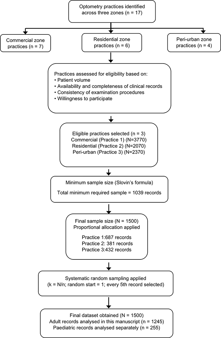 |
Figure 1 Flow diagram depicting the selection of optometry practices, determination of sample size, proportional allocation, systematic random sampling, and segmentation of datasets. |
Data Analysis
The data was entered into Microsoft Excel (Microsoft 365) for verification and cleaning, then exported to IBM SPSS Statistics version 28 for analysis. Spearman correlation coefficient was used to analyse data of the right and left eye, and a statistically significant and high correlation was found (Rs: 0.82; p < 0.001), which is the reason why statistical analysis was made only with data from the right eye of each patient, as was done in similar studies.22–24 Descriptive statistics, including means, standard deviations, frequencies, and percentages, were used to summarise demographic variables and the prevalence of refractive errors. Chi-square tests were applied to determine associations between refractive errors and demographic variables. A p-value of <0.05 was considered statistically significant. Effect sizes for practical significance (Phi and Cramer’s V =Small (0.2), medium (0.5), and large (0.8)) were also used.
Refractive errors were classified based on spherical equivalent refraction (SER) calculated using the standard formula, in which half of the cylindrical power is added to the spherical power. Refractive errors were classified according to commonly applied epidemiological thresholds: myopia was defined as an SER of less than –0.75 dioptres, hyperopia as an SER greater than +0.75 dioptres, emmetropia as an SER between –0.50 and +0.50 dioptres, and astigmatism as a cylindrical error of less than –0.75 dioptres. Presbyopia was defined as near addition in individuals aged 40 years and above. Participants younger than 40 who exhibited early near-vision symptoms were classified as pre-presbyopic and excluded from the presbyopia prevalence estimates.
Ethical Consideration
Ethical approval for this retrospective cross-sectional study was obtained from the School of Health Care Sciences Research Committee (SREC), the Faculty of Health Sciences (Proposal No: FHDC2024/120), and the University of Limpopo Turfloop Research Ethics Committee (Reference No: TREC/185/2025:PG). The study adhered to the principles outlined in the Declaration of Helsinki. Authorization to access clinical records was granted by the participating practices’ manager. This study involved a retrospective analysis of routinely collected clinical records, conducted without any direct contact with patients; therefore, informed consent was not required. All data were anonymized prior to analysis and are presented in aggregate form to ensure confidentiality.
Results
Demographics
A total of 1245 clinical records were reviewed. The mean age was 44.58 ± 13.9 years, with an age range from 18 to 101 years. Among these participants, 635 (51%) were female, and 610 (49%) were male, with their mean ages as illustrated in Figure 2. Figure 3 shows the ethnicity of participants, with most being black African (n=899; 72%).
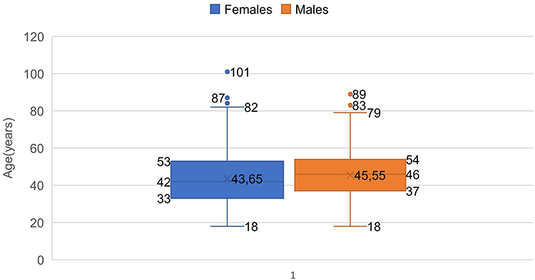 |
Figure 2 Age distribution of participants by gender. |
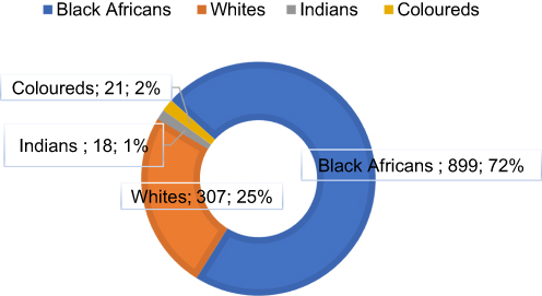 |
Figure 3 Ethnic composition of the study sample. |
Refractive Error
Among the 1245 participants, 827 (66%) had refractive errors, and their mean age was 45.51 ± 14.7 years. Of these, 167 (13%) had myopia, 124 (10%) had hyperopia, and 536 (43%) had astigmatism. Astigmatism was further classified as hyperopic astigmatism (n=60;11%) participants and myopic astigmatism (n=342;64%) participants. Compound myopic astigmatism was the most common, with 272 (51%) cases, as illustrated in Figure 4. Myopia ranged from −0.75 to −6.25 D, hyperopia from +0.625 to +4.25 D, and astigmatism from −0.75 to −6.50 D.
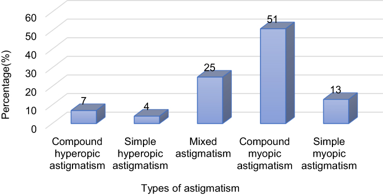 |
Figure 4 Distribution of astigmatism subtypes among participants with astigmatism. |
Table 1 outlines the age distribution of participants categorized by their type of refractive error. The mean age for individuals with astigmatism was 44.60 ± 14.41 years, with ages ranging from 18 to 89 years. In contrast, those with myopia had a mean age of 39.29 ± 11.865 years, spanning from 18 to 67 years. Participants with hyperopia displayed a higher average age of 57.82 ± 11.93 years, with a range of 20 to 84 years. Statistical analysis revealed a significant age difference among myopic participants (p < 0.001; Cramer’s V=0.559). However, no statistically significant associations were observed between age and astigmatism (p = 0.992; Cramer’s V = 0.338) or hyperopia (p = 0.905; Cramer’s V = 0.555).
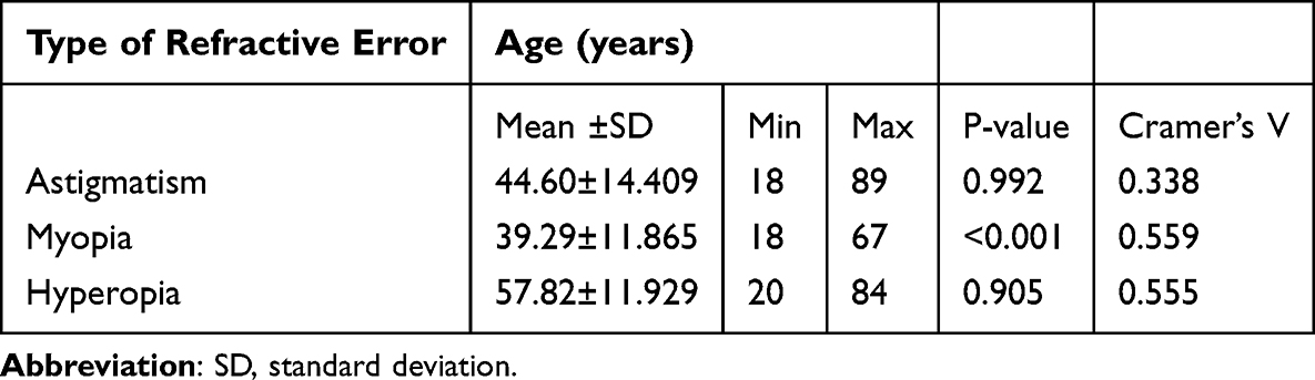 |
Table 1 Age Characteristics of Participants with Different Types of Refractive Error |
Table 2 summarises the distribution of refractive errors by gender and ethnicity. The prevalence of myopia was equal in both males and females (13%), while hyperopia and astigmatism were slightly more common among males (11% and 45%, respectively). Statistical analysis revealed no significant gender differences in the prevalence of myopia (p = 0.376; Cramer’s V=0.437), hyperopia (p = 0.284; Cramer’s V=0.400), and astigmatism (p = 0.888; Cramer’s V=0.159). Similarly, no significant difference was found across the different ethnic groups.
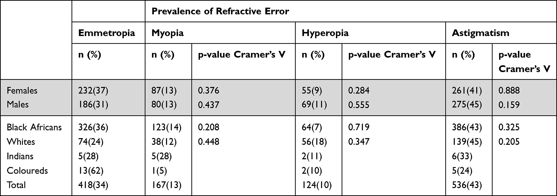 |
Table 2 Prevalence of Emmetropia and Refractive Errors by Gender and Ethnicity |
Eight hundred seventy-three participants (70%) presented with visual acuity (VA) of 6/12 or better, while three hundred seventy-two participants (30%) had VA of 6/12 or worse. Among the myopic participants, 34% (n=59) had uncorrected visual acuity (UVA) ranging from 6/18 to 6/60, whereas 14% of hyperopic participants (n=17) fell within the same UVA range. Additionally, 16% of astigmatic participants (n=86) demonstrated UVA between 6/12 and 6/18, as shown in Table 3.
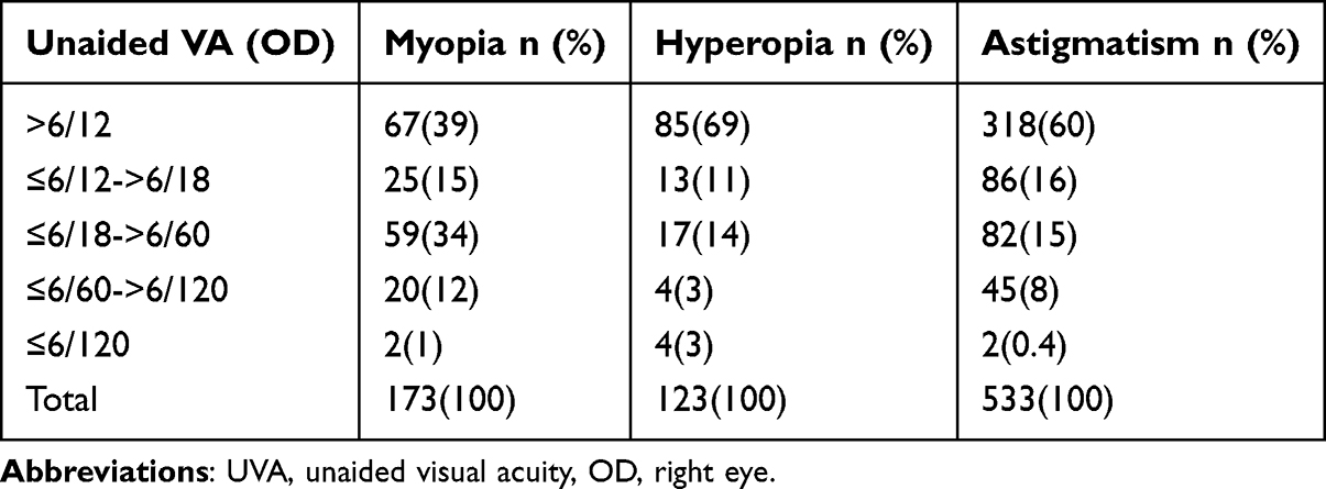 |
Table 3 Impact of Refractive Error on UVA |
Presbyopia
Of the 785 (63%) participants aged 40 and above, 665 (84.7%) had presbyopia. Table 4 illustrates the prevalence of presbyopia by demographic characteristics.
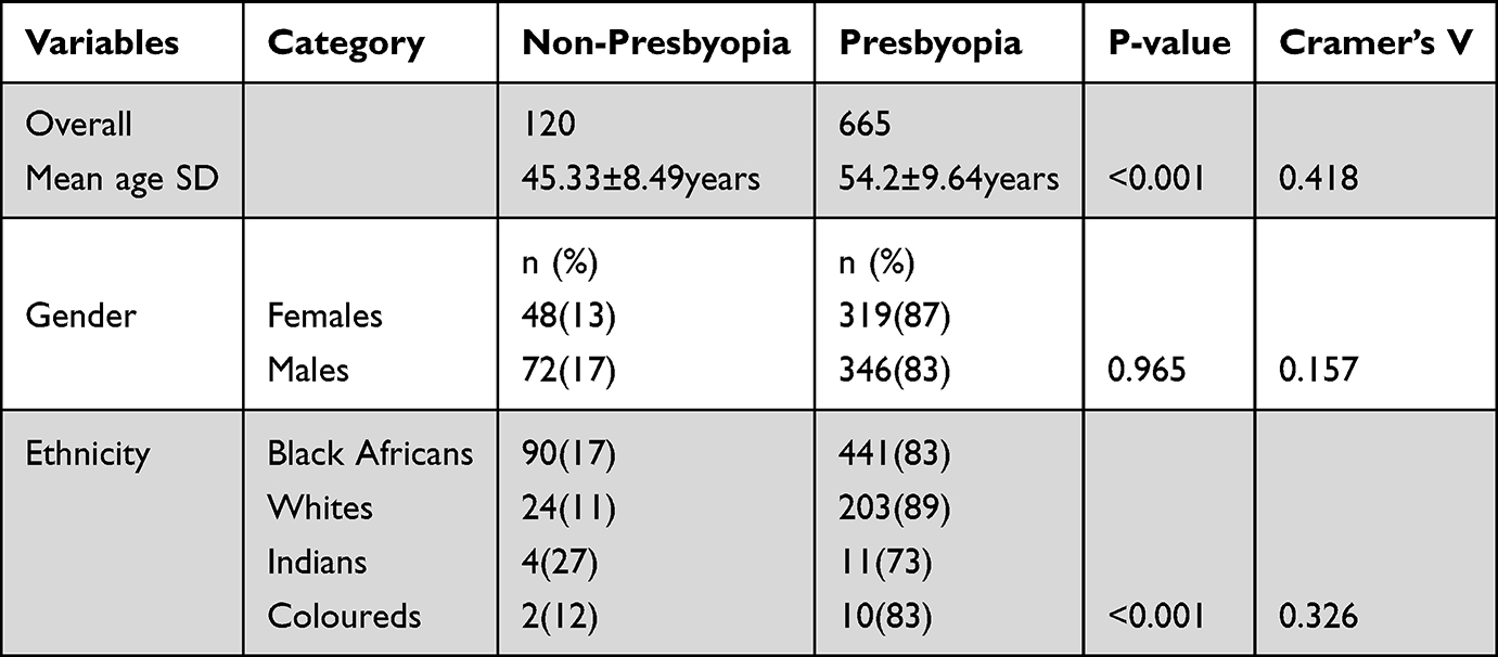 |
Table 4 Prevalence of Presbyopia by Demographic Characteristics |
Discussion
This study aimed to determine the prevalence and distribution of refractive errors (REs) among adults attending selected private optometry practices in Witbank, Mpumalanga. The findings provide insights into the profile of a health-seeking urban population. The significant prevalence of refractive error (66%) in this study aligns with trends commonly observed in clinic-based populations, where individuals are more likely to seek assistance due to visual symptoms or functional impairment.14,25 Research indicates that such environments tend to capture a greater proportion of individuals with correctable visual conditions compared to community-based studies, as the behaviour of seeking care influences the distribution observed.25,26 Therefore, these results should be interpreted as indicative of the demand for refractive services within the private practices, rather than reflecting the overall population-level prevalence.
Astigmatism emerged as the most common refractive error in this study, which is consistent with findings from similar clinical research.27 However, a study conducted by Malu and Ojabo reported that myopia was the most prevalent refractive error among patients in a private hospital in Nigeria.26 This difference may stem from a mix of demographic diversity among the study population and various environmental influences. Furthermore, compound myopic astigmatism was the most frequently observed type of astigmatism in this study, aligning with established patterns in adult refractive profiles where combined refractive components are common.28 This pattern underscores the critical importance of precise detection and effective correction of astigmatism in routine optometric care. The trends identified may result from a combination of genetic factors and environmental influences, including prolonged near work and increased reliance on digital devices, as highlighted by Chuck et al.5 This observation is further supported by global trends showing that uncorrected astigmatism significantly contributes to functional visual impairment in adult populations.29
The age-related trends observed in this study are strongly supported by existing epidemiological evidence. Notably, myopia is most prevalent among younger adults in this study, a phenomenon closely linked to various environmental and behavioral factors.8,9 Urbanization, rising educational demands, and extended periods of near work among younger individuals are likely contributors to this trend, aligning with global observations and research conducted in countries such as Iran and Zimbabwe.30,31 In contrast, the higher incidence of hyperopia in older individuals in this study can be attributed to age-related physiological changes, including a decline in accommodative capacity and modifications in lens structure, as noted by Mian et al.4 Interestingly, this study did not uncover significant age-related associations for astigmatism. This finding aligns with the work of Wajuihian and Mashige (2021),10 who reported that astigmatism may exhibit a stable age gradient in certain populations, while in others, it varies depending on corneal morphology and environmental influences. Similar age-related patterns have also been documented in studies from South Korea and Nigeria.32,33
There were no significant gender differences in the prevalence of myopia, hyperopia, and astigmatism in the current study. These results are consistent with findings from Poland,34 Ethiopia,8 and Nigeria,33 yet they contrast with the INK study, which indicated that hyperopia was significantly more prevalent among females.13 This discrepancy may be partially attributed to differences in gender distribution among the study populations. The INK study included a significantly higher proportion of female participants (approximately 75%), while the current study achieved a nearly equal representation of males and females. Consequently, the overrepresentation of females in the INK study may have influenced the observed predominance of hyperopia among female participants.
The predominance of Black African participants in this study aligns with the demographic composition of the eMalahleni (Witbank) region and reflects national population trends in South Africa, where Black Africans represent the majority.35 However, it is essential to acknowledge that the utilization of private eye care services may differ among population groups, which may also influence patterns of healthcare access and health-seeking behavior. In this context, the lack of statistically significant differences in the distribution of refractive errors across ethnic groups should be interpreted with caution. While global evidence suggests that ethnicity can impact refractive error patterns, the absence of an association in this study likely results from the clinical, practice-based nature of the sample rather than indicating a genuine lack of ethnic variation.36 Collectively, these findings suggest that while the sample is demographically consistent with the broader population, the relationships between ethnicity and refractive error warrant further exploration through population-based studies.
The prevalence of presbyopia (84.7%) reported in this study is higher than that found in several other African countries, which range from 46.5% to 50.3%,37,38 but similar to the 80.8% reported by other researchers.18,39 Additionally, the absence of significant gender differences aligns with global trends noted by Bodunde et al;40 however, some African studies suggest a higher prevalence among women, which may be linked to biological and hormonal factors.41,42 Visual acuity assessments indicate that many patients with astigmatism and myopia have unaided vision worse than 6/12 in this study, emphasizing the considerable impact of uncorrected refractive errors on daily life. This finding reinforces the American Academy of Ophthalmology’s recommendation for timely refractive management to prevent avoidable visual impairment.5 Furthermore, regional studies have highlighted the high prevalence of unaffordable spectacles.29,39 The patterns observed in Witbank must also be contextualised within regional barriers to spectacle use. Studies from Ethiopia highlight challenges such as poor knowledge of refractive errors, stigma, financial limitations, and misconceptions about spectacles.16,17
Considering the industrial and urban context of Witbank, the notable prevalence of refractive errors indicates a significant demand for refractive services, which may impact visual function and overall productivity. These findings underscore the vital role of private optometry practices in meeting the visual needs of this population. However, it is important to note that this sample consists of individuals actively seeking care at a clinic, meaning that the results should be understood within this specific context and may not be applicable to the wider population.
Limitations
The primary limitation of this study lies in the potential for selection bias, as the sample was exclusively comprised of individuals attending private optometry practices. This demographic represents a health-seeking population that is more likely to present with visual complaints, potentially leading to an overestimation of the prevalence of refractive errors compared to the general population. As a result, the findings may not be applicable to individuals who depend on public healthcare services or those who do not seek eye care; this should be considered when interpreting the reported prevalence estimates. Furthermore, the study did not account for behavioral and lifestyle factors such as digital device usage, occupational visual demands, outdoor exposure, and educational background. This oversight limits the capacity to explore potential influences on the observed patterns of refractive errors.
Additionally, the cross-sectional and retrospective design of the study restricts the ability to establish causal relationships between exposures and outcomes. The reliance on routinely collected clinical records introduces the risk of information bias due to incomplete or inconsistent documentation. Variability in examination techniques and equipment across different practices may have also impacted the consistency of the measurements. Moreover, the use of a single eye (right eye) in the analysis may overlook interocular differences in some patients.
Conclusion
The findings of this study reveal a significant prevalence of refractive errors in the urban private practice population examined. Astigmatism accounted for nearly half of all identified cases, followed by myopia and hyperopia. Furthermore, refractive errors did not show a significant association with gender or ethnicity. Although this study is limited to a specific population and its findings cannot be generalized to the wider community, it provides valuable insights for the planning and optimization of eye care services in the region. Additionally, the results offer essential baseline data that may guide future research and the development of vision care strategies in similar clinical settings.
Acknowledgments
The authors wish to acknowledge and express their gratitude to all parties who permitted the conduct of this study within their practice.
Disclosure
The authors declare that there are no financial or personal conflicts of interest related to this work.
References
1. Kim HM, Son SM. Impacts of daily life and job satisfaction on social participation of persons with visual impairment. Occup Ther Int. 2023;2023:6475756. doi:10.1155/2023/6475756
2. Marques AP, Ramke J, Cairns J, et al. Global economic productivity losses from vision impairment and blindness. EClinicalMedicine. 2021;35:100852. doi:10.1016/j.eclinm.2021.100852
3. Gkioka M, Almpanidou S, Lioti N, Almaliotis D, Karampatakis V. Daily functionality of people with low vision: the impact of visual acuity, depression, and life orientation-a cross-sectional study. Behav Neurol. 2024;2024:4366572. doi:10.1155/2024/4366572
4. Mian SI, Trobe J, Givens J. Visual impairment in adults: refractive disorders and presbyopia. Waltham, MA: UpToDate Inc. 2018. Available from: https://www.uptodate.com/contents/visual-impairmentin-adults-refractive-disorders-and-presbyopia.
5. Chuck RS, Jacobs DS, Lee JK, et al. Refractive errors & refractive surgery preferred practice pattern®. Ophthalmology. 2018;125(1):P1–10. doi:10.1016/j.ophtha.2017.10.003
6. Pesudovs K, Lansingh VC, Kempen JH; Vision Loss Expert Group of the Global Burden of Disease Study. GBD 2019 Blindness and Vision Impairment Collaborators. Global estimates on the number of people blind or visually impaired by cataract: a meta-analysis from 2000 to 2020. Eye. 2024;38(11):2156–2172. doi:10.1038/s41433-024-02961-1
7. World Health Organization. Blindness and vision impairment. World Health Organization; 2021. Available from: https://www.who.int/news-room/fact-sheets/detail/blindness-and-visual-impairment.
8. Worku S, Getachew T, Nagarchi K, Shewangizaw M. The magnitude of refractive error and its associated factors among patients visiting ophthalmology clinics in Southern Ethiopia, 2022. Clin Ophthalmol. 2023;17:1801–1811. doi:10.2147/OPTH.S408610
9. Hashemi H, Fotouhi A, Yekta A, Pakzad R, Ostadimoghaddam H, Khabazkhoob M. Global and regional estimates of prevalence of refractive errors: systematic review and meta-analysis. J Curr Ophthalmol. 2017;30(1):3–22. doi:10.1016/j.joco.2017.08.009
10. Wajuihian SO, Mashige KP. Gender and age distribution of refractive errors in an optometric clinical population. J Optom. 2021;14(4):315–327. doi:10.1016/j.optom.2020.09.002
11. Shiferaw Alemu D, Desalegn Gudeta A, Tsega Ferede A, Woretaw Alemu H. Prevalence and degrees of myopia and hyperopia at Gondar university hospital tertiary eye care and training center, Northwest Ethiopia. Clin Optom. 2016;Volume 8:85–91. doi:10.2147/OPTO.S116535
12. Besufikad B, Hailemichael W, Tilahun L, Yimam W, Anteneh S. Refractive errors and associated factors among patients visiting BoruMeda Hospital’s secondary eye Unit in Dessie Town, South Wollo Zone, Ethiopia. BMC Ophthalmol. 2022;22(1):312. doi:10.1186/s12886-022-02539-z
13. Mashige KP, Jaggernath J, Ramson P, Martin C, Chinanayi FS, Naidoo KS. Prevalence of refractive errors in the INK Area, Durban, South Africa. Optom Vis Sci. 2016;93(3):243–250. doi:10.1097/OPX.000000000000077
14. Maluleke KD, Hasrod N, Rubin A. Prevalence and determinants of refractive errors at Sekororo District Hospital in Limpopo Province, South Africa. Afr Vision Eye Health. 2024;83(1):890. doi:10.4102/AVEH.v83i1.890
15. Ebrahim Khan N, Mahomedy M, Mngadi L, et al. Knowledge, attitudes and practices towards refractive error amongst students. Afr Vision Eye Health. 2022;81(1):633. doi:10.4102/aveh.v81i1.633
16. Desalegn A, Tsegaw A, Shiferaw D, Woretaw H. Knowledge, attitude, practice and associated factors towards spectacles use among adults in Gondar town, northwest Ethiopia. BMC Ophthalmol. 2016;16(1):184. doi:10.1186/s12886-016-0357-3
17. Gebresellassie MG, Sisay H, Desta K, et al. Spectacles under utilization and associated factors among adults in Hawassa, South Ethiopia, June, 2022. Clin Optom. 2023;15:303–311. doi:10.2147/OPTO.S426032
18. Statistics South Africa (2023b) census 2022 municipal fact sheet. Report 03-01-82.
19. Department of Cooperative Governance and Traditional Affairs (COGTA), 2019, Mpumalanga spatial development framework. Available from: https://cogta.mpg.gov.za/documents/SpatialDevFramework/PSDF%20Final%20Report.pdf.
20. Spatial development framework review phase 3: spatial proposals draft report 2024. Available from: https://www.emalahlenilm.gov.za/wp-content/uploads/2023/11/DRAFT-Emalahleni-Local-Municipality-Spatial-Development-Framework-2024-.pdf.
21. Medpages.Statistics–optometry practices in witbank. 2020. Available from: http://www.medpages.info/sf/index.php?page=listing&servicecode=549&suburbcode=742.
22. Sengo DB, Dos Santos IIDB, Faquihe MF, et al. The prevalence of visual impairment and refractive errors among a youth population in Mozambique: evidence of the need for intervention. Children. 2021;8(10):892. doi:10.3390/children8100892
23. Bourne RR, Dineen BP, Ali SM, Noorul Huq DM, Johnson GJ. Prevalence of refractive error in Bangladeshi adults: results of the national blindness and low vision survey of Bangladesh. Ophthalmology. 2004;111(6):1150–1160. doi:10.1016/j.ophtha.2003.09.046
24. Mahjoob M, Heydarian S, Nejati J, Ansari-Moghaddam A, Ravandeh N. Prevalence of refractive errors among primary school children in a tropical area, Southeastern Iran. Asian Pac J Trop Biomed. 2016;6(2):181–184. doi:10.1016/j.apjtb.2015.10.008
25. Owusu-Afriyie B, Kombra M, Gende T, Kia A, Mou I. Prevalence and distribution of refractive errors among ophthalmic patients in Madang Province, Papua New Guinea. J Curr Ophthalmol. 2022;34(2):223–228. doi:10.4103/joco.joco_337_21
26. Malu K, Ojabo C. Refractive errors in patients attending a private hospital in Jos, Nigeria. Niger J Clin Pract. 2014;17(1):106–111. doi:10.4103/1119-3077.122863
27. Kaiti R, Pradhan A, Dahal HN, Shrestha P. Pattern and prevalence of refractive error and secondary visual impairment in patients attending a tertiary hospital in Dhulikhel, Nepal. Kathmandu Univ Med J. 2018;16(62):114–119. PMID: 30636750.
28. Ajibode HA, Fakolujo VO, Bodunde OT. Prevalence and patterns of refractive error among adults in Sagamu, South-West Nigeria. Afr Vision Eye Health. 2022;81(1):724. doi:10.4102/aveh.v81i1.724
29. Das S, Shergill SS. Prevalence of uncorrected refractive errors among adults attending at a tertiary care hospital–a retrospective study. Med J Dr DY Patil Vidyapeeth. 2023;16(1):71–75. doi:10.4103/mjdrdypu.mjdrdypu_145_21
30. Kahoto VE, Kwarteng MA, Owusu-Afriyie B, Zaabaar E, Kyei S. Epidemiology and trends in the uptake of refractive error services in Harare, Zimbabwe: a hospital-based retrospective study. Malawi Med J. 2023;35(4):214–219. doi:10.4314/mmj.v35i4.3
31. Alipour F, Mohammadzadeh M, Jafari F, et al. Distribution and prevalence of refractive error in Iranian adult population results of the PERSIAN eye cohort study PECS. Sci Rep. 2024;14(1):14515. doi:10.1038/s41598-024-65328-2
32. Yoo YC, Kim JM, Park KH, Kim CY, Kim TW; Namil Study Group, Korean Glaucoma Society. Refractive errors in a rural Korean adult population: the Namil Study. Eye. 2013;27(12):1368–1375. doi:10.1038/eye.2013.195
33. Ezelum C, Razavi H, Sivasubramaniam S, et al. Refractive error in Nigerian adults: prevalence, type, and spectacle coverage. Invest Ophthalmol Vis Sci. 2011;52(8):5449–5456. doi:10.1167/iovs.10-6770
34. Nowak MS, Jurowski P, Grzybowski A, Smigielski J. Characteristics of refractive errors in a population of adults in the Central Region of Poland. Int J Environ Res Public Health. 2018;15(1):90. doi:10.3390/ijerph15010090
35. Statistics South Africa. Census 2022 statistical release. Report P0301.4. Pretoria: Statistics South Africa; 2023. Available from: https://census.statssa.gov.za/assets/documents/2022/P03014_Census_2022_Statistical_Release.pdf.
36. Pan CW, Klein BE, Cotch MF, et al. Racial variations in the prevalence of refractive errors in the United States: the multi-ethnic study of atherosclerosis. Am J Ophthalmol. 2013;155(6):1129–1138.e1. doi:10.1016/j.ajo.2013.01.009
37. Mashayo ER, Chan VF, Ramson P, Chinanayi F, Naidoo KS. Prevalence of refractive error, presbyopia and spectacle coverage in Kahama District, Tanzania: a rapid assessment of refractive error. Clin Exp Optom. 2015;98(1):58–64. doi:10.1111/cxo.12207
38. Nsubuga N, Ramson P, Govender P, Chan V, Wepo M, Naidoo KS. Uncorrected refractive errors, presbyopia and spectacle coverage in Kamuli District, Uganda. Afr Vision Eye Health. 2016;75(1):1–6. doi:10.4102/aveh.v75i1.327
39. Otutu M, Nachega J, Harvey J, Meyer D. The prevalence of refractive error in three communities of Cape Town, South Africa. Afr Vision Eye Health. 2012;71(1):32–38. doi:10.4102/aveh.v71i1.65
40. Bodunde OT, Ajibod HA, Jagun OO, Fafiolu VO, Otulana TO, Onabolu OO. The impact of presbyopia on the quality of life in a semi-urban community in Southwest Nigeria. Afr Vision Eye Health. 2020;79(1):1–6.
41. Ajibode HA, Fakolujo VO, Onabolu OO, Jagun O, Ogunlesi TA, Abiodun OA. A community-based prevalence of presbyopia and spectacle coverage in Southwest Nigeria. J West Afr Coll Surg. 2016;6(4):66–82. doi:10.1111/cxo.12402
42. Burke AG, Patel I, Munoz B, et al. Population-based study of presbyopia in rural Tanzania. Ophthalmology. 2006;113(5):723–727. doi:10.1016/j.ophtha.2006.01.030
 © 2026 The Author(s). This work is published and licensed by Dove Medical Press Limited. The
full terms of this license are available at https://www.dovepress.com/terms
and incorporate the Creative Commons Attribution
- Non Commercial (unported, 4.0) License.
By accessing the work you hereby accept the Terms. Non-commercial uses of the work are permitted
without any further permission from Dove Medical Press Limited, provided the work is properly
attributed. For permission for commercial use of this work, please see paragraphs 4.2 and 5 of our Terms.
© 2026 The Author(s). This work is published and licensed by Dove Medical Press Limited. The
full terms of this license are available at https://www.dovepress.com/terms
and incorporate the Creative Commons Attribution
- Non Commercial (unported, 4.0) License.
By accessing the work you hereby accept the Terms. Non-commercial uses of the work are permitted
without any further permission from Dove Medical Press Limited, provided the work is properly
attributed. For permission for commercial use of this work, please see paragraphs 4.2 and 5 of our Terms.
Recommended articles

Evaluation of the EVO/EVO+ Sphere and Toric Visian ICL: Six Month Results from the United States Food and Drug Administration Clinical Trial
Packer M
Clinical Ophthalmology 2022, 16:1541-1553
Published Date: 21 May 2022
Photorefractive Keratectomy Enhancement (PRK) After Small-Incision Lenticule Extraction (SMILE)
Moshirfar M, Parsons MT, Chartrand NA, Lau CK, Stapley S, Bundogji N, Ronquillo YC, Hoopes PC
Clinical Ophthalmology 2022, 16:3033-3042
Published Date: 12 September 2022

A Comprehensive Retrospective Analysis of EVO/EVO+ Implantable Collamer Lens: Evaluating Refractive Outcomes in the Largest Single Center Study of ICL Patients in the United States
Albo C, Nasser T, Szynkarski DT, Nguyen N, Mueller B, Libfraind L, Parkhurst G
Clinical Ophthalmology 2024, 18:69-78
Published Date: 9 January 2024
Prevalence of Refractive Errors Among School-Age Children and Adolescents in Saudi Arabia: A Systematic Review and Meta-Analysis
Al Khathami A, Baklola M, Alshehri AA, Alnasser LH, Alshehri RS, Salawi MA, Alwadai RH, Al Ghazwi M, Alanazi AAQ, Alshammari AM, Al-bawah N, Hafez M, Abu Melha A
Clinical Ophthalmology 2025, 19:2117-2132
Published Date: 5 July 2025
Prevalence of Refractive Errors, Myopic Macular Degeneration, and Associated Risk Factors in a Maltese Population-Based Study
Agius D, Mamo J, Calleja N, Cassar D, Marku X, Nappa MC, Zammit M, Pace ME, Carbonaro F
Clinical Optometry 2026, 18:561426
Published Date: 7 February 2026