
Back to Journals » Journal of Inflammation Research » Volume 19
White Blood Cell-to-Hemoglobin Ratio, a Promising Indicator for Diabetic Kidney Disease: An Observational Cross-Sectional Study
Authors Zhang M, Wang L, Lin Y, Wu M, Tan J  , Chen Y, Zhao J, Xu H, Qiang W , Guo H, Wang Y, Shi B , He M
Received 31 October 2025
Accepted for publication 17 March 2026
Published 9 April 2026 Volume 2026:19 572888
DOI https://doi.org/10.2147/JIR.S572888
Checked for plagiarism Yes
Review by Single anonymous peer review
Peer reviewer comments 5
Editor who approved publication: Dr Quan Zhang
Meng Zhang,* Ling Wang,* Yuxuan Lin, Mengzhi Wu, Jing Tan, Yu Chen, Jiarui Zhao, Huayang Xu, Wei Qiang, Hui Guo, Yue Wang, Bingyin Shi, Mingqian He
Department of Endocrinology, the First Affiliated Hospital of Xi’an JiaoTong University, Xi’an, Shaanxi, People’s Republic of China
*These authors contributed equally to this work
Correspondence: Mingqian He, Department of Endocrinology, the First Affiliated Hospital of Xi’an JiaoTong University, No. 277, West Yanta Road, Xi’an, Shaanxi, 710061, People’s Republic of China, Tel +86-(029)85323974, Fax +86-(029)85323974, Email [email protected]
Background: Diabetic kidney disease (DKD), a prevalent microvascular complication of type 2 diabetes (T2DM), is frequently diagnosed late with irreversible renal damage, underscoring the need for early-stage biomarkers. Anemia commonly occurs in DKD due to impaired erythropoiesis and reduced hemoglobin levels, while chronic inflammation also plays a key role in its progression. The white blood cell-to-hemoglobin ratio (WBCHR), derived from full blood examination (FBE), integrates inflammation and anemia signals in DKD. This study investigates WBCHR’s role in early DKD risk stratification.
Materials and Methods: This observational cross-sectional study included 6257 patients with T2DM from Northwest China from 2013 to 2022 (1721 DKD, 4536 non-DKD). Demographic, metabolic, FBE indices, and renal function parameters were compared. Spearman correlation and logistic regression analysis were performed to assess the correlation between FBE indices of renal impairment, and the relationship between WBCHR and DKD occurrence. Receiver operating characteristic (ROC) analysis was used to calculate the discriminatory performance of WBCHR for DKD presence.
Results: Age, male proportion, T2DM duration, insulin usage, blood pressure, fasting blood glucose and triglycerides were greater in patients with DKD (P < 0.05). Patients with DKD were more likely to exhibit lower hemoglobin levels and higher inflammatory FBE indices (P < 0.001). Among all the inflammatory indices, WBCHR showed the strongest association with DKD occurrence and positively correlated with multiple renal impairment indicators. After adjusting for confounding factors, WBCHR remained independently associated with DKD progression (OR: 3.669, 95% CI: 2.407– 5.592, P< 0,001). Moreover, ROC analysis identified WBCHR as a potential risk factor for DKD, particularly in patients with T2DM aged> 40, with a cut-off value of 0.529 (AUC = 0.641, P< 0.001).
Conclusion: WBCHR was significantly associated with an increased risk of DKD, suggesting its potential as a diagnostic and management indicator for DKD.
Highlights: WBCHR was significantly associated with increased DKD risk in patients with T2DM and remained an independent factor after confounder adjustment. WBCHR could be adopted as a low-cost screening tool for early DKD risk in routine diabetes management.
Keywords: diabetic kidney disease, complete blood count, white blood cell-to-hemoglobin ratio
Introduction
Diabetic kidney disease (DKD), also known as diabetic nephropathy (DN), is a major microvascular complication of diabetes and affects approximately 40% of patients with type 2 diabetes (T2DM).1,2 It is projected that by 2040, the global diabetic population will reach approximately 642 million,3 with over 35% of these individuals developing kidney damage.4 As the leading attributable cause of end-stage renal disease, DKD not only contributes to high rates of disability and mortality in patients with diabetes,5 but also imposes a substantial economic burden on healthcare systems.6 Current diagnostic approaches for DKD often detect kidney damage only after significant morphological changes have occurred, which makes it too late for effective intervention.1 Therefore, identifying accessible biomarkers and modifiable risk factors is critical for early detection and timely diagnosis of DKD. Such efforts would facilitate targeted preventive strategies, optimize disease management, and ultimately improve clinical outcomes while preserving patients’ quality of life.
Inflammatory factors play a pivotal role in the pathogenesis of both T2DM and DKD.2,7 Chronic inflammation acts as a core driver of DKD progression, involving dysregulated immune pathways and disrupted intercellular communication.8 Crucially, immune cells, such as macrophages, T cells, and neutrophils, directly mediate renal damage through tissue infiltration, cytokine release, and bidirectional crosstalk with resident kidney cells, thereby establishing a self-perpetuating inflammatory loop.9 On the other hand, anemia is common in CKD across a wide spectrum of aetiologies, typically observed CKD of stages 3 or higher (estimated glomerular filtration rate, eGFR < 60 mL/min/1.73 m2) and becoming more prevalent with worsening renal function.10
Blood cell variables and their derived indices, such as monocyte-to-lymphocyte ratio (MLR), platelet-to-lymphocyte ratio (PLR), neutrophil-to-lymphocyte ratio (NLR) and systemic immune-inflammation index (SII), serve as fundamental indicators of inflammation and have been linked to the development of T2DM or chronic kidney disease (CKD).11–14 Additionally, hemoglobin (Hb), another component of the full blood examination (FBE), reflects renal tubulointerstitial impairment and has been recognized as a predictive biomarker for early-stage DKD (stage 1–2), even when values remain within the normal range.15–18 Recently, the white blood cell-to-hemoglobin ratio (WBCHR), a novel systemic index, has emerged as a potential marker capable of concurrently reflecting inflammatory activity and anemia status.19–21 As a composite metric, WBCHR may offer incremental value beyond its individual components, particularly hemoglobin, by capturing their synergistic interactions. Thus, WBCHR is positioned as a candidate for detecting earlier, concomitant pathological processes of DKD, although current clinical markers (eg eGFR) are essential for diagnosing functional decline and structural damage. While existing studies on WBCHR have primarily focused on its prognostic value in conditions such as heart failure and malignancies, its utility in DKD remains underexplored. Building on our previous research into endocrine biomarkers for DKD,22 we shift our focus to the potential of novel hematological indices, specifically investigating the WBCHR as an integrative marker of inflammation and anemia in this condition.
To comprehensively investigate the relationship between FBE indices and DKD pathogenesis, we conducted a cross-sectional study involving over 6000 patients with T2DM stratified by the presence of DKD. We hypothesized that FBE indices are significantly associated with DKD in this population, and that may hold promise as potential predictive marker for DKD.
Materials and Methods
Study Design and Participants
This cross-sectional analysis retrospectively screened adult inpatients with a confirmed diagnosis of T2DM based on standard clinical criteria,23 admitted to the Department of Endocrinology at The First Affiliated Hospital of Xi’an Jiaotong University over a 10-year period (January 2013 to December 2022). To ensure a clearly defined study population, individuals meeting any of the following criteria were excluded: (1) presence of recent acute diabetic complications; (2) presence of non-diabetic renal diseases, such as chronic nephritis, primary glomerulonephritis, urinary tract infection, urinary tract stones or tumors caused by acute infection and other factors; (3) diagnosis of hematologic diseases, such as agranulocytosis, aplastic anemia or leukemia; (4) coexistence of major systemic diseases, such as severe hepatic dysfunction, malignancy, autoimmune diseases, inflammatory disease or immunodeficiency syndromes; (5) incomplete medical records of renal function, FBE or other important covariables. Following this screening process, a total of 6257 patients were included in the final cohort (Figure 1).
 |
Figure 1 Flowchart of the inclusion and exclusion of participants. Abbreviation: DKD, diabetic kidney disease. |
This study adhered to the ethical principles outlined in the Declaration of Helsinki and received approval from the Institutional Review Board of the First Affiliated Hospital of Xi’an Jiaotong University (XJTU1AF2018LSK-055). Furthermore, this study was conducted in accordance with the REporting of studies Conducted using Observational Routinely-collected health Data (RECORD) guidelines. All participants provided written informed consent with full knowledge of the study protocol.
The Definition of DKD
Renal function was obtained from fast venous blood samples, urinalysis results were obtained from urine samples included fasting morning specimens and 24-hour total collections. A LABOSPECT 008AS automatic biochemical analyzer (HITACHI, Japan) were used.
Study participants were categorized based on the presence of DKD. DKD was diagnosed based on standard clinical criteria (ICD-10 code, E11.2),24 defined as a reduced estimated glomerular filtration rate (eGFR < 60 mL/min/1.73 m2) and/or an elevated urinary albumin-to-creatinine ratio (UACR > 30 mg/g) for ≥3 months. The eGFR was determined using the Chronic Kidney Disease Epidemiology Collaboration (CKD–EPI) equations.25 Based on this classification, the cohort was stratified into a non-DKD group (n = 4,536) and a DKD group (n = 1,721).
FBE Parameters and Calculated Indices
FBE parameters were assessed using an XN-20 automated hematology analyzer (SYSMEX, Japan) from fast venous blood samples: red cell count (RBC), Hb, white cell count (WBC), neutrophil (NEUT), lymphocyte (LYMPH), monocyte (MONO), erythrocyte sedimentation rate (ESR) and C-reactive protein (CRP) using a standard assay.
Furthermore, the following indices were derived from the baseline laboratory data:
WBCHR = WBC (×109/L) / Hb (g/dL),
NLR = NEUT (×109/L) / LYMPH (×109/L),
MLR = MONO (×109/L) / LYMPH (×109/L).
The Collection of Covariates
Clinical data were systematically extracted from electronic medical records. Demographic information included age, sex, and T2DM duration. Medication history included the use of insulin and oral hypoglycemic drugs, including biguanides, sulfonylureas, meglitinides, thiazolidinediones, dipeptidyl peptidase-4 (DPP4) inhibitors, glucagon-like peptide-1 (GLP-1) analogues, α-glucosidase inhibitors, sodium-glucose cotransporter-2 (SGLT-2) inhibitors. The laboratory examination results mainly included metabolic parameters: glycated hemoglobin (HbA1c), fasting blood glucose (FBG), glycated albumin (GA), total cholesterol (TC), triglycerides (TGs), high-density lipoprotein cholesterol (HDL-c) and low-density lipoprotein cholesterol (LDL-c). The Triglyceride-glucose (TyG) index was derived as ln[TG (mg/dL) × FBG (mg/dL)]/2.
Statistical Analysis
All statistical analyses were conducted using SPSS software. Continuous variables are presented as mean ± standard deviation (SD) and median with interquartile range (IQR) for normally and non-normally distributed data, respectively. Group differences were analyzed using the Student’s t-test and the Mann–Whitney U-test for normal and non-normally distributed data, respectively. Categorical variables are summarized as frequencies and percentages, and group differences were assessed with the Chi-squared test.
Correlations between FBE-derived indices and renal impairment markers were evaluated using Spearman correlation analysis. Univariate and multivariate logistic regression analyses were performed to identify potential independent risk factors associated with DKD. The discriminatory power of the WBCHR for detecting DKD was quantified by the area under the receiver operating characteristic curve (AUC-ROC). A two-tailed P-value of less than 0.05 was considered statistically significant for all tests.
Results
Clinical Characteristics of the Study Participants
A total of 6257 patients with T2DM were enrolled, including 1721 with DKD and 4536 without DKD. Baseline clinical characteristics of the two groups are summarized in Table 1. Compared to patients without DKD, patients with DKD were significantly older, had a longer duration and exhibited a higher male predominance (P < 0.001, P < 0.001 and P = 0.043, respectively). Regarding medication use, patients with DKD showed a higher prevalence of insulin and sulfonylurea usage (both P < 0.001), but lower utilization of biguanides, meglitinides, DPP4 inhibitors (P < 0.001, P = 0.017 and P < 0.001, respectively).
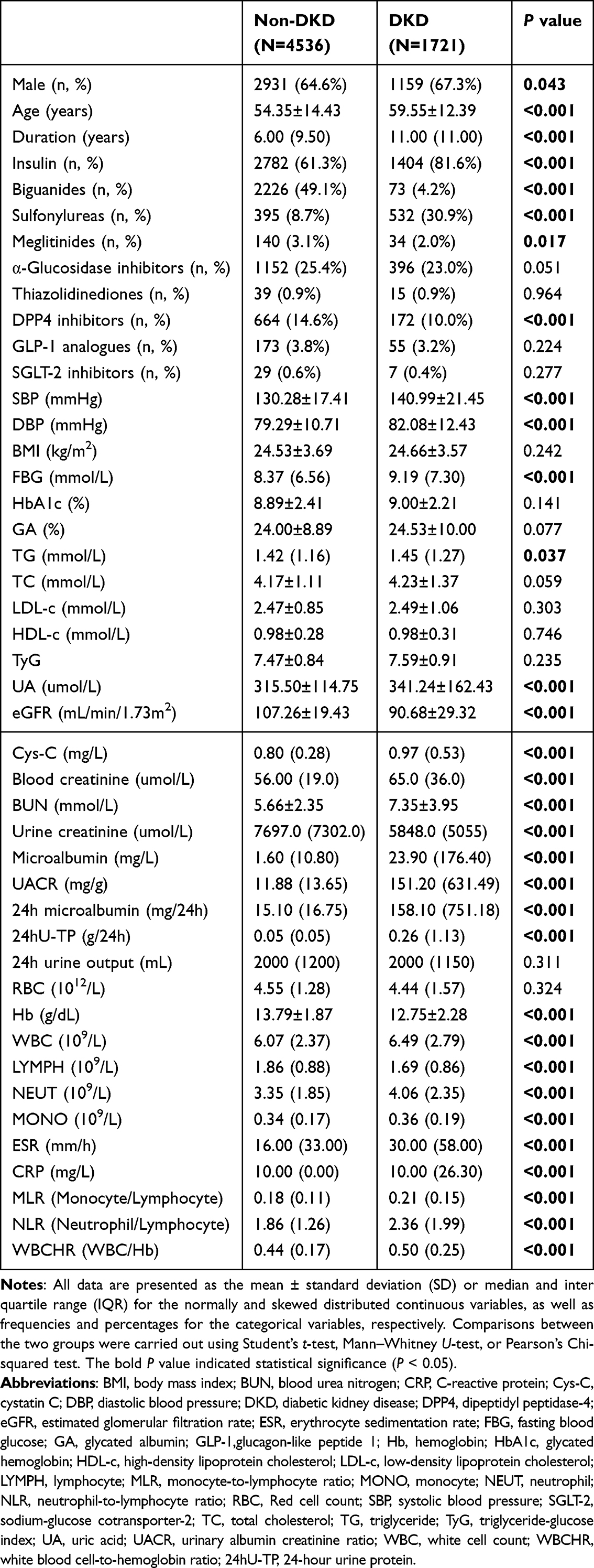 |
Table 1 Clinical Characteristics of the Study Participants |
As shown in Table 1, patients with DKD demonstrated significantly elevated systolic blood pressure (SBP), diastolic blood pressure (DBP), FBG and TG levels compared to patients without DKD (P < 0.001, P < 0.001, P < 0.001 and P = 0.037, respectively). Renal function markers further highlighted differences: patients with DKD had the higher uric acid (UA), cystatin C (Cys-C), serum creatinine, blood urea nitrogen (BUN), urinary albumin-to-creatinine ratio (UACR), urinary macroglobulin, 24h microalbumin and 24 h urine protein (24hU-TP), alongside reduced eGFR, and urinary creatinine levels (all P < 0.001). No significant between-group differences were observed in HbA1c, GA, TC, LDL-c, HDL-c, or TyG.
Hematologic analysis revealed higher levels of WBC, NEUT, and MONO in patients with DKD, whereas Hb and LYMPH were lower (all P < 0.001). Additionally, patients with DKD showed elevated ESR and CPR levels (all P < 0.001). RBC did not differ between groups. Furthermore, FBE-derived indices, including MLR (0.18 [0.11] vs. 0.21 [0.15]), NLR (1.86 [1.26] vs. 2.36 [1.99]) and WBCHR (0.44 [0.17] vs. 0.50 [0.25]) were significantly higher in the DKD group (all P < 0.001).
Univariate Analysis of FBE Indices on the Occurrence of DKD
Univariate logistic regression analyses were performed to assess the association of FBE indices and DKD. As shown in Figure 2, WBCHR emerged as the most significant positive predictor of DKD risk (odds ratio (OR): 4.736, 95% confidence interval (CI): 3.745–5.989, P < 0.001). Other indices associated with increased risk included MONO (OR: 2.615, 95% CI: 1.935–3.535), NEUT (OR: 1.115, 95% CI: 1.091–1.139), WBC (OR: 1.085, 95% CI: 1.064–1.107), NLR (OR: 1.037, 95% CI: 1.023–1.052), ESR (OR: 1.012, 95% CI: 1.008–1.018) and CRP (OR: 1.004, 95% CI: 1.002–1.007) (all P < 0.001). In contrast, Hb (OR: 0.781, 95% CI: 0.759–0.803) and LYMPH (OR: 0.673, 95% CI: 0.617–0.733) were negatively correlated with DKD risk. No significant association was observed between MLR and DKD. It is worth noting that the relatively high OR value for WBCHR may also be associated with its small numerical range in this population.
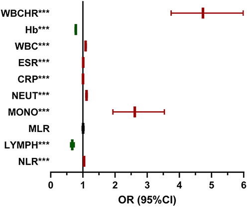 |
Figure 2 Forest graph showing the results of univariate logistic regression analyses of the association between the FBE indices and DKD. *** P < 0.001: significant correlation between FBE indices and DKD. Abbreviations: CI, confidence interval; CRP, C-reactive protein; DKD, diabetic kidney disease; ESR, erythrocyte sedimentation rate; FBE, complete blood cell; Hb, hemoglobin; LYMPH, lymphocyte; MLR, monocyte-to-lymphocyte ratio; MONO, monocyte; NEUT, neutrophil; NLR, neutrophil-to-lymphocyte ratio; OR, odds ratio; WBC, white cell count; WBCHR, white blood cell-to-hemoglobin ratio. |
Correlation of FBE Indices and Indicators of Renal Impairment
Spearman correlation analysis was conducted to evaluate the associations between FBE indices and renal impairment indicators (Figure 3 and Tables S1–2). The results revealed that WBCHR, NLR, WBC, NEUT, MONO, CRP, and ESR exhibited significant positive correlations with Cys-C, UACR, 24hU-TP, 24h microalbumin, BUN, blood creatinine and UA, as well as significant negative correlations with eGFR and urine creatinine. Conversely, Hb and LYMPH showed negative correlations with most of these renal impairment indicators. Additionally, WBCHR, NLR, WBC and NEUT were positively associated with HDL-C, HbA1c and FBG levels.
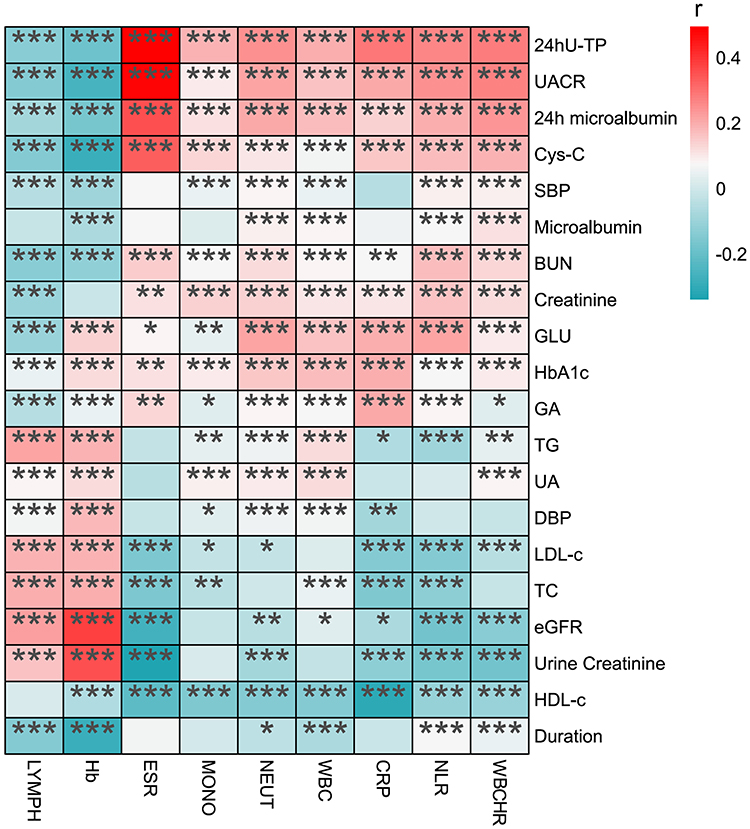 |
Figure 3 Heatmap showing the results of correlations between FBE indices and indicators of renal impairment. *P < 0.05, **P < 0.01, ***P < 0.001: significant correlation between FBE indices and renal impairment indicators. Abbreviations: BUN, blood urea nitrogen; CRP, C-reactive protein; Cys-C, cystatin C; DBP, diastolic blood pressure; eGFR, estimated glomerular filtration rate; ESR, erythrocyte sedimentation rate; FBE, full blood examination; FBG, fasting blood glucose; GA, glycated albumin; Hb, hemoglobin; HbA1c, glycated hemoglobin; HDL-c, high-density lipoprotein cholesterol; LDL-c, low-density lipoprotein cholesterol; LYMPH, lymphocyte; MONO, monocyte; NEUT, neutrophil; NLR, neutrophil-to-lymphocyte ratio; SBP, systolic blood pressure; TC, total cholesterol; TG, triglyceride; UA, uric acid; UACR, urinary albumin creatinine ratio; WBC, white cell count; WBCHR, white blood cell-to-hemoglobin ratio; 24hU-TP, 24-hour urine protein. |
Multivariate Analysis of the Independent Effect of FBE Indices on the Risk of DKD
To account for confounding factors, multivariate logistic regression models were performed to evaluate the independent effects of FBE indices on the risk of DKD. Three models were constructed: the Model 1 adjusted for age, gender and body mass index (BMI); Model 2 adjusted for variables in Model 1 plus duration of T2DM, and HbA1c; and Model 3 adjusted for variables in Model 2 plus SBP, DBP and UACR.
The results of the regression analyses were presented in Table 2. In the fully adjusted Model 3, WBCHR demonstrated the strongest positive association with DKD (OR = 3.669, 95% CI: 2.407–5.592, P < 0.001), followed by MONO, NEUT, WBC and NLR. Conversely, Hb exhibited a significant correlation with DKD.
 |
Table 2 Multivariate Analysis of the Association Between FBE Indices and DKD |
Receiver Operating Characteristic Analysis to Determine the WBCHR Cut-Off Value
Considering that a higher WBCHR is an independent risk factor for DKD, we performed a ROC analysis (Figure 4a). The analysis yielded an AUC of 0.627, with an optimal cut-off value of 0.529 (P < 0.001, Youden index = 0.190, sensitivity = 44.89%, specificity = 74.13%).
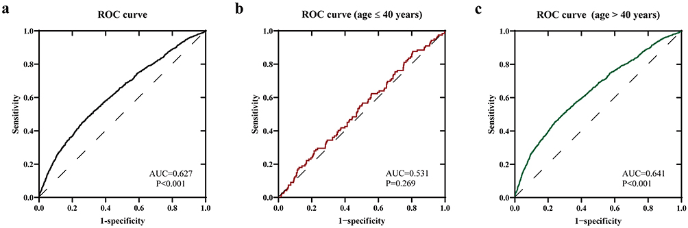 |
Figure 4 ROC analysis of the effect of the WBCHR on the risk of DKD in patients of different ages. (a) ROC analysis of all the patients. (b) ROC analysis of the patients aged ≤40 years. (c) ROC analysis of the patients aged >40 years. Abbreviations: AUC, area under the curve; DKD, diabetic kidney disease; ROC, receiver operating characteristic; WBCHR, white blood cell-to-hemoglobin ratio. |
We then performed ROC analysis for age-stratified subgroups, with 40 years selected as the cutoff value (Figure 4b and c). Age 40 serves as a commonly used clinical threshold for defining early-onset T2DM, a condition whose pathophysiological features often differ from those of late-onset T2DM, and whose incidence has risen notably in recent years.26 Furthermore, previous research indicated that hematological inflammatory markers are closely associated with the development and progression of T2DM in patients over 40.27 The results demonstrated that WBCHR had a potential predictive value for DKD risk in patients aged > 40 years, with an AUC of 0.641 (P < 0.001, Youden index = 0.213, sensitivity = 45.48%, and specificity = 75.83%), and a consistent cut-off value of 0.529.
Discussion
In this cross-sectional study involving a cohort of over 6000 patients with T2DM, we identified significant associations between FBE indices and DKD occurrence. Notably, WBCHR emerged as an independent risk factor for DKD after multivariable adjustment. Furthermore, WBCHR > 0.529 demonstrated potential predictive performance for DKD in patients with T2DM aged > 40 years. These findings offer valuable indicators and evidence-based strategies for early risk stratification and precision management of DKD in patients with diabetes.
Multiple immune-inflammatory FBE indices were positively correlated with DKD occurrence, including WBCHR, NLR, MONO, WBC and NEUT. Previous clinical studies have linked NEUT and NLR to DKD progression in patients with diabetes.28,29 A UK Biobank-based study demonstrated that NEUT, WBC and MONO were positively associated with the risk of developing T2DM, and significantly mediated the relationship between dietary patterns and T2DM risk.11 However, evidence on the association between WBCHR and DKD remains scarce. To our knowledge, this is the first large-scale clinical study (n > 6000) to demonstrate that WBCHR is an independently risk factor for DKD (OR = 3.669, 95% CI 2.407–5.592), even after rigorous adjustment for confounders including glycemic control, hypertension and UACR. Furthermore, WBCHR exhibited a strong predictive value for DKD risk in patients aged >40 years with a cut-off value of 0.529 (AUC=0.641), which may be attributed to the age-related changes in hematological parameters.30,31 These findings pioneer novel insights into the dual-pathway pathophysiology linking systemic inflammation (via leukocytes) and anemia (via hemoglobin) to DKD.
Chronic inflammation and leukocyte activity play pivotal roles in DKD pathogenesis.7,32,33 Hyperglycemia and hemodynamic abnormalities activate intracellular signaling cascades in renal cells,34,35 including activation of NF-κB and JAK/STAT pathways,36,37 which drive excessive production of pro-inflammatory cytokines (eg., TNF-α, IL-1β) and chemokines (eg., CCL2).38,39 Concurrently, endothelial cell activation upregulates the expression of chemokine receptors and adhesion molecules,34,40 facilitating leukocyte transendothelial migration into renal interstitium.41,42 Infiltrated leukocytes exacerbate renal damage through direct interactions with renal cells, sustained production of reactive oxygen species (ROS), and release of pro-inflammatory cytokines (TNF-α, IL-1β and IL-17a) and chemokines (CCL2).43–45 All these events amplify the inflammatory response in a positive feedback loop that enhances renal damage.7,33
Anemia is prevalent among patients with DKD, and is associated with accelerated progression of renal dysfunction, cardiovascular complications, and increased mortality.16,46 Studies have shown that patients with advanced-stage CKD (stages 4–5) are more susceptible to anemia and present with a higher proportion of females.47 In DKD, renal tubulointerstitial damage, characterized by impaired erythropoietin (EPO) production, serves as the primary driver of anemia.46 Biopsy-based studies further indicate that Hb levels are negatively correlated with renal pathological features, particularly interstitial fibrosis severity, suggesting Hb is a predictive marker for early-stage DKD.16 Our findings align with prior evidence,15 demonstrating that even marginally reduced Hb levels within the normal range independently correlate with incident DKD. Mechanistically, Hb is critical for oxygen transport; its decline predisposes oxygen-dependent renal tissues to chronic hypoxia.48 Hypoxia triggers pathological cascades involving HIF-1α activation, intermediate metabolite accumulation, ROS overproduction, and fibrotic remodeling, collectively exacerbating renal injury.49–52
Hypoxia and inflammation are hypothesized to exhibit potent synergism in DKD pathogenesis. Pro-inflammatory cytokines (eg., IL-6, TNF-α) are known to suppress EPO synthesis, accelerate erythrocyte apoptosis, and stimulate hepatic hepcidin production, thereby inhibiting iron absorption and macrophage iron release.53–55 Chronic inflammation further disrupts iron dysregulation by enhancing ferritin degradation and reducing bone marrow responsiveness to EPO.53,54 It is plausible that hypoxia-induced HIF activation may stimulate the NF-κB pathway, potentially driving inflammatory cytokine production.56 Hypoxia might also amplify ROS generation, which could activate the NLRP3 inflammasome and promote IL-1β and IL-18 secretion.57 Furthermore, hypoxia and inflammation could synergistically activate the TGF-β/Smad pathway, which is implicated in accelerating renal tubular epithelial-mesenchymal transition and extracellular matrix deposition, ultimately contributing to fibrosis and DKD progression.7,33,58
WBCHR, integrating inflammatory (leukocytes) and anemic (Hb) signals, emerges as a promising biomarker for DKD progression. Derived from FBE parameters, WBCHR is a widely accessible, non-invasive, and cost-effective method. Furthermore, our ROC analysis determined an optimal WBCHR cut-off value of 0.529, providing a simple and easily memorable threshold for screening of early-stage DKD. Additionally, through age-stratified analysis, we identified individuals over 40 years as the target population for WBCHR-based screening, and enhanced its precision in clinical application.
Our study has several strengths. First, the large sample size and representative selection of participants enhance the statistical power and validity of the findings. Second, rigorous adjustments for confounders such as glycemic status, hypertension, and urinary UACR improve the reliability of the results. However, limitations should be acknowledged. As a cross-sectional observational study, our design precludes establishing causal relationships between WBCHR and DKD progression. Additionally, the single-center recruitment of participants from Northwest China may limit the generalizability of findings to other ethnic or regional populations. Excluding patients with missing data may cause selection bias. Furthermore, as this study focused on patients with early-stage DKD primarily managed in endocrinology department, detailed stratification of DKD stages was not conducted, which may affect the granular interpretation of risk across disease progression. Moreover, the modest sensitivity and specificity of WBCHR limit its potential utility as a stand-alone screening tool in clinical practice. Although key confounders were adjusted for, residual confounding from unmeasured variables cannot be excluded. Future prospective cohort studies are warranted to validate the causal role of WBCHR in DKD onset.
Conclusion
In summary, this study demonstrates that multiple immune-inflammatory FBE indices are positively associated with the occurrence of DKD, while Hb exhibits an inverse correlation. Notably, WBCHR, a novel composite index integrating inflammation and hypoxia, emerged a strong positive association with kidney damage progression, and suggested potential value in early-stage DKD. These findings enhanced our understanding of DKD pathogenesis and highlighted WBCHR as an easily obtainable biomarker worthy of further investigation for early risk stratification. With additional validation, WBCHR could support timely clinical assessment and tailored management in patients with diabetes.
Data Sharing Statement
The data that support the findings of this study are available from the corresponding author upon reasonable request.
Ethics Approval and Consent to Participate
This study was approved by the Institutional Review Board of the First Affiliated Hospital of Xi’an Jiaotong University (XJTU1AF2018LSK-055). All participants provided written informed consent with full knowledge of the study protocol.
Author Contributions
Meng Zhang (First author), Funding acquisition, Resources, Investigation, Data curation, Writing - Original Draft. Ling Wang (Co-first author), Conceptualization, Methodology, Investigation, Writing - Original Draft. Yuxuan Lin, Conceptualization, Methodology, Writing - Original Draft. Mengzhi Wu, Conceptualization, Methodology, Validation, Writing - Original Draft. Jing Tan, Conceptualization, Methodology, Validation, Writing - Original Draft. Yu Chen, Investigation and Writing - Original Draft. Jiarui Zhao, Investigation and Writing - Original Draft. Huayang Xu, Conceptualization, Methodology, Validation and Writing - Review & Editing. Wei Qiang, Resources, Investigation, Writing - Review & Editing. Hui Guo, Resources, Investigation, Writing - Review & Editing. Yue Wang, Supervision, Resources, Investigation, Writing - Review & Editing. Bingyin Shi, Supervision, Resources, Investigation, Writing - Review & Editing. Mingqian He (Corresponding author), Supervision, Resources, Data curation, Writing - Review & Editing, Project administration. All authors took part in drafting, revising or critically reviewing the article; gave final approval of the version to be published; have agreed on the journal to which the article has been submitted; and agree to be accountable for all aspects of the work.
Funding
This study was supported by the Clinical Research Award of the First Affiliated Hospital of Xi’an Jiaotong University, China (No. XJTU1AF-CRF-2023-008).
Disclosure
The authors declare that there is no conflict of interest in this study.
References
1. Luo Y, Zhang W, Qin G. Metabolomics in diabetic nephropathy: unveiling novel biomarkers for diagnosis. Mol Med Rep. 2024;30(3). doi:10.3892/mmr.2024.13280
2. Tuttle KR, Agarwal R, Alpers CE, et al. Molecular mechanisms and therapeutic targets for diabetic kidney disease. Kidney Int. 2022;102(2):248–12. doi:10.1016/j.kint.2022.05.012
3. Ogurtsova K, da Rocha Fernandes JD, Huang Y, et al. IDF diabetes atlas: global estimates for the prevalence of diabetes for 2015 and 2040. Diabet Res Clin Pract. 2017;128:40–50. doi:10.1016/j.diabres.2017.03.024
4. Liu C, Yang J, Li H, et al. Association between life’s essential 8 and diabetic kidney disease: a population-based study. Ren Fail. 2025;47(1):2454286. doi:10.1080/0886022X.2025.2454286
5. Bikbov B, Purcell CA, Levey AS. Global, regional, and national burden of chronic kidney disease, 1990-2017: a systematic analysis for the Global burden of disease study 2017. Lancet. 2020;395(10225):709–733. doi:10.1016/S0140-6736(20)30045-3
6. Nichols GA, Ustyugova A, Déruaz-Luyet A, O’Keeffe-Rosetti M, Brodovicz KG. Health care costs by type of expenditure across eGFR stages among patients with and without diabetes, cardiovascular disease, and heart failure. J Am Soc Nephrol. 2020;31(7):1594–1601. doi:10.1681/ASN.2019121308
7. Rayego-Mateos S, Rodrigues-Diez RR, Fernandez-Fernandez B, et al. Targeting inflammation to treat diabetic kidney disease: the road to 2030. Kidney Int. 2023;103(2):282–296. doi:10.1016/j.kint.2022.10.030
8. Lin S, Shu Z, Zhou M, Jia Z, Wei T, Zhou X. Inflammatory mechanisms in diabetic nephropathy: emerging insights and targeted therapeutics. Front Med. 2026;12:1722159. doi:10.3389/fmed.2025.1722159
9. Zhang RM, Caramori ML. Inflammation and diabetic kidney disease. Int J Mol Sci. 2026;27(2). doi:10.3390/ijms27021097
10. Fishbane S, Coyne DW. How I treat renal anemia. Blood. 2020;136(7):783–789. doi:10.1182/blood.2019004330
11. Yang G, Du X, Wang J, et al. Unveiling the roles of immune function and inflammation in the associations between dietary patterns and incident type 2 diabetes. J Am Nutr Assoc. 2025;44(1):59–67. doi:10.1080/27697061.2024.2401053
12. Dávila-Collado R, Jarquín-Durán O, Solís-Vallejo A, Nguyen MA, Espinoza JL. Elevated monocyte to lymphocyte ratio and increased mortality among patients with chronic kidney disease hospitalized for COVID-19. J Pers Med. 2021;11(3):224. doi:10.3390/jpm11030224
13. Zeng M, Liu Y, Liu F, Peng Y, Sun L, Xiao L. J-shaped association of platelet-to-lymphocyte ratio with 5-year mortality among patients with chronic kidney disease in a prospective cohort study. Int Urol Nephrol. 2020;52(10):1943–1957. doi:10.1007/s11255-020-02548-1
14. Guo W, Song Y, Sun Y, et al. Systemic immune-inflammation index is associated with diabetic kidney disease in Type 2 diabetes mellitus patients: evidence from NHANES 2011-2018. Front Endocrinol. 2022;13:1071465. doi:10.3389/fendo.2022.1071465
15. Wang W, Wang Y, Tang F, et al. Low hemoglobin, even within the normal range, is associated with diabetic kidney disease. Diabetes Metab. 2024;50(6):101580. doi:10.1016/j.diabet.2024.101580
16. Yamanouchi M, Furuichi K, Shimizu M, et al. Serum hemoglobin concentration and risk of renal function decline in early stages of diabetic kidney disease: a nationwide, biopsy-based cohort study. Nephrol Dial Transplant. 2022;37(3):489–497. doi:10.1093/ndt/gfab185
17. Lin J, Liu G, Lin Y, Wei C, Liu S, Xu Y. Ultrasonography combined with blood biochemistry on the early diagnosis of diabetic kidney disease. Dis Markers. 2022;2022:4231535. doi:10.1155/2022/4231535
18. Zou Y, Zhao L, Zhang J, et al. Development and internal validation of machine learning algorithms for end-stage renal disease risk prediction model of people with type 2 diabetes mellitus and diabetic kidney disease. Ren Fail. 2022;44(1):562–570. doi:10.1080/0886022X.2022.2056053
19. Shin KW, Park EB, Jo W-Y, Lee H-C, Park H-P, Oh H. Association between high preoperative white blood cell-to-hemoglobin ratio and postoperative symptomatic cerebral vasospasm in patients with aneurysmal subarachnoid hemorrhage. J Neurosurg Anesthesiol. 2025;37(2):216–224. doi:10.1097/ANA.0000000000000977
20. Li K, Yan J, Zhang H, et al. Prognostic value of preoperative white blood cell to hemoglobin ratio and fibrinogen to albumin ratio in patients with colorectal cancer. Medicine. 2024;103(3):e37031. doi:10.1097/MD.0000000000037031
21. Liu L, Dong X, Liu Y, et al. Predictive value of white blood cell to hemoglobin ratio for 30-day mortality in patients with severe intracerebral hemorrhage. Front Neurol. 2024;14:1222717. doi:10.3389/fneur.2023.1222717
22. Shang J, Zheng Y, Zhang M, et al. Lower free thyroxine levels are associated with diabetic kidney disease in males with type 2 diabetes mellitus: an observational cross-sectional study. Biomedicines. 2024;12(10):2370. doi:10.3390/biomedicines12102370
23. Bajaj M, McCoy RG, Balapattabi K. 2 diagnosis and classification of diabetes: standards of care in diabetes. Diabetes Care. 2026;49(Supplement_1):S27–S49. doi:10.2337/dc26-S002
24. KDIGO. Clinical practice guideline for diabetes management in chronic kidney disease. Kidney Int. 2022;102(5s):S1–s127. doi:10.1016/j.kint.2022.06.008
25. Inker LA, Eneanya ND, Coresh J, et al. New creatinine- and cystatin C–based equations to estimate GFR without race. N Engl J Med. 2021;385(19):1737–1749. doi:10.1056/NEJMoa2102953
26. Hu S, Zhang Y, Poon ACY, et al. Risk factors of early-onset type 2 diabetes: a systematic review and meta-analysis. Diab Vasc Dis Res. 2025;22(6):14791641251400358. doi:10.1177/14791641251400358
27. Wu Y, Wu H, Wang T, et al. Association between inflammatory biomarkers and type 2 diabetes in adults aged ≥40 years: a cross-sectional study. Neuro Endocrinol Lett. 2025;46(8):428–440.
28. Zhang R, Chen J, Xiong Y, et al. Increased neutrophil count Is associated with the development of chronic kidney disease in patients with diabetes. J Diabetes. 2022;14(7):442–454. doi:10.1111/1753-0407.13292
29. Huang W, Huang J, Liu Q, et al. Neutrophil–lymphocyte ratio is a reliable predictive marker for early-stage diabetic nephropathy. Clin Endocrinol. 2015;82(2):229–233. doi:10.1111/cen.12576
30. Kasten-Jolly J, Lawrence DA. Differential blood leukocyte populations based on individual variances and age. Immunol Res. 2022;70(1):114–128. doi:10.1007/s12026-021-09257-6
31. Mazzucco S, Li L, Tuna MA, Rothwell PM. Age-specific sex-differences in cerebral blood flow velocity in relation to haemoglobin levels. Eur Stroke J. 2024;9(3):772–780. doi:10.1177/23969873241245631
32. Pérez-Morales RE, Del Pino MD, Valdivielso JM, Ortiz A, Mora-Fernández C, Navarro-González JF. Inflammation in diabetic kidney disease. Nephron. 2019;143(1):12–16. doi:10.1159/000493278
33. Rayego-Mateos S, Morgado-Pascual JL, Opazo-Ríos L, et al. Pathogenic pathways and therapeutic approaches targeting inflammation in diabetic nephropathy. Int J Mol Sci. 2020;21(11). doi:10.3390/ijms21113798
34. Anders H-J, Huber TB, Isermann B, Schiffer M. CKD in diabetes: diabetic kidney disease versus nondiabetic kidney disease. Nat Rev Nephrol. 2018;14(6):361–377. doi:10.1038/s41581-018-0001-y
35. Alicic RZ, Rooney MT, Tuttle KR. Diabetic kidney disease: challenges, progress, and possibilities. Clin J Am Soc Nephrol. 2017;12(12):2032–2045. doi:10.2215/CJN.11491116
36. Tanase DM, Gosav EM, Anton MI, et al. Oxidative stress and NRF2/KEAP1/ARE pathway in diabetic kidney disease (DKD): new perspectives. Biomolecules. 2022;12(9):1227. doi:10.3390/biom12091227
37. Liu Y, Wang W, Zhang J, Gao S, Xu T, Yin Y. JAK/STAT signaling in diabetic kidney disease. Front Cell Dev Biol. 2023;11:1233259. doi:10.3389/fcell.2023.1233259
38. Xiong Y, Zhou L. The signaling of cellular senescence in diabetic nephropathy. Oxid Med Cell Longev. 2019;2019:7495629. doi:10.1155/2019/7495629
39. Sakai N, Wada T. Revisiting inflammation in diabetic nephropathy: the role of the Nlrp3 inflammasome in glomerular resident cells. Kidney Int. 2015;87(1):12–14. doi:10.1038/ki.2014.322
40. Chen D, Shao M, Song Y, et al. Single-cell RNA -seq with spatial transcriptomics to create an atlas of human diabetic kidney disease. FASEB J. 2023;37(6):e22938. doi:10.1096/fj.202202013RR
41. Sarakpi T, Mesic A, Speer T. Leukocyte–endothelial interaction in CKD. Clin Kidney J. 2023;16(11):1845–1860. doi:10.1093/ckj/sfad135
42. Moreno JA, Moreno S, Rubio-Navarro A, Gómez-Guerrero C, Ortiz A, Egido J. Role of chemokines in proteinuric kidney disorders. Expert Rev Mol Med. 2014;16e3.
43. Lavoz C, Rayego-Mateos S, Orejudo M, et al. Could IL-17A be a novel therapeutic target in diabetic nephropathy? J Clin Med. 2020;9(1):272. doi:10.3390/jcm9010272
44. Sinha SK, Carpio MB, Nicholas SB. Fiery connections: macrophage-mediated inflammation, the journey from obesity to type 2 diabetes mellitus and diabetic kidney disease. Biomedicines. 2024;12(10):2209. doi:10.3390/biomedicines12102209
45. Wang N, Zhang C. Oxidative stress: a culprit in the progression of diabetic kidney disease. Antioxidants. 2024;13(4). doi:10.3390/antiox13040455
46. Yamazaki T, Mimura I, Tanaka T, Nangaku M. Treatment of diabetic kidney disease: current and future. Diabetes Metab J. 2021;45(1):11–26. doi:10.4093/dmj.2020.0217
47. Wen Q, Ye Q, Sun Y, et al. Biochemical indicators, physical activity, and functional performance in elderly CKD patients: a cross-sectional study. Sci Prog. 2026;109(1):368504261417773. doi:10.1177/00368504261417773
48. Jiang J, Lan L, Zhou X, Peng L, Ren W. The relationship between haemoglobin level and type 1 diabetic nephropathy in Han patients in Anhui, China. Intern Med J. 2018;48(7):830–835. doi:10.1111/imj.13767
49. Pan S-Y, Chiang W-C, Chen Y-M. The journey from erythropoietin to 2019 nobel prize: focus on hypoxia-inducible factors in the kidney. J Formos Med Assoc. 2021;120(1):60–67. doi:10.1016/j.jfma.2020.06.006
50. Console L, Scalise M, Giangregorio N, Tonazzi A, Barile M, Indiveri C. The link between the mitochondrial fatty acid oxidation derangement and kidney injury. Front Physiol. 2020;11:794. doi:10.3389/fphys.2020.00794
51. Zhao H, Han Y, Jiang N, et al. Effects of HIF-1α on renal fibrosis in cisplatin-induced chronic kidney disease. Clin Sci. 2021;135(10):1273–1288. doi:10.1042/CS20210061
52. Nakagawa T, Sanchez-Lozada LG, Andres-Hernando A, et al. Endogenous fructose metabolism could explain the warburg effect and the protection of SGLT2 inhibitors in chronic kidney disease. Front Immunol. 2021;12:694457. doi:10.3389/fimmu.2021.694457
53. Gluba-Brzózka A, Franczyk B, Olszewski R, Rysz J. The influence of inflammation on anemia in CKD patients. Int J Mol Sci. 2020;21(3):725. doi:10.3390/ijms21030725
54. Matsuoka T, Abe M, Kobayashi H. Iron metabolism and inflammatory mediators in patients with renal dysfunction. Int J Mol Sci. 2024;25(7):3745. doi:10.3390/ijms25073745
55. Kanamori Y, Murakami M, Sugiyama M, Hashimoto O, Matsui T, Funaba M. Hepcidin and IL-1β. Vitam Horm. 2019;110:143–156.
56. Korbecki J, Simińska D, Gąssowska-Dobrowolska M, et al. Chronic and cycling hypoxia: drivers of cancer chronic inflammation through HIF-1 and NF-κB activation: a review of the molecular mechanisms. Int J Mol Sci. 2021;22(19):10701. doi:10.3390/ijms221910701
57. Ma X, Jiao J, Aierken M, Sun H, Chen L. Hypoxia inducible factor-1α through ROS/NLRP3 pathway regulates the mechanism of acute ischemic stroke microglia scorching mechanism. Biologics. 2023;17:167–180. doi:10.2147/BTT.S444714
58. Yeh TH, Tu KC, Wang HY, Chen JY. From acute to chronic: unraveling the pathophysiological mechanisms of the progression from acute kidney injury to acute kidney disease to chronic kidney disease. Int J Mol Sci. 2024;25(3):1755. doi:10.3390/ijms25031755
 © 2026 The Author(s). This work is published and licensed by Dove Medical Press Limited. The
full terms of this license are available at https://www.dovepress.com/terms
and incorporate the Creative Commons Attribution
- Non Commercial (unported, 4.0) License.
By accessing the work you hereby accept the Terms. Non-commercial uses of the work are permitted
without any further permission from Dove Medical Press Limited, provided the work is properly
attributed. For permission for commercial use of this work, please see paragraphs 4.2 and 5 of our Terms.
© 2026 The Author(s). This work is published and licensed by Dove Medical Press Limited. The
full terms of this license are available at https://www.dovepress.com/terms
and incorporate the Creative Commons Attribution
- Non Commercial (unported, 4.0) License.
By accessing the work you hereby accept the Terms. Non-commercial uses of the work are permitted
without any further permission from Dove Medical Press Limited, provided the work is properly
attributed. For permission for commercial use of this work, please see paragraphs 4.2 and 5 of our Terms.