
Back to Journals » Journal of Inflammation Research » Volume 19
Wei-Mi-Shu, a Stomach-Harmonizing Herbal Prescription, Alleviates Chronic Gastritis with Liver‑Stomach Disharmony by Modulating IL-6/STAT3 Signaling
Authors Zou X, Wei M, Jia S, Qi Y, Li H, Liu Y  , Li J
Received 25 July 2025
Accepted for publication 26 December 2025
Published 8 January 2026 Volume 2026:19 556234
DOI https://doi.org/10.2147/JIR.S556234
Checked for plagiarism Yes
Review by Single anonymous peer review
Peer reviewer comments 3
Editor who approved publication: Dr Alberto Caminero
Xiaoyun Zou,1 Minmin Wei,2 Shouning Jia,3 Yongfu Qi,3 Hailing Li,4 Yan Liu,5 Junru Li6
1Department of Hepatopathy, Qinghai Provincial Hospital of Traditional Chinese Medicine, Xining, 810000, People’s Republic of China; 2Department of Cardiology, Qinghai Provincial Hospital of Traditional Chinese Medicine, Xining, 810000, People’s Republic of China; 3Department of Research Institute of Traditional Chinese Medicine, Qinghai Provincial Hospital of Traditional Chinese Medicine, Xining, 810000, People’s Republic of China; 4Department of Pharmacy, Qinghai Provincial Hospital of Traditional Chinese Medicine, Xining, 810000, People’s Republic of China; 5Medical College, Qinghai University, Xining, 810016, People’s Republic of China; 6Department of Spleen and Stomach Diseases, Qinghai Provincial Hospital of Traditional Chinese Medicine, Xining, 810000, People’s Republic of China
Correspondence: Junru Li, Department of Spleen and Stomach Diseases, Qinghai Provincial Hospital of Traditional Chinese Medicine, Xining, 810000, People’s Republic of China, Email [email protected]
Objective: Chronic gastritis, with the liver-stomach disharmony (CG-LSD) type being particularly common, is a prevalent digestive disorder. The Chinese herbal prescription “Wei-Mi-Shu (WMSP)” demonstrated positive therapeutic outcomes in clinical practice for CG-LSD. This study aimed to verify the therapeutic effect of WMSP on CG-LSD and reveal its molecular mechanism based on IL-6/STAT3 pathway.
Methods: Network pharmacology analysis was employed to predict the target genes of WMSP for CG. Following this, a CG-LSD rat model was treated with WMSP, and the changes in body weight, syndrome score, and motor ability were analyzed. The gastric mucosal damage was examined by HE, AB-PAS, and scanning electron microscopy. Serum levels of inflammatory factors and mucosal injury factors were measured via ELISA. Apoptosis was evaluated by TUNEL staining, and the protein expression related to apoptosis and the IL-6/STAT3 pathway was determined by WB. Additionally, a Helicobacter pylori (H. pylori)-infected GES-1 cell model was established to measure cell activity, inflammatory factors levels, and IL-6/STAT3 pathway activation.
Results: Network pharmacology identified 674 common targets between WMSP and CG, including key genes such as TP53, AKT1, TNF, IL-6, and STAT3. In CG-LSD rat models, WMSP significantly improved general health (body weight, symptoms, motor ability), suppressed serum inflammatory factors, and ameliorated gastric mucosal damage (P< 0.05). And it specifically up-regulated the expressions of PG I, GAS17, PGE2, sIgA, and GSH, while down-regulated the expressions of PG II, NOS, ET, GSSG (P< 0.05). Furthermore, WMSP inhibited gastric mucosal cell apoptosis by regulating Bcl-2/Bax (P< 0.05), and suppressed the IL-6/JAK/STAT3 pathway (P< 0.05). In H. pylori-infected GES-1 cell, WMSP enhanced cell viability, and inhibited inflammation and IL-6/JAK/STAT3 activation (P< 0.05). Critically, the protective effects of WMSP on the H. pylori-induced GES-1 cell were inhibited by a STAT3 activator (P< 0.05).
Conclusion: WMSP alleviated inflammation, apoptosis, and mucosal injury in CG-LSD by targeting the IL-6/STAT3 axis.
Keywords: Wei-Mi-Shu prescription, chronic gastritis with liver- stomach disharmony, inflammatory response, IL-6/STAT3 pathway
Introduction
Chronic gastritis (CG) is a highly prevalent digestive disorder worldwide, characterized by its persistent and recurrent nature. Its primary pathological features include inflammatory infiltration and tissue damage of the gastric mucosa.1 According to Western medical classification, CG is categorized into chronic non-atrophic gastritis and chronic atrophic gastritis.2 In the framework of Traditional Chinese Medicine (TCM), liver-stomach disharmony (LSD) stands as one of the common syndromes of CG, which is closely associated with gastrointestinal dysmotility and dysregulation of the neuro-endocrine-immune network as described in modern medicine.3,4 The incidence of CG has been rising annually, significantly impacting patients’ health and quality of life.5 However, the true disease burden is likely underestimated due to the absence of overt or specific symptoms in many patients during the early stages.6 Current Western medical treatments primarily focus on symptomatic management, which often shows limitations in providing sustained relief and modifying disease progression, alongside potential side effects from certain medications.7 Thus, discovering more effective and safe treatment options is clinically important.
Recent studies have increasingly explored the therapeutic potential of TCM for digestive disorders.8–10 TCM ameliorates chronic gastritis via several molecular mechanisms, including the inhibition of Helicobacter pylori (H. pylori) infection, reduction of oxidative stress, enhancement of gastric function, repair of the gastric mucosa, and suppression of inflammatory responses and cellular apoptosis.11 Wei-Mi-Shu Prescription (WMSP) is a Chinese herbal formula developed by Prof. Junru Li, based on the classical TCM strategy of “soothing the liver and harmonizing the stomach,” combined with years of clinical experience. Composed in accordance with the “Sovereign, Minister, Assistant, and Envoy” principle of TCM formulation, WMSP collectively acts to soothe the liver, harmonize stomach function, clear heat, activate blood circulation, and alleviate pain. It has demonstrated encouraging therapeutic outcomes in clinical practice, effectively improving patients’ clinical symptoms. However, the underlying pharmacologic mechanisms, particularly its regulatory effects on specific pathways, have not been fully elucidated. This knowledge gap limits its further clinical promotion, standardization, and development.
Network pharmacology analysis suggested that the IL-6/STAT3 pathway, a central regulator of inflammation, apoptosis, and mucosal repair, is a potential key target of WMSP. This study integrated network pharmacology prediction with in vivo experiments using a CG-LSD rat model and in vitro assays on H. pylori-infected GES-1 cell. By systematically investigating the specific targets of WMSP and molecular mechanisms from the whole-animal level down to the cellular level, our research aimed to provide a solid scientific foundation for its clinical efficacy and to contribute new insights and experimental data to the modernization of TCM compound research.
Material and Methods
Network Pharmacology
The active constituents of WMSP were initially identified using the TCMSP database, and the corresponding effective active compound targets were determined based on data from the TCMID database. Subsequently, disease target information related to gastritis was gathered from GeneCards, OMIM, TTD, DisGeNET, and DrugBank. A protein-protein interaction (PPI) network for gastric Mi Shu and chronic gastritis was then constructed. Finally, the David platform was utilized to conduct Gene Ontology (GO) enrichment analysis and Kyoto Encyclopedia of Genes and Genomes (KEGG) pathway analysis on the core target genes of WMSP in the treatment of chronic gastritis. The GO analysis included Biological Process (BP), Molecular Function (MF), and Cellular Component (CC).
Preparation of WMSP
WMSP is a TCM compound formulated by Prof. Junru Li based on the classic treatment method of “relieving liver and soothing stomach”, combined with her extensive clinical experience. WMSP comprises 11 distinct Chinese medicinal ingredients, including Chai-hu (Bupleuri Radix) 20 g, Zhi-qiao (Aurantii Fructus) 16 g, Bai-zhi (Angelicae Dahuricae Radix) 10 g, Bai-ji (Bletillae Rhizoma) 16 g, San-qi (Notoginaeng Radix ET Rhizoma) 5 g, Ru-xiang (Olibanum) 6 g, Mo-yao (Myrrha) 6 g, Wa-leng-zi (Arcae Concha) 30 g, Huang-qi (Aatragali Radix) 30 g, Bing-lang (Arecae Semen) 10 g, Bai-jiang-cao (Herba Patriniae) 16 g. All the herbs are sourced from the same supplier with GMP qualifications, provided by the TCM Pharmacy of Qinghai Provincial Hospital of Traditional Chinese Medicine, and verified by Prof. Junru Li according to the “Chinese Pharmacopoeia” (National Pharmacopoeia Committee, China, version 2025). The entire preparation process - from raw material identification to extraction and concentration - follows the standards stipulated in the “Chinese Pharmacopoeia”, and the Preparation center of Qinghai Provincial Hospital of Traditional Chinese Medicine uniformly executed the standard procedures using the same extraction parameters. The formula follows the compatibility principle of “Sovereign, Minister, Assistant, and Envoy” principle of TCM formulation. Specifically, Chai-hu (Bupleuri Radix) and Zhi-qiao (Aurantii Fructus) serves as the Sovereign herb to soothe the liver and resolve depression, regulate qi and relieve flatulence, directly addressing the root cause of LSD. Bai-zhi (Angelicae Dahuricae Radix), Bai-ji (Bletillae Rhizoma), San-qi (Notoginaeng Radix ET Rhizoma), and Bai-jiang-cao (Herba Patriniae) act as the Minister herbs to resolve dampness, activate blood circulation, resolve stasis, clear heat, and alleviate pain. Ru-xiang (Olibanum), Mo-yao (Myrrha), Huang-qi (Aatragali Radix), Bing-lang (Arecae Semen), and Wa-leng-zi (Arcae Concha) serve as Assistant herbs reinforce the effects of the sovereign and minister herbs by invigorating blood, tonifying qi, guiding stagnation downward, and counteracting acidity. The entire prescription achieves the therapeutic effects of relieving liver, soothing stomach, clearing heat, relieving pain, activating blood circulation, and promoting tissue regeneration and wound healing.
Animals
Thirty-six male Sprague-Dawley rats (SPF grade, 230 ± 10 g) were sourced from Chengdu Dashuo Experimental Animal Co., Ltd. (SCXK[chuan]2020–0030). The animals were housed under controlled conditions (20-26°C, 30-70% humidity) with a 12-hour light/dark cycle. Food and water were available ad libitum throughout the study. All animal procedures received approval from the Animal Ethics Committee of Qinghai Provincial Hospital of Traditional Chinese Medicine (QZYEC20241223-021), and were conducted in accordance with the National Institutes of Health Guide for the Care and Use of Laboratory Animals.
Animal Experiments
SD rats were randomly allocated into six groups, each comprising six rats (n = 6 per group): (1) control group, (2) model group, (3) positive control group treated with omeprazole (#O815322, Macklin, Shanghai, China), (4) low-dose WMSP (WMSP-L) group, (5) medium-dose WMSP (WMSP-M) group, and (6) high-dose WMSP (WMSP-H) group. All groups, except the control group, were subjected to a CG-LSD model. Initially, CG modeling was conducted as described in previous studies.12,13 For this purpose, all groups except the control group received 56% ethanol (8 g/kg) via gavage every three days over a four-week period to induce CG. Subsequently, a model of liver-stomach disharmony was established. Anxiety and depression were induced in the rats through tail-clip stimulation to mimic the human condition of liver-stomach disharmony.14,15 After three weeks of CG induction, the middle third of each rat’s tail was clamped using a standardized size and force to provoke inter-rat aggression. Care was taken to avoid skin breakage and bleeding; any abrasions were treated with 0.5% iodophor to prevent infection. The clamp position was altered after 15 min to prevent injury from prolonged ischemia, with the stimulation procedure lasting 30 min daily for seven consecutive days. After establishing the model, we administered pharmacological treatments. Control and model group rats received equivalent volumes of saline by oral gavage for four weeks, while the OME group received 20 mg/kg omeprazole (Catalog No. O815322, Macklin, China) under the same conditions. The WMSP-L, WMSP-M, and WMSP-H groups were treated with WMSP at doses of 8.7, 17.3, and 34.6 g/kg/day by gavage, respectively, for four weeks.
Score of the CG-LSD Model Rats
Following the modeling process, the CG-LSD model was evaluated using four indicators: emotional condition, hair condition, fecal condition, and food intake condition.16 Emotional condition was rated as follows: 0 for calm and active with alert eyes; 1 for irritability and aggression; 2 for lethargy and dullness; 3 for listlessness and reduced aggression. Hair condition was rated: 0 for smooth, shiny hair with firm skin; 1 for slightly loose skin and dull hair; 2 for loose skin and dry hair; 3 for very loose skin, visible bones, and dry, loose hair. Fecal condition was rated: 0 for dry, well-formed stools; 1 for sticky, soft stools; 2 for unformed stools; 3 for sparse, sticky, malodorous stools. 4 for food intake condition: A score of 0 means normal eating with a strong appetite and increasing consumption; 1 indicates reduced appetite and slight decrease in intake; 2 shows significant decrease in consumption; 3 represents near cessation of eating with no appetite and major intake reduction.
Open Field Test
The spontaneous locomotor activities of the rats were evaluated by the open field test. During testing, the laboratory was kept quiet with constant room temperature and uniform light, and the rats were placed in an open field box (100 cm × 100 cm × 40 cm). The bottom of the box was composed of 4 small squares of 50 cm × 50 cm. The rats of each group were placed back in the center of the open field box. The total distance traveled by rats for 5 min was recorded, and the average speed was calculated.
Enzyme Linked Immunosorbent Assay (ELISA)
The concentrations of interleukin-6 (IL-6, #ZC-32446), C-reactive protein (CRP, #ZC-31853), tumor necrosis factor-alpha (TNF-α, #ZC-35733), and interleukin-1 beta, IL-1β (#32420) in serum or GES-1 cells, as well as the levels of pepsinogen I (PG I, #ZC-37308), pepsinogen II (PG II, #ZC-37309), gastrin-17 (GAS 17, #ZC-55591), prostaglandin E2 (PGE2, #ZC-37100), nitric oxide synthase (NO, #ZC-37503), endothelin (ET, #ZC-37021), secretory immunoglobulin A (sIgA, #ZC-36586), glutathione disulfide (GSSG, #ZC-37498), and glutathione (GSH, #ZC-36690) in serum, were quantified using ELISA kits (ZCIBIO, Shanghai, China) according to the manufacturer’s protocols.
Hematoxylin and Eosin (HE) Staining
The gastric tissues were fixed overnight in 4% paraformaldehyde, processed, and embedded in paraffin. Serial sections were stained with HE, and lesion formation was evaluated at 400× magnification using a digital slide scanner (Pannoramic MIDI, 3DHistech; Budapest, Hungary).
Alcian Blue Periodic Acid–Schiff (AB-PAS) Staining
Gastric tissue sections were deparaffinized and rehydrated to water. The sections were then washed with distilled water for 2 min. Alcian blue staining solution was applied for a duration of 10 to 20 min, followed by three washes with distilled water, each lasting 1 to 2 min. The sections were subsequently immersed in an oxidizing agent for 5 to 8 min for oxidation. After oxidation, the sections were rinsed with tap water and dipped in distilled water twice, and then stained with Schiff reagent for 20 min. Following staining, the Schiff reagent was removed, and the sections were washed twice with distilled water for 5 min each. The sections were dehydrated using a series of graded alcohols, rendered transparent with a clearing agent, and mounted with neutral gum. Image acquisition of the sections was conducted using a microcamera system.
Scanning Electron Microscopy (SEM)
SEM was employed to examine morphology of gastric mucosal epithelial cells in rats. Cell samples were initially fixed in a 3% glutaraldehyde solution, followed by post-fixation in a 1% osmium tetroxide solution for 2 h. The samples were then dehydrated through a graded series of acetone solutions. The cover glass was attached to the sample stage using conductive adhesive and subsequently placed in an ion sputtering device for gold coating. The morphological characteristics of the samples were examined using a JSM-IT700HR scanning electron microscope (JEOL, Japan Electronics Corporation). Initial observations were conducted at low magnification, followed by the selection of specific regions of interest for high-resolution imaging and detailed structural analysis.
TUNEL Staining
Paraffin-embedded sections of gastric tissue were prepared and subjected to routine deparaffinization and hydration procedures. TUNEL staining was subsequently conducted in accordance with the manufacturer’s protocol for the Fluorescein (FITC) TUNEL Cell Apoptosis Detection Kit (#G1501, Servicebio, Wuhan, China). The sections underwent deparaffinization, followed by incubation in a proteinase K solution at 37°C for 20 min. They were then incubated with 100 μL 1 × Equilibration Buffer at room temperature for 20 min and incubated in the 50 μL TdT incubation buffer at 37°C for 1 hour. The sections were incubated in PBS for 5 min at room temperature, with the process repeated once. Excess PBS was gently removed with filter paper. Samples were stained with 4’,6-diamidino-2-phenylindole (DAPI) in the dark for 8 min, then washed with deionized water. Anti-fluorescence quenching sealant was added dropingly, and image acquisition of the sections was performed using a microcamera system (3DHISTECH, Budapest, Hungary).
Preparation of WMSP-Containing Serum
One hour after the final intragastric dose of 34.6 g/kg/day WMSP administered over three days, rats were anesthetized. Blood drawn from the abdominal aorta was left at room temperature for two hours before centrifugation at 3000 RPM for 10 min to isolate the supernatant, which was combined across samples from the same experimental group. The serum underwent heat inactivation in a 56°C water bath for 30 min, followed by sterile filtration through a 0.22 μm membrane.
Cell Culture and Treatment
The GES-1 cell line was sourced from the Cell Bank of the Chinese Academy of Sciences (Shanghai, China), while *Helicobacter pylori* SS1 was acquired from the Guangdong Microbiological Culture Collection Centre (Guangzhou, China). GES-1 cells were cultured in RPMI-1640 medium containing 10% fetal bovine serum (FBS; Invitrogen, Carlsbad, CA, USA) and 100 μg/mL penicillin-streptomycin. To establish a gastritis model, GES-1 cells were co-cultured with H. pylori at a 100:1 ratio for 12 h. The experimental groups comprised: (1) Control (Blank + 10% blank serum), (2) Model (Model + 10% blank serum), (3) WMSP (Model + 10% medicated serum), (4) Colivelin TFA (Model + 10% blank serum + 25 μg/mL STAT3 activator Colivelin TFA), and (5) WMSP + Colivelin TFA (Model + 10% medicated serum + Colivelin TFA). After seeding in 6-well plates, cells received their respective treatments for 24 h before supernatant and cell collection.
Cell Proliferation Assay
GES-1 cell viability was assessed with a Cell Counting Kit-8 (CCK-8, #BS350D, Biosharp, Hefei, China) following the manufacturer’s protocol. Absorbance measurements at 450 nm were recorded using a BioTek ELx800 microplate reader (Winooski, VT, USA).
Western Blot Analysis
Total protein was extracted from 0.2 g of gastric tissue and GES-1 cells using RIPA lysate (Servicebio, Wuhan, China, #G2002). The extracted proteins (30 μg) were separated by electrophoresis on a 10% SDS-PAGE gel. After separation, proteins were transferred onto a polyvinylidene difluoride (PVDF) membrane (Millipore Corporation, USA, #IPVH00010) and blocked with 5% skim milk powder (Biosharp, Beijing, China, #BS102-500g) for 2 h at room temperature. Primary antibodies, diluted according to manufacturer specifications (Table 1), were incubated with the membrane overnight at 4°C with gentle shaking. The membrane was then washed three times with TBST buffer (Solaibao, Beijing, China, #T8220) before incubation with horseradish peroxidase-conjugated secondary antibody for 2 h at room temperature. Protein bands were visualized using enhanced chemiluminescence after final washes.
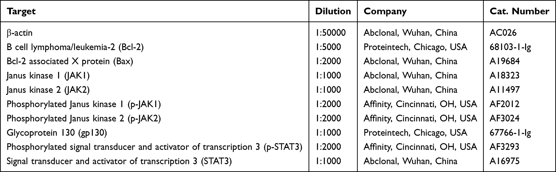 |
Table 1 Antibodies Used in Western Blot Analysis |
Real-Time Polymerase Chain Reaction (PCR)
Total RNA was isolated with the Total RNA Kit I (Omega Bio-Tek, Georgia, USA, #R6834-01) according to the manufacturer’s protocol. RNA concentration and purity were determined by UV spectrophotometry. A 1 µL aliquot of total RNA was then reverse-transcribed into complementary DNA (cDNA) using the Evo M-MLV RT Premix kit (Accurate Biology, China, #AG11728) for downstream amplification. PCR assays were performed in a 25 µL reaction volume with the GAPDH gene serving as an internal control. The cycle threshold (Ct) values obtained from the PCR instrument were used for the relative quantification of the initial templates.
Statistical Analysis
A mean and standard deviation are presented for the results. Data analysis was performed using SPSS 25.0 (IBM Corp., Armonk, NY, USA) with one-way ANOVA, followed by least significant difference post hoc testing. Statistical significance was set at P < 0.05.
Results
Target Analysis of WMSP and Chronic Gastritis
Network pharmacology was used to analyze the targets of WMSP and chronic gastritis. A total of 137 effective components were obtained after removing repetitive active ingredients, and the number of drug-related targets was 4874 (Figure 1A). There were 1475 targets related to chronic gastritis, and the number of common targets was 674 (Figure 1A). The PPI analysis results showed that the top ten targets were TP53, AKT1, TNF, EGFR, IL-1β, ALB, MYC, CTNNB1, IL-6, and STAT3 (Figure 1B). GO enrichment analysis of the core targets of WMSP in the treatment of chronic gastritis showed that in the category of BP, the gene targets were associated with positive regulation of transcription from RNA polymerase II promoter, signal transduction, positive regulation of gene expression, negative regulation of apoptotic process, apoptotic process, etc (Figure 1C). In the category of CC, the gene targets were associated with cytosol, cytoplasm, plasma membrane, nucleoplasm, extracellular exosome, etc (Figure 1C). In the category of MF, the gene targets were associated with protein binding, identical protein binding, enzyme binding, protein homodimerization activity, protein kinase binding, etc (Figure 1C). KEGG enrichment analysis of the core targets of WMSP in the treatment of chronic gastritis showed that the gene targets were associated with pathways in cancer, lipid and atherosclerosis, toxoplasmosis, chagas disease, hepatitis C, etc (Figure 1D).
 |
Figure 1 Target analysis of WMSP and chronic gastritis. (A) A network pharmacology approach was employed to analyze the targets of WMSP and chronic gastritis. (B) Network pharmacology analysis was further utilized to identify the top ten common targets shared by WMSP and chronic gastritis. (C) Gene Ontology (GO) enrichment analysis. (D) Kyoto Encyclopedia of Genes and Genomes (KEGG) pathway enrichment analysis. |
Effects of WMSP on Body Weight, Syndrome Scores, and Locomotor Ability in CG-LSD Rats
Rats were weighed weekly. Before treatment began, the model group showed significantly lower body weight than controls (Figure 2A). By weeks 3–4 post-intervention, WMSP administration at all three doses substantially increased body weight in CG-LSD rats (Figure 2A). The syndrome scores of CG-LSD rats exceeded those of controls according to the standardized assessment (Figure 2B), though medium and high WMSP doses significantly lowered these scores. In open field testing, CG-LSD rats displayed slower walking speeds than controls—an impairment that medium and high WMSP doses effectively reversed (Figure 2C and D).
 |
Figure 2 Target analysis of WMSP and chronic gastritis and its effects on relevant parameters in CG-LSD rats. (A) The body weight of the rats was recorded. (B) Utilizing an animal syndrome scoring system, the impact of WMSP on the syndrome score of CG-LSD rats was assessed. (C) Representative locomotor trajectories from the open field test. (D) Quantitative analysis of the average velocity in the open field test. **P < 0.01 vs Control. #P < 0.05, ##P < 0.01 vs Model. |
WMSP Ameliorated Inflammatory Response, Gastric Mucosal Injury, and Serum Indices in CG-LSD
The serum concentrations of IL-6, CRP, TNF-α, and IL-1β in rats were quantified using specific assay kits (Figure 3A). In the CG-LSD model group, these inflammatory markers were significantly elevated compared to the blank control group (Figure 3A). Following a 4-week treatment period, the serum levels of these cytokines in the various treatment groups exhibited a reduction to differing extents (Figure 3A). Notably, the levels of IL-6, CRP, TNF-α, and IL-1β in the high-dose WMSP group closely resembled those observed in the positive control group (Figure 3A). AB-PAS staining results indicated that, relative to the control group, the CG-LSD model group exhibited impaired gastric mucosal epithelial cell function and a reduction in positive expression (Figure 3B and C). Treatment with WMSP ameliorated gastric mucosal injury in a dose-dependent manner, as evidenced by a marked increase in positive expression (Figure 3B and C). Histological examination through HE staining revealed substantial infiltration of neutrophils and lymphocytes in the gastric mucosa of the model group compared to the control group (Figure 3D). After 4 weeks of treatment, all administration groups demonstrated a decrease in the degree of inflammatory cell infiltration in the gastric mucosa (Figure 3D). SEM analysis demonstrated that in the model group, the morphological architecture of gastric mucosal epithelial cells showed pronounced damage, characterized by extensive cellular shedding and significant structural impairment, including the presence of numerous vesicles on the cell membrane surface. Conversely, the WMSP intervention group exhibited a notable decrease in structural abnormalities within the gastric mucosal epithelial cells. The cells were predominantly well-organized, uniform in size, and only a minority displayed signs of fragmentation, perforation, or vesicle formation (Figure 3E). Furthermore, in CG-LSD rats, there was a reduction in serum PG I levels, an increase in PG II levels, and a decrease in GAS17 levels (Figure 3F). Following a 4-week treatment period, these serum indices showed improvement to varying extents across the different treatment groups (Figure 3F). Concurrently, upon successful model induction, the serum PGE2 levels in the model group decreased, while the levels of NOS and ET increased (Figure 3G). After 4 weeks of WMSP administration, there was a dose-dependent increase in serum PGE2 levels and a decrease in NOS and ET levels in CG-LSD rats (Figure 3G). In addition, compared with the control group, the serum levels of sIgA and GSH in the model group were significantly reduced, while the level of GSSG was markedly increased (Figure 3H). However, these alterations could be substantially reversed by the intervention of WMSP (Figure 3H).
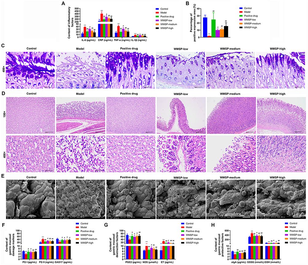 |
Figure 3 WMSP ameliorated inflammatory response, gastric mucosal injury, and serum indices in CG-LSD. (A) The serum concentrations of interleukin-6 (IL-6), C-reactive protein (CRP), tumor necrosis factor-α (TNF-α), and interleukin-1β (IL-1β) were quantified utilizing specific detection kits. (B) Statistical chart showing the percentage of positive expression areas in the Alcian blue-periodic acid-Schiff (AB-PAS) staining. (C) Representative images of AB-PAS staining. Magnification, 400 ×. (D) Histological examination through hematoxylin-eosin (HE) staining was conducted to detect the infiltration of neutrophils and lymphocytes within the gastric mucosa. Magnification, 100 ×, and 400 ×. (E) Scanning electron microscopy (SEM) was employed to examine morphology of gastric mucosal epithelial cells in rats. Magnification, 2000 ×. (F) Serum levels of pepsinogen I (PG I), pepsinogen II (PG II), and gastrin-17 (GAS17) were determined using commercial kits. (G) Serum levels of prostaglandin E2 (PGE2), nitric oxide synthase (NOS), and endothelin (ET) were measured with commercial kits. (H) Serum levels of secretory immunoglobulin A (sIgA), glutathione (GSH), and oxidized glutathione (GSSG) were quantified using commercial kits. **P < 0.01 vs Control. #P < 0.05, ##P < 0.01 vs Model. |
WMSP Inhibited Apoptosis of Gastric Mucosal Cells in CG-LSD Rats
TUNEL staining analysis revealed a significant increase in apoptosis of gastric mucosal cells in the model group compared to the control group, as evidenced by a marked rise in the number of apoptotic cells (Figure 4A and B). In contrast, treatment with various drug groups, including the positive drug group and low, medium, and high doses of WMSP, resulted in a reduction in the number of apoptotic cells in the gastric mucosa of CG-LSD rats, with the high-dose group demonstrating the most pronounced decrease, comparable to the effects observed in the positive drug group (Figure 4A and B). Furthermore, Western blot analysis indicated that, relative to the control group, the model group exhibited a significant reduction in Bcl-2 expression and a significant increase in Bax expression within gastric mucosal tissues (Figure 4C–E). These alterations were notably reversed following treatment with WMSP (Figure 4C–E).
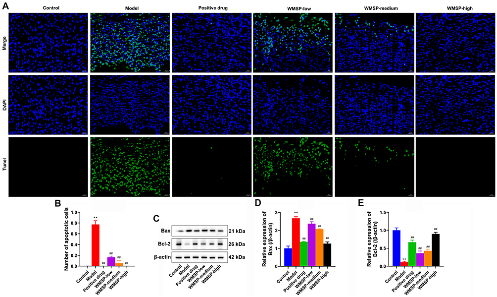 |
Figure 4 WMSP inhibited apoptosis of gastric mucosal cells in CG-LSD rats. (A) TUNEL staining analysis was employed to assess the apoptosis of gastric mucosal cells in CG-LSD rats. Magnification, 400 ×. (B) Statistical results of the number of apoptotic cells in TUNEL staining. (C) The expression levels of B cell lymphoma/leukemia-2 (Bcl-2) and Bcl-2 associated X protein (Bax) in the gastric mucosa were quantified using Western blot analysis. (D) The statistical results of the relative expression levels of Bax. (E) The statistical results of the relative expression levels of Bcl-2. **P < 0.01 vs Control. ##P < 0.01 vs Model. |
WMSP Suppressed the Activity of the IL-6/JAK/STAT3 Pathway in the Gastric Mucosa of CG-LSD Rats
RT-qPCR and Western blot analyses revealed comparable mRNA and protein expression levels of JAK1, JAK2, and STAT3 in gastric mucosa across all experimental groups (Figure 5A–C). The model group showed markedly increased phosphorylation of JAK1, JAK2, and STAT3 compared to controls, with WMSP treatment reversing these effects (Figure 5A–C). Additionally, both gp130 expression (mRNA and protein) and IL-16 mRNA levels were significantly higher in the model group than in controls, with WMSP administration producing reductions in these markers (Figure 5D–F).
 |
Figure 5 WMSP suppressed the activity of the IL-6/JAK/STAT3 signaling pathway in the gastric mucosa of CG-LSD rats. (A) Real-time fluorescent quantitative polymerase chain reaction (RT-qPCR) were employed to detect the mRNA of Janus kinase 1 (JAK1), Janus kinase 2 (JAK2), and signal transducer and activator of transcription 3 (STAT3) in the gastric mucosa. (B) Western blot analysis was used to tested the protein expression of phosphorylated Janus kinase 1 (p-JAK1), JAK1, phosphorylated Janus kinase 2 (p-JAK2), JAK2, phosphorylated signal transducer and activator of transcription 3 (p-STAT3) and STAT3 in the gastric mucosa. (C) The statistical results of relative protein expression levels. (D) The mRNA of IL-16 and glycoprotein 130 (gp130) in the gastric mucosa of rats were detected using RT-qPCR. (E) The protein level of gp130 in the gastric mucosa of rats were tested using Western blot analysis. (F) The statistical results of the relative expression levels of gp130. **P < 0.01 vs Control. #P < 0.05, ##P < 0.01 vs Model. |
WMSP Promoted the Viability of H. Pylori-Infected GES-1 Cell and Inhibited Cellular Inflammation Through IL-6/STAT3 Signaling Pathway
The CCK-8 assay revealed significantly lower cell viability in the model group, while treatment with WMSP-containing serum substantially restored viability (Figure 6A and B). Combining WMSP-containing serum with a STAT3 activator further diminished cell viability compared to WMSP treatment alone (Figure 6A and B). ELISA measurements showed elevated IL-6, CRP, TNF-α, and IL-1β levels in the model group, which WMSP-containing serum effectively suppressed (Figure 6C). The addition of a STAT3 activator to WMSP-containing serum treatment increased inflammatory factor levels beyond those observed with WMSP alone (Figure 6C). Western blot analysis detected no significant changes in JAK1, JAK2, or STAT3 expression across treatment groups (Figure 6D–F). Phosphorylated JAK1, JAK2, and STAT3 levels rose sharply in the model group but decreased significantly following WMSP-containing serum treatment (Figure 6E and F). Co-treatment with a STAT3 activator elevated phosphorylation levels above those achieved by WMSP-containing serum alone (Figure 6E and F). Both Western blot and RT-qPCR analyses indicated increased gp130 expression in the model group (Figure 6G–I), which WMSP-containing serum effectively reduced (Figure 6F–H). The STAT3 activator further enhanced gp130 expression relative to WMSP-containing serum treatment (Figure 6G–I)).
 |
Figure 6 WMSP promoted the viability of H. pylori-infected GES-1 cells and inhibited cellular inflammation through IL-6/STAT3 signaling pathway. (A) The morphology of GES-1 cells was observed by inverted microscope. (B) The viability of GES-1 cells infected with H. pylori was assessed using the Cell Counting Kit-8 (CCK-8) assay. (C) The concentrations of IL-6, CRP, TNF-α, and IL-1β in cell culture supernatants were measured by ELISA. (D) RT-qPCR were used to detect the mRNA of JAK1, JAK2, and STAT3 in GES-1 cells. (E) The expression levels of p-JAK1, JAK1, p-JAK2, JAK2, p-STAT3, and STAT3 in each treatment group were analyzed via Western blot. (F) The statistical results of relative protein expression levels. (G) The gene expression level of gp130 in cells was investigated using RT-qPCR. (H) The protein expression level of gp130 in cells was investigated using and Western blot. (I) The statistical results of the relative expression levels of gp130. **P < 0.01 vs Control. ##P < 0.01 vs Model. &P < 0.05, &&P < 0.01 vs WMSP. |
Discussion
This study demonstrated a significant improvement in body weight, comprehensive scores, and motor abilities in CG-LSD rats following WMSP intervention. From the perspective of TCM, CG-LSD is often associated with impaired functions of the spleen and stomach, as well as a deficiency in “qi” (conceptualized in TCM as vital energy and the foundation of normal physiological functions). This condition is linked to the TCM principle that “the liver governs the smooth flow of bodily processes, while the spleen is responsible for digestion and nutrient transport.” Liver-stomach disharmony, often stemming from emotional stress or functional imbalance of the liver, can disrupt stomach function, leading to symptoms such as distension, pain, belching, and acid reflux. Concurrently, spleen-stomach dysfunction, often manifesting as a deficient state, weakens the digestive capacity, resulting in symptoms like poor appetite, abdominal distension, and loose stools.3 The findings of this study suggest that WMSP alleviates these symptoms by modulating liver function, enhancing digestive capacity, and improving systemic functional status, which corresponds to the TCM therapeutic aim of “soothing the liver, regulating qi, and strengthening the spleen and stomach.”
WMSP is a formula that has been clinically verified, prepared from 11 TCM including Chai-hu (Bupleuri Radix), Zhi-qiao (Aurantii Fructus), Bai-zhi (Angelicae Dahuricae Radix), Bai-ji (Bletillae Rhizoma), San-qi (Notoginaeng Radix ET Rhizoma), Ru-xiang (Olibanum), Mo-yao (Myrrha), Wa-leng-zi (Arcae Concha), Huang-qi (Aatragali Radix), Bing-lang (Arecae Semen), and Bai-jiang-cao (Herba Patriniae). Studies have shown that the main herb in WMSP, Chai-hu (Bupleuri Radix), has various biological activities such as anti-inflammatory, antibacterial, antiviral, and immunomodulation.17 Zhi-qiao (Aurantii Fructus) is a common traditional edible herb for regulating internal organ functions and has ideal therapeutic effects on digestive system diseases.18 Bai-zhi (Angelicae Dahuricae Radix) has the main active components of coumarin and volatile oil, and also exhibits various pharmacological activities such as anti-inflammatory, antioxidant, and analgesic.19 WMSP follows the Chinese medicine principle of “Sovereign, Minister, Assistant, and Envoy”, and this structure ensures that the medicinal materials can work synergistically, thereby generating an overall effect through multiple target mechanisms. Therefore, we believe that WMSP is a multi-component combined Chinese herbal formula, and each component in the formula works synergistically to exert therapeutic effects on CG-LSD.
Research indicates that Helicobacter pylori infection is a critical contributor to chronic gastritis, as it induces inflammation of the gastric mucosa and may potentially lead to gastric cancer.20,21 In the present study, inflammatory markers such as IL-6, CRP, TNF-α, and IL-1β were significantly elevated in the serum of rats within the CG-LSD model group, mirroring the heightened levels of inflammatory factors observed in patients with chronic gastritis in clinical settings.22,23 Herbal treatments have demonstrated substantial efficacy in managing chronic gastritis, particularly through the modulation of inflammatory responses, restoration of gastric mucosa, and enhancement of gastric function.11,24 Notably, the Piwei Peiyuan Prescription has been shown to decelerate the progression of chronic atrophic gastritis (CAG) and inhibit the formation of precancerous lesions by promoting mitochondrial autophagy via MAPK10 mediation.25 Similarly, the Yangyin Huowei mixture mitigates inflammation by suppressing the IL-10/JAK1/STAT3 pathway, thereby reducing gastric mucosal damage and enhancing gastric secretion function, ultimately preventing the onset and advancement of CAG.26 Furthermore, the Yiqi Jiedu Huayu decoction has been shown to significantly mitigate pathological changes and precancerous lesions in the gastric mucosa of CAG rats by inhibiting NLRP3 inflammasome-mediated cell pyroptosis.27 Our findings indicate that after four weeks of WMSP treatment, there was a reduction in serum levels of inflammatory cytokines in CG-LSD rats. This provides compelling evidence that WMSP exhibits a pronounced anti-inflammatory effect and can effectively suppress the inflammatory response in CG-LSD rats in vivo.
Gastric mucosal injury represents a significant pathological characteristic of chronic gastritis.1 Histological analyses, utilizing AB-PAS and HE staining, revealed compromised functionality of gastric mucosal epithelial cells in the CG-LSD model group. This was accompanied by substantial infiltration of neutrophils and lymphocytes, indicating pronounced damage and an inflammatory response within the gastric mucosa. Treatment with WMSP demonstrated a dose-dependent amelioration of gastric mucosal injury. The protection and repair of the gastric mucosa constitute a complex process involving various biochemical markers and mechanisms. PGE2 has been identified as a pivotal factor in maintaining gastric mucosal integrity. It exerts protective effects by inhibiting gastric acid secretion, enhancing mucus and bicarbonate secretion, and augmenting gastric mucosal blood flow.28 GSH, a critical intracellular antioxidant, neutralizes free radicals and shields cells from oxidative stress. Studies suggest that elevated GSH levels can mitigate oxidative damage to the gastric mucosa, thereby facilitating its repair.29 NOS is integral to the protection of the gastric mucosa. By facilitating vasodilation, NO enhances blood flow to the gastric mucosa, thereby bolstering its defensive mechanisms.30 ET, a potent vasoconstrictor, has been linked to gastric mucosal injury when present at elevated levels. However, research indicates that modulating ET levels can improve blood flow to the gastric mucosa and promote its healing.31 Furthermore, sIgA, a critical component of the mucosal immune system, enhances the gastric mucosa’s defense capabilities by neutralizing pathogens and toxins.32 Studies have demonstrated a strong correlation between increased sIgA levels and the protection and repair of the gastric mucosa.32 Our study observed a reduction in serum levels of PG I, GAS17, PGE2, sIgA, and GSH, all of which are associated with gastric mucosal protection and repair, in CG-LSD rats. Conversely, there was an increase in the levels of PG II, NOS, ET, and GSSG. Following a 4-week treatment with WMSP, these serum parameters showed improvement to varying extents. These findings suggest that WMSP may facilitate the repair of gastric mucosa, potentially through mechanisms involving the regulation of proliferation, differentiation, and apoptosis of gastric mucosal cells, as well as enhancements in blood circulation and immune function within the gastric mucosa.
The IL-6/STAT3 signaling pathway is pivotal in the pathogenesis and progression of chronic gastritis.33 IL-6, a multifunctional cytokine, orchestrates various biological processes through the activation of the STAT3 signaling pathway. In the context of chronic gastritis, there is a marked upregulation of IL-6 expression, which is intricately associated with the inflammatory response and cellular proliferation within the gastric mucosa.34 Empirical evidence indicates that IL-6 interacts with its receptor to activate the JAK/STAT3 signaling pathway, thereby facilitating the release of inflammatory mediators and contributing to gastric mucosal damage.35,36 Moreover, the IL-6/STAT3 signaling pathway is significantly implicated in the oncogenesis and progression of gastric cancer. In individuals with chronic gastritis, elevated activation levels of IL-6 and STAT3 exhibit a positive correlation with the advancement of gastric cancer.37,38 Consequently, the aberrant activation of the IL-6/STAT3 signaling pathway may expedite pathological alterations in the gastric mucosa, culminating in carcinogenesis. Our study revealed that in CG-LSD rats, the hyperactivation of the IL-6/STAT3 signaling pathway could contribute to the sustained inflammatory response and damage to gastric mucosal cells. By inhibiting the activity of this signaling pathway, WMSP may attenuate the production and release of inflammatory mediators, thereby mitigating the inflammatory response in the gastric mucosa and facilitating its repair.
While this study yields significant findings, it is not without limitations. Although the study clearly demonstrates the inhibitory effect of WMSP on the IL-6/STAT3 signaling pathway, the specific active components of WMSP and their mechanisms of action within this pathway remain unidentified. Future research should focus on a detailed chemical analysis of WMSP to isolate its principal active components and elucidate the interaction mechanisms between these components and key molecules in the IL-6/STAT3 signaling pathway using molecular biology techniques.
Conclusion
This study demonstrated that WMSP exerted a notable therapeutic effect on CG-LSD rats by attenuating the inflammatory response and apoptosis in gastric mucosal cells, thereby facilitating the repair of gastric mucosal damage. Additionally, WMSP enhanced the viability and suppressed cellular inflammation in Helicobacter pylori-infected GES-1 cell in vitro, with the underlying mechanism linked to the inhibition of the IL-6/STAT3 signaling pathway. These findings provide experimental evidence for the potential application of WMSP in the treatment of CG-LSD.
Data Sharing Statement
The datasets used or analyzed during the current study are available from the corresponding author on reasonable request.
Funding
This study was supported by a grant from the 2023 Central Fiscal Transfer Payment Local Project-Traditional Chinese Medicine Innovation Capability Enhancement Project and the 2024 Qinghai Province “Kunlun Talents High - End Innovative and Entrepreneurial Talents” Top Notch Talent Project (QHKLYC-GDCXCY-2024-152).
Disclosure
The authors declare that they have no competing interests.
References
1. Azer SA, Awosika AO, Akhondi H. Gastritis. In: StatPearls. StatPearls Publishing LLC.; 2025.
2. Yao H, Liu T, Chen Y, et al. Dysregulated gastric microbial communities and functional shifts in chronic atrophic versus non-atrophic gastritis: a Helicobacter pylori-Negative observational study. BMC Gastroenterol. 2025;25(1):304. doi:10.1186/s12876-025-03900-4
3. Bai CY, Tian W, Zhang Q. Clinical study on microscopic syndrome differentiation and traditional Chinese medicine treatment for liver stomach disharmony in chronic gastritis. World J Gastrointestinal Surg. 2024;16(5):1377–16. doi:10.4240/wjgs.v16.i5.1377
4. Wang C, Zhu M, Xia W, Jiang W, Li Y. Meta-analysis of traditional Chinese medicine in treating functional dyspepsia of liver-stomach disharmony syndrome. J Traditional Chin Med. 2012;32(4):515–522. doi:10.1016/s0254-6272(13)60063-1
5. Yang L, Liu X, Zhu J, et al. Progress in traditional Chinese medicine against chronic gastritis: from chronic non-atrophic gastritis to gastric precancerous lesions. Heliyon. 2023;9(6):e16764. doi:10.1016/j.heliyon.2023.e16764
6. Chinese Society of Gastroenterology. Guidelines for diagnosis and treatment of chronic gastritis in China (2022, Shanghai). J Digest Dis. 2023;24(3):150–180. doi:10.1111/1751-2980.13193
7. Dilaghi E, Carabotti M, Annibale B. Perspectives on the current pharmacological strategies for chronic and atrophic gastritis: can more be done? Expert Opinion Pharmacother. 2024;25(9):1107–1110. doi:10.1080/14656566.2024.2373348
8. Shao G, Liu Y, Lu L, Wang L, Ji G, Xu H. Therapeutic potential of traditional Chinese medicine in the prevention and treatment of digestive inflammatory cancer transformation: portulaca oleracea L. as a promising drug. J Ethnopharmacol. 2024;327:117999. doi:10.1016/j.jep.2024.117999
9. Liu Y, Li BG, Su YH, et al. Potential activity of Traditional Chinese Medicine against Ulcerative colitis: a review. J Ethnopharmacol. 2022;289:115084. doi:10.1016/j.jep.2022.115084
10. Li K, Ma X, Li Z, et al. A Natural Peptide from A Traditional Chinese Medicine Has the Potential to Treat Chronic Atrophic Gastritis by Activating Gastric Stem Cells. Adv Sci. 2024;11(20):e2304326. doi:10.1002/advs.202304326
11. Chen L, Wei S, He Y, et al. Treatment of Chronic Gastritis with Traditional Chinese Medicine: pharmacological Activities and Mechanisms. Pharmaceuticals. 2023;16(9):1308. doi:10.3390/ph16091308
12. Liu F, Nong X, Qu W, Li X. Pharmacokinetics and tissue distribution of 12 major active components in normal and chronic gastritis rats after oral administration of Weikangling capsules. J Ethnopharmacol. 2023;316:116722. doi:10.1016/j.jep.2023.116722
13. Ji W, Liang K, An R, Wang X. Baicalin protects against ethanol-induced chronic gastritis in rats by inhibiting Akt/NF-κB pathway. Life Sci. 2019;239:117064. doi:10.1016/j.lfs.2019.117064
14. Wang X, Liu X, Wang Y, et al. Chaihu Shugan Powder inhibits interstitial cells of cajal mitophagy through USP30 in the treatment of functional dyspepsia. J Ethnopharmacol. 2024;323:117695. doi:10.1016/j.jep.2023.117695
15. Hou LW, Fang JL, Zhang JL, et al. Auricular vagus nerve stimulation ameliorates functional dyspepsia with depressive-like behavior and inhibits the hypothalamus-pituitary-adrenal axis in a rat model. Dig Dis Sci. 2022;67(10):4719–4731. doi:10.1007/s10620-021-07332-4
16. Fan J. Mechanism of electroacupuncture in regulating visceral hypersensitivity in functional dyspepsia rats based on EPAC1/PIEZO2 axis. Hubei Univ Chin Med. 2022;2022:2.
17. Yang F, Dong X, Yin X, Wang W, You L, Ni J. Radix Bupleuri: a Review of Traditional Uses, Botany, Phytochemistry, Pharmacology, and Toxicology. Biomed Res Int. 2017;2017:7597596. doi:10.1155/2017/7597596
18. Gao T, Jiang M, Deng B, Zhang Z, Fu Q, Fu C. Aurantii Fructus: a systematic review of ethnopharmacology, phytochemistry and pharmacology. Phytochem Rev. 2021;20(5):909–944. doi:10.1007/s11101-020-09725-1
19. Zhao H, Feng YL, Wang M, Wang JJ, Liu T, Yu J. The Angelica dahurica: a Review of Traditional Uses, Phytochemistry and Pharmacology. Front Pharmacol. 2022;13:896637. doi:10.3389/fphar.2022.896637
20. Shukla GT, Yadav S, Shukla A, et al. Histopathological Features of Chronic Gastritis and its Association with Helicobacter pylori Infection. Korean J Gastroenterol. 2024;84(4):153–159. doi:10.4166/kjg.2024.063
21. Zhou X, Zhu Y, Liu J, Liu J. Effects of Helicobacter pylori Infection on the Development of Chronic Gastritis. Turk J Gastroenterol. 2023;34(7):700–713. doi:10.5152/tjg.2023.22316
22. Zhou J, Li J, Chen J, et al. Decoding inflammatory mediators in the Correa’s cascade: from chronic gastritis to carcinogenesis and targeted therapies. Int Immunopharmacol. 2025;162:115191. doi:10.1016/j.intimp.2025.115191
23. Altun E, Yildiz A, Cevik C, Turan G. The role of high sensitive C-reactive protein and histopathological evaluation in chronic gastritis patients with or without Helicobacter pylori infection. Acta Cir Bras. 2019;34(3):e201900310. doi:10.1590/s0102-865020190030000010
24. Ma X, Wang Y, Kong L, et al. FZHWT alleviates chronic atrophic gastritis by inhibiting inflammatory pathways and promoting mucosal repair. Int Immunopharmacol. 2025;153:114473. doi:10.1016/j.intimp.2025.114473
25. Zhang Y, Wu Y, Pei B, et al. Piwei Peiyuan Prescription Attenuates the Progression of Chronic Atrophic Gastritis by Eliciting MAPK10-Mediated Mitochondrial Autophagy. Cell Biol Int. 2025;2025:1. doi:10.1002/cbin.70016
26. Xie SS, Zhi Y, Shao CM, Zeng BF. Yangyin Huowei mixture alleviates chronic atrophic gastritis by inhibiting the IL-10/JAK1/STAT3 pathway. World J Gastrointestinal Surg. 2024;16(7):2296–2307. doi:10.4240/wjgs.v16.i7.2296
27. Zhou P, Zheng ZH, Wan T, Liao CW, Wu J. Yiqi Jiedu Huayu decoction inhibits precancerous lesions of chronic atrophic gastritis by inhibiting NLRP3 inflammasome-mediated pyroptosis. World J Gastrointestinal Oncol. 2024;16(7):3158–3168. doi:10.4251/wjgo.v16.i7.3158
28. Takeuchi K, Amagase K. Roles of Cyclooxygenase, Prostaglandin E2 and EP Receptors in Mucosal Protection and Ulcer Healing in the Gastrointestinal Tract. Curr Pharm Des. 2018;24(18):2002–2011. doi:10.2174/1381612824666180629111227
29. Baskerville MJ, Kovalyova Y, Mejías-Luque R, Gerhard M, Hatzios SK. Isotope tracing reveals bacterial catabolism of host-derived glutathione during Helicobacter pylori infection. PLoS Pathogens. 2023;19(7):e1011526. doi:10.1371/journal.ppat.1011526
30. Magierowski M, Magierowska K, Kwiecien S, Brzozowski T. Gaseous mediators nitric oxide and hydrogen sulfide in the mechanism of gastrointestinal integrity, protection and ulcer healing. Molecules. 2015;20(5):9099–9123. doi:10.3390/molecules20059099
31. Fu X, Huang X, Lin Z, et al. Protective effect of teprenone on gastric mucosal injury induced by dual antiplatelet therapy in rats. Am J Transl Res. 2021;13(4):2702–2709.
32. Xie L, Luo M, Li J, et al. Gastroprotective mechanism of modified lvdou gancao decoction on ethanol-induced gastric lesions in mice: involvement of Nrf-2/HO-1/NF-κB signaling pathway. Front Pharmacol. 2022;13:953885. doi:10.3389/fphar.2022.953885
33. Wang Y, Li D, Zhao L, et al. Mechanism of Yinxu Weitong Capsule in the treatment of precancerous lesions of gastric cancer based on network pharmacology and experimental validation. J Ehnopharmacol. 2025;11(341):119303. doi:10.1016/j.jep.2024.119303
34. Zhao CN, Xiao LL, Zhang Y. Effects of Helicobacter pylori Infection on the Prognosis of Chronic Atrophic Gastritis by Inducing the Macrophage Polarization. Gastroenterol Res. 2023;16(4):226–233. doi:10.14740/gr1636
35. Ma Q, Jiang Z, Zhang Z, et al. Qizhiweitong granule alleviates diarrhea-predominant irritable bowel syndrome via inhibition of the IL-6/Src/STAT3 feedback loop. J Ethnopharmacol. 2025;356:120772. doi:10.1016/j.jep.2025.120772
36. Jiao Z, Zheng J, Yang X, et al. Yangweishu Ameliorates Chronic Atrophic Gastritis with Stomach Yin Deficiency Syndrome Through IL-6/STAT3 Signaling Pathway. Drug Des Devel Ther. 2025;19:7865–7885. doi:10.2147/dddt.s529330
37. Fan H, Ou Q, Su Q, et al. ZIPK activates the IL-6/STAT3 signaling pathway and promotes cisplatin resistance in gastric cancer cells. FEBS Open Bio. 2021;11(9):2655–2667. doi:10.1002/2211-5463.13270
38. Ding LL, Zhang M, Zhang T, Liu H, Pf L. MFGE8 promotes gastric cancer progression by activating the IL-6/JAK/STAT3 signaling. Cell Signalling. 2025;125:111486. doi:10.1016/j.cellsig.2024.111486
 © 2026 The Author(s). This work is published and licensed by Dove Medical Press Limited. The
full terms of this license are available at https://www.dovepress.com/terms
and incorporate the Creative Commons Attribution
- Non Commercial (unported, 4.0) License.
By accessing the work you hereby accept the Terms. Non-commercial uses of the work are permitted
without any further permission from Dove Medical Press Limited, provided the work is properly
attributed. For permission for commercial use of this work, please see paragraphs 4.2 and 5 of our Terms.
© 2026 The Author(s). This work is published and licensed by Dove Medical Press Limited. The
full terms of this license are available at https://www.dovepress.com/terms
and incorporate the Creative Commons Attribution
- Non Commercial (unported, 4.0) License.
By accessing the work you hereby accept the Terms. Non-commercial uses of the work are permitted
without any further permission from Dove Medical Press Limited, provided the work is properly
attributed. For permission for commercial use of this work, please see paragraphs 4.2 and 5 of our Terms.