
Back to Journals » Clinical Ophthalmology » Volume 19
Vitreoretinal Injury Associated with Sports Ball Ocular Trauma
Authors Rohowetz LJ  , Fan J , Flynn HW Jr
Received 19 November 2024
Accepted for publication 17 February 2025
Published 23 June 2025 Volume 2025:19 Pages 1931—1943
DOI https://doi.org/10.2147/OPTH.S507399
Checked for plagiarism Yes
Review by Single anonymous peer review
Peer reviewer comments 3
Editor who approved publication: Dr Scott Fraser
Landon J Rohowetz, Jason Fan, Harry W Flynn Jr
Department of Ophthalmology, Bascom Palmer Eye Institute, University of Miami Miller School of Medicine, Miami, FL, USA
Correspondence: Harry W Flynn Jr, Department of Ophthalmology, Bascom Palmer Eye Institute, University of Miami Miller School of Medicine, 900 NW 17th St, Miami, FL, USA, Tel +1 305 326-6148, Fax +1 305 326-6417, Email [email protected]
Abstract: Sports are a common cause of ocular trauma. Vitreoretinal injuries secondary to sports ball trauma can cause permanent vision loss and may be a source of significant morbidity. Trauma most frequently occurs in young males and over half of injuries involve the posterior segment. Injuries typically occur as a result of direct contact with the globe either before or after initial contact with the orbit and are dependent on the size, mass, and velocity of the ball. Compressive forces and subsequent equatorial expansion lead to direct tissue damage and dehiscence of posterior segment layers. Smaller balls with relatively high mass and velocity such as golf balls and paintballs are associated with rupture injuries while larger balls frequently cause contusive trauma and distortion of the globe which may lead to vitreoretinal traction and damage to posterior segment layers. Retinal detachment may occur and is frequently associated with retinal dialysis and giant retinal tears. Traumatic macular hole is uncommon and, unlike idiopathic macular hole, frequently closes spontaneously. The institution of eye protection standards in select sports have dramatically reduced the incidence of eye injuries. Continued evaluation of the most appropriate and logical protective measures is essential to promote the safety of all athletes and reduce the incidence of frequently-disabling ocular injury.
Keywords: ocular trauma, sports injury, open globe injury, retinal detachment, traumatic macular hole
Introduction
Ocular trauma secondary to sports ball injury is a significant cause of morbidity.1 There are over 40,000 emergency department visits per year for sports-related ocular trauma in the United States.2 Injuries most commonly occur in young males.3,4 Anterior segment injuries include corneal abrasion, corneal laceration, traumatic iritis, hyphema, angle recession glaucoma, and lens subluxation or dislocation.5 Over half of eye injuries involve the posterior segment and may include commotio retinae, open globe injury (OGI), vitreous hemorrhage (Figure 1A), retinal detachment (Figure 1B), choroidal rupture, retinal hemorrhage, and retinal tear (Figure 2A and B).6,7 In the current manuscript, we review the clinical characteristics and outcomes of vitreoretinal injuries secondary to sports ball trauma.
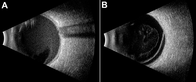 |
Figure 1 B-scan ultrasonography of vitreous hemorrhage and retinal detachment B-scan ultrasonography demonstrating (A) vitreous hemorrhage and (B) retinal detachment. |
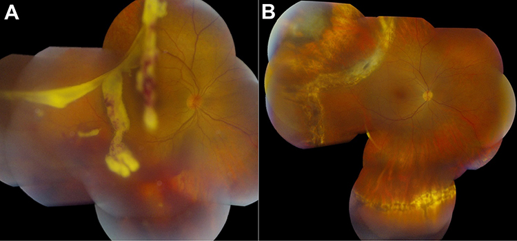 |
Figure 2 Giant retinal tear secondary to baseball injury (A) A 36-year-old male with a history of baseball-related ocular trauma presented with a giant retinal tear in the superotemporal quadrant of the right eye with associated vitreous hemorrhage. The patient underwent scleral buckling, pars plana vitrectomy, and silicone oil insertion. (B) The retina was attached at last follow-up 2 years later and best-corrected visual acuity was 20/20. Reprinted from Am J Ophthalmol Case Rep, volume 6, Read SP, Young RC, Flynn HW Jr. Outcomes in bullous retinal detachment. 18–20, copyright 2016, Creative Commons.8 |
Pathophysiology
While the bony orbit provides protection of the globe against many objects, some sports balls such as golf balls, paintballs, and badmintons are small enough to contact the globe directly. Furthermore, larger balls are frequently able to contact the globe by molding to the orbit in the setting of sufficient force. Vitreoretinal injury occurs as a result of blunt forces directed in the anterior-posterior direction, causing compression of the anterior chamber and coup-countrecoup injuries such as commotio retina. This compression also causes equatorial expansion and stretching of the posterior eye wall which can lead to choroidal rupture, macular hole, retinal dialysis, vitreous hemorrhage, retinal tear, and scleral rupture. Injuries that disrupt the integrity of the globe and those that lead to uveal or retinal prolapse are typically associated with a poor prognosis.9 Vitreous hemorrhage and optic nerve injury have also been demonstrated to be associated with worse visual outcomes.9
The institution of standards for eye protection in select sports has drastically reduced the incidence of sports-related ocular trauma. In 2006, the American Hockey League instituted a mandatory requirement for the use of protective visors and in 2013, the National Hockey League made protective visors mandatory for all new players in the league. Accordingly, the number of eye injuries in the National Hockey League decreased from 10 in the 2010–2011 season to 0 in the 2017–2018 season.10
Furthermore, the introduction of rules on protective eyewear in racquetball have decreased the incidence of ocular injury. Organizations such as the American Society for Testing and Materials (ASTM) and European Committee for Standardization have been influential in developing and recommending standards for protective eyewear in racquetball and other sports. While racquetball players initially wore eye guards without lens (“open” eye guards), injuries continued to occur as the ball was able to deform and fit through the gap between the rims.11,12 Furthermore, the use of CR39 lenses frequently led to injury as a result of an opponent’s racquet striking and shattering the lens.11,12 However, in 1983, many racquetball clubs began to mandate the use of ASTM-compliant eyewear, which led to a drastic reduction in eye injuries. Indeed, there have been no reports of racquetball-related eye injuries in individuals wearing eye protective devices compliant with the ASTM F803 standard.7,12 While the use of eyewear approved by the ASTM and other organizations has been demonstrated to be beneficial, eyewear not compliant with these standards such as prescription eyeglasses and sunglasses may actually be detrimental due to injury caused by the devices themselves.13–16 As such, it is important for patients and providers to be aware of these standards.
Epidemiology
Sports are one of the most common causes of traumatic eye injuries worldwide. In a study using data from the Eye Injury Registry of Alabama from 1982 to 1986 blunt injury, which includes ball injuries, was the most common type of injury reported. The retina and/or vitreous were involved in nearly half of cases, the sclera in 8%, and the optic nerve in 5%.9 In 2003, Filipe et.al reported 25 cases of eye injuries caused by “modern sports.” Over half of eyes experienced posterior segment injury and the most common cause was paintball. More recently, large database studies evaluating emergency room encounters for sports-related injuries in the United States have demonstrated that the most common sports causing eye injury include basketball, baseball, soccer, and air gun sports.2,5 Out of all injuries, the rate of posterior segment involvement is around 2.5%, with commotio retina, globe rupture, and retinal detachment as the most common reported injuries. Soccer, paintball, and golf, specifically, are the most likely to cause major posterior segment injury in the setting of ocular trauma.5
The prevalence of sports-related injuries is dependent on the corresponding geographic popularity of various sports. A 1986 study from the Sussex Eye Hospital in England demonstrated that squash was the most frequent cause of sports-related ocular injury followed by soccer and badminton.14 On the other hand, the most common sports causing ocular injury in Korea in 2021 were soccer, baseball, and badminton.3 Handball, soccer, and floorball are the most common causes of sports-related ocular trauma in China, Australia, and Sweden, respectively (Table 1).17–19 As such promotion of awareness, education, and counseling should be directed according to the popularity of individual sports.
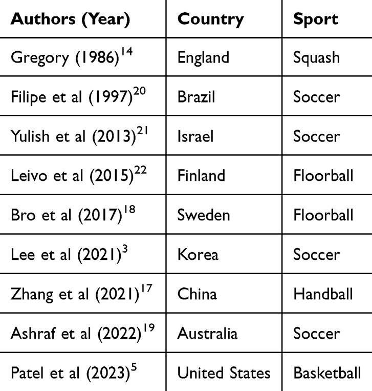 |
Table 1 Most Common Causes of Sports-Related Eye Injury by Country |
Types of Vitreoretinal Injury
Retinal Tear/Detachment
Retinal tear and detachment may occur following sports ball injury as a result of equatorial expansion of the globe with resultant vitreoretinal traction.9 Retinal tears are frequently located in the infratemporal or supratemporal quadrants and are occasionally associated with a full-thickness macular hole.23,24 Initial visual acuity is a predictor of postoperative visual acuity and myopia may be a risk factor.25 Common symptoms include flashes, floaters, blurred vision, decreased peripheral vision, and a curtain-like shadow in the field of view.26 Concurrent injuries include retinal dialysis, giant retinal tear, or avulsion of the vitreous base.8,24,27,28
Retinal Dialysis
Retinal dialysis is defined as the disinsertion of the retina at the ora serrata.29 The condition most frequently occurs in young males and may be either symptomatic or asymptomatic.29 Retinal dialysis has been reported in associated with tennis, badminton, racquetball, basketball, and soccer injuries.27,28,30,31 When associated with retinal detachment, scleral buckling alone is generally the treatment of choice for retinal dialysis (Figure 3A and B).29
 |
Figure 3 Retinal dialyses secondary to soccer ball injury (A) A 22-year-old male soccer player presented with bilateral retinal dialyses. Best-corrected visual acuity was 20/20 in the right eye and 20/300 in the left eye. Examination revealed a macula-sparing retinal detachment in the right eye and a macula-involving retinal detachment in the left. Scleral buckling with external drainage of subretinal fluid was performed in both eyes. (B) At postoperative year 2, the retina was attached in both eyes and best-corrected visual acuity was 20/20 and 20/200 in the right eye and left eyes, respectively. |
Open Globe Injury
Open globe injury frequently occurs in sports ball-related ocular trauma, particularly in those involving small balls with high impact velocity such as golf and paintball.13,32 Injuries generally involve globe rupture due to the blunt configuration of the ball unless there is simultaneous contact with an adjacent object such as eyeglasses.13 Visual acuity outcomes are generally poor and patients often require multiple surgeries including evisceration or enucleation.32
Commotio Retinae
Commotio retinae is a common injury following blunt ocular trauma, characterized by photoreceptor outer segment disruption or loss manifesting as retinal whitening.33 Damage may occur at the site of injury (coup) or at distant sites (countrecoup) and may result in traumatic retinal pigment epitheliopathy.34 The presence of commotio retinae within the macula (Berlin’s edema) is associated with poor visual acuity outcomes.35
Macular Hole
Macular hole is a common complication of sports-related retinal injury. Unlike non-traumatic macular holes, traumatic macular holes may resolve spontaneously without treatment due to the tangential forces of the attached vitreous and as such may be observed for a period of time.36–40 Some authors have reported the presence of parafoveal cysts as a poor prognostic factor caused by radial traction from the internal limiting membrane and contraction of activated Müller cells.41 In refractory cases, pars plana vitrectomy generally results in good anatomic outcomes and modest improvements in visual acuity.42 Shallow macular hole-related retinal detachment may occur and has been reported to resolve spontaneously.37
Traumatic Retinal Pigment Epitheliopathy
Traumatic retinal pigment epitheliopathy may be identified early by hyperautofluorescence and the presence of leakage on fluorescein with ellipsoid zone loss on OCT. The finding has been reported in many causes of sports ball-related ocular trauma and is an indicator of poor long-term visual prognosis.36,41
Other Injuries
Central retinal vein and central retinal artery occlusion have been reported and are theorized to occur as a result of compressive forces transmitted to the vitreous and retina.43 Retinal pigment epithelial tears and detachments may occur due to the aforementioned equatorial expansion and stretching of posterior segment layers.41 Additional reported injuries include choroidal rupture, choroidal neovascularization (Figure 4A and B), sclopetaria (Figure 5), subretinal hemorrhage, sub-pigment epithelium hemorrhage, optic neuropathy, and optic nerve avulsion.44–51
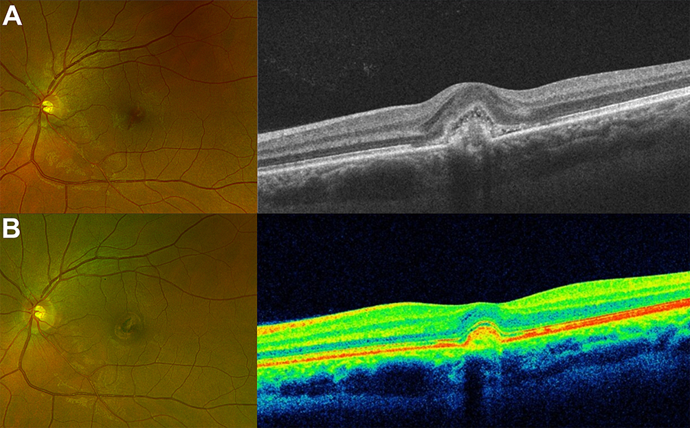 |
Figure 4 Choroidal neovascularization secondary to tennis ball injury (A) Fundus photography and optical coherence tomography of a 52-year-old male with a history of tennis-related ocular trauma presenting with a pigment epithelial detachment and subretinal fluid consistent with choroidal neovascularization. (B) 1 month after receiving intravitreal bevacizumab, the subretinal fluid had resolved although the pigment epithelial detachment persisted. |
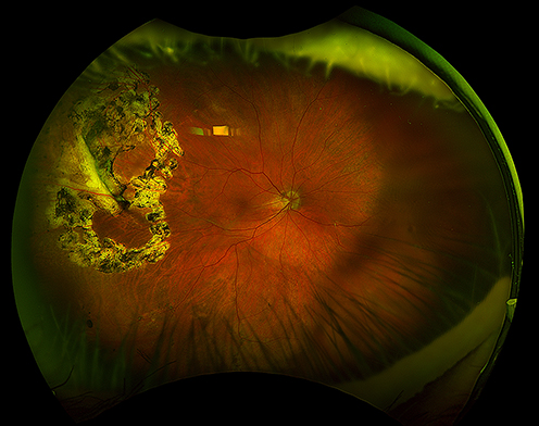 |
Figure 5 Retinal detachment and sclopetaria secondary to golf ball injury A 69-year-old male presented after being struck by a golf ball in the right eye. Best-corrected visual acuity was hand motions and examination demonstrated dense vitreous hemorrhage. Pars plana vitrectomy was pursued and intraoperative examination revealed temporal sclopetaria and a localized temporal retinal detachment. Endolaser was performed and air tamponade was placed. At postoperative year 4, the retina was attached and best-corrected visual acuity was 20/25. |
Sports
Soccer
In the past, it was thought that injury due to soccer balls were rare because of the ball’s relatively large and relatively soft shape. However, frequent reports of ophthalmic injury from soccer ball trauma led Vinger et.al to perform a study in which they simulated soccer ball injuries using artificial orbits and demonstrated that while the depth of penetration is less in soccer ball-related injury compared to injury caused by other balls, the ball remains in contact with the globe 2.5 to 10 times longer. As a result, while the peak force of a soccer ball is less than that of smaller balls, the duration of globe impact can lead to significant distortion and resultant vitreoretinal trauma.52 Furthermore, a suction effect of the ball can cause further globe distortion and corresponding injury.52 Indeed, soccer-related ocular injury has been associated with a higher rate of visual impairment compared to injuries caused by other sports.2,3 Common injuries include retinal dialysis, giant retinal tear, retinal detachment, and macular hole. Injuries frequently occur in the superior hemisphere due to the upward trajectory of the ball. In a study evaluating sports-related ocular injuries in Korea, soccer was the most common cause of OGI. Notably, eye protection was worn by all patients who experienced OGI, demonstrating the importance of using eye protection adhering to certain standards such as those developed and published by the ASTM.4
Golf
Golf ball injuries, while rare, are typically severe due to the high velocity and resultant force of the small ball.53 Open globe injuries (OGIs) have been reported in over half of cases and visual acuity outcomes are generally poor with about one-third of cases resulting in enucleation or evisceration.13,14,32,54 In a report of 22 golf ball-related ocular injuries, the mean number of total surgeries per eye was 1.5. All patients with a rupture injury eventually underwent evisceration and the final visual acuity in all eyes with penetrating injury was 20/100 or worse.32 All patients with zone III OGIs either underwent enucleation or had a visual acuity of no light perception at last follow-up examination.32 Reported closed globe injuries include lens subluxation, commotio retinae, retinal dialysis, and choroidal rupture.32 In one study, 9% of injuries occurred in spectators, demonstrating the importance of appropriate protective measures in fans and players alike.54
Tennis
As the popularity of tennis increased in the United States in the 1970s, Seelenfreund and Freilich published a report describing 10 eyes with vitreoretinal trauma secondary to tennis ball injury. All 10 eyes in their report had vitreous hemorrhage. Retinal tear and retinal dialysis also occurred and 5 (50%) eyes required surgery.27 However, the authors point out that the rate of retinal tear and retinal detachment was low compared to the overall incidence of tennis ball injury and reported that retinal detachment after trauma in the absence of a predisposition is rare.27 In 2007, Qureshi et.al reported 28 cases of retinal detachment secondary to tennis ball injury, 34% of which had more than 1 break. Fifty-nine percent of breaks were in the infratemporal quadrant and 10% of retinal detachments were associated with a full-thickness macular hole. All patients underwent scleral buckling only with a single operation success rate of 75%. Visual acuity of 20/60 or better was achieved in 29% of eyes at last follow-up examination.23
In 2016, Karimi et.al used magnetic resonance imaging to model tennis-related eye injuries and demonstrated that the majority of the induced stress was localized to the anterior chamber while the vitreous experienced the lowest perceived stress. However, the authors emphasize that globe deformation rather than transmitted stress may cause the majority of tennis-related injuries.55 In 2019, Patel et.al reviewed over 15,000 emergency department visits for tennis-related ocular injury between 2000 and 2019. Injury was most frequent in individuals under the age of 20 years. The incidence of OGI, retinal detachment, and posterior vitreous detachment was 1.5%, 0.9%, and 0.2%, respectively. There was a decrease in the rate of ocular injury between the start and end of the study.44
Tennis ball-related injuries associated with poor visual prognosis include retinal detachment, choroidal rupture, and vitreous hemorrhage.45 Other reported injuries after tennis ball trauma include macular hole, choroidal rupture, choroidal neovascularization (Figure 3), subretinal hemorrhage, sub-pigment epithelium hemorrhage, and OGI.44–50
Squash
Squash, a popular sport in many parts of the world, is frequently associated with eye injury in the absence of adequate eye protection due to the ball’s small size and high velocity. In 1978, Easterbrook described 23 cases of ocular trauma secondary to squash injury over 2.5 years, 11 of which led to hospitalization of the patient. Injuries included retinal hemorrhage, OGI, vitreous hemorrhage, and a macular cyst. Five cases resulted in a permanent decrease in vision and there was no association between the odds of injury and player experience.56
Squash was the leading cause of sports-related ocular trauma in a 1986 study by Gregory et.al at the Sussex Eye Hospital, causing 24 of 92 (26%) injuries. Four cases involved retinal abnormalities although there were no cases of vision loss.14 Other previously-reported injuries include retinal hemorrhage and retinal edema.7
Retinal detachment has been reported in squash ball accidents.24 Knorr and Jonas described a cohort of 26 eyes with retinal detachment caused by squash ball injury. Detachments were frequently associated with large retinal tears near the ora. The supratemporal quadrant was the most frequent location followed by the infratemporal quadrant. Avulsion of the vitreous base was noted in half of patients and final visual acuity was 20/40 or better in 42% of eyes.24 Capão Filipe et.al also described 7 cases of squash-related eye injury, 2 of which involved retinal tears. Other previously-reported injuries included retinal hemorrhage and retinal edema.7
Badminton
Badminton may be associated with severe eye injury due to the small size and high velocity of the shuttlecock. Injury may also occur due to contact with another player’s racquet given the relatively small court size and the players’ close proximity to each other. Eye injuries secondary to badminton have been associated with higher rates of visual impairment when compared to other sports.3
In 1974, Chandran described 63 cases of badminton-related eye injury. Reported vitreoretinal injuries included commotio retinae and vitreous hemorrhage. Visual acuity was 20/60 or worse in 27% of eyes at last follow-up.57 Kelly described 6 cases of badminton-associated ocular trauma, 4 of which occurred due to close-range shuttlecock injury from an opponent’s smash hit. Injuries included OGI, IOFB, retinal dialysis-associated retinal detachment, choroidal rupture, and vitreous hemorrhage. Surgery was performed in 3 cases.30 In 2020, Yu et.al reported the largest series badminton-related ocular trauma involving 85 eyes. There were 5 penetrating injuries in their study. Sixteen patients underwent pars plana vitrectomy and 2 required silicone oil tamponade. Eighty-six percent of cases occurred in doubles matches and 61% of injuries were caused by the patient’s partner rather than the opponent.16
Racquetball
Racquetball, a popular sport in the United States and internationally, is frequently associated with eye injury in the absence of adequate eye protection due to the close proximity of players and the ball’s small size, high velocity, and significant flexibility. In 1980, Doxanas and Soderstrom reported 37 cases of ocular injury in a 3-month period. No patients in the cohort were wearing protective eyewear and there was no correlation with player experience.58
Although eye protection began to be worn shortly thereafter, the initial standard eye protective device in racquetball was an open eye guard without lenses. The flexibility of the racquetball allowed it to come into contact with the globe despite the surrounding protection afforded by the lensless eyeguards.11,12 Indeed, in 1981, Easterbrook et.al described 18 racquetball-associated eye injuries including 1 OGI, 1 vitreous hemorrhage, and 1 retinal dialysis. Open eye guards were used by 10 patients at the time of injury.31 An updated report was published in 1988 involving 292 ocular injuries from racquetball, revealing 21 cases of vitreous/retinal hemorrhage, 5 macular scars, and 3 retinal detachments, with the author noting that injuries frequently occurred with open eyeguards.59
Following the subsequent institution of ASTM-compliant eyewear mandates, including eye guards with impact resistant lenses, there was a significant reduction in racquetball-related eye injuries. Indeed, there have been no reports of injury in patients using eye protective devices compliant with the ASTM F803 standard.7,12
Basketball
Basketball is the leading cause of sports eye injury in the United States, with 1 in 10 college basketball players sustaining eye injuries each year.2 While general eye injuries are frequent in basketball due to contact with other players, vitreoretinal injuries specifically are less common due to the low velocity and large size of the ball. In a study examining high school sports injuries, baseball and basketball accounted for most ocular injuries. Most basketball injuries were caused by an elbow or finger and there was 1 case of retinal detachment after a patient was hit in the eye by a rebound.60
The National Basketball Association reported 59 and 18 eye injuries in the 1992–1993 and 2018–2019 seasons, respectively. Notably, none of the injuries involved the posterior segment.61,62 Similarly, a retrospective review of the Duke University men’s basketball injuries over 16 seasons revealed only 1 vitreoretinal injury – a case of retinal dialysis and traumatic optic nerve head avulsion.28
In a retrospective review of 13 cases of retinal detachment associated with basketball injury, 70% of patients had moderate to high myopia, with −5.2 diopters as the average refractive error. Seventy percent of injuries were caused by direct blunt contusion from the basketball and the majority of breaks were superior. Most patients were young and the authors argue that the relatively undeveloped orbital rim and cavity compared to the fully-developed globe of young patients, in addition to the increased axial length characteristic of myopia, may cause relative protrusion of the eye making it more prone to injury. Concurrent injuries included retinal detachment, choroidal detachment, retinal dialysis, and giant retinal tear and there was a 76% reattachment rate after the first operation. Visual acuity was 20/200 or better in 54% of patients and the only prognostic factor was initial visual acuity although there was trend towards better visual acuity at last examination in patients with shorter symptom-to-diagnosis interval.25
Baseball
Given its popularity, baseball is one of the most frequent causes of sports ball-related ocular trauma in the United States.2,5 The incompressibility and large size of a baseball typically prevents significant globe compression during injury. As such, most baseball injuries involve the orbital bones rather than the globe itself. Injury may occur from a throw, as in the case of a batter being hit by a pitch, or from a hit with a wooden or metal bat.
In 1993, Zagelbaum et.al reported all ocular injuries in Major League Baseball over a 1-year period. There were 24 total injuries, none of which involved the posterior segment. However, 1 fan suffered a ruptured globe from a foul ball.63 Traumatic macular hole has been described in 4 cases, all of which demonstrated spontaneous closure.39,40 In 2017, Flynn et.al described 2 patients with giant retinal tear-related retinal detachments requiring pars plana vitrectomy.8 Other previously-reported vitreoretinal injuries include choroidal rupture, choroidal neovascularization, choroidal folds, and retinal pigment epithelial detachment.39,64
Pickleball
Pickleball is the fastest growing sport in the United States, with over 13.6 million individuals in the United States participating in the sport as of 2023.65 Pickleball-related injuries have increased in recent years alongside the sport’s increase in popularity, particularly among elderly individuals.66 Over 500 injuries occurred in the United States between 2010 and 2019, 5 (0.7%) of which were eye injuries. Most of the injuries were due to contact with the ball and were generally less severe than tennis injuries.67 In 2022, Atkinson et.al reported 2 patients with retinal tears after direct blunt ocular trauma with a pickleball. One patient had a localized retinal detachment and vitreous hemorrhage that was successfully treated with cryopexy while the other had a single retinal tear that was treated with laser retinopexy.68 Continued identification and reporting of pickleball-related ocular injury is essential as the sport continues to gain popularity to better understand its risks.
Paintball
Paintball was first played in 1981 in New Hampshire and has since became a popular game played around the globe.69–71 The game involves shooting opponents with paintballs fired from carbon dioxide-powered guns.72 The small size, relatively high mass, and high velocity of paintballs result in a sharply localized impact that typically causes significant ocular damage.73 As paintballs are designed to rupture on impact, all of the ball’s energy is released at the site of contact, contributing further to the severe tissue damage characteristic of these injuries.74 Indeed, in a 2016 study evaluating all patients presenting to an emergency department with sports-related eye injury, the odds of impaired vision were greatest for paintball injuries, accounting for 26% of all cases of impaired vision, while only accounting for 10% of all injuries.2
At the time of the inception of paintball in the 1980s, the game was frequently played in unregulated settings without adequate protection. In 1985, the first study evaluating eye injuries in paintball demonstrated that over half of 26 eyes had a final best-corrected visual acuity of less than 20/800. Notably, no players in this study were wearing eye protection at the time of injury and there was only 1 reported OGI.75 Since then, over 500 cases of paintball-related ocular injury have been reported.
In 1996, Zwaan et.al performed a literature review of all paintball-related ocular injuries. The most common posterior segment injuries were “retinal damage”, vitreous hemorrhage, and macular hole. Best-corrected visual acuity was 20/300 or worse at last follow-up in 30% of eyes and 4% suffered an OGI.74 Thach et.al described 13 eyes with paintball-related ocular injury, demonstrating an improvement in visual acuity at last follow-up in only about half of eyes. The most common reasons for poor visual acuity were posterior segment injuries including recurrent retinal detachment due to proliferative vitreoretinopathy, epiretinal membrane, choroidal neovascularization, and retinal necrosis.76 Similarly, Peñaranda et.al described 14 eyes with paintball injury, 9 of which had no visual acuity improvement at last follow-up. Reported causes of poor vision included macular alterations, choroidal rupture, retinal tears, and retinal folds.77 In 2008, Taban et.al described 9 eyes with paintball-related injuries, 4 of which manifested with chorioretinitis sclopetaria, demonstrating that sclopetaria can occur with soft projectiles even when they do not penetrate the orbit. The authors hypothesized that sclopetaria was previously unrecognized by ophthalmologists as it is typically associated with high speed projectiles that penetrate the orbit. The most common causes of decreased vision were traumatic maculopathy, optic neuropathy, and optic nerve avulsion.51
In 2009, Allman et.al described 36 eyes with paintball-related ocular injury over a 6-year period demonstrating that nearly half of eyes underwent surgical intervention within 3 days of the injury while 67% of eyes underwent surgical intervention after 3 days, either for the first or second time. Eleven percent of eyes required 3 or more surgeries and over one-quarter of eyes had a visual acuity of no light perception at last follow-up. Open globe injury occurred in one-quarter of patients and there was a strong correlation between initial visual acuity and visual acuity at last follow-up. Enucleation was performed in 7 eyes. Notably, no injuries occurred at a formal, sponsored event.78 Furthermore, in 2022, Amin et.al reported 20 cases of ocular injury from drive-by paintball shootings and demonstrated a 30% ruptured globe rate, with 50% of eyes requiring evisceration and 38% of eyes demonstrating a final visual acuity of no light perception. Visual acuity was no light perception at last follow-up in 5 of the 6 eyes requiring surgical intervention, demonstrating the particularly poor prognosis associated with OGI.79
In 2000, Fineman et.al examined 35 paintball injuries over a 13-year period. The most common posterior segment injuries were vitreous hemorrhage, commotio retinae, and choroidal rupture (Table 2). Open globe injury occurred in 6% of cases. Injuries sustained after 1995 were 5.8 times more likely to occur in a non-commercial setting than those in 1995 or before, possibly related to the ASTM’s institution of standard specifications for eye protection devices in paintball sports in 1997.73
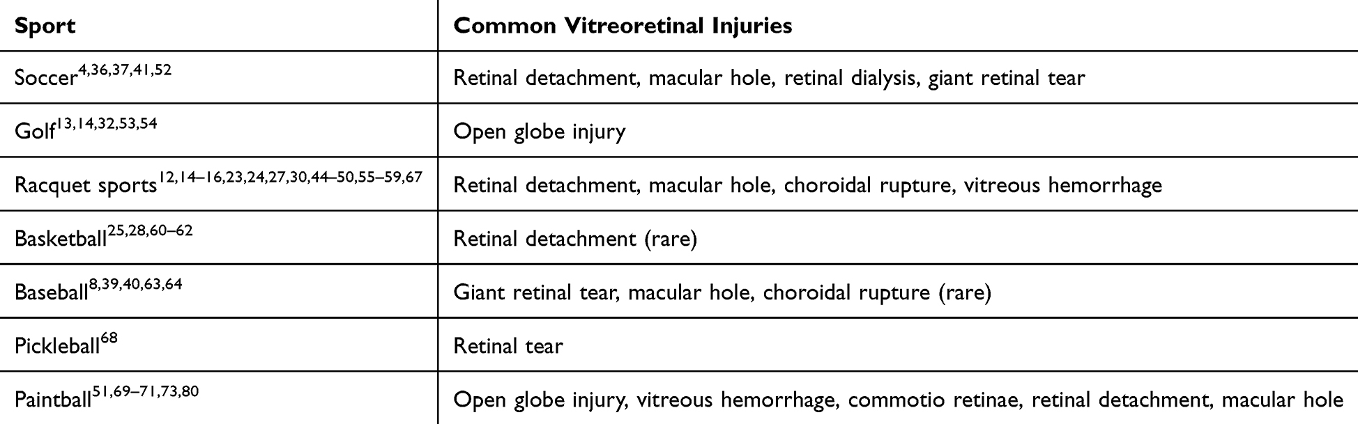 |
Table 2 Common Vitreoretinal Injuries by Sport |
Indeed, a 2001 review of all reported paintball injuries demonstrated no ocular injuries in individuals wearing eye protection devices meeting ASTM standards. However, injuries have been reported in individuals wearing non-ASTM-approved eye protection devices as a result of the paintball entering under the mask, displacing the mask, or causing the goggle or a goggle fragment to be projected into the eye.73,80–83 Along these lines, Keles et.al described 10 cases of paintball-related ocular injury in 2014, all of which occurred after the injured player thought the game was over and had taken off their eye protection device.69 Indeed, most cases of modern ocular paintball injury occur in unregulated settings with inadequate eye protection. The use of ASTM-approved eye protection is generally very effective in preventing frequently-devastating eye injuries associated with paintball-related ocular trauma.
Conclusion
Vitreoretinal injury due to sports ball trauma is a significant source of visual morbidity and disability. Injury is typically dependent on the size, mass, and velocity of the ball. As such, the extent and types of injury vary from sport to sport. Injuries due to sports with smaller balls with relatively high mass and velocity such as golf and paintball are associated with more severe damage including OGI. Larger balls frequently cause contusive injury and brief distortion of the globe which may lead to vitreoretinal traction and damage to posterior segment tissue layers. Trauma-induced retinal detachment is frequently associated with retinal dialysis and giant retinal tear and treatment is dependent on the etiology and age of the patient. Traumatic macular holes are common but, unlike idiopathic macular holes, frequently close spontaneously. The institution of eye protection standards in select sports has dramatically reduced the incidence of eye injuries. The use of eye protection should be considered based on the sport and individual player with special consideration being given to those participating in sports with a high rate of ocular trauma and those with a history of ocular disease. Continued evaluation of the most appropriate and logical protective measures is prudent in promoting the safety of all athletes and reducing the incidence of frequently-disabling ocular injury.
Funding
Research to Prevent Blindness - Unrestricted Grant (GR004596-1; New York, NY). The sponsor had no role in the design of execution of the study.
Disclosure
The authors report no conflicts of interest in this work.
References
1. Ohana O, Alabiad C. Ocular related sports injuries. J Craniofac Surg. 2021;32(4):1606–1611. doi:10.1097/SCS.0000000000007618
2. Haring RS, Sheffield ID, Canner JK, Schneider EB. Epidemiology of sports-related eye injuries in the United States. JAMA Ophthalmol. 2016;134(12):1382–1390. doi:10.1001/jamaophthalmol.2016.4253
3. Lee DE, Ryoo HW, Moon S, Ahn JY, Kim JH, Kim JY. Epidemiology and risk factors for sports- and recreation-related eye injury: a multicenter prospective observational study. Int J Ophthalmol. 2021;14(1):133–140. doi:10.18240/ijo.2021.01.19
4. Moon S, Ryoo HW, Ahn JY, et al. Analysis on sports and recreation activity-related eye injuries presenting to the emergency department. Int J Ophthalmol. 2016;9(10):1499–1505. doi:10.18240/ijo.2016.10.22
5. Patel V, Pakravan P, Mehra D, Watane A, Yannuzzi NA, Sridhar J. Trends in sports-related ocular trauma in United States emergency departments from 2010 to 2019: multi-center cross-sectional study. Semin Ophthalmol. 2023;38(4):333–337. doi:10.1080/08820538.2022.2107400
6. Patel PS, Uppuluri A, Zarbin MA, Bhagat N. Epidemiologic trends in pediatric ocular injury in the USA from 2010 to 2019. Graefes Arch Clin Exp Ophthalmol. 2022;260(4):1387–1394. doi:10.1007/s00417-021-05368-w
7. Capao Filipe JA, Rocha-Sousa A, Falcao-Reis F, Castro-Correia J. Modern sports eye injuries. Br J Ophthalmol. 2003;87(11):1336–1339. doi:10.1136/bjo.87.11.1336
8. Read SP, Young RC, Flynn HW Jr. Outcomes in bullous retinal detachment. Am J Ophthalmol Case Rep. 2017;6:18–20. doi:10.1016/j.ajoc.2016.12.008
9. Morris RE, Witherspoon CD, Helms HA Jr, Feist RM, Byrne JB Jr. eye injury registry of Alabama (preliminary report): demographics and prognosis of severe eye injury. South Med J. 1987;80(7):810–816. doi:10.1097/00007611-198707000-00004
10. Pradeep T, Arun S, Ravipati A, Poudel B, Aradhya A, Pradeep K. Eye injuries in the national hockey league from 2010 to 2018: an analysis of injury rates, mechanisms, and the national hockey league visor policy. Can J Ophthalmol. 2021;56(1):17–23. doi:10.1016/j.jcjo.2020.08.003
11. Fong LP. Sports-related eye injuries. Med J Aust. 1994;160(12):743–747,750. doi:10.5694/j.1326-5377.1994.tb125941.x
12. Mazarelo JFD, Winter SL, Fong DTP. A systematic review on the effectiveness of eyewear in reducing the incidence and severity of eye injuries in racket sports. Phys Sportsmed. 2024;52(2):115–124. doi:10.1080/00913847.2023.2196934
13. Crane ES, Kolomeyer AM, Kim E, Chu DS. Comprehensive review of golf-related ocular injuries. Retina. 2016;36(7):1237–1243. doi:10.1097/IAE.0000000000001119
14. Gregory PT. Sussex eye hospital sports injuries. Br J Ophthalmol. 1986;70(10):748–750. doi:10.1136/bjo.70.10.748
15. Kahle G, Dach T, Wollensak J. Eye injuries in squash. Klin Monbl Augenheilkd. 1993;203(3):195–199. doi:10.1055/s-2008-1045667
16. Yu J, Chen Y, Miao J, et al. Doubles trouble-85 cases of ocular trauma in badminton: clinical features and prevention. Br J Sports Med. 2020;54(1):23–26. doi:10.1136/bjsports-2018-099496
17. Zhang J, Zhu X, Sun Z, et al. Epidemiology of sports-related eye injuries among athletes in Tianjin, China. Front Med. 2021;8:690528. doi:10.3389/fmed.2021.690528
18. Bro T, Ghosh F. Floorball-related eye injuries: the impact of protective eyewear. Scand J Med Sci Sports. 2017;27(4):430–434. doi:10.1111/sms.12653
19. Ashraf G, Arslan J, Crock C, Chakrabarti R. Sports-related ocular injuries at a tertiary eye hospital in Australia: a 5-year retrospective descriptive study. Emerg Med Australas. 2022;34(5):794–800. doi:10.1111/1742-6723.13982
20. Filipe JA, Barros H, Castro-Correia J. Sports-related ocular injuries. A three-year follow-up study. Ophthalmology. 1997;104(2):313–318. doi:10.1016/S0161-6420(97)30318-2
21. Yulish M, Reshef N, Lerner A, Pikkel J. Sport-related eye injury in northern Israel. Isr Med Assoc J. 2013;15(12):763–765.
22. Leivo T, Haavisto AK, Sahraravand A. Sports-related eye injuries: the current picture. Acta Ophthalmol. 2015;93(3):224–231. doi:10.1111/aos.12633
23. Qureshi N, Abbas M, Miah M, Ishaq N, Mumtaz M, Khan W. Traumatic retinal detachment due to tennis ball injury. Pak J Ophthalmol. 2007;41(3):151–154.
24. Knorr HL, Jonas JB. Retinal detachments by squash ball accidents. Am J Ophthalmol. 1996;122(2):260–261. doi:10.1016/S0002-9394(14)72019-7
25. Lee TH, Chen YH, Kuo HK, et al. Retinal detachment associated with basketball-related eye trauma. Am J Ophthalmol. 2017;180:97–101. doi:10.1016/j.ajo.2017.05.025
26. Yılmaz F, Baltacıoğlu B, Akça E, Yavuz YF. A case of retinal detachment resulting from air bag deployment. Emerg Care J. 2023;19(1). doi:10.4081/ecj.2023.11039
27. Seelenfreund MH, Freilich DB. Rushing the net and retinal detachment. JAMA. 1976;235(25):2723–2726. doi:10.1001/jama.1976.03260510017016
28. Wisely CE, Legault G, Kim T. Retrospective review of Duke men’s basketball eye care: annual screenings and traumatic injuries. Phys Sportsmed. 2021;49(3):337–341. doi:10.1080/00913847.2020.1835137
29. Rohowetz LJ, Jabbehdari S, Smiddy WE, et al. Retinal detachment associated with retinal dialysis: clinical features and outcomes of surgery in a 10-year study. Ophthalmol Retina. 2023;7(10):857–861. doi:10.1016/j.oret.2023.06.013
30. Kelly SP. Serious eye injury in badminton players. Br J Ophthalmol. 1987;71(10):746–747. doi:10.1136/bjo.71.10.746
31. Easterbrook M. Eye Injuries in racket sports: a continuing problem. Phys Sportsmed. 1981;9(1):91–101. doi:10.1080/00913847.1981.11710991
32. Park SJ, Park KH, Heo JW, Woo SJ. Visual and anatomic outcomes of golf ball-related ocular injuries. Eye. 2014;28(3):312–317. doi:10.1038/eye.2013.283
33. Hart JC, Blight R. Commotio retinae. Arch Ophthalmol. 1979;97(9):1738. doi:10.1001/archopht.1979.01020020296025
34. Park JY, Nam WH, Kim SH, Jang SY, Ohn YH, Park TK. Evaluation of the central macula in commotio retinae not associated with other types of traumatic retinopathy. Korean J Ophthalmol. 2011;25(4):262–267. doi:10.3341/kjo.2011.25.4.262
35. Blanch RJ, Good PA, Shah P, Bishop JR, Logan A, Scott RA. Visual outcomes after blunt ocular trauma. Ophthalmology. 2013;120(8):1588–1591. doi:10.1016/j.ophtha.2013.01.009
36. Horn EP, McDonald HR, Johnson RN, et al. Soccer ball-related retinal injuries: a report of 13 cases. Retina. 2000;20(6):604–609. doi:10.1097/00006982-200011000-00003
37. Lai MM, Joshi MM, Trese MT. Spontaneous resolution of traumatic macular hole-related retinal detachment. Am J Ophthalmol. 2006;141(6):1148–1151. doi:10.1016/j.ajo.2006.01.035
38. Stepankova J, Dotrelova D. Treatment of pediatric traumatic macular holes. Cesk Slov Oftalmol. 2015;71(1):30–36.
39. Kusaka S, Fujikado T, Ikeda T, Tano Y. Spontaneous disappearance of traumatic macular holes in young patients. Am J Ophthalmol. 1997;123(6):837–839. doi:10.1016/S0002-9394(14)71136-5
40. Takahashi Y, Takahira M, Ohkubo S, Kakizaki H. Concomitant traumatic macular hole and orbital blowout fracture with a baseball injury. Orbit. 2015;34(3):164–165. doi:10.3109/01676830.2015.1014511
41. Carrera W, Ng C, Koppinger J, et al. Multimodal imaging of soccer ball-related ocular posterior segment injuries. Eur J Ophthalmol. 2022;32(6):3527–3535. doi:10.1177/11206721221086230
42. Kothari N, Read SP, Baumal CR, et al. A multicenter study of pediatric macular holes: surgical outcomes with microincisional vitrectomy surgery. J Vitreoretin Dis. 2020;4(1):22–27. doi:10.1177/2474126419887555
43. Rosine Matsanga O, Boumehdi I, A RKN, et al. Central retinal artery occlusion and central retinal vein occlusion after severe contusive trauma: multimodal imaging. J Fr Ophtalmol. 2020;43(3):e113–e114. doi:10.1016/j.jfo.2019.07.031
44. Patel PS, Uppuluri A, Oydanich M, Langer PD, Zarbin MA, Bhagat N. Epidemiology of United States tennis-related ocular injuries from 2000 to 2019. Int Ophthalmol. 2022;43:997–1003. doi:10.1007/s10792-022-02502-3
45. Mahapatra SK, Malhotra K, Mendke RG. A 3-year prospective study on ocular injuries with tennis or cricket ball while playing cricket: a case series. Indian J Ophthalmol. 2018;66(2):256–261. doi:10.4103/ijo.IJO_458_17
46. Wong M, Frank JH. Multiple choroidal ruptures after tennis ball injury. Ophthalmol Retina. 2019;3(2):160. doi:10.1016/j.oret.2018.10.013
47. Cornut T, Seguy C, Rougier MB, Delyfer MN, Morillon C, Korobelnik JF. Multimodal imaging in multiple traumatic choroidal ruptures. J Fr Ophtalmol. 2018;41(7):676–678. doi:10.1016/j.jfo.2017.11.034
48. Moinul P, Dodd MM, Murphy PH. Modified tennis ball-induced ocular trauma. Can J Ophthalmol. 2015;50(2):e30–31. doi:10.1016/j.jcjo.2014.12.004
49. Sampangi R, Chandrakumar HV, Somashekar SE, Joshi GR, Ganesh S. SD-OCT to differentiate traumatic submacular hemorrhage types using automatic three-dimensional segmentation analysis. Ophthalmic Surg Lasers Imaging. 2011;42:e32–36. doi:10.3928/15428877-20110217-04
50. Soutome N, Sugahara M, Okada AA, Hida T. Subretinal hemorrhages after blunt trauma in pseudoxanthoma elasticum. Retina. 2007;27(6):807–808. doi:10.1097/IAE.0b013e318054693c
51. Taban M, Sears JE, Sears JE. Ocular findings following trauma from paintball sports. Eye. 2008;22(7):930–934. doi:10.1038/sj.eye.6702773
52. Vinger PF, Capao Filipe JA. The mechanism and prevention of soccer eye injuries. Br J Ophthalmol. 2004;88(2):167–168. doi:10.1136/bjo.2003.026229
53. Portis JM, Vassallo SA, Albert DM. Ocular sports injuries: a review of cases on file in the Massachusetts eye and ear infirmary pathology laboratory. Int Ophthalmol Clin. 1981;21(4):1–19. doi:10.1097/00004397-198102140-00003
54. Jenkins E, Hawkes R, Murray A. A scoping review of the associations of golf with eye injuries in adults and children. J Sports Med. 2016;2016:7216325. doi:10.1155/2016/7216325
55. Karimi A, Razaghi R, Navidbakhsh M, Sera T, Kudo S. Quantifying the injury of the human eye components due to tennis ball impact using a computational fluid–structure interaction model. Sports Engineering. 2016;19(2):105–115. doi:10.1007/s12283-015-0192-4
56. Easterbrook M. Eye injuries in squash: a preventable disease. Can Med Assoc J. 1978;118(3):298,303–295.
57. Chandran S. Ocular hazards of playing badminton. Br J Ophthalmol. 1974;58(8):757–760. doi:10.1136/bjo.58.8.757
58. Doxanas MT, Soderstrom C. Racquetball as an ocular hazard. Arch Ophthalmol. 1980;98(11):1965–1966. doi:10.1001/archopht.1980.01020040817003
59. Easterbrook M. Eye protection in racquet sports. Clin Sports Med. 1988;7(2):253–266. doi:10.1016/S0278-5919(20)30933-9
60. Huffman EA, Yard EE, Fields SK, Collins CL, Comstock RD. Epidemiology of rare injuries and conditions among United States high school athletes during the 2005-2006 and 2006-2007 school years. J Athl Train. 2008;43(6):624–630. doi:10.4085/1062-6050-43.6.624
61. Go JA, Lin SY, Williams KJ, et al. Eye injuries in the national basketball association. Ophthalmology. 2020;127(5):696–697. doi:10.1016/j.ophtha.2019.12.016
62. Zagelbaum BM, Starkey C, Hersh PS, Donnenfeld ED, Perry HD, Jeffers JB. The national basketball association eye injury study. Arch Ophthalmol. 1995;113(6):749–752. doi:10.1001/archopht.1995.01100060075035
63. Zagelbaum BM, Hersh PS, Donnenfeld ED, Perry HD, Hochman MA. Ocular trauma in major-league baseball players. N Engl J Med. 1994;330(14):1021–1023. doi:10.1056/NEJM199404073301421
64. Higashide T, Sugiyama K. Optical coherence tomography characteristics of a hemorrhagic detachment of the retinal pigment epithelium after blunt trauma. Am J Ophthalmol. 2003;136(3):567–569. doi:10.1016/S0002-9394(03)00316-7
65. Greiner N. Pickleball: injury considerations in an increasingly popular sport. Mo Med. 2019;116(6):488–491. doi:10.1016/j.jsams.2018.06.006
66. Forrester MB. Pickleball-related injuries treated in emergency departments. J Emerg Med. 2020;58(2):275–279. doi:10.1016/j.jemermed.2019.09.016
67. Weiss H, Dougherty J, DiMaggio C. Non-fatal senior pickleball and tennis-related injuries treated in United States emergency departments, 2010-2019. Inj Epidemiol. 2021;8(1):34. doi:10.1186/s40621-021-00327-9
68. Atkinson CF, Patron ME, Joondeph BC. Retinal tears due to pickleball injury. Retin Cases Brief Rep. 2022;16(3):312–313. doi:10.1097/ICB.0000000000000965
69. Keles S, Ondas O, Ekinci M, et al. Paintball-related ocular trauma: paintball or painball? Med Sci Monit. 2014;20:564–568. doi:10.12659/MSM.890319
70. Kruger LP, Acton JK. Paintball ocular injuries. S Afr Med J. 1999;89(3):265–268.
71. Kitchens JW, Danis RP. Increasing paintball related eye trauma reported to a state eye injury registry. Inj Prev. 1999;5(4):301–302. doi:10.1136/ip.5.4.301
72. Tardif D, Little J, Mercier M, Podtetenev M, Labelle P. Ocular trauma in war games. Phys Sportsmed. 1986;14(3):90–94. doi:10.1080/00913847.1986.11709012
73. Fineman MS, Fischer DH, Jeffers JB, Buerger DG, Repke C. Changing trends in paintball sport-related ocular injuries. Arch Ophthalmol. 2000;118(1):60–64. doi:10.1001/archopht.118.1.60
74. Zwaan J, Bybee L, Casey P. Eye injuries during training exercises with paint balls. Mil Med. 1996;161(12):720–722. doi:10.1093/milmed/161.12.720
75. Easterbrook M, Pashby TJ. Eye injuries associated with war games. CMAJ. 1985;133(5):415–417,419.
76. Thach AB, Ward TP, Hollifield RD, et al. Ocular injuries from paintball pellets. Ophthalmology. 1999;106(3):533–537. doi:10.1016/S0161-6420(99)90112-4
77. Penaranda AC, Montoya A, Arciniegas AP, Lopez-de-Mesa C. Opthalmological sequelae due to paintball injuries: case studies. Arch Soc Esp Oftalmol. 2018;93(8):375–380. doi:10.1016/j.oftal.2018.03.001
78. Alliman KJ, Smiddy WE, Banta J, Qureshi Y, Miller DM, Schiffman JC. Ocular trauma and visual outcome secondary to paintball projectiles. Am J Ophthalmol. 2009;147(2):239–242e231. doi:10.1016/j.ajo.2008.08.007
79. Amin SV, Otti VE, Farooq AV, Shah HA. Ocular injuries from drive-by paintball shootings. Am J Ophthalmol. 2022;242:139–143. doi:10.1016/j.ajo.2022.05.004
80. Fineman MS. Ocular paintball injuries. Curr Opin Ophthalmol. 2001;12(3):186–190. doi:10.1097/00055735-200106000-00007
81. Acheson JF, Griffiths MF, Cooling RJ. Serious eye injuries due to war games. BMJ. 1989;298(6665):26. doi:10.1136/bmj.298.6665.26
82. Wrenn KD, White SJ. Injury potential in “paintball” combat simulation games: a report of two cases. Am J Emerg Med. 1991;9(4):402–404. doi:10.1016/0735-6757(91)90069-V
83. Martin PL, Magolan JJ Jr. Eye injury during “war games” despite the use of goggles. case report. Arch Ophthalmol. 1987;105(3):321–322. doi:10.1001/archopht.1987.01060030035013
 © 2025 The Author(s). This work is published and licensed by Dove Medical Press Limited. The
full terms of this license are available at https://www.dovepress.com/terms
and incorporate the Creative Commons Attribution
- Non Commercial (unported, 4.0) License.
By accessing the work you hereby accept the Terms. Non-commercial uses of the work are permitted
without any further permission from Dove Medical Press Limited, provided the work is properly
attributed. For permission for commercial use of this work, please see paragraphs 4.2 and 5 of our Terms.
© 2025 The Author(s). This work is published and licensed by Dove Medical Press Limited. The
full terms of this license are available at https://www.dovepress.com/terms
and incorporate the Creative Commons Attribution
- Non Commercial (unported, 4.0) License.
By accessing the work you hereby accept the Terms. Non-commercial uses of the work are permitted
without any further permission from Dove Medical Press Limited, provided the work is properly
attributed. For permission for commercial use of this work, please see paragraphs 4.2 and 5 of our Terms.
Recommended articles

Open Globe Injuries: Review of Evaluation, Management, and Surgical Pearls
Zhou Y, DiSclafani M, Jeang L, Shah AA
Clinical Ophthalmology 2022, 16:2545-2559
Published Date: 10 August 2022
Comparison of Time-to-Surgery and Outcomes in Transferred Vs Non-Transferred Open Globe Injuries

Fernandez EO, Miller HM, Pham VQ, Fleischman D
Clinical Ophthalmology 2022, 16:2733-2742
Published Date: 22 August 2022
Patterns and Prevention of Occupational Eye Injuries: A Narrative Review
Vought V, Zarbin F, Vought R, Khouri AS
Clinical Ophthalmology 2025, 19:4257-4268
Published Date: 20 November 2025