
Back to Journals » Breast Cancer: Targets and Therapy » Volume 17

Utility of Volatile Organic Compounds and Electronic Nose Technology for Breast Cancer Detection: A Systematic Review
Authors Orduña-Medina FM  , Díaz de León-Martinez L, Alarcón-Rivera GDD , Prieto-Gómez NA , Mizaikoff B, Alcántara-Quintana LE
Received 27 February 2025
Accepted for publication 21 August 2025
Published 11 September 2025 Volume 2025:17 Pages 805—817
DOI https://doi.org/10.2147/BCTT.S525265
Checked for plagiarism Yes
Review by Single anonymous peer review
Peer reviewer comments 2
Editor who approved publication: Professor Harikrishna Nakshatri
Video abstract of “Volatile organic compounds in breast cancer” [525265].
Views: 80
Fernanda M Orduña-Medina,1 Lorena Díaz de León-Martinez,2 Grecia DD Alarcón-Rivera,3 Nancy Angélica Prieto-Gómez,3 Boris Mizaikoff,4 Luz E Alcántara-Quintana5
1Medical Intern of Social Service, School of Medicine, Universidad Autónoma de San Luis, Lomas los Filtros, San Luis Potosí, C.P. 78210, Mexico; 2Institute of Analytical and Bioanalytical Chemistry, Ulm University, Ulm, 89081, Germany; 3General Hospital of Soledad de Graciano Sanchez, Soledad de Graciano Sánchez, 78436, Mexico; 4Institute of Analytical and Bioanalytical 983 Chemistry, Ulm University, Ulm, 89081, Germany; Hahn- 984 Schikard, Ulm, 89077, Germany; 5Unit of Innovation and Diagnosis in Cellular and Molecular Biology of the Coordination for the Innovation and Application of Science and Technology (CIACyT), Universidad Autonoma de San Luis Potosí, San Luis Potosí, C.P. 78200, Mexico
Correspondence: Luz E Alcántara-Quintana, Email [email protected]; [email protected]
Abstract: Breast cancer is a leading cause of mortality in women worldwide, primarily due to challenges in early detection and limited access to timely treatment. While mammography is widely used, it may produce false positives and lead to overdiagnosis. Recent advancements suggest that electronic nose technology, based on the detection of volatile organic compounds (VOCs), may offer a complementary non-invasive approach to breast cancer screening. This systematic review evaluates current detection methods and explores the feasibility and diagnostic value of the electronic nose, assessing its integration into existing clinical strategies.Methods. Study design: A systematic review was conducted following PRISMA guidelines. Eligibility criteria: Seventy-six original articles were included, alongside data from eight additional studies. Eligible studies were published in English or Spanish, evaluated VOCs as a breast cancer screening method, and reported identified VOCs. Systematic reviews, duplicates, editorials, and articles without full-text access were excluded. Information sources and search strategy: Searches were conducted in PubMed, Web of Science, Wiley Online Library, and Science Direct between September and October 2024. Keywords included: volatile organic compounds, breath biomarkers, volatolomics, breast cancer, breast carcinoma, screening, detection, and electronic nose. A total of 581 articles were retrieved: 64 from PubMed, 44 from Web of Science, 152 from Wiley, and 321 from Science Direct. Study selection: Zotero was used for reference management and duplicate removal. Two reviewers independently screened titles and abstracts; eligible full texts were reviewed, and discrepancies resolved by consensus. Data extraction: A standardized form was used to collect author, publication year, population, intervention, comparator, main results, and analysis-relevant data. Three reviewers performed the extraction independently.
Keywords: volatile organic compounds, electronic nose, breast cancer, screening, early detection
Introduction
Breast cancer is one of the leading causes of cancer-related death among women worldwide, with approximately 670,000 deaths reported in 2022.1,2 Early detection remains the cornerstone of improving prognosis and survival rates. Conventional screening methods such as mammography, ultrasound, and biopsy have proven valuable, but present limitations related to accessibility, radiation exposure, overdiagnosis, and patient discomfort. Therefore, there is a growing interest in alternative, non-invasive diagnostic strategies.3–7 Screening and diagnosis for breast cancer in Mexico are based on clinical examination and breast self-examination, together with the use of mastographys, ultrasound, and biopsies; however, there is a deficient number of available mastographys and specialist radiologists who interpret these studies, together with a poor culture of prevention in the population, which contributes to late diagnosis even in those with suggestive clinical symptoms.3,8,9 With the above, there is a need to implement screening tools that are simpler, faster, cheaper, and more convenient for users, taking into account that early detection is, so far, the most critical point in the fight against breast cancer.10
Among these, the analysis of volatile organic compounds (VOCs) has emerged as a promising approach. VOCs are low molecular weight compounds that readily evaporate at room temperature and are produced as metabolic byproducts during normal and pathological processes. In the context of cancer, cellular alterations such as hypoxia, oxidative stress, and dysregulated metabolism result in specific VOC profiles, which can be detected in exhaled breath, urine, sweat, and other biofluids.
The detection of VOCs can be performed using gas chromatography-mass spectrometry (GC-MS), which allows detailed molecular identification, or through electronic noses (E-noses)—devices that mimic human olfaction by recognizing patterns in complex VOC mixtures. E-noses offer the advantages of portability, rapid analysis, and cost-effectiveness, making them suitable for potential implementation in clinical or population-level screening programs. Despite encouraging preliminary results, challenges remain. These include the lack of standardized sampling protocols, variability in sensor performance, and insufficient validation across diverse populations. This review aims to evaluate the current evidence regarding VOCs as biomarkers for breast cancer detection and assess the role of E-nose technology in this context. By identifying knowledge gaps and emerging trends, the study contributes to the growing field of metabolomic-based cancer diagnostics.11,12
Materials and Methods
Source of Data
Our systematic review was performed following PRISMA (Preferred Reporting Items for Systematic Reviews and Meta-Analyses) guidelines as shown in Figure 1. The information search was performed in PubMed, Web Of Science, Wiley Online Library and Science Direct databases, with studies published from January 1, 2018 to October 1, 2024. The keywords used were the English terms: volatile organic compounds, breath biomarkers, volatolomics, breast cancer, breast carcinoma, screening, detection, artificial intelligence and electronic nose, identifying synonyms for each MeSH term. The search was conducted in September and October 2024, where a total of 581 articles were found, of which 64 were obtained from PubMed, 44 from Web Of Science, 152 from Wiley Online Library and 321 from Science Direct.
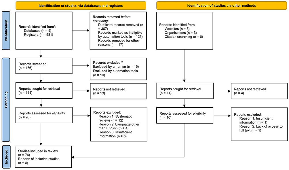 |
Figure 1 PRISMA flowchart of selection of reports analyzed for breast cancer detection by volatile organic compounds. Source: Page MJ, et al. BMJ 2021;372:n71. doi: 10.1136/bmj.n71. This work is licensed under CC BY 4.0. To view a copy of this license, visit https://creativecommons.org/licenses/by/4.0/. |
Study Selection
All retrieved records were managed using Zotero software and duplicates were removed. Two reviewers, Orduña and Alcántara, independently evaluated the titles and abstracts of the retrieved studies, and the selected articles were reviewed in full text to determine their eligibility. Any discrepancies were resolved by consensus with the work team.
Eligibility Criteria
To successfully retrieve useful articles, duplicate and Portuguese-language articles were eliminated. Seventy articles and information from 3 studies obtained from websites and organizations were included, meeting the following criteria: original research published in English or Spanish, evaluation of the efficacy of VOCs as a screening method for breast cancer, and reporting of identified VOCs. Systematic reviews, duplicate studies, letters to the editor, and articles without access to the full text were excluded. We examined references used in previous systematic reviews and included additional relevant studies needed, these being 5 references.
Data Extraction
A form was used to extract data from the included studies. The information collected included: authors, year of publication, study population, intervention, comparison, main results, and data relevant to the analysis. Data were extracted independently by two reviewers and a third reviewer corroborated the information on the form.
Results and Discussion
Advantages and Disadvantages of Current Methods for Breast Cancer Diagnosis and Screening
The World Health Organization defines screening as applying tests or studies in an asymptomatic population to detect signs of cancer as early as possible.8 Screening seeks to include the population that meets certain specific criteria to avoid leading to false positives.13 The main objective of screening is to detect a pathology in a Timely manner, without the need to wait for symptoms to begin to manifest themselves.14,15 Table 1 shows the advantages and disadvantages of current methods for diagnosing and screening breast cancer.16
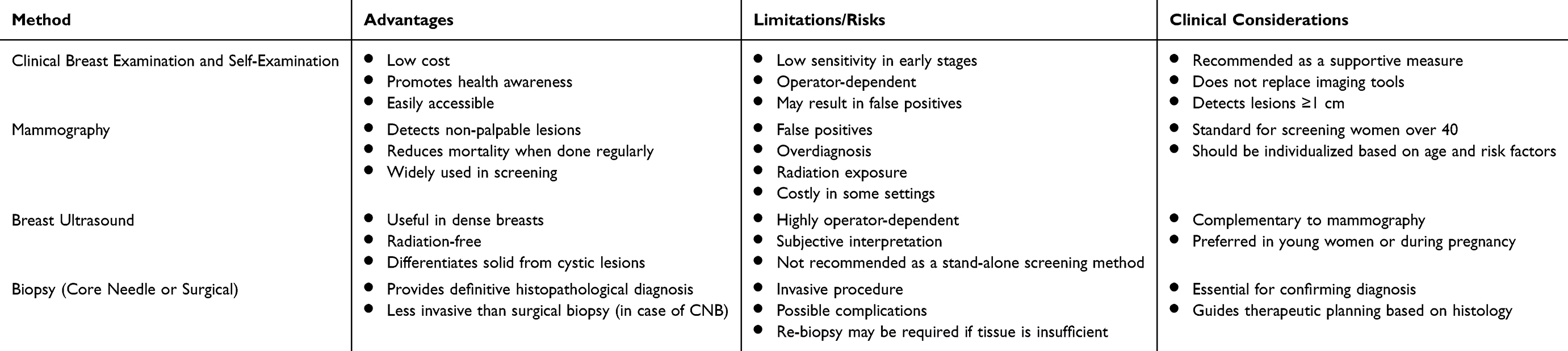 |
Table 1 Conventional Methods for Breast Cancer Detection |
Electronic Nose (E-nose)
The early history of the electronic nose takes us back to the 1960s when the Bacharac company built a device called a “sniffer” that consisted of just a gas sensor for testing.17 While that device was not considered an electronic nose, it is the first approach to what we now consider an “E-nose.”During the 1980s, the first intelligent devices were developed to mimic human olfactory ability.18,19 An electronic nose, or “E-nose”, is an elaborate device for detecting and recognizing volatile organic compounds (VOCs), mimicking the functioning of the human sense of smell but with increased sensitivity.20 In general, electronic noses translate chemical information captured by nanosensors using automated recognition algorithms, interpreting “chemical fingerprints”.20,21 By having an electronic sensory system, electronic noses can capture various elements and recognize them within a sample.22
Current analytical methods for the analysis of exhaled breath can be divided into two main groups, the first being the “classical” identification and quantification techniques, such as proton transfer reaction mass spectrometry (PTR-MS) and gas chromatography coupled to mass spectrometry (GC-MS), and the second, methods associated with pattern recognition or also called “electronic nose” (E-nose) and ion mobility spectroscopy (IMS) methods.23,24 Electronic noses can improve the quality of life by offering simple devices such as prevention and screening methods for diagnosing international diseases such as breast cancer and using breath analysis and other human biofluids. Methods for exhaled breath analysis are concentrated in Table 2.
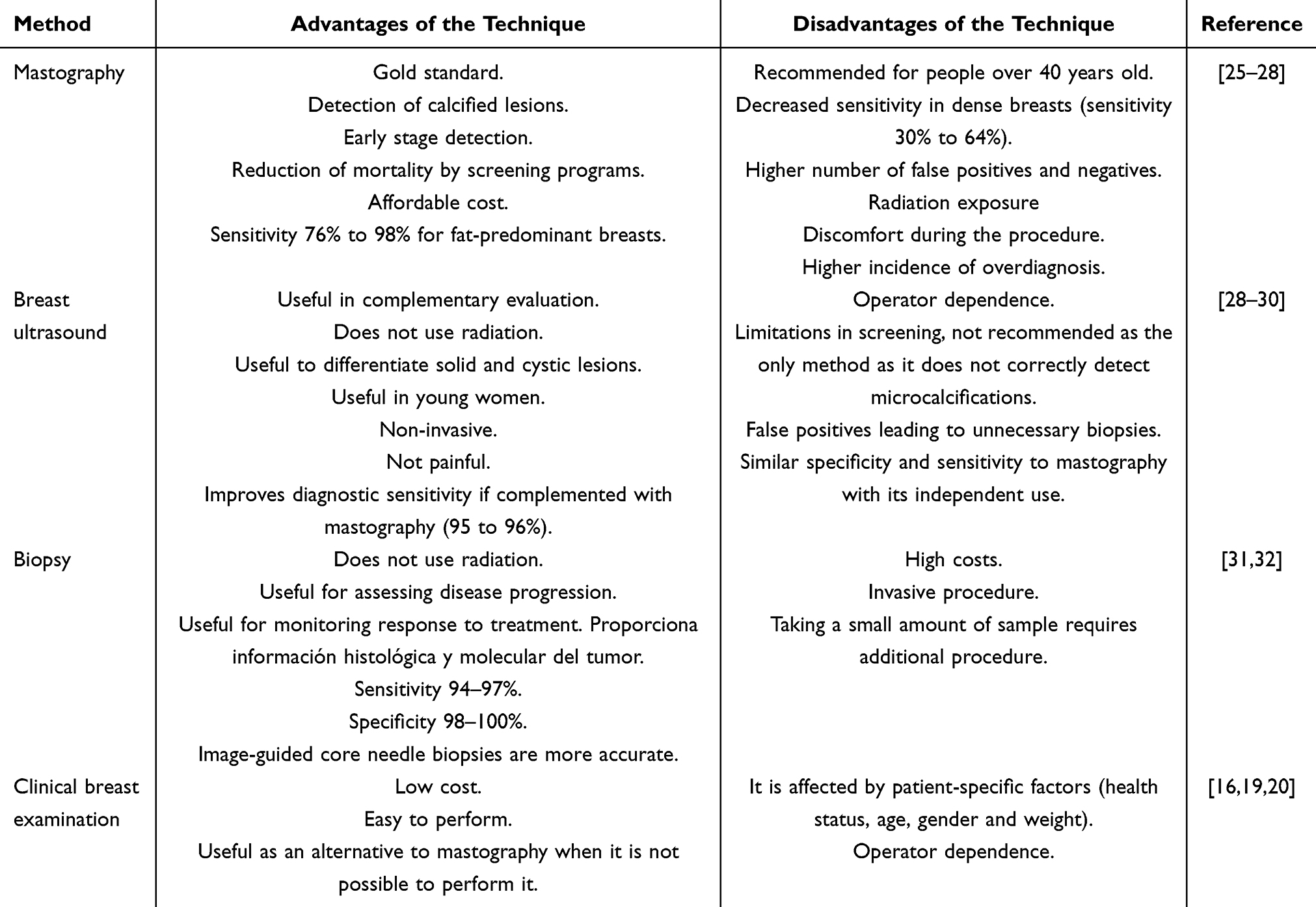 |
Table 2 Comparison of Advantages and Disadvantages of Diagnostic Methods for Breast Cancer Screening Worldwide |
Composition and Operation
E-nose works as a multi-sensor system composed of a panel or matrix containing gas--sensitive nanosensors. There are sensors based on metal oxide and carbon nanoparticles containing conductive polymers that achieve an analysis or creation of respiratory fingerprint by capturing a change in the electrical resistances detected by the metals. The function of the sensors is to detect components of a sample and transform them into a physical magnitude, such as resistance, thus reflecting how much exposure or affinity the sensors have to the “odorous” sample to which they were subjected.
Electronic noses work with a pattern recognition approach, where the data set obtained from each sample is analyzed as an image that is unique and individual to each patient, considered as a “breath print” or respiratory fingerprint, as if it were a fingerprint.33–35 As a type of pattern recognition training is created, a mathematical model can be constructed that can distinguish between healthy and sick patients by classifying them according to their breath print.22 It is common to use the analogy of the functioning of the human nose with that of the electronic nose, since in our olfactory system, odors are received by millions of receptor cells in the nose, which translate the information and send it to our brain in the form of electrical impulses to be analyzed and interpreted. The system of an electronic nose uses sensors that interact with molecules of the exposed gases, converting that information into a sequence of data thanks to an internal resistor.23
Methods such as multiple linear regression, linear discriminant analysis, clustering, k-nearest neighbor (kNN) algorithm, artificial neural networks, and fuzzy logic approaches are employed for the analysis of samples obtained by an electronic nose.24 Electronic nose analysis methods have advanced to provide fast, accurate, and cost-effective solutions in a variety of applications. The integration of sophisticated sensors, data processing techniques and machine learning has facilitated the effective use of these technologies in quality control, environmental monitoring and, potentially, medical diagnostics.25,26
Influence of Molecular Biology in Breast Cancer Screening
The odor of a patient’s skin, breath, urine, and even blood and saliva has been considered a diagnostic sign since ancient times. Kononov et al, mention the existence of evidence regarding the use of odors as a form of diagnosis since 400 B.C., with Hippocrates being one of the first people to work on this subject.36 Multiple aromatic compounds, such as phenolic acid, and some acid metabolites, such as phenol derivatives or benzene, have been found in human blood.27 Gong et al, identified some key metabolites that correlate with breast cancer: N-acetyl-D-tryptophan, 2-arachidonoylglycerol, pipecolic acid, and oxoglutaric acid.37 On the other hand, in a study by Park Jiwon, L-octanoyl carnitine, 5-oxoproline, hypoxanthine, and docosahexaenoic acid were found to be potential biomarkers for breast cancer.28 A wide variety of volatile organic compounds and metabolites with high predictive value for breast cancer detection have been found in urine, including 2-propanol and 2-butanone.38 The development of new, more economical, noninvasive, simple and compact screening tools for the early diagnosis of breast cancer is a relevant task for clinical analytical chemistry.
Volatolomics
Volatolomics is a subfield of metabolomics focused on the detection and analysis of volatile organic compounds (VOCs) released in gaseous form, which are the product of cellular processes released to the external environment by pathways such as air exchange in the lungs or water exchange in the kidneys, allowing their detection in breath and urine, respectively.39 Volatolomics studies have a promising future in diagnosis and treatment monitoring, applied in biomedical research, the food industry,toxicological analysis.29 However, the biggest challenge faced is the lack of standards in the technique of obtaining samples and their analysis to reduce the variance between the results of the studies that have been performed. The results obtained in the samples to extract VOCs become unreliable due to the factors involved in each person, such as dietary habits, environmental pollution to which they are exposed, work situations, and even alcohol or tobacco consumption.30 Volatolomics promises to be a new tool for identifying volatile biomarkers in various biological matrices; however, its integration into clinical practice requires further research.
Volatile Organic Compounds
The fluids we excrete from our bodies contain hundreds of volatile organic compounds (VOCs), which originate from various biochemical and metabolic pathways. If a metabolic pathway is altered, it can result in an altered VOC profile that can be perceived in some human biofluids. Volatile organic compounds are carbon-containing chemicals that evaporate quickly at room temperature and can be considered valuable biomarkers in medicine. Biomarkers are biological molecules that can be detected in fluids, tissues, or blood. They include proteins, nucleic acids, and carbohydrates, which indicate the onset or progression of diseases such as cancer.31
A volatile biomarker refers to a compound or substance with high volatile characteristics, or in gaseous form, applicable to interpret an individual’s current health status. We Continuously produce volatile organic compounds (VOCs) responsible for our chemical footprint.40
The production of VOCs originates from cellular metabolism and can vary in composition or quantity when cells experience pathophysiological conditions that lead to neoplastic transformation. This can occur, for example, due to hypoxia, increased energy expenditure due to hyperproliferation, or the production of reactive oxygen species.32 Several studies have shown that VOCs can differentiate pre- and post-disease states in breast cancer patients, being useful in the detection of endogenous cancer-related metabolites.
Techniques for the Detection of Volatile Organic Compounds
Exhaled breath analysis is becoming a topic of recent interest due to the need to create faster and simpler diagnostic methods. Gas chromatography (GC) is used in the context of breast cancer mainly for the analysis of volatile organic compounds that can serve as potential biomarkers in the diagnosis and monitoring of this pathology. Gas chromatography combined with mass spectrometry (GC-MS) has been used to identify VOCs that could provide information on the characteristics of cancer cells, aiding in the optimization and improvement of low-cost diagnostic devices. Although the technology of an electronic nose is portable and easy to use, its detection parameters are often insufficient for accurate quantitative or qualitative analysis of volatile organic compounds. Gas chromatography allows the identification of VOCs that could improve current diagnostic tools.41
Infrared spectroscopy (IS) identifies compounds based on their molecular vibrations. This analytical technique measures how molecules interact with infrared light, giving information about their vibrational states.42 IE is used to identify and characterize each molecule’s chemical composition and structure.43
Electrochemical sensors are advanced tools for detecting biomarkers associated with diseases such as breast cancer. These electrochemical sensors enable the rapid and accurate detection of biomarkers in body fluids, facilitating noninvasive diagnosis. Biosensors based on nanocomposites of polymers and metal nanoparticles have improved the linear detection range by developing homogeneous electrochemical platforms to detect biomarkers simultaneously.44
Table 3 aims to present the clinical studies on breast cancer diagnosis and detection included in this review.
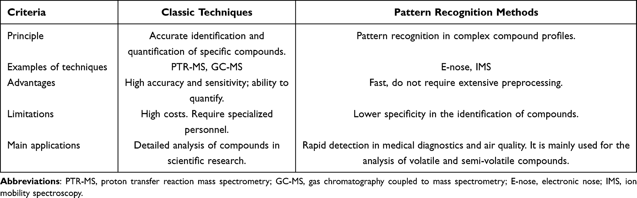 |
Table 3 Methods for Exhaled Breath Analysis |
Time-of-flight mass spectrometry (TOF-MS) is considered a widely used analytical technique to determine the composition and structure of various chemical compounds. In this an electric field accelerates technique, ions generated from a sample towards a detector, the key to which is the separation of ions according to their mass-to-charge ratio, where lighter ions reach the detector faster than heavier ions, thus determining the component masses.45 The combination of time-of-flight mass spectrometry with other similar techniques have broadened the landscape for the detection of complex biological molecules, such as proteins and even microorganisms, allowing for greater precision and accuracy in the identification of biological compounds. Zhang et al use time-of-flight mass spectrometry with high-pressure photon ionization to detect VOCs in breath samples, showing high sensitivity and specificity for differentiating between patients with and without breast cancer, but with limited performance for differentiating pathological or molecular subtypes.46 The diagnostic performance of the tests evaluated in the studies included in this review are found in Table 4.
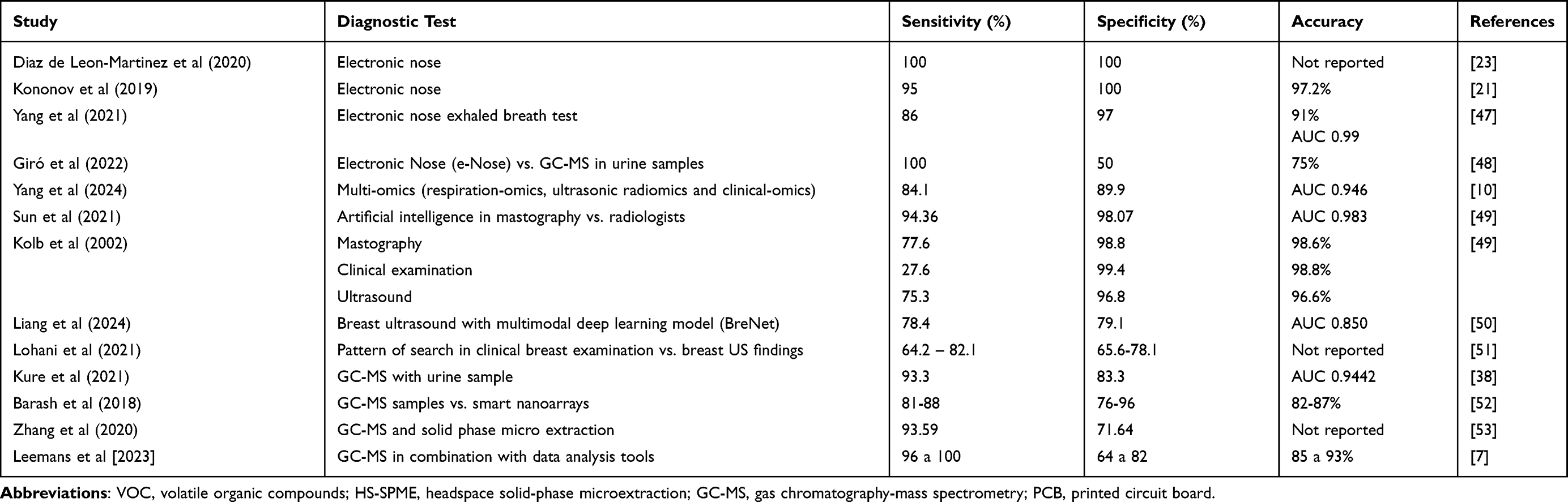 |
Table 4 Clinical Studies on Breast Cancer Diagnosis and Screening |
Volatile Organic Compounds Related to Breast Cancer
Volatile organic compounds (VOCs) have been investigated in breast cancer because they are potentially diagnostic biomarkers. Several studies have shown that VOC profiles in breath and other body fluids may be able to differentiate between healthy and breast cancer patients.33
VOCs related to breast cancer are a variety of metabolites detected in different contexts, as shown in Table 5. Compounds such as 2-propanol and 2-butanone have been found in urine samples, with ample potential to distinguish breast cancer. It is essential to mention that compounds such as 2-ethyl-1-hexanol, isolongifolenone, furan, dodecanoic acid, and 2-methoxyphenol have been identified in patients with invasive ductal carcinoma.54 However, the evidence is not entirely consistent, with discrepancies being found regarding the Collection and analysis of VOCs.34 VOCs present promising potential as diagnostic and prognostic tools in breast cancer, although more research is needed to standardize methods and clinically validate these findings.
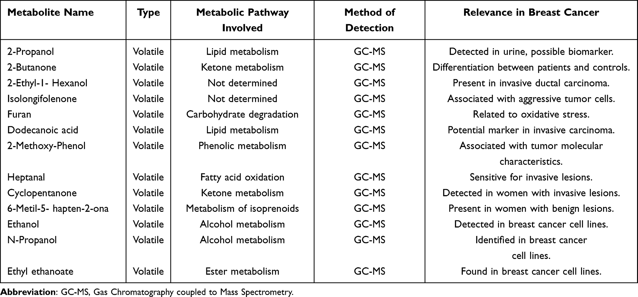 |
Table 5 Volatile Metabolites Probably Related to Breast Cancer |
Use of Nanotechnology for Disease Detection
In recent years, various nanomaterials have been created and discovered to improve the quality of life in the field of health, known as “nanomedicine”.35 Nanomedicine is a branch of medicine that uses nanotechnology to diagnose and treat diseases, including breast cancer. In breast cancer treatment, nanomedicine offers advantages over conventional therapies.55 Nanomedicines allow for more precise and controlled delivery and affinity of therapeutic agents, ie, better penetration into tumor tissues and excellent retention at the tumor site, improving treatment efficacy and reducing side effects. Due to their composition, nanoparticles can be enveloped with proteins and peptides of importance for the efficient detection and measurement of specific cancer biomarkers.52
Nanotechnology has significant applications in diagnosing, treating, and preventing disease.56 It can be used to increase the efficacy of radiotherapy, improve the use of chemotherapy drugs, detect biomarkers in vitro, or increase the efficiency of image interpretation and disease detection tools.57 Nanotechnology has been considered a promising tool in treating breast cancer, covering topics such as drug delivery, photothermal therapy, and immunotherapy.58 Immunotherapy is another branch that benefits from the use of nanotechnology, as it improves the delivery of immune checkpoint inhibitors and agents that modulate the tumor microenvironment, enhancing the T-cell response against cancer.59
Nanomedicine has advanced in the treatment of bone metastases from breast cancer, allowing the combination of therapy with diagnostic agents, which facilitates treatment monitoring and early detection of this disease.60 Despite these advances, nanomedicine in the context of breast cancer continues to face challenges such as complexity in manufacturing and regulation of practice. However, it This represents a promising strategy to improve the efficacy and safety of breast cancer treatment, bypassing some limitations of current conventional therapies.61
Application of Machine Learning Models
A comparative analysis by Onakpojeruo et al shows a study on the classification of brain tumor images using automated recognition tools, developing a model known as Conditional Deep Convolutional Neural Network (C-DCNN) that achieved an accuracy of 99% for tumor identification. This study highlights the potential of data generation and recognition to improve the training of machine learning models in medical image classification, especially in situations where access to important data or background is very limited.62 Artificial intelligence (AI) is performing an increasingly important role in disease diagnosis, and breast cancer is no exception. The use of machine learning tools has been seen to support tools for mastographic detection and classification of different subtypes of lesions.63
Using mastography as the screening method of choice implies the need to improve the false positive and false negative rates that occur at the time of interpretation. Cancers that are not identified early may not be diagnosed until advanced stages due to an error in the interpretation of the images.64 In detection and screening, artificial intelligence has shown comparable or even superior performance to that of radiologists in the interpretation of mastographic images. A study by Yoel Shoshan et al shows the work obtained in two health care centers that retrospectively collected 13,043 digital tomosynthesis images as well as background information on each patient using a cohort of 9,919 women and 5 radiologists specializing in breast imaging. The results showed a 39.6% (95% CI: 38.0, 41.7) reduction in workload for those interpreting the results with the help of AI, obtaining a sensitivity of 90% and specificity of 93.6% when automated learning was combined with the experience and skills of each radiologist.65 Kyung Park and coworkers developed a multicenter study, evaluating whether the use of AI could improve the diagnostic accuracy of radiologists. The study spanned from 2010 to 2021, conducted at 14 institutions with the support of 15 radiologists (7 breast imaging specialists and 8 general radiologists), resulting in the analysis of 258 breast tomosynthesis images, of which 65 had a confirmed diagnosis of cancer. AI alone showed an AUC of 0.93, indicating a high accuracy in detecting breast cancer. The AUC of radiologists improved from 0.90 to 0.92 with the help of AI, which would indicate an increase in diagnostic accuracy. The specificity of AI was higher at 89.64% than that of radiologists who resulted in 77.34%, suggesting that AI reduced false positives. The interpretation time decreased from 54.41 to 48.52 seconds with AI (p <0.001), demonstrating greater efficiency when used in combination with the radiologist’s experience.66
On the other hand, the retrospective comparison study conducted by Rodriguez-Ruiz in 2019 included 2,652 mastographics analyzed by 101 radiologists, using an AI system that assigned a level of suspicion for breast cancer on a scale of 1 to 10 where they compared the diagnostic performance of AI vs radiologists by measuring the AUC, obtaining an AI AUC of 0. 840 (95% CI: 0.820–0.860) and AUC of radiologists of 0.814 (95% CI: 0.787–0.841), the small difference between both measurements indicates that AI was not inferior with respect to the diagnostic ability of radiologists and that their diagnostic accuracy could be similar to that of an average radiologist.67
AI has also been used to identify subtypes of breast lesions in the analysis of images, achieving a higher accuracy in the prediction of malignancy diagnosis. However, to think of an accurate diagnosis of molecular or histological subtypes just by observing the image study without performing the biopsy procedure is still under debate. Another useful application of AI in breast cancer has been in the area of genetic alterations, specifically to classify invasive lobular carcinoma of the breast, demonstrating a high accuracy in the prediction of bi-allelic mutations of the CDH1 germline.68
Conclusions
This systematic review highlights the potential of volatile organic compounds (VOCs) and electronic nose (E-nose) technology as promising non-invasive tools for breast cancer detection. The reviewed studies demonstrate that exhaled breath and other biofluids contain VOC profiles capable of distinguishing between healthy individuals and those with breast cancer, with variable sensitivity and specificity depending on the methodology employed. The integration of pattern recognition algorithms and machine learning further enhances the diagnostic performance of E-nose systems. The findings support the notion that VOC analysis could complement current screening methods, especially in low-resource settings where access to mammography or expert radiology is limited. This approach offers advantages in terms of speed, patient comfort, and potential for population-level screening. However, current studies are limited by small sample sizes, lack of standardized sampling and analysis protocols, and variability in the identified VOCs. These factors hinder clinical translation and comparability between studies. Moreover, many investigations remain in proof-of-concept stages, requiring validation in larger and more diverse cohorts. Future research should aim to establish standardized protocols for VOC collection and analysis, evaluate cost-effectiveness, and explore the integration of E-nose systems into existing healthcare frameworks. Clinical trials are essential to assess real-world performance and user acceptance.
In summary, while VOC-based technologies are not yet ready to replace current diagnostic methods, they hold significant promise as adjunctive tools in early breast cancer detection. Advancing this field could improve diagnostic accuracy, enable earlier intervention, and ultimately reduce mortality from breast cancer.
Author Contributions
Medical Intern. Fernanda Michelle Orduña Medina. School of Medicine, Autonomous University of San Luis Potosi. Data collection, manuscript preparation, article writing, table and figure design. Ph D. Lorena Díaz de León-Martínez, and Ph D. Boris Mizaikoff Institute of Analytical and Bioanalytical Chemistry, Ulm University, Germany. Supervise and verify the manuscript and correct information technical analytical. Medical Specialist. Grecia D. D. Alarcon Rivera, and Nancy Angelica Prieto Soledad de Graciano Sanchez General Hospital, S.L.P. Supervise and verify the manuscript and correct information. Ph D. Luz Eugenia Alcántara Quintana, Unit of Innovation and Diagnosis in Cellular and Molecular Biology at the Coordination for Innovation and Application of Science and Technology (CIACyT) of the Autonomous University of San Luis Potosi. Data collection, preparation of the manuscript, supervision, and verification of the manuscript, correction of information. All authors made a significant contribution to the work reported, whether that is in the conception, study design, execution, acquisition of data, analysis and interpretation, or in all these areas; took part in drafting, revising or critically reviewing the article; gave final approval of the version to be published; have agreed on the journal to which the article has been submitted; and agree to be accountable for all aspects of the work.
Funding
Sources SMEO 2024 Project.
Disclosure
The authors declare that they have no conflicts of interest in this work.
References
1. Breast Cancer Facts and Statistics 2024 [Internet]; 2024. Available from: https://www.breastcancer.org/facts-statistics?gad_source=1&gclid=Cj0KCQjwr9m3BhDHARIsANut04bPkbQYsZg9r-LlddKuHtGcVG1E3UTF_x-mkgTPSk7ikJSTut_kHkAaAoYJEALw_wcB.
2. Breast cancer [Internet]; 2024. Available from: https://www.who.int/news-room/fact-sheets/detail/breast-cancer.
3. Li J, Guan X, Fan Z, et al. Non-invasive biomarkers for early detection of breast cancer. Cancers. 2020;12(10):2767.
4. Gözüm S, Tuzcu A. Elapsed time between the first symptoms of breast cancer and medical help-seeking behavior and the affecting factors. Cancer Nurs. 2018;41(3):E21–9. doi:10.1097/NCC.0000000000000498
5. Al-Ajmi K, Lophatananon A, Ollier W, Muir KR. Risk of breast cancer in the UK biobank female cohort and its relationship to anthropometric and reproductive factors. PLoS One. 2018;13(7):e0201097. doi:10.1371/journal.pone.0201097
6. Aguilar-Torres CR, Cisneros-Castolo M, Stener-Lechuga T, et al. Panorama actual del tamizaje para detección del cáncer de mama en el estado de Chihuahua, México. Ginecología y obstetricia de México. 2021;89(2):91–99.
7. Leemans M, Cuzuel V, Bauer P, et al. Screening of breast cancer from sweat samples analyzed by 2-dimensional gas chromatography-mass spectrometry: a preliminary study. Cancers. 2023;15(11):2939. doi:10.3390/cancers15112939
8. Mittra I, Mishra GA, Dikshit RP, et al. Effect of screening by clinical breast examination on breast cancer incidence and mortality after 20 years: prospective, cluster randomised controlled trial in Mumbai. BMJ. 2021;372:n256. doi:10.1136/bmj.n256
9. Monticciolo DL, Newell MS, Moy L, Niell B, Monsees B, Sickles EA. Breast cancer screening in women at higher-than-average risk: recommendations from the ACR. J Am Coll Radiol. 2018;15(3 Pt A):408–414. doi:10.1016/j.jacr.2017.11.034
10. Yang Y, Long H, Feng Y, Tian S, Chen H, Zhou P. A multi-omics method for breast cancer diagnosis based on metabolites in exhaled breath, ultrasound imaging, and basic clinical information. Heliyon. 2024;10(11):e32115.
11. Hanna GB, Boshier PR, Markar SR, Romano A. Accuracy and methodologic challenges of volatile organic compound-based exhaled breath tests for cancer diagnosis: a systematic review and meta-analysis. JAMA Oncol. 2019;5(1):e182815.
12. Liu J, Chen H, Li Y, et al. A novel non-invasive exhaled breath biopsy for the diagnosis and screening of breast cancer. J Hematol Oncol. 2023;16(1):63. doi:10.1186/s13045-023-01459-9
13. Abo Al-Shiekh SS, Ibrahim MA, Alajerami YS. Breast cancer knowledge and practice of breast self-examination among female university students, Gaza. Sci World J. 2021;2021:6640324.
14. Cáncer [Internet]; 2024. Available from: https://www.who.int/es/news-room/fact-sheets/detail/cancer.
15. Broza YY, Zhou X, Yuan M, et al. Disease detection with molecular biomarkers: from chemistry of body fluids to nature-inspired chemical sensors. Chem Rev. 2019;119(22):11761–11817.
16. da Costa Vieira RA, Biller G, Uemura G, Ruiz CA, Curado MP. Breast cancer screening in developing countries. Clinics. 2017;72(4):244–253.
17. Rocco G, Pennazza G, Tan KS, et al. A real-world assessment of stage I lung cancer through electronic nose technology. J Thorac Oncol. 2024;19(9):1272–1283. doi:10.1016/j.jtho.2024.05.006
18. Gashimova E, Osipova A, Temerdashev A, et al. Study of confounding factors influence on lung cancer diagnostics effectiveness using gas chromatography-mass spectrometry analysis of exhaled breath. Biomarker Med. 2021;15(11):821–829. doi:10.2217/bmm-2020-0828
19. De Vietro N, Aresta A, Rotelli MT, et al. Relationship between cancer tissue derived and exhaled volatile organic compound from colorectal cancer patients. Preliminary results. J Pharm Biomed Anal. 2020;180(180):113055. doi:10.1016/j.jpba.2019.113055
20. Abideen ZU, Arifeen WU, Bandara YMNDY. Emerging trends in metal oxide-based electronic noses for healthcare applications: a review. Nanoscale. 2024;16(19):9259–9283.
21. K A, K B, J I, et al. Online breath analysis using metal oxide semiconductor sensors (electronic nose) for diagnosis of lung cancer. J Breath Res. 2019;14(1).
22. van de Goor RMGE, Hardy JCA, van Hooren MRA, Kremer B, Kross KW. Detecting recurrent head and neck cancer using electronic nose technology: a feasibility study. Head Neck. 2019;41(9):2983–2990. doi:10.1002/hed.25787
23. de León-Martínez L D, Rodríguez-Aguilar M, Gorocica-Rosete P, et al. Identification of profiles of volatile organic compounds in exhaled breath by means of an electronic nose as a proposal for a screening method for breast cancer: a case-control study. J Breath Res. 2020;14(4):046009. doi:10.1088/1752-7163/aba83f
24. Kiani P, Vatankhahan H, Zare-Hoseinabadi A, et al. Electrochemical biosensors for early detection of breast cancer. Clin Chim Acta. 2025;564:119923.
25. Rodríguez-Aguilar M, Ramírez-García S, Ilizaliturri-Hernández C, et al. Ultrafast gas chromatography coupled to electronic nose to identify volatile biomarkers in exhaled breath from chronic obstructive pulmonary disease patients: a pilot study. Biomed Chromatogr. 2019;33(12):e4684.
26. Chiorcea-Paquim AM. Advances in electrochemical biosensor technologies for the detection of nucleic acid breast cancer biomarkers. Sensors. 2023;23(8):4128. doi:10.3390/s23084128
27. Sung J, Rajendraprasad SS, Philbrick KL, et al. The human gut microbiome in critical illness: disruptions, consequences, and therapeutic frontiers. J Crit Care. 2024;79:154436.
28. Park J, Shin Y, Kim TH, Kim DH, Lee A. Plasma metabolites as possible biomarkers for diagnosis of breast cancer. PLoS One. 2019;14(12):e0225129.
29. Giannoukos S, Agapiou A, Brkić B, Taylor S. Volatolomics: a broad area of experimentation. J Chromatogr B Analyt Technol Biomed Life Sci. 2019;(1105):136–147.
30. Hu W, Wu W, Jian Y, et al. Volatolomics in healthcare and its advanced detection technology. Nano Res. 2022;15(9):8185–8213.
31. Rocco G, Pennazza G, Santonico M, et al. Breathprinting and early diagnosis of lung cancer. J Thorac Oncol. 2018;13(7):883–894.
32. Chaudhary V, Taha BA, null L, et al. Nose-on-chip nanobiosensors for early detection of lung cancer breath biomarkers. ACS Sens. 2024;9(9):4469–4494.
33. Silva CL, Perestrelo R, Silva P, Tomás H, Câmara JS. Volatile metabolomic signature of human breast cancer cell lines. Sci Rep. 2017;(7):43969.
34. Li X, Wen X, Luo Z, et al. Development of a headspace-solid phase microextraction gas chromatography-high resolution mass spectrometry method for analyzing volatile organic compounds in urine: application in breast cancer biomarker discovery. Clin Chim Acta. 2023;540:117236. doi:10.1016/j.cca.2023.117236
35. Ashrafizadeh M, Zarrabi A, Bigham A, et al. (Nano)platforms in breast cancer therapy: drug/gene delivery, advanced nanocarriers and immunotherapy. Med Res Rev. 2023;43(6):2115–2176. doi:10.1002/med.21971
36. Jiang Y, Jiang Z, Wang M, Ma L. Current understandings and clinical translation of nanomedicines for breast cancer therapy. Adv Drug Deliv Rev. 2022;180:114034.
37. Gong S, Wang Q, Huang J, et al. LC-MS/MS platform-based serum untargeted screening reveals the diagnostic biomarker panel and molecular mechanism of breast cancer. Methods. 2024;222:100–111.
38. Kure S, Satoi S, Kitayama T, et al. A prediction model using 2-propanol and 2-butanone in urine distinguishes breast cancer. Sci Rep. 2021;11(1):19801.
39. Capuano R, Ciotti M, Catini A, Bernardini S, Di Natale C. Clinical applications of volatilomic assays. Crit Rev Clin Lab Sci. 2024;1–20.
40. null N, Sharma M, Thakur P, et al. Cancer treatment and toxicity outlook of nanoparticles. Environ Res. 2023;237(Pt 1):116870.
41. Hadi NI, Jamal Q, Iqbal A, Shaikh F, Somroo S, Musharraf SG. Serum metabolomic profiles for breast cancer diagnosis, grading and Staging by gas chromatography-mass spectrometry. Sci Rep. 2017;7(1):1715. doi:10.1038/s41598-017-01924-9
42. Edington SC, Liu S, Baiz CR. Infrared spectroscopy probes ion binding geometries. Methods Enzymol. 2021;651:157–191.
43. Mokari A, Guo S, Bocklitz T. Exploring the steps of infrared (IR) spectral analysis: pre-processing, (Classical) data modelling, and deep learning. Molecules. 2023;28(19):6886.
44. Zhang Y, Ma W, Li N, Yang M, Hou C, Huo D. A clinically feasible diagnostic typing of breast cancer built on a homogeneous electrochemical biosensor for simultaneous multiplex detection. Anal Chem. 2024.
45. Brais CJ, Ibañez JO, Schwartz AJ, Ray SJ. Recent advances in instrumental approaches to time-of-flight mass spectrometrY. Mass Spectrom Rev. 2021;40(5):647–669.
46. Zhang J, He X, Guo X, et al. Identification potential biomarkers for diagnosis, and progress of breast cancer by using high-pressure photon ionization time-of-flight mass spectrometry. Anal Chim Acta. 2024;1320:342883. doi:10.1016/j.aca.2024.342883
47. Yang H-Y, Wang Y-C, Peng H-Y, Huang C-H. Breath biopsy of breast cancer using sensor array signals and machine learning analysis. Sci Rep. 11(1):103. doi:10.1038/s41598-020-80570-0.
48. Giró Benet J, Seo M, Khine M, Gumà Padró J, Pardo Martínez A, Kurdahi F. Breast cancer detection by analyzing the volatile organic compound (VOC) signature in human urine. Sci Rep. 2022;12(1):14873. doi:10.1038/s41598-022-19237-1.
49. Sun Y, Qu Y, Wang D, et al. Deep learning model improves radiologists’ performance in detection and classification of breast lesions. Chin J Cancer Res. 2021;33(6):682–693. doi:10.21147/j.issn.1000-9604.2021.06.05.
50. Liang F, Song Y, Huang X, et al. Assessing breast disease with deep learning model using bimodal bi-view ultrasound images and clinical information. iScience. 2024;27(7):110279. doi:10.1016/j.isci.2024.110279.
51. Lohani KR, Srivastava A, Jeyapradha DA, et al. “Dial of a clock” search pattern for clinical breast examination. J Surg Res. 2021;260:10–19. doi:10.1016/j.jss.2020.04.029.
52. Broza YY, Vishinkin R, Barash O, Nakhleh MK, Haick H. Synergy between nanomaterials and volatile organic compounds for non-invasive medical evaluation. Chem Soc Rev. 2018;47(13):4781–4859.
53. Zhang Y, Guo L, Qiu Z, Lv Y, Chen G, Li E. Early diagnosis of breast cancer from exhaled breath by gas chromatography-mass spectrometry (GC/MS) analysis: a prospective cohort study. J Clin Lab Anal. 2020;34(12). doi:10.1002/jcla.23526.
54. Taunk K, Taware R, More TH, et al. A non-invasive approach to explore the discriminatory potential of the urinary volatilome of invasive ductal carcinoma of the breast. RSC Adv. 2018;8(44):25040–25050. doi:10.1039/C8RA02083C
55. Rahman M, Afzal O, Ullah SNMN, et al. Nanomedicine-based drug-targeting in breast cancer: pharmacokinetics, clinical progress, and challenges. ACS Omega. 2023;8(51):48625–48649. doi:10.1021/acsomega.3c07345
56. Karahmet Sher E, Alebić M, Marković Boras M, et al. Nanotechnology in medicine revolutionizing drug delivery for cancer and viral infection treatments. Int J Pharm. 2024;660:124345. doi:10.1016/j.ijpharm.2024.124345
57. Rajana N, Mounika A, Chary PS, et al. Multifunctional hybrid nanoparticles in diagnosis and therapy of breast cancer. J Control Release. 2022;352:1024–1047. doi:10.1016/j.jconrel.2022.11.009
58. Mugundhan SL, Mohan M. Nanoscale strides: exploring innovative therapies for breast cancer treatment. RSC Adv. 2024;14(20):14017–14040. doi:10.1039/d4ra02639j
59. Gong C, Yu X, Zhang W, et al. Regulating the immunosuppressive tumor microenvironment to enhance breast cancer immunotherapy using pH-responsive hybrid membrane-coated nanoparticles. J Nanobiotechnol. 2021;19(1):58. doi:10.1186/s12951-021-00805-8
60. Morad G, Daisy CC, Out HH, Libermann TA, Dillon ST, Moses MA. Cdc42-dependent transfer of mir301 from breast cancer-derived extracellular vesicles regulates the matrix modulating ability of astrocytes at the blood-brain barrier. Int J Mol Sci. 2020;21(11):3851. doi:10.3390/ijms21113851
61. Wu SG, Wang J, Lian CL, et al. Evaluation of the 8th edition of the American joint committee on cancer’s pathological staging system in prognosis assessment and treatment decision making for stage T1-2N1 breast cancer after mastectomy. Breast. 2020;51:2–10.
62. Onakpojeruo EP, Mustapha MT, Ozsahin DU, Ozsahin I. A comparative analysis of the novel conditional deep convolutional neural network model, using conditional deep convolutional generative adversarial network-generated synthetic and augmented brain tumor datasets for image classification. Brain Sci. 2024;14(6):559. doi:10.3390/brainsci14060559
63. Freeman K, Geppert J, Stinton C, et al. Use of artificial intelligence for image analysis in breast cancer screening programmes: systematic review of test accuracy. BMJ. 2021;374:n1872. doi:10.1136/bmj.n1872
64. Topol EJ. High-performance medicine: the convergence of human and artificial intelligence. Nat Med. 2019;25(1):44–56.
65. Shoshan Y, Bakalo R, Gilboa-Solomon F, et al. Artificial intelligence for reducing workload in breast cancer screening with digital breast tomosynthesis. Radiology. 2022;303(1):69–77. doi:10.1148/radiol.211105
66. Park EK, Kwak S, Lee W, Choi JS, Kooi T, Kim EK. Impact of AI for digital breast tomosynthesis on breast cancer detection and interpretation time. Radiol Artif Intell. 2024;6(3):e230318. doi:10.1148/ryai.230318
67. Rodriguez-Ruiz A, Lång K, Gubern-Merida A, et al. Stand-alone artificial intelligence for breast cancer detection in mammography: comparison with 101 radiologists. J Natl Cancer Inst. 2019;111(9):916–922. doi:10.1093/jnci/djy222
68. Pareja F, Dopeso H, Wang YK, et al. A genomics-driven artificial intelligence-based model classifies breast invasive lobular carcinoma and discovers CDH1 inactivating mechanisms. Cancer Res. 2024;84(20):3478–3489.
 © 2025 The Author(s). This work is published and licensed by Dove Medical Press Limited. The
full terms of this license are available at https://www.dovepress.com/terms
and incorporate the Creative Commons Attribution
- Non Commercial (unported, 4.0) License.
By accessing the work you hereby accept the Terms. Non-commercial uses of the work are permitted
without any further permission from Dove Medical Press Limited, provided the work is properly
attributed. For permission for commercial use of this work, please see paragraphs 4.2 and 5 of our Terms.
© 2025 The Author(s). This work is published and licensed by Dove Medical Press Limited. The
full terms of this license are available at https://www.dovepress.com/terms
and incorporate the Creative Commons Attribution
- Non Commercial (unported, 4.0) License.
By accessing the work you hereby accept the Terms. Non-commercial uses of the work are permitted
without any further permission from Dove Medical Press Limited, provided the work is properly
attributed. For permission for commercial use of this work, please see paragraphs 4.2 and 5 of our Terms.