
Back to Journals » International Journal of Women's Health » Volume 17
Uterine NK Cell Polarization Associates with Chronic Endometritis and Predisposition to Recurrent Implantation Failure
Authors Xie Q, Zhang L, Yang J, Yang M, Li Q, Chen Q
Received 16 July 2025
Accepted for publication 25 October 2025
Published 7 November 2025 Volume 2025:17 Pages 4255—4266
DOI https://doi.org/10.2147/IJWH.S554069
Checked for plagiarism Yes
Review by Single anonymous peer review
Peer reviewer comments 3
Editor who approved publication: Dr Matteo Frigerio
Qiuyin Xie,1,2,* Lu Zhang,3,4,* Jing Yang,4 Mengjie Yang,3,4 Qiyuan Li,4 Qionghua Chen1,3,4
1The School of Clinical Medicine, Fujian Medical University, Fuzhou, Fujian, People’s Republic of China; 2Department of Obstetrics and Gynecology, Xiamen Third Hospital, Xiamen, People’s Republic of China; 3Laboratory of Research and Diagnosis of Gynecological Diseases of Xiamen City, Clinical Medical Research Center for Obstetrics and Gynecology Diseases of Fujian Province, Department of Obstetrics and Gynecology, The First Affiliated Hospital of Xiamen University, School of Medicine, Xiamen University, Xiamen, People’s Republic of China; 4National Institute for Data Science in Health and Medicine, Xiamen University, Xiamen, People’s Republic of China
*These authors contributed equally to this work
Correspondence: Qiyuan Li, Email [email protected] Qionghua Chen, Email [email protected]
Background: Disturbances in the endometrial immune microenvironment, particularly uterine natural killer (uNK) cell polarization, are linked to chronic endometritis (CE) and recurrent implantation failure (RIF). However, the underlying mechanisms and a lack of reliable biomarkers hinder effective clinical diagnosis and treatment.
Methods: We integrated public single-cell RNA sequencing (scRNA-seq) datasets to characterize endometrial immune cell dynamics. Single-cell regulatory network inference identified key transcription factors (TFs) regulating uNK subtypes, and their functions were explored via pathway enrichment. A diagnostic model was subsequently developed and validated using bulk RNA-seq data from CE and RIF cohorts.
Results: Our analysis identified two functionally distinct uNK subtypes: cytotoxic uNK2 cells regulated by TFs EOMES and ELF4, and uNK3 cells involved in platelet activation and tight junctions, driven by ELK4 and IRF1. The abundance of these TFs correlated with their respective uNK subtype proportions moderately (p< 0.001). Key marker genes—AFAP1L2, KLRC1, and SOCS1 for uNK2, and SAMD3 for uNK3—were identified and demonstrated altered expression patterns in samples from patients with CE and RIF. In a bulk RNA-seq dataset comprising 51 endometrial samples (18 CE and 33 normal), the ratio of uNK2/uNK3 signature expression was notably upregulated in CE samples. This finding was corroborated in an independent RIF dataset. The diagnostic model based on these markers demonstrated strong predictive power. For CE, the AUC for SDC1 was 0.48, the uNK2/uNK3 ratio was 0.675, and the logistic model reached 0.822. For RIF, the uNK2/uNK3 ratio had an AUC of 0.823, while the logistic model achieved 0.83.
Conclusion: Our findings suggested that an imbalance in uNK cell polarization was a key feature of immune dysregulation in CE and RIF, with the uNK2/uNK3 signature ratio emerging as a potential biomarker. Extensive validation in prospective clinical cohorts and through functional experiments is essential to confirm the clinical utility and therapeutic potential of targeting these uNK subtypes to improve reproductive health.
Keywords: immune microenvironment, single-cell sequencing, chronic endometritis, recurrent implantation failure, NK cell polarization
Introduction
The human endometrium undergoes dynamic remodeling throughout the menstrual cycle, governed by hormonal fluctuations that modulate its immune microenvironment.1,2 This carefully balanced immune landscape is critical not only for host defense against pathogens but also for tissue repair, successful embryo implantation, and pregnancy maintenance. However, disturbances in immune homeostasis can disrupt endometrial function, contributing to pathological conditions such as chronic endometritis (CE) and recurrent implantation failure (RIF). While these conditions present formidable barriers to successful reproduction, their impact extends further, imposing significant physical and psychological burdens on patients. For individuals with RIF, this distress is often compounded by the need to undergo repetitive invasive medical procedures.3
Disruptions in the immune microenvironment of the endometrium often stem from bacterial infections or other local perturbations, altering immune cell recruitment and activity. In CE, bacterial invasion may trigger the selective migration of peripheral blood B cells into the endometrium and is linked to impaired stromal decidualization.4 Immune cell profiling of patients with CE also reveals heightened populations of CD68+ macrophages, CD83+ mature dendritic cells, CD8+ T cells, and FoxP3+ regulatory T cells.5
In addition to these cellular changes, the endometrium of infertile patients with CE exhibited a marked upregulation of pro-inflammatory cytokines, including interleukin (IL)-1β, IL-4, IL-6, IL-10, interferon-gamma, and tumor necrosis factor-alpha, with expression levels up to four times higher than in controls.6 Furthermore, transcriptomic analyses via RNA sequencing have expanded our understanding of CE’s molecular pathology, for instance, by identifying the upregulation of TVP23A.7
Central to this immune dysregulation was the polarization of natural killer (NK) cells, which played a pivotal role at the maternal-fetal interface. Among the diverse immune cell populations in the endometrium, uterine natural killer (uNK) cells were particularly dominant, comprising up to 70% of endometrial leukocytes during the window of implantation and in early pregnancy.8,9 In a physiological setting, these cells were critical for successful embryo implantation. Far from being merely cytotoxic, they adopt a pro-pregnancy phenotype, secreting a range of cytokines and growth factors that are essential for spiral artery remodeling—a process vital for supplying blood to the developing placenta. Furthermore, they played a key role in regulating the invasion of fetal trophoblast cells, ensuring a secure yet controlled attachment to the uterine wall.10,11 However, in pathological states, this delicate balance was lost. Immune impairment could lead to a shift in uNK cell polarization from this supportive, decidual phenotype towards a more aggressive, cytotoxic phenotype. This dysregulation created a hostile, pro-inflammatory microenvironment that was non-receptive to the embryo. The altered cytokine profile could disrupt endometrial receptivity markers, and hyperactive uNK cells may even erroneously attack the semi-allogeneic embryo, directly contributing to RIF.11
Despite the recognized link between immune dysregulation and reproductive disorders, diagnosing CE and RIF remains fraught with challenges. The current gold standard for CE diagnosis involves histopathological examination for plasma cells, typically confirmed by CD138 immunohistochemical staining.12 However, widespread clinical adoption is hampered by inconsistencies in staining protocols and sampling variability.13 Similarly, immune-related factors underlying RIF are often overlooked in standard reproductive evaluations. CE is increasingly associated with adverse in vitro fertilization and embryo transfer outcomes, contributing to RIF, recurrent pregnancy loss, and habitual miscarriage. Reports indicate a broad range of RIF prevalence in patients with CE (7.7–44%) which underscores the critical influence of immune factors on implantation and pregnancy success.13,14 Given the diagnostic and therapeutic limitations in current practice, there is a pressing need to develop reliable molecular markers and robust immune profiles for CE and RIF. Identifying markers that reflect NK cell subtype polarization and other immune dysregulations may pave the way for more precise diagnostic tools and targeted therapies aimed at restoring endometrial receptivity, thereby improving reproductive outcomes.
Advances in single-cell RNA sequencing (scRNA-seq) have transformed our capacity to unravel the complex cellular heterogeneity and transcriptional landscapes of the endometrial immune microenvironment.15 This cutting-edge technology provides high-resolution profiling of individual cells, allowing the identification of rare and previously uncharacterized cell populations that are instrumental in tissue homeostasis, immune regulation, and disease pathogenesis. A major milestone in this field is the Human Endometrial Cell Atlas, which integrates data from over 300,000 cells across multiple donors, offering a comprehensive view of the endometrial tissue’s cellular architecture and dynamic interactions.15 The Human Endometrial Cell Atlas has been pivotal in enhancing our understanding of normal physiological processes, such as menstrual cycle progression, and in identifying dysregulated cellular subtypes associated with pathological conditions. Further expanding this knowledge, the profiling of more than 50,000 uterine cells from healthy premenopausal women, alongside previously published datasets, culminated in the construction of a detailed uterine cell atlas encompassing over 167,000 cells.16 This expansive resource characterizes a broad spectrum of cell types, including lymphatic and blood endothelial cells, stromal cells, ciliated and unciliated epithelial cells, and diverse immune cell populations. These foundational insights have significantly advanced our understanding of uterine function and dysregulation, particularly in the context of reproductive disorders.
In addition to these developments, integrative single-cell analyses of NK cells across various tumor microenvironments have illuminated tumor-type-specific heterogeneity in NK cell populations.17 Applying similar high-resolution mapping techniques to the endometrial immune landscape enables the discovery of previously unrecognized or transient immune cell subtypes. This comprehensive approach offers a unique window into immune cell dynamics within the endometrium, furthering our grasp of its intricate immune regulation.
Given the diagnostic challenges for endometrial disorders and the pivotal role of uNK cells, we hypothesized that an imbalance in uterine NK cell polarization is a core pathological feature of CE and RIF that could serve as a robust biomarker. To test this, we used single-cell RNA sequencing (scRNA-seq) and bioinformatics to investigate the endometrial immune microenvironment and identify the key drivers of NK cell subtypes. Based on these insights, we developed and validated a diagnostic model, establishing a specific uNK polarization signature as a novel predictive marker. Our findings thus offer a promising new strategy for improving diagnostic accuracy and guiding potential therapeutic interventions to enhance reproductive outcomes.
Methods
ScRNA-Seq Data Preprocessing
We obtained publicly available data from databases published by Wang et al3 and Alonso et al2 which included 21 cases of normal endometrial tissue (Table S1). Each sample underwent individual filtering based on UMI counts and mitochondrial proportions as part of the quality control process (Table S1). After applying quality control measures, a total of 100,291 cells were selected for further analysis.
Dimensionality Reduction, Clustering, and Annotation
Following normalization, we identified highly variable genes using the FindVariableGenes function in Seurat, which were then used for principal component analysis.18 We applied UMAP to generate a two-dimensional representation using the first 40 principal components.19 Clustering was performed using the FindClusters algorithm at a resolution of 0.3, and the results were visualized with UMAP. Through UMAP visualization, strong inter-mixing of cells from both datasets was observed, indicating a minimal batch effect. Therefore, to avoid over-correction and preserve the biological variance of the menstrual cycle, the merged, uncorrected data were used for downstream analysis. Cell types were annotated based on known marker genes and differential expression profiles. The FindAllMarkers function in Seurat was used to identify marker genes, defined as those expressed in more than 40% of cells with a minimum log-fold change of 0.6.
Analysis of RNA Sequencing Data
To collect original data from patients with CE and RIF, we conducted a data mining process. We downloaded bulk RNA sequencing data from patients with CE from the Japanese Genotype-phenotype Archive (accession number JGAD000750).7 In this source study, CE was diagnosed based on the histological criterion of identifying five or more CD138-positive plasma cells per high-power field. Upon reviewing the clinical data, we identified one patient (CE36___6_m41) with incorrect menstrual history and excluded five patients (CE_34___5_m12, CE_37___7_m8, CE_44__18_m27, CE_48__34_m12, and CE_49__38_m12) due to discrepancies in plasma cell proportions reported by Cibersort, which contradicted expected patterns. Therefore, the final dataset included 51 patients, consisting of 33 healthy controls and 18 patients with CE. Bulk data for patients with RIF were obtained from the Gene Expression Omnibus20 (accession number GSE106602), which included 50 patients: 31 with RIF and 19 healthy controls. As all data were publicly available, no ethical approval was required for this study.
Cell Communication Analysis
We utilized the CellChat tool21 to infer cell-cell communication based on scRNA-seq data, enabling the analysis of intercellular communication. CellChat was applied to investigate critical cellular pathways involved in cell clustering. The netVisual_circles function was used to illustrate the network of cell clusters in relation to each other, while netVisual_bubble was employed to display interactions of ligand-receptor crosstalk between different cell clusters.
SCENIC Analysis
We conducted SCENIC analysis using the pySCENIC package (version 0.9.19) and the hg19-500bp-upstream-10species database, along with RcisTarget, GRNBoost, and AUCell. The input matrix comprised the normalized expression data from Seurat. To compute regulon activity scores (RAS) for each cell, we leveraged the pySCENIC Python package for SCENIC analysis.22 Initially, we applied GRNBoost2 to infer co-expression modules between TFs and their candidate target genes. We then used RcisTarget to analyze the genes within each co-expression module to identify enriched motifs, defining TFs and their potential direct target genes as regulons. Finally, we assessed the activity of each regulon in individual cells using AUCell.
Statistical Analysis
Bioinformatics analyses were performed using R version 4.3.2, with data visualization carried out using the ggplot2 package. Spearman correlation was used to examine relationships between TF activity and uterine natural killer (uNK) cell subtypes (uNK2 and uNK3) across the menstrual cycle, as well as between TF-target genes and cell-type markers. To minimize bias, the coefficient of variation was included in the analyses. Statistical significance was set at an adjusted p-value of <0.05. The Wilcoxon rank-sum test was applied for group comparisons. To assess the diagnostic performance of the uNK2/uNK3 ratio, a logistic regression model was developed. To ensure robustness and generalizability, the model was trained exclusively on a discovery cohort (CE dataset, n = 51) and then validated on a completely separate, independent cohort (RIF dataset, n = 50). The diagnostic accuracy in both cohorts was quantified using Receiver Operating Characteristic (ROC) curve analysis and the Area Under the Curve (AUC).
Results
Dynamic Regulation and NK Cell Polarization in the Immune Microenvironment Across Menstrual Cycles
Consistent with the findings of the parent studies from which the data were derived, our re-analysis of the integrated datasets distinguished 10 major cell types, including lymphocytes, macrophages, stromal fibroblasts (such as eS, dS, PV MYH11, Fibro C7, and uSMC), endothelial cells, ciliated epithelial cells, and unciliated epithelial cells (Figures 1A, B and S1A, B). This concordance confirms a successful alignment and robust integration of the two independent datasets. Among these, the epithelial cells were further categorized into eight subtypes, encompassing pre-ciliated, ciliated, SOX9+ proliferative, and various glandular (glandular, glandular secretory) and luminal (luminal 1 and luminal 2) cells (Figure S1C and D). The proportions of these cell types exhibited significant variations across different menstrual cycle phases, reflecting the dynamic changes within the endometrial microenvironment (Figure 1C).
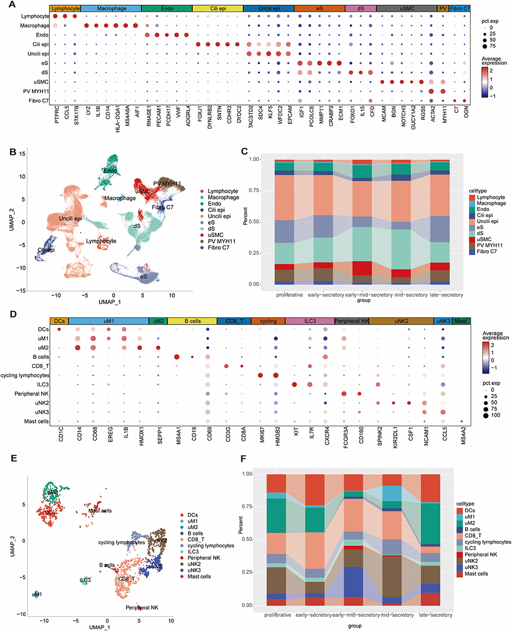 |
Figure 1 Single-cell transcriptomic atlas of the human endometrium revealed dynamic cellular composition across the menstrual cycle. Single-cell RNA sequencing analysis of 100,291 cells from 21 healthy donors identified 10 major cell lineages, including a detailed atlas of 11 distinct immune cell subtypes. (A) Dot plot showing the expression of selected marker genes used to annotate the 10 major cell types. (B) UMAP visualization of the 10 annotated cell type clusters. (C) Stacked bar chart illustrating the dynamic changes in the relative proportions of the 10 major cell types across five phases of the menstrual cycle. (D) Dot plot detailing the expression of key marker genes used to identify 11 immune cell subtypes. (E) UMAP visualization of the 11 immune cell sub-clusters. (F) Stacked bar chart showing the changing proportions of the 11 immune cell subtypes throughout the menstrual cycle, highlighting the dynamic nature of the endometrial immune microenvironment. |
Focusing on immune cells, re-clustering revealed 11 distinct immune subtypes, including dendritic cells, uM1 and uM2 macrophages, B cells, CD8+ T cells, cycling lymphocytes, ILC3, peripheral NK cells, mast cells, and two subtypes of uNK cells (uNK2 and uNK3) (Figure 1D and E). Annotation of these subtypes was performed in accordance with the canonical marker genes defined in the original studies from which our data were sourced. Of particular interest, uNK3 cells showed a substantial increase during the early-to-mid-secretory phase, followed by a marked decrease in subsequent phases. Similarly, uNK2 cells displayed statistically significant fluctuations across the menstrual cycle (Figures 1F and 2A). The proportions of other immune cell types throughout the menstrual cycle are shown in Figure S2A. Correlation analysis revealed that the proportions of uNK2 and uNK3 cells were dynamically reshaped across different phases, with significant interactions between these subtypes and other immune cell types (Figure S2B).
 |
Figure 2 uNK2 and uNK3 subtypes exhibit distinct temporal dynamics, cellular interactions, and regulatory programs across the menstrual cycle. (A) Line graph showing the dynamic proportions of uNK2 (teal) and uNK3 (gold) cells across five phases of the menstrual cycle. Points represented the mean ratio, and error bars indicate the standard error of the mean. Asterisks denoted statistical significance between the two groups at each phase (***p < 0.001). (B) Chord diagrams illustrating the predicted cell-cell communication strength between uNK subtypes, B cells, CD8+ T cells, and DCs in each phase. The thickness of the connecting lines is proportional to the calculated interaction strength. (C) Box plots comparing the activity scores of key TFs between uNK2 and uNK3 cells. EOMES and ELF4 show significantly higher activity in uNK2, while ELK4 and IRF1 are more active in uNK3. (D) Heatmap displaying the Z-scaled activity scores of the four TFs in individual cells, visually confirming the differential TF activity between the uNK2 and uNK3 subtypes. |
Our analysis of intercellular communication networks revealed highly dynamic and phase-specific remodeling of the endometrial immune microenvironment (Figures 2B and S2C). A key observation was that the overall interaction strength among immune cells peaked during the early-mid-secretory phase, a critical period immediately preceding the uterine window of implantation. Robust signaling between uNK2 and uNK3 cells during the early-secretory phase. This interaction peaked during the early-to-mid-secretory phase, after which it gradually declined. Throughout the cycle, uNK2 cells emerged as a central communication hub, maintaining robust interactions with CD8+ T cells. Specifically, the communication strength between uNK2 and CD8+ T cells reached its maximum in the early-mid-secretory phase. These dynamic interactions suggest that NK cell subtypes play a pivotal role in modulating the immune microenvironment throughout the menstrual cycle, particularly during the secretory phase.
Key TFs EOMES, ELF4, ELK4, and IRF1 Drive Distinct Roles of uNK2 and uNK3 Subtypes in Immune Microenvironment Across Menstrual Cycles
We identified the top five TFs with high activity specific to various immune cell clusters (Figure S3A). Analysis of NK cell polarization across the menstrual cycle revealed distinct TFs and marker genes associated with uNK2 and uNK3 subtypes. Notable differences in TF activity were observed between the two uNK subtypes (Figure 2C and D). Specifically, for uNK2, the TF activities of EOMES and ELF4 were significantly higher than those for uNK3, while for uNK3, the TF activities of ELK4 and IRF1 were significantly higher than those for uNK2 (Figure 2C). To investigate the regulatory mechanisms governing uNK cell dynamics, we analyzed the activity of key TFs within each uNK subtype across the menstrual cycle (Figure S4). For the uNK2 subtype, the activity of TFs EOMES and ELF4 showed a modest but highly significant positive correlation with cyclical progression, peaking during the mid-secretory phase before sharply declining (R = 0.19 and R = 0.20, respectively; p < 0.001). In contrast, the uNK3-associated TFs, ELK4 and IRF1, exhibited a stronger correlation with the cycle, with activity increasing from the proliferative phase to a peak in the early- and mid-secretory phases (R = 0.29 and R = 0.34, respectively; p < 0.001). These distinct temporal patterns and effect sizes underscored that uNK2 and uNK3 cells were governed by phase-dependent regulatory programs.
KEGG pathway enrichment analysis provided valuable insights into the functional roles of uNK2 and uNK3 cells. uNK2 was primarily associated with pathways related to cytotoxicity, highlighting its crucial role in immune defense and surveillance within the endometrium (Figure 3A). In contrast, uNK3 cells were enriched in pathways linked to platelet activation and tight junction formation, suggesting a role in tissue remodeling and vascular regulation (Figure 3B). We identified cell marker genes from the single-cell data (Table S2), and genes related to regulon activity that were significantly associated with cell proportions are shown (Figure S5 and Tables S3, S4). Marker genes for uNK2 and uNK3 were selected by intersecting regulon-related genes significantly associated with cell proportions across menstrual cycles with known marker genes for these cells (Figure 3C and D). For uNK2, 34 genes were identified in the intersection, while only 4 genes were found for uNK3. To refine the gene selection and minimize potential bias, we incorporated correlation coefficients and the coefficient of variation of gene expression. This led to the final selection of 4 key marker genes for uNK2 and 1 for uNK3, which were subsequently used to construct gene expression signatures for evaluating the uNK subtype balance. For uNK2, these marker genes included AFAP1L2, KLRC1, SAMD3, and SOCS1 (Figure 3E), while SAMD3 was also identified as a prominent marker for uNK3 (Figure 3F). The expression levels of these marker genes were strongly correlated with the changes in the proportions of uNK2 and uNK3 cells, further confirming their role as distinctive features of these subtypes.
 |
Figure 3 Functional analysis and marker gene identification for uNK2 and uNK3 subtypes. (A and B) KEGG pathway enrichment analysis for genes targeted by the primary regulons of uNK2 (A) and uNK3 (B) cells. Dot size was proportional to the number of genes enriched in a pathway, color corresponded to the statistical significance (p-value), and the Rich Factor indicated the degree of enrichment. (C) Representation of uNK2 cell-specific genes selected by considering an intersection between regulon-related genes significantly associated with uNK2 cell proportion and marker genes in uNK2 cells by using a Venn diagram. (D) Representation of uNK3 cell-specific genes selected by considering an intersection between regulon-related genes significantly associated with uNK3 cell proportion and marker genes in uNK3 cells by using a Venn diagram. (E) An illustration of the expression of uNK2 marker genes selected based on a correlation with cell proportion across menstrual cycles >0.2 and a coefficient of variation (CV) in the gene expression >0.02 across different menstrual phases. (F) An illustration of the expression of uNK3 marker genes was selected based on a correlation with cell proportion across menstrual cycles >0.2 and a CV in gene expression >0.02 across different menstrual phases. |
uNK2/uNK3 Ratio as a Diagnostic Biomarker for CE and RIF
The ratio of uNK2 to uNK3 signatures, reflecting NK cell polarization, was used as a more effective measure of immune microenvironment imbalance. In bulk endometrial data from 51 samples (18 patients with CE and 33 healthy controls), the average uNK2/uNK3 ratio was significantly higher in CE samples, suggesting dysregulation of the immune microenvironment in CE (Figure 4A). A similar trend was observed in an independent bulk dataset of 50 samples (31 patients with RIF and 19 healthy controls), where the uNK2/uNK3 ratio was also significantly elevated in patients with RIF, further confirming the robustness of this biomarker (Figure 4B).
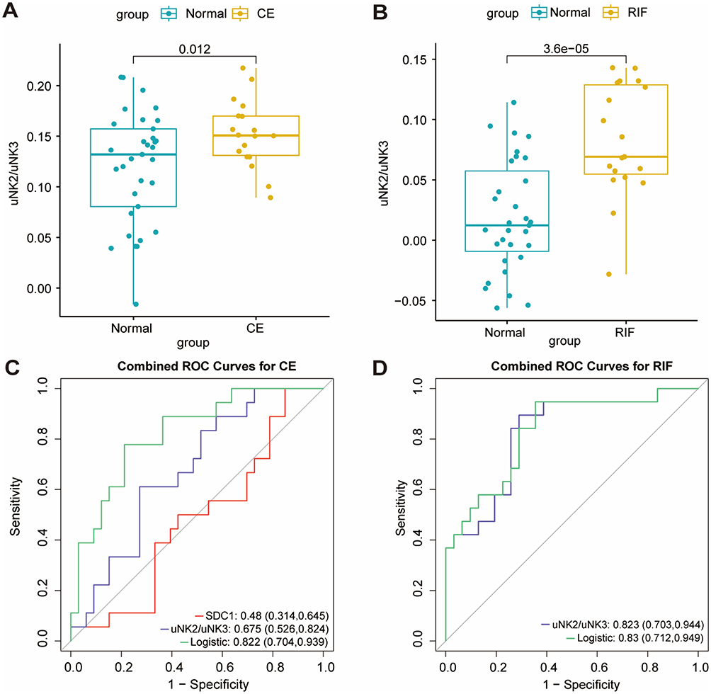 |
Figure 4 The uNK2/uNK3 signature ratio served as a robust diagnostic biomarker for CE and RIF. (A and B) Box plots comparing the uNK2/uNK3 signature ratio in endometrial bulk RNA-seq samples from Normal versus patients with CE (A) and RIF (B). The ratio was significantly elevated in both patient cohorts. Each dot represents an individual sample; the box indicated the interquartile range (IQR), and the central line represents the median. (C) A display of the ROC curve analysis for the diagnostic model based on the uNK2/uNK3 ratio, SDC1 marker genes, and logistic regression in the diagnosis of CE. (D) A display of the ROC curve analysis for the diagnostic model based on the uNK2/uNK3 ratio, the CE-based model, and logistic regression in the diagnosis of RIF. The AUC and 95% confidence intervals were provided for each model, with values closer to 1.0 indicating higher diagnostic accuracy. |
The diagnostic model based on these markers demonstrated robust predictive performance. For CE, SDC1 alone achieved an area under the curve (AUC) of 0.48 (0.314–0.645), while the uNK2/uNK3 ratio yielded an AUC of 0.675 (0.526–0.824). The logistic regression model, constructed using these markers, demonstrated the best performance, with an AUC of 0.822 (0.704–0.939) (Figure 4C). In the context of RIF, the uNK2/uNK3 ratio alone produced an AUC of 0.823 (0.703–0.945). Moreover, a logistic regression model built using RIF-specific data achieved an AUC of 0.83 (0.712–0.949), considerably outperforming the other models (Figure 4D).
Discussion
The endometrial immune microenvironment is essential for maintaining the balance required for successful embryo implantation and pregnancy. Disruptions to this equilibrium can lead to immune dysregulation, which is a key contributor to various reproductive disorders, such as CE and RIF. This study focuses on the dynamic fluctuations of specific uNK cell subtypes—uNK2 and uNK3—throughout the menstrual cycle. While uNK1 is recognized as another key uNK subtype, it was not resolved as a distinct population in our integrated analysis. This is likely attributable to the high degree of transcriptional similarity between uNK1 and uNK2 cells, with their main distinction in parent studies often being proliferation-related markers. The process of integrating two independent datasets can merge such closely related clusters. Consequently, our study concentrates on the relationship between the two clearly separable subtypes, uNK2 and uNK3. By examining these variations, we provide valuable insights into how imbalances within the immune microenvironment contribute to reproductive failure, with a particular emphasis on RIF as a manifestation of these disturbances.
Changes in the reproductive microbiota may worsen immune dysregulation in the endometrium, further complicating the immune environment.23 For example, microbial genera such as Leptotrichia and Sneathia have been associated with the modulation of immune cells in the endometrium. These microbes influence metabolic pathways and immune responses, contributing to alterations in the tissue’s immune profile. Specifically, an increased abundance of Sneathia correlated with higher uNK cell presence, supporting the hypothesis that microbial dysbiosis contributes to immune dysregulation in the endometrium. Moreover, previous studies have emphasized changes in the proportions of uNK cell subtypes and other immune cells, such as CD3+ T lymphocytes, in women with CE. These alterations were accompanied by a reduction in macrophage inflammatory protein 1β levels, which signaled disrupted immune signaling pathways essential for embryo implantation.24 Such immune imbalances can impair critical processes, including uterine vascular remodeling, preventing the formation of a supportive immune environment necessary for successful implantation.
NK cells are fundamental components of the innate immune system, serving as the first line of defense against pathogens while also maintaining tissue homeostasis.25 During pregnancy, uNK cells play a crucial role not only in defending against infections but also in regulating the immune environment to ensure successful embryo implantation. This requires a delicate balance, as uNK cells must tolerate the semi-allogeneic fetus while simultaneously protecting the endometrium from infection.26 Moreover, uNK cell numbers fluctuate throughout the menstrual cycle, with a notable peak occurring during the mid-luteal and mid-secretory phases, aligning with the implantation window.27,28 Our study corroborated this pattern, showing dynamic changes in uNK cell proportions across the menstrual cycle and emphasizing their role in regulating immune responses during implantation. However, disruptions to the immune microenvironment, as observed in disorders like CE and RIF, can impair these delicate processes. The regulation of uNK cell subtypes, particularly uNK2 and uNK3, is crucial in maintaining this immune balance. Our findings suggested that uNK2 and uNK3 cells have distinct functions throughout the menstrual cycle, with their dysregulation playing a key role in the immune microenvironment imbalances observed in conditions such as CE and RIF. KEGG pathway analysis revealed that uNK2 cells may be mainly involved in cytotoxic pathways during the proliferative phase, where they act to protect against infections. However, during the implantation window, these cytotoxic activities must be downregulated to prevent harm to the embryo. In contrast, uNK3 cells are more involved in processes like platelet activation and tight junction formation, both of which are crucial for vascular remodeling and creating a supportive uterine environment for pregnancy. In conditions such as RIF, the altered expression of uNK2 and uNK3 markers indicates a more widespread disturbance in immune regulation, which may hinder successful implantation.29,30
Our findings both aligned with and extend previous research on immune dysregulation in reproductive failure. The observation of an elevated uNK2 signature, which was enriched for cytotoxic pathways, was consistent with earlier studies that reported an increase in cytotoxic NK cell markers in women experiencing implantation failure.31 This suggested a common pathological theme of a pro-inflammatory shift in the uterine microenvironment. However, our study provided a more nuanced perspective. For example, some prior investigations using methods like flow cytometry found no significant change in the total uNK cell population in RIF patients.11,32 Our results clarified this apparent contradiction by demonstrating that the critical factor is not the overall cell count, but rather the functional polarization and ratio between the uNK2 and uNK3 subtypes. This revealed a subtle but crucial imbalance that conventional methods, which rely on a limited set of surface markers, may overlook. By defining subtypes based on their complete transcriptional profiles, we could detect a functional shift that is more indicative of pathology than simple cell enumeration.
The primary innovation of our study was that we derived a quantifiable gene signature which predicted the risk of CE and RIF by measuring uNK cell polarization. This signature was based on the expression ratio of two functionally distinct uNK subtypes—cytotoxic uNK2 and tissue-remodeling uNK3—which we identified using single-cell transcriptomics. We then validated the predictive power of this signature in independent bulk RNA-seq datasets from CE and RIF cohorts. A logistic regression model incorporating our signature demonstrated high diagnostic accuracy for both conditions (AUC > 0.82), establishing a clear path toward clinical application.
Limitations and Future Directions
Despite its promising findings, our study had key limitations that directly inform future research directions. First, our reliance on in silico analysis meant our findings represent associations, not causation. Second, the modest sample sizes of the validation cohorts limited the generalizability of our diagnostic model. Third, while many of our reported correlations were statistically significant, their effect sizes were modest, which necessitates a cautious interpretation of their immediate biological impact.
These limitations highlight a clear path forward. Mechanistic studies—using in vitro co-culture systems or in vivo animal models—are essential to dissect the causal role of the uNK2/uNK3 imbalance in implantation failure. Furthermore, a crucial next step is the prospective clinical validation of the uNK2/uNK3 signature ratio in large, multicenter trials to confirm its real-world diagnostic accuracy. Ultimately, these validation and mechanistic studies could pave the way for novel therapeutic interventions aimed at modulating uNK cell polarization to restore endometrial receptivity and enhance pregnancy outcomes.
Conclusion
In conclusion, our study proposed a candidate biomarker based on the ratio of uNK2 to uNK3 signature gene expression, which appeared to reflect the immune microenvironment imbalance in CE and RIF. The key strengths of our research included the use of high-resolution single-cell data to define functionally distinct immune subtypes, the innovative concept of a ratiometric biomarker, and the bioinformatic validation of our model in two independent patient cohorts. However, we acknowledged several limitations. Our analysis was retrospective and relied on publicly available datasets, which limited control over clinical variables. Furthermore, while our study demonstrated an association, it did not establish causality, and the proposed diagnostic model requires extensive validation before any clinical implementation. Looking forward, future research should focus on prospectively validating this uNK2/uNK3 signature ratio in large, multicenter clinical trials to confirm its potential diagnostic utility. Moreover, functional studies are essential to elucidate the causal mechanisms by which this uNK cell imbalance leads to implantation failure.
Data Sharing Statement
The datasets analysed during the current study are available in the NCBI’s Gene Expression Omnibus (series accession code GSE111976), ArrayExpress under accession numbers E-MTAB-10287, the Japanese Genotype-phenotype Archive (accession number JGAD000750) and the Gene Expression Omnibus (accession number GSE106602).
Ethics Statement
The requirement for ethical approval for this study was waived by the Institutional Review Board of the First Affiliated Hospital of Xiamen University. The study was based on de-identified and publicly accessible data and is exempt from ethical review in accordance with Article 32 of the Measures for Ethical Review of Life Science and Medical Research Involving Human Subjects dated February 18, 2023, China. This article specified that ethical review can be exempted for research under circumstances including: (1) utilizing legally obtained public data, or data generated through observation without interfering with public behavior; and (2) using anonymized information data for research.
Author Contributions
All authors made a significant contribution to the work reported, whether that is in the conception, study design, execution, acquisition of data, analysis and interpretation, or in all these areas; took part in drafting, revising or critically reviewing the article; gave final approval of the version to be published; have agreed on the journal to which the article has been submitted; and agree to be accountable for all aspects of the work.
Funding
This work was supported by the National Natural Science Foundation of China (No. 82271678), the Natural Science Foundation of Fujian Province (No. 2022J02058 and No. 2023J01623).
Disclosure
All authors have no conflicts to disclose for this work.
References
1. Greenbaum S, Averbukh I, Soon E, et al. A spatially resolved timeline of the human maternal-fetal interface. Nature. 2023;619(7970):595–605. doi:10.1038/s41586-023-06298-9
2. Garcia-Alonso L, Handfield LF, Roberts K, et al. Mapping the temporal and spatial dynamics of the human endometrium in vivo and in vitro. Nat Genet. 2021;53(12):1698–1711. doi:10.1038/s41588-021-00972-2
3. Wang W, Vilella F, Alama P, et al. Single-cell transcriptomic atlas of the human endometrium during the menstrual cycle. Nat Med. 2020;26(10):1644–1653. doi:10.1038/s41591-020-1040-z
4. Kitaya K, Yasuo T. Aberrant expression of selectin E, CXCL1, and CXCL13 in chronic endometritis. Mod Pathol. 2010;23(8):1136–1146. doi:10.1038/modpathol.2010.98
5. Li Y, Yu S, Huang C, et al. Evaluation of peripheral and uterine immune status of chronic endometritis in patients with recurrent reproductive failure. Fertil Steril. 2020;113(1):187–196. doi:10.1016/j.fertnstert.2019.09.001
6. Tortorella C, Piazzolla G, Matteo M, et al. Interleukin-6, interleukin-1β, and tumor necrosis factor α in menstrual effluents as biomarkers of chronic endometritis. Fertil Steril. 2014;101(1):242–247. doi:10.1016/j.fertnstert.2013.09.041
7. Oshina K, Kuroda K, Nakabayashi K, et al. Gene expression signatures associated with chronic endometritis revealed by RNA sequencing. Front Med. 2023;10:1185284. doi:10.3389/fmed.2023.1185284
8. Wira CR, Rodriguez-Garcia M, Patel MV. The role of sex hormones in immune protection of the female reproductive tract. Nat Rev Immunol. 2015;15(4):217–230. doi:10.1038/nri3819
9. Zeng S, Liu X, Liu D, et al. Research update for the immune microenvironment of chronic endometritis. J Reprod Immunol. 2022;152:103637. doi:10.1016/j.jri.2022.103637
10. Andreotti JP, Paiva AE, Prazeres PHDM, et al. The role of natural killer cells in the uterine microenvironment during pregnancy. Cell Mol Immunol. 2018;15(11):941–943. doi:10.1038/s41423-018-0023-1
11. Von Woon E, Greer O, Shah N, et al. Number and function of uterine natural killer cells in recurrent miscarriage and implantation failure: a systematic review and meta-analysis. Hum Reprod Update. 2022;28(4):548–582. doi:10.1093/humupd/dmac006
12. Yasuo T, Kitaya K. Challenges in clinical diagnosis and management of chronic endometritis. Diagnostics. 2022;12(11):2711. doi:10.3390/diagnostics12112711
13. Song D, Feng X, Zhang Q, et al. Prevalence and confounders of chronic endometritis in premenopausal women with abnormal bleeding or reproductive failure. Reprod Biomed Online. 2018;36(1):78–83. doi:10.1016/j.rbmo.2017.09.008
14. Vomstein K, Feil K, Strobel L, et al. Immunological risk factors in recurrent pregnancy loss: guidelines versus current state of the art. J Clin Med. 2021;10(4):869. doi:10.3390/jcm10040869
15. Ren X, Kang B, Zhang Z. Understanding tumor ecosystems by single-cell sequencing: promises and limitations. Genome Biol. 2018;19(1):211. doi:10.1186/s13059-018-1593-z
16. Ulrich ND, Vargo A, Ma Q, et al. Cellular heterogeneity and dynamics of the human uterus in healthy premenopausal women. Proc Natl Acad Sci U S A. 2024;121(45):e1890192175. doi:10.1073/pnas.2404775121
17. Tang F, Li J, Qi L, et al. A pan-cancer single-cell panorama of human natural killer cells. Cell. 2023;186(19):4235–4251. doi:10.1016/j.cell.2023.07.034
18. Sun S, Zhu J, Ma Y, et al. Accuracy, robustness and scalability of dimensionality reduction methods for single-cell RNA-seq analysis. Genome Biol. 2019;20(1):269. doi:10.1186/s13059-019-1898-6
19. Becht E, McInnes L, Healy J, et al. Dimensionality reduction for visualizing single-cell data using UMAP. Nat Biotechnol. 2018. doi:10.1038/nbt.4314
20. Suhorutshenko M, Kukushkina V, Velthut-Meikas A, et al. Endometrial receptivity revisited: endometrial transcriptome adjusted for tissue cellular heterogeneity. Hum Reprod. 2018;33(11):2074–2086. doi:10.1093/humrep/dey301
21. Jin S, Guerrero-Juarez CF, Zhang L, et al. Inference and analysis of cell-cell communication using CellChat. Nat Commun. 2021;12(1):1088. doi:10.1038/s41467-021-21246-9
22. Aibar S, González-Blas CB, Moerman T, et al. SCENIC: single-cell regulatory network inference and clustering. Nat Methods. 2017;14(11):1083–1086. doi:10.1038/nmeth.4463
23. Chen P, Chen P, Guo Y, et al. Interaction between chronic endometritis caused endometrial microbiota disorder and endometrial immune environment change in recurrent implantation failure. Front Immunol. 2021;12:748447. doi:10.3389/fimmu.2021.748447
24. Ticconi C, Pietropolli A, Di Simone N, et al. Endometrial immune dysfunction in recurrent pregnancy loss. Int J Mol Sci. 2019;20(21):5332. doi:10.3390/ijms20215332
25. Thiruchelvam U, Wingfield M, O’Farrelly C. Increased uNK progenitor cells in women with endometriosis and infertility are associated with low levels of endometrial stem cell factor. Am J Reprod Immunol. 2016;75(4):493–502. doi:10.1111/aji.12486
26. Lapides L, Varga I, Csöbönyeiová M, et al. The neglected uterine NK cells/hamperl cells/endometrial stromal granular cell, or K cells: a narrative review from history through histology and to medical education. Int J Mol Sci. 2023;24(16):12693. doi:10.3390/ijms241612693
27. Russell P, Sacks G, Tremellen K, et al. The distribution of immune cells and macrophages in the endometrium of women with recurrent reproductive failure. III: further observations and reference ranges. Pathology. 2013;45(4):393–401. doi:10.1097/PAT.0b013e328361429b
28. Bulmer JN, Morrison L, Longfellow M, et al. Granulated lymphocytes in human endometrium: histochemical and immunohistochemical studies. Hum Reprod. 1991;6(6):791–798. doi:10.1093/oxfordjournals.humrep.a137430
29. Huhn O, Zhao X, Esposito L, et al. How do uterine natural killer and innate lymphoid cells contribute to successful pregnancy? Front Immunol. 2021;12:607669. doi:10.3389/fimmu.2021.607669
30. Sacks G. Enough! Stop the arguments and get on with the science of natural killer cell testing. Hum Reprod. 2015;30(7):1526–1531. doi:10.1093/humrep/dev096
31. Béquet YLBN, Lashley EELO, Goddijn M, et al. The role of uterine natural killer cells in recurrent pregnancy loss and possible treatment options. Fertil Steril. 2023;120(5):945–947. doi:10.1016/j.fertnstert.2023.08.949
32. Shimada S, Kato EH, Morikawa M, et al. No difference in natural killer or natural killer T-cell population, but aberrant T-helper cell population in the endometrium of women with repeated miscarriage. Hum Reprod. 2004;19(4):1018–1024. doi:10.1093/humrep/deh159
 © 2025 The Author(s). This work is published and licensed by Dove Medical Press Limited. The
full terms of this license are available at https://www.dovepress.com/terms
and incorporate the Creative Commons Attribution
- Non Commercial (unported, 4.0) License.
By accessing the work you hereby accept the Terms. Non-commercial uses of the work are permitted
without any further permission from Dove Medical Press Limited, provided the work is properly
attributed. For permission for commercial use of this work, please see paragraphs 4.2 and 5 of our Terms.
© 2025 The Author(s). This work is published and licensed by Dove Medical Press Limited. The
full terms of this license are available at https://www.dovepress.com/terms
and incorporate the Creative Commons Attribution
- Non Commercial (unported, 4.0) License.
By accessing the work you hereby accept the Terms. Non-commercial uses of the work are permitted
without any further permission from Dove Medical Press Limited, provided the work is properly
attributed. For permission for commercial use of this work, please see paragraphs 4.2 and 5 of our Terms.
Recommended articles
Spontaneous Conception After Integrated Traditional Chinese and Western Medicine Treatment for Chronic Endometritis with Recurrent Implantation Failure: A Case Report and Literature Review
Jin Y, Shen T, Hong R, Yu X, Liang Y, Jin B
Infection and Drug Resistance 2026, 19:579367
Published Date: 4 February 2026