
Back to Journals » Advances in Medical Education and Practice » Volume 16
Ultrasound as a Foundational Tool in Medical Education: A Literature Review
Authors Provenzano SM  , Mageto I , González Frontera A , Uter C, Casey S, Badillo A, Fakoya AOJ
Received 7 July 2025
Accepted for publication 16 November 2025
Published 24 November 2025 Volume 2025:16 Pages 2167—2179
DOI https://doi.org/10.2147/AMEP.S552069
Checked for plagiarism Yes
Review by Single anonymous peer review
Peer reviewer comments 3
Editor who approved publication: Prof. Dr. Balakrishnan Nair
Stephanie M Provenzano,1 Ivy Mageto,2 Adriana González Frontera,2 Chase Uter,2 Samantha Casey,2 Antonio Badillo,2 Adegbenro OJ Fakoya3
1Department of Medical Education, Louisiana State University Health Sciences Center at Shreveport, Shreveport, LA, USA; 2Department of Medical Education, University of Medicine and Health Sciences, St. Kitts, St Kitts and Nevis; 3Department of Cellular Biology and Anatomy, Louisiana State University (LSU) Health Sciences Center at Shreveport, Shreveport, LA, USA
Correspondence: Adegbenro OJ Fakoya, Email [email protected]
Abstract: The integration of ultrasound into undergraduate medical education (UGME) has garnered increasing attention due to its potential to enhance clinical training; however, a standardized framework for its implementation and curricular standards has yet to be established. This study aims to review how medical schools have implemented ultrasound training, evaluate its benefits in helping students develop clinical decision-making skills, and identify barriers that limit its broader adoption. A literature review was conducted using PubMed, MEDLINE, and Google Scholar databases to identify peer-reviewed studies published between 1993 and 2025, focusing on curricula design, implementation strategies, educational outcomes, and challenges. Results show that early and longitudinal exposure to ultrasound improves students’ understanding of anatomy, strengthens clinical reasoning, and increases preparedness for residency; however, curricular time constraints, faculty training gaps, and financial barriers continue to deter some programs from introducing ultrasound curriculum early. While growing evidence supports the value of ultrasound in UGME, adoption remains inconsistent, underscoring the need for standardized, evidence-based frameworks to ensure equitable access and skill development across institutions. Future efforts should focus on developing and implementing national standards that promote consistent and uniform integration of ultrasound training into UGME.
Keywords: ultrasonography, undergraduate medical education, point-of-care ultrasound, anatomy, medical curriculum
Introduction
Ultrasound, or sonography, has evolved from a niche research tool in the early 1970s to a fundamental, non-invasive diagnostic modality that plays a pivotal role in medical education and patient care1–3 by providing real-time imaging without the use of ionizing radiation, making it an ideal tool for learning and practice. Unlike X-rays and CT scans, ultrasound’s safety profile, particularly for vulnerable populations such as pregnant women and children, further underscores its value.2,4 By emitting sound waves from a transducer and converting the reflected waves into visual images, ultrasound enables healthcare professionals to assess organ structure and function, guide procedures, and detect pathologies quickly and precisely. Its ability to provide structural and functional insights in real-time has revolutionized diagnostic medicine, enabling faster and more accurate clinical decision-making.4,5
Given these advantages, the integration of ultrasound into medical education has become increasingly important, enabling students to gain essential hands-on experience early in their training. Medical schools began adopting ultrasound curricula in the 1990s, which have since become crucial for meeting the demand for comprehensive, hands-on, and patient-centered training of medical students.1,3,6,7 Traditionally, medical education has emphasized theoretical knowledge and radiation-based imaging techniques, but the practical advantages of ultrasound significantly outweigh its pitfalls. Ultrasound is accessible, affordable, and can be easily integrated into preclinical and clinical curricula, with significant positive implications for patient care.1,8,9 Furthermore, learning ultrasound techniques enhances the development of diagnostic skills in medical students, facilitates a deeper understanding of human anatomy and improves their clinical decision-making.10–12
The range and variety of ultrasound clinical applications have significantly increased, and it is now employed across nearly every medical specialty and subspecialty, aiding in bedside diagnoses of various pathologies.1,2,13–15 This growing interest has coincided with advances in ultrasound technology and the development of artificial intelligence (AI) and AI-assisted image display. These innovations have led to user-friendly technology that produces high-quality images, making it feasible to incorporate ultrasound education into medical schools. However, some limitations remain, such as the need for specialized training, the potential for misinterpreting images, and the risk of over-reliance on ultrasound.1,16,17
This review aims to highlight the importance of early ultrasound education, the benefits it affords students, the limitations of incorporating it into the curriculum, and our recommendations for a more standardized curriculum, as well as the need for national guidelines.
Methods
To capture the current landscape of ultrasound education within undergraduate medical programs, we conducted a comprehensive literature search using PubMed, Medline, and Google Scholar. Our objective was to explore how ultrasound has been integrated into medical curricula, with a focus on curriculum design, implementation strategies, educational outcomes, and reported challenges. The search was limited to peer-reviewed publications from 1993 through 2025 to reflect developments over the past three decades.
Search terms included combinations of the following keywords: “ultrasound education”, “benefits”, “POCUS”, “medical schools”, “limitations”, “curriculum”, “standards”, and “recommendations.” This process initially identified 150 articles.
Each article was screened for relevance based on its title and abstract, with full-text review conducted when necessary. Exclusion criteria included papers that were primarily opinion pieces, studies that did not address student learning outcomes, and those that discussed ultrasound in contexts outside undergraduate medical education. Following this process, 66 articles met the inclusion criteria and formed the basis for this review.
Institutions with Undergraduate Medical Education (UGME) Ultrasound Education
While several medical schools have incorporated ultrasound education into their 4-year curricula, many programs have yet to adopt early training.9,18,19 To bridge this gap, understanding the barriers to its widespread adoption and the benefits of early integration remains essential. Across the United States (U.S)., several medical schools have pioneered innovative approaches to incorporating ultrasound into their curricula, employing diverse methods that reflect the evolving nature of ultrasound education.19,20 These approaches range from early integration into preclinical courses to more advanced, specialty-specific training. To illustrate the various strategies for undergraduate medical education (UGME) ultrasound curricula in the United States, we included a selection of a few institutions from 2006 to 2022, along with their methods for implementing ultrasound education (Table 1). There is significant variation in how ultrasound is taught in UGME curricula across US medical schools, making it difficult to ensure that students receive similar training. Despite the increasing adoption of UGME ultrasound curricula, this high variability limits the usefulness of ultrasound education with significant impacts on medical student competency and preparedness for residency.20
 |
Table 1 Implementation of UGME Ultrasound Curriculum in Some of the United States Institutions |
Similarly, UGME ultrasound education has been implemented in several other countries, including Australia,21 Austria,22 China,23 Germany,24 Canada, and the United Kingdom (UK), with significant variability between programs, necessitating further study to improve overall ultrasound education. To better understand how the overall landscape of medical education in other countries has been shaped, we further investigated the integration of ultrasound in early medical education within Canada and the UK.
Following a 2014 national survey in Canada of UGME POCUS education, only 50% of Canadian medical schools were found to have integrated ultrasound education, with a wide range of strategies implemented, and the majority of schools offered only 1–5 hours of instruction each year. These universities reported an array of practical instructional formats, including hands-on teaching and clinical problem-based approaches, either alone or in combination with procedural, anatomical, and physiologic-based approaches.25,26 While there is still a wide-variability and lack of standardization of ultrasound education in Canada, there is a commonality of the curriculum at each of the schools including 1) implementation in all years of medical school with at least 1–5 hours/year, 2) anatomy or medical simulation site with a small instructor to student ratio, and 3) combination of textbooks and online resources as well as hands on practice utilized.26
In the United Kingdom, a study by Tandon et al showed that only 55% of medical schools have a formal UGME ultrasound curriculum.27 As seen with US and Canadian UGME curricula, there was a large variety in implementation across the 23 schools that typically taught ultrasound in their anatomy course. Of these, 74% of schools taught normal ultrasound anatomy while only 26% included pathology.27 There was also a wide range of teaching methods utilized, including didactic demonstrations, hands-on experience, and combinations.28
The progressive integration of ultrasound across various medical schools highlights its growing importance in medical education. However, the evident lack of consistency in how ultrasound is integrated into curricula, how students are assessed for competency, and its application in clinical settings emphasizes the need for a more standardized approach to ultrasound education, enabling students to maximize the benefits of their medical education. The following sections will review the benefits, challenges, and potential solutions to achieving this standardization.
Demonstrable Benefits of Ultrasound Integration into the Medical Curriculum
The introduction of ultrasound education into medical training has significantly enhanced the practical competencies of medical students, including proficiency in operating ultrasound equipment, image acquisition and interpretation, anatomic understanding, procedural skills, clinical decision-making, and improved patient interactions.1,3,6–8,13 Several studies have indicated that the most significant barrier to ultrasound utilization is the user’s comfort with the technique, which can be mitigated through active practice and continuous instruction.2,14 By integrating ultrasound education early in the curriculum, students learn to recognize normal anatomy and pathologies and develop the critical thinking and hands-on skills necessary for accurate patient assessments during clinical rotations.10,11,29 Several studies support the idea that the early incorporation of ultrasonography into the medical curriculum has enhanced students’ understanding and visualization of different organs, enabling them to make more accurate clinical diagnoses.10,12,30
Anatomy
For students not afforded early ultrasound education, a gap often exists between when they learn basic anatomy and when they begin interpreting medical imaging, causing difficulties in applying this knowledge. This further supports the need for the early incorporation of ultrasound education, allowing students to reinforce basic anatomy while learning essential clinical skills. A common theme in medical education is that to understand what is abnormal and possibly indicative of a specific pathology, one must first thoroughly understand the typical structures and presentations. To firmly grasp what normal is using ultrasonography, students must have ample opportunities to scan repeatedly using consistent reference points. As they gain more comfort, they will begin to notice and differentiate the nuances of the anatomy, allowing them to detect when something should not be there outside of normal anatomic variation.10,29 Therefore, by incorporating ultrasound into the curriculum alongside anatomy, students are not only allowed to apply their anatomy knowledge and better visualize relationships within the body, but also to solidify their understanding of normal anatomy.11,12,30,31
Compared to students who learn anatomy traditionally, using ultrasound as an adjunct to the anatomy curriculum has improved students’ understanding of anatomical relationships, helping them develop enhanced spatial orientation.31 The real-time dynamic images ultrasound affords, as opposed to dissections alone, more accurately portray the physiologic relations of structures, aiding in students’ understanding. In a study by Brown et al, students reported improved comprehension of anatomy, clinical concerns, and the ability to orient themselves after MSK workshops were incorporated into their anatomy course.16 Students who were taught ultrasound while learning anatomy were also found to have better retention of the information than those who did not have the training.9 Furthermore, ultrasound affords students a hands-on experience, similar to cadaveric dissections, which is far more interactive than passive learning methods established through lectures, thereby increasing engagement and enthusiasm for learning anatomy.32
POCUS
Point-of-Care Ultrasound (POCUS) refers to the use of ultrasound imaging at the patient’s bedside, enabling providers to obtain real-time imaging that facilitates immediate diagnoses and rapid treatment decisions.9 As POCUS becomes increasingly important in clinical and hospital medicine, it is essential that students not only feel confident in their abilities but also utilize this skill to optimize patient care and accurate diagnosis. Early ultrasound training has been shown to improve competence and accuracy in diagnosing common medical conditions compared to those without this training.9 Ultrasound education also equips students with other valuable skills by encouraging independent lifelong learning and sparking curiosity, as evident in the independent investigations undertaken by students outside of required workshops.4,14,16 Competency in ultrasonography not only aids students in their medical education but also sets them up for success in their future careers as physicians by enhancing patient care and allowing for more robust clinical workups without significant additional expenses for the patients.
Cardiac
Cardiac POCUS has become an indispensable bedside tool for the rapid and accurate assessment of the heart in emergency and critical care settings. Unlike delayed or off-site imaging, POCUS provides immediate, real-time visualization of cardiac structures and function, enabling clinicians to make prompt diagnoses and management decisions.33 For example, in a patient with undifferentiated shock, a focused cardiac ultrasound can quickly evaluate overall cardiac function (qualitative ejection fraction and ventricular contractility) and identify life-threatening causes such as cardiac tamponade or acute wall motion abnormalities indicative of myocardial infarction.29 Importantly, early exposure to cardiac POCUS during medical school has been shown to bolster students’ clinical skills, diagnostic reasoning, and confidence as future physicians.
For instance, visualizing cardiac structures and function via ultrasound can reinforce the understanding of heart sounds and hemodynamics, thereby sharpening diagnostic thinking. As a result, ultrasound-trained students are not only more confident but can also achieve notable diagnostic accuracy even with limited training.33 Studies have shown that a brief focused cardiac ultrasound course enables students to detect significant cardiac findings with reasonable accuracy. In one study, just 4 hours of cardiac POCUS training allowed medical students to identify moderate mitral regurgitation with much higher sensitivity (69% with ultrasound vs 29% by auscultation alone). The same training achieved a 90% sensitivity for detecting left ventricular systolic dysfunction, a critical finding in heart failure.33 Similarly, a systematic review of focused cardiac ultrasound training programs reported that learners (including medical students) reached near-perfect agreement with expert echocardiographers in recognizing key pathologies like reduced ejection fraction and pericardial effusions after only a few hours of hands-on training,29 suggesting that medical students, when taught cardiac ultrasound, can rapidly acquire skills to visualize cardiac pathology, enhancing their diagnostic decision-making.
A study by Russell et al assessing second-year medical students’ ability to integrate cardiac POCUS into a case-based simulation after a short intervention further supports the importance of early exposure.31 The intervention consisted of 10 minutes of one-on-one, hands-on instruction and 35 minutes of ultrasound video instruction. Despite 95% of participants being novices to POCUS and cardiac pathology, 61% were able to diagnose a pericardial effusion on a parasternal long-axis (PSLA) cardiac image and identify pertinent anatomy, including the left ventricle, right ventricle, and pericardium. However, 28% of students were unable to acquire an image, and 39% could not correctly identify and diagnose pericardial effusion and cardiac tamponade, suggesting that while the short intervention was effective for many, additional time may be needed for comprehensive learning.31
Lungs
POCUS has also emerged as a critical bedside tool for the rapid and accurate assessment of pulmonary conditions in emergency and critical care settings. Unlike traditional chest radiography or CT scans, which may involve delays and require patient transport, lung POCUS offers the advantage of immediate, real-time, bedside visualization of pulmonary structures and pathology.
For example, in a patient presenting with acute respiratory distress or undifferentiated dyspnea, lung POCUS can rapidly identify life-threatening conditions such as pneumothorax, pulmonary edema, pneumonia, or pleural effusion. An observational study at the University of Wisconsin demonstrated the effectiveness of early ultrasound integration, where medical students attended structured lectures and simulated clinical scenarios involving dyspnea cases.32 Students effectively utilized lung POCUS to identify characteristic ultrasound findings, such as B-lines (indicative of pulmonary edema).32 Even brief and focused lung ultrasound training substantially enhances students’ diagnostic accuracy. A study by Lum et al reported that short, structured lung ultrasound training enabled students to accurately differentiate between congestive heart failure and chronic obstructive pulmonary disease (COPD) in simulated clinical scenarios.34 This study demonstrated that medical students can not only rapidly acquire proficiency in identifying common lung pathologies after minimal hands-on instruction34 but also illustrate the practical benefits of ultrasound education in clinical reasoning and diagnostic precision.34
Given the documented advantages of lung ultrasound and the positive outcomes associated with early instructional exposure, an increasing number of medical schools are integrating lung POCUS training into their educational programs. However, like cardiac ultrasound education, significant challenges remain for broader adoption.30
Nerve Blocks
Ultrasound-guided peripheral nerve blocks (UGPNBs) have become an essential component in pain management, allowing clinicians to visualize anatomical structures in real-time, increasing precision and improving patient safety during procedures. The study by Situ-LaCasse et al demonstrated that a single-day ultrasound training workshop had a significant and positive impact on third-year medical students with limited prior ultrasound experience.35 After the session, students reported being comfortable locating key peripheral nerves (median, ulnar, radial, femoral, and sciatic) using ultrasound for nerve blocks. Nearly all participants agreed the workshop significantly improved their understanding of how nerve blocks can be effectively applied in acute pain management.35
Further research supports the efficacy of simulation-based training in UGPNBs. Walsh et al developed a low-cost, low-fidelity simulation-based training curriculum for emergency physicians, resulting in improved procedural competence and confidence. Participants demonstrated increased knowledge and competency scores immediately following the intervention, with a trend toward sustained improvement over time.36 To standardize UGPNB education, Tucker et al proposed a comprehensive curriculum for emergency medicine physicians, emphasizing the importance of structured training in various nerve block techniques. This curriculum aims to enhance the proficiency of practitioners in performing UGPNBs safely and effectively.37 The integration of UGPNB training into medical curricula not only enhances students’ practical skills but also equips them with valuable tools for effective pain management, thereby improving their overall preparedness for clinical practice. As UGPNBs continue to gain traction in emergency departments, standardized training and education will be pivotal in ensuring their safe and effective implementation.38
Musculoskeletal Palpation
A recent study by Cowan et al demonstrated that introducing ultrasound early in undergraduate medical training is primarily used as a tool for anatomical learning and soft tissue differentiation, helping students build a strong foundation rather than focusing solely on diagnostic outcomes.39 The study integrated a brief physician-led ultrasound session into the second-year musculoskeletal block, during which students used ultrasound to identify structures of the shoulder (eg, the long head of the biceps tendon, rotator cuff tendons, and acromioclavicular joint) and the knee (eg, the patellar ligament, meniscus, and joint line). This session improved students’ ability to correlate imaging with palpable landmarks and enhanced confidence in both anatomy recall and physical examination. Integrating musculoskeletal ultrasound training into medical curricula is likely to enhance both immediate palpation skills and long-term clinical proficiency, thereby effectively preparing students for successful clinical rotations and future medical practice.39
Improved Retention, Student Satisfaction, and Residency Preparedness
One of the many benefits of early implementation of ultrasound education into the undergraduate medical curriculum is that this skill requires honing over time through dedicated practice. Many newly graduated doctors entering residency do not possess the competency or prior ultrasound training, limiting the effectiveness of ultrasound in aiding their diagnoses.40–42 Early exposure in medical school, however, provides a considerable advantage by enhancing not only undergraduate medical education but also improving and helping new doctors excel in their graduate medical education (GME) training.40–42
Part of the hesitation in introducing ultrasound so early is the concern that students may struggle to retain this information alongside the multitude of other coursework and skills already being taught in the curriculum.42 However, a study revealed that most participants enrolled in their school’s ultrasonography courses were students in the early stages of their preclinical education. The results showed an overall increase in understanding of ultrasonography by 84.5% following the training from a baseline of 63.7%, supporting the ability of students to acquire and retain knowledge of ultrasound even in the earliest stages of medical school.43 Several studies also support the idea that students and residents are more likely to have higher scores in baseline knowledge and skills assessments when they are given more opportunities to practice.43,44 This idea is further supported by a consensus of emergency residency program directors that, at a minimum, 50 emergency ultrasounds must be performed to gain proficiency in performing these exams independently.17 To develop clinical proficiency, repetitive practice is essential for achieving functional autonomy and the ability to diagnose pathologies accurately.
The early introduction of ultrasonography training into the undergraduate curriculum has been well received, aiding students in the retention and understanding of anatomy, diagnosing common pathologies, and, more importantly, providing proficiency to help them excel in their residency programs.45 A study reported that students scored better than residents and interns, likely due to previous ultrasound training that was not reliant on the effectiveness of the ultrasound curriculum.45
Ultrasound training enhances traditional teaching by providing real-time visualization of anatomy and physiology, leading many educators to describe it as an “extension of the senses”, similar to the stethoscope. Growing evidence supports the integration of ultrasound into the undergraduate curriculum.45 A 2022 systematic review involving over 1,600 students reported that, in 88.9% of included studies, students who received ultrasound instruction demonstrated superior physical exam proficiency compared to their peers who did not.45 By augmenting a student’s ability to perform a targeted and accurate physical exam, students can better integrate physical findings, such as ultrasound examinations, into their overall clinical problem-solving, greatly enhancing their clinical judgment.45
Several studies have revealed that students feel significantly more prepared for their clinical rotations and residency after participating in ultrasound courses.7,46,47 Students who participated in clinical ultrasound electives reported feeling significantly more prepared for rotations and residency, and also reported an improved understanding of anatomy,46 especially when taking electives in ER, ICU, OB, internal medicine, family medicine, and surgery. Prior ultrasound training has also proven beneficial for some students participating in away rotations, allowing them to stand out among their peers.47
Moreover, in all studies that surveyed learners, students overwhelmingly reported that learning ultrasound improved their confidence in clinical examination and helped them better understand what they were feeling or hearing during a physical exam.45,47,48 Across the board, students who participate in early ultrasonography education feel they gain proficiency, confidence, and a deeper understanding of ultrasound beyond their ultrasound examinations. There is a significant desire for further exposure to ultrasound within medical education.44,49
Furthermore, as POCUS becomes more widely used across various fields, it may be reasonable for residency programs to expect students to have some experience with ultrasound before starting their postgraduate training.49 As such, UGME programs must understand the implications and benefits for students by providing early hands-on ultrasound education, leading to improved clinical reasoning (as students learn to correlate imaging with clinical signs) and greater autonomy and confidence during patient care encounters.48 As a result, trainees enter residency with enhanced preparedness, able to contribute meaningfully to bedside diagnostics and potentially lead the adoption of POCUS in their teams. Studies have consistently shown that ultrasound enhances students’ spatial understanding, engagement, retention, and development of clinical skills, while also increasing diagnostic accuracy.45,47 These advantages underscore the importance of ultrasound training in medical education, preparing students for their future roles as competent and confident clinicians.
Limitations/Drawbacks
Ultrasound technology has become increasingly popular in clinical practice and has been progressively integrated into medical education, despite significant challenges that remain. Key limitations identified in the literature include high costs, the potential loss of physical examination skills, difficulties fitting ultrasound into an already crowded curriculum, lack of institutional support, and the absence of standardized curricular guidelines.
Cost
One of the most significant limitations medical education institutions face is the high cost of incorporating ultrasound training into their curricula. Ultrasound machines typically range from $17,000 to $40,000 each, excluding the cost of ongoing maintenance. For institutions with over 200 students per cohort, upfront costs usually range from $150,000 to $200,000, as multiple machines must be purchased to accommodate the students.50–52 To reduce financial burden, some schools use refurbished machines or rely on donations.53,54 However, these solutions often come with trade-offs such as high maintenance requirements and repeated repairs.53,54 Institutions must also invest in faculty and staff training to ensure both effective teaching and the upkeep of equipment. Due to these high initial costs, some programs defer ultrasound education to clinical clerkships, especially in OB/GYN.1 This approach has limitations: students may miss foundational ultrasound skills, and the absence of a standardized curriculum in clerkships results in uneven student experiences. Despite the financial hurdles, early integration is a worthwhile investment,55 providing students with a well-rounded clinical education that better prepares them for rotations, residency, and practice.50
Physical Examination Skills
Some faculty have expressed concern that integrating ultrasound may detract from students’ development of traditional physical examination skills. There is a concern that students may become overly reliant on ultrasound for diagnosis, rather than using it to support a working diagnosis or understand normal anatomy.56 Educators emphasize the importance of maintaining a strong foundation in medical history-taking and physical examination, with ultrasound serving as a supplementary diagnostic tool. The concern is that without proper balance, students may neglect honing their physical examination skills and instead default to imaging, even in scenarios where it is not clinically necessary.57
Lack of Institutional Support
Institutional support is not only beneficial but essential for successfully integrating ultrasound into the medical curriculum. This includes providing funding, faculty development, curriculum resources, research opportunities, standardization efforts, and quality assurance.18 A national survey of US medical schools revealed that the two most frequently reported barriers were a lack of funding for faculty and equipment (52.9%) and insufficient curricular time (50.0%).9 These findings highlight the pressing need for institutional investment in both the financial and curricular development of ultrasound programs. A notable example is The Ohio State University College of Medicine, which implemented a vertical curriculum that embeds ultrasound training throughout all four years of medical education. This model requires a sustained commitment of resources and faculty, and without such support, many programs fail to take root or expand meaningfully.58
Curricular Time
Even with strong institutional support, fitting ultrasound education into an already overcrowded curriculum poses a significant challenge.9 Faculty often question where to allocate time for didactic instruction and hands-on practice. Some programs have addressed this by offering ultrasound training as elective courses or optional workshops, which provide valuable exposure but are limited in reach and do not ensure universal competency across the cohort.46 By contrast, some programs have addressed this by integrating ultrasound into the core curriculum, particularly alongside anatomy and within organ system blocks, which ensures that all students receive early, consistent, and reinforced exposure59 this model not only alleviates scheduling concerns by leveraging existing course structures but also aligns ultrasound training with foundational learning.56 Moreover, the amount of time required for students to become proficient with ultrasound has been a topic of broad debate.19 It is well recognized that increased “time-on-probe” and hands-on practice correlate with improved skill and confidence.1,5,18,25 It is established that repeated exposure to ultrasound throughout the curriculum enhances skill retention and long-term competency.25,60
Curricular Standards
Another critical barrier is the lack of standardized ultrasound curricula across medical schools.17 Currently, emergency medicine is one of the few specialties with explicit ultrasound-related residency requirements.17 The absence of consensus across specialties contributes to a lack of clarity regarding what should be taught at the undergraduate level. Efforts to address this include expert-led conferences involving medical educators, students, and residents.1 Participants discussed core competencies, instructional methods, and the scope of ultrasound education.50 While specific topics, such as probe types and scanning techniques, received broad support, regional differences and institutional limitations make standardization difficult.1 Another key issue is the variability in instructor expertise, which limits what can realistically be taught.61 Conference participants also emphasized the importance of uniform terminology to ensure seamless transitions for students between institutions and training phases. There was a strong consensus against including transvaginal ultrasound and advanced modalities, such as power and spectral Doppler, in the undergraduate curriculum.56
The integration of ultrasound technology into medical education presents clear benefits, but also significant limitations. Addressing these challenges requires a multi-faceted strategy involving institutional investment, thoughtful curriculum design, interdepartmental collaboration, and ongoing efforts toward national standardization.
Our Recommendations
The integration of ultrasound into undergraduate medical education (UGME) has gained global momentum due to its utility in enhancing anatomical understanding, clinical examination skills, and procedural proficiency. Despite growing adoption, variability in curricular implementation and educational outcomes persists. To address this, we present evidence-based recommendations for a standardized ultrasound curriculum at the UGME level, emphasizing collaboration among educators, curriculum developers, and accrediting bodies.
Evidence-Based Recommendations and Consensus: In 2022, an International Consensus Conference convened 64 multidisciplinary ultrasound experts from 16 countries, 50 consultants, and 21 medical trainees to establish standardized recommendations for the UGME ultrasound curriculum. These findings and guidelines from the American Institute of Ultrasound in Medicine (AIUM) inform our proposed framework. The AIUM provides comprehensive guidance to ensure safe and effective ultrasound use in medical education and practice.50
The most critical components of an effective curriculum include a standardized curricular framework, emphasis on hands-on training with regular assessment and feedback, peer-based instruction, specific core competencies that can assess students’ long-term knowledge acquisition, and longitudinal integration throughout the four years of medical school. It is also crucial to prioritize the use of current technology and address specific barriers to its successful integration.
Kern’s six-step model of curricular development (Table 2) offers a standardized framework that schools can utilize to establish their curriculum. The significant components stressed for the importance of curricular development include identification of the problem, assessment of the needs of the students and the increasing requirement of basic understanding of ultrasonography by many residency programs, the establishment of clear goals and objectives to help students achieve proficiency with anatomic recognition and point-of-care ultrasound, identification of specific educational strategies which will be implemented, and a system for frequent evaluation of student’s knowledge and progress as well as feedback of how students/faculty believe the curriculum can be improved50,51(Table 3).
 |
Table 2 Key Components of an Effective UGME Ultrasound Curriculum |
 |
Table 3 Framework for Curriculum Development – Kern’s Six-Step Model |
The most successful UGME ultrasound curricula adopt a modular framework, ensuring a progressive increase in complexity that aligns with the student’s educational level.62,63 Minimum competency areas are also crucial for providing a more standardized ultrasound education from school to school, allowing students from different schools to gain a similar experience. We recommend an emphasis on the following core content throughout the four years of medical school (Table 4). In the first and second years, we recommend a focus on anatomy and physiology, utilizing living anatomy and an understanding of dynamic processes, as well as ultrasound, to augment physical exams.63 In the third and fourth years, we recommend a more procedural integration, allowing students to learn basic ultrasound-guided procedures, along with an emphasis on POCUS.18
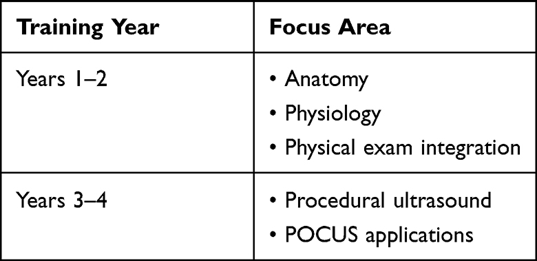 |
Table 4 Minimum Competency Areas and Progression by Year |
The minimum POCUS modules we recommend are basic cardiac views, identification of B-lines, pleural effusions, and pneumothorax on lung examination, Focused Assessment with Sonography in Trauma (FAST) exam, aortic aneurysm screening, gallbladder abdominal exams, IVC assessment, and DVT screening for vascular examination, bladder volume, and hydronephrosis assessment (Table 4). These common skills utilized throughout the medical setting will provide sufficient competency for students as they progress through their medical education and residency programs. To assess competency in these areas, we recommend scheduling hands-on sessions with structured feedback, such as objective structured clinical examinations (OSCEs).50,64–66 These assessment tools objectively evaluate students’ capabilities in acquiring and interpreting ultrasound images, ensuring consistency and reliability.62–64 Moreover, this approach enables institutions to integrate multiple evaluation tools over time, creating opportunities for formative feedback and ongoing learner development.47–50 These competencies should be mastered by graduation, thereby allowing the assessment and evaluation of the curriculum’s longitudinal success in preparing students for clinical practice.
Practical experience is essential for skill acquisition. A comprehensive curriculum, including early hands-on sessions and exposure to various equipment, significantly improves learning outcomes.50,52 We recommend two hours of didactic instruction and three hours of hands-on training per instructional unit. For example, suppose the topic is echocardiography in the emergency room setting. In that case, students should have 2 hours of lectures on the basics, technique, and normal/pathologic findings, followed by a minimum of 3 hours of hands-on practice. These sessions should align with foundational science courses and clinical rotations. Supplemental lectures should cover ultrasound physics, normal anatomy, and common pathological findings.
Peer teaching, an educational tool that leverages students to teach and learn from each other, has emerged as a promising strategy in ultrasound training. Several institutions are exploring their effectiveness in enhancing medical student engagement, hands-on experience, and overall learning outcomes. This method capitalizes on peers’ shared experiences and understanding to facilitate learning, often improving comprehension and retention. It has been widely utilized throughout medical education and is particularly valuable for improving learning outcomes, promoting collaboration, and reinforcing skill acquisition.65 While challenges are associated with implementing peer-to-peer instruction, faculty oversight, constant feedback from students and faculty, and the implementation of standardized training sessions for peer instructors with clear goals and objectives for each session can help bridge these gaps. Furthermore, peer-to-peer training applies the concept “see one, do one, teach one” to reinforce students’ understanding and allow the development of competency and skill reinforcement.63
Conclusion
While there are limitations to the introduction of ultrasound into the early UGME curriculum, there are clear benefits associated with its incorporation for students’ understanding, skill acquisition, and the development of well-versed future physicians capable of utilizing a low-cost, radiation-free tool at the bedside to diagnose pathologies properly. Challenges such as the high cost of equipment, limited faculty expertise, and the lack of standardized guidelines, however, remain barriers to its widespread integration. Importantly, early ultrasound training has also been shown to enhance long-term retention of skills and increase students’ confidence during clinical rotations. There is a need for a standardized curriculum as more residency programs begin to expect students to have undergone UGME ultrasound exposure. Therefore, we recommend a modular longitudinal curriculum that provides students with adequate hands-on experience, accompanied by periodic assessments to determine their understanding and evaluate their ability to meet specific competencies before graduation. Implementing this curriculum alongside anatomy courses and clinical rotations can help alleviate the stress of allocating additional time for ultrasound within the curriculum, while enhancing students’ understanding of anatomy and allowing for real-time identification of relationships between structures and clinical pathologies. Future research should focus on larger, more diverse studies to validate and confirm the conclusions presented in this review, ensuring generalizability across different institutions and student populations.
Acknowledgment
We gratefully acknowledge the Department of Cellular Biology and Anatomy, under the leadership of Dr. Kevin McCarthy, for securing one GE Venue Go R4 Premier Ultrasound machine and 4 VScan Air devices. We also wish to thank Dr. David DeSha and the LSU Health Shreveport (LSUHS) Alumni for securing funding for 20 VScan units from the David DeSha Endowment for Medical Education, under the LSUHS Foundation. This support has been instrumental in overcoming the financial barriers to acquiring these ultrasound devices.
Disclosure
The authors report no conflicts of interest in this work.
References
1. Hoppmann RA, Mladenovic J, Melniker L, et al. International consensus conference recommendations on ultrasound education for undergraduate medical students. Ultrasound J. 2022;14(1):31. doi:10.1186/s13089-022-00279-1
2. European Society of R. Position statement and best practice recommendations on the imaging use of ultrasound from the European society of radiology ultrasound subcommittee. Insights Imaging. 2020;11(1):115. doi:10.1186/s13244-020-00919-x
3. Rao S, van Holsbeeck L, Musial JL, et al. A pilot study of comprehensive ultrasound education at the Wayne State university school of medicine: a pioneer year review. J Ultrasound Med. 2008;27(5):745–749. doi:10.7863/jum.2008.27.5.745
4. Moore CL, Copel JA. Point-of-care ultrasonography. N Engl J Med. 2011;364(8):749–757. doi:10.1056/NEJMra0909487
5. Bahner DP, Hughes D, Royall NA. I-AIM: a novel model for teaching and performing focused sonography. J Ultrasound Med. 2012;31(2):295–300. doi:10.7863/jum.2012.31.2.295
6. Kameda T, Taniguchi N, Konno K, Koibuchi H, Omoto K, Itoh K. Ultrasonography in undergraduate medical education: a comprehensive review and the education program implemented at Jichi medical university. J Med Ultrason. 2022;49(2):217–230. doi:10.1007/s10396-021-01178-z
7. Kondrashova T, Kondrashov P. Integration of ultrasonography into the undergraduate medical curriculum: seven years of experience. Mo Med. 2018;115(1):38–43.
8. Zeitouni F, Matejka C, Boomer M, et al. Integration of point of care ultrasound into undergraduate medical education at Texas tech university health sciences center school of medicine: a 6 year review. BMC Med Educ. 2024;24(1):1476. doi:10.1186/s12909-024-06483-y
9. Bahner DP, Goldman E, Way D, Royall NA, Liu YT. The state of ultrasound education in U.S. medical schools: results of a national survey. Acad Med. 2014;89(12):1681–1686. doi:10.1097/ACM.0000000000000414
10. Johnson CD, Davison L, Graham EC, Sweeney EM. Ultrasound technology as a tool to teach basic concepts of physiology and anatomy in undergraduate and graduate courses: a systematic review. Adv Physiol Educ. 2025;49(1):11–26. doi:10.1152/advan.00199.2023
11. Smith CF, Barfoot S. Implementation of ultrasound in anatomy education. Adv Exp Med Biol. 2021;1317:111–130. doi:10.1007/978-3-030-61125-5_6
12. Chen WT, Kang YN, Wang TC, et al. Does ultrasound education improve anatomy learning? Effects of the parallel ultrasound hands-on (PUSH) undergraduate medicine course. BMC Med Educ. 2022;22(1):207. doi:10.1186/s12909-022-03255-4
13. Rempell JS, Saldana F, DiSalvo D, et al. Pilot point-of-care ultrasound curriculum at harvard medical school: early experience. West J Emerg Med. 2016;17(6):734–740. doi:10.5811/westjem.2016.8.31387
14. Swamy M, Searle RF. Anatomy teaching with portable ultrasound to medical students. BMC Med Educ. 2012;12:99. doi:10.1186/1472-6920-12-99
15. Brown CC, Arrington SD, Olson JF, Finch CA Jr, Nydam RL. Musculoskeletal ultrasound training encourages self-directed learning and increases confidence for clinical and anatomical appreciation of first-year medical students. Anat Sci Educ. 2022;15(3):508–521. doi:10.1002/ase.2145
16. Hoppmann R, Blaivas M, Elbarbary M. Better medical education and health care through point-of-care ultrasound. Acad Med. 2012;87(2):134. doi:10.1097/ACM.0b013e31823f0e8f
17. Hammoudi N, Arangalage D, Boubrit L, et al. Ultrasound-based teaching of cardiac anatomy and physiology to undergraduate medical students. Arch Cardiovasc Dis. 2013;106(10):487–491. doi:10.1016/j.acvd.2013.06.002
18. Akhtar S, Theodoro D, Gaspari R, et al. Resident training in emergency ultrasound: consensus recommendations from the 2008 council of emergency medicine residency directors conference. Acad Emerg Med. 2009;16(Suppl 2):S32–6. doi:10.1111/j.1553-2712.2009.00589.x
19. Hoppmann RA, Rao VV, Poston MB, et al. An integrated ultrasound curriculum (iUSC) for medical students: 4-year experience. Crit Ultrasound J. 2011;3(1):1–12. doi:10.1007/s13089-011-0052-9
20. Trembley L, Radomski M. Use of ultrasound in introducing anatomical pathology to preclinical medical students, in correlation with physical exam curricula. MedEdPORTAL. 2020;16:10950. doi:10.15766/mep_2374-8265.10950
21. Moscova M, Bryce DA, Sindhusake D, Young N. Integration of medical imaging including ultrasound into a new clinical anatomy curriculum. Anat Sci Educ. 2015;8(3):205–220. doi:10.1002/ase.1481
22. Wicke W, Brugger PC, Firbas W. Teaching ultrasound of the abdomen and the pelvic organs in the medicine curriculum in Vienna. Med Educ. 2003;37(5):476. doi:10.1046/j.1365-2923.2003.01502_3.x
23. Cheng WC, Lin XZ, Chen CY. Using modern teaching strategies to teach upper abdominal sonography to medical students. J Chin Med Assoc. 2013;76(7):395–400. doi:10.1016/j.jcma.2013.03.011
24. Heinzow HS, Friederichs H, Lenz P, et al. Teaching ultrasound in a curricular course according to certified EFSUMB standards during undergraduate medical education: a prospective study. BMC Med Educ. 2013;13:84. doi:10.1186/1472-6920-13-84
25. Steinmetz P, Dobrescu O, Oleskevich S, Lewis J. Bedside ultrasound education in Canadian medical schools: a national survey. Can Med Educ J. 2016;7(1):e78–86. doi:10.36834/cmej.36646
26. Weill S, Picard DA, Kim DJ, Woo MY. Recommendations for POCUS curriculum in canadian undergraduate medical education: consensus from the Inaugural Seguin Canadian POCUS Education Conference. POCUS J. 2023;8(1):13–18. doi:10.24908/pocus.v8i1.16153
27. Tandon A, Moneim J, Hector L, et al. A national survey on the use of ultrasound as an educational tool to complement anatomy teaching at UK medical schools. Clin Anat. 2025;38(1):90–96. doi:10.1002/ca.24229
28. McCormick E, Flanagan B, Johnson CD, Sweeney EM. Ultrasound skills teaching in UK medical education: a systematic review. Clin Teach. 2023;20(5):e13635. doi:10.1111/tct.13635
29. Gibson LE, White-Dzuro GA, Lindsay PJ, Berg SM, Bittner EA, Chang MG. Ensuring competency in focused cardiac ultrasound: a systematic review of training programs. J Intensive Care. 2020;8(1):93. doi:10.1186/s40560-020-00503-x
30. Russell FM, Zakeri B, Herbert A, Ferre RM, Leiser A, Wallach PM. The state of point-of-care ultrasound training in undergraduate medical education: findings from a national survey. Acad Med. 2022;97(5):723–727. doi:10.1097/ACM.0000000000004512
31. Russell FM, Herbert A, Peterson D, Wallach PM, Ferre RM. Assessment of medical students’ ability to integrate point-of-care cardiac ultrasound into a case-based simulation after a short intervention. Cureus. 2022;14(7):e27513. doi:10.7759/cureus.27513
32. Pereira ROL, Convissar DL, Montgomery S, et al. Point-of-care lung ultrasound in adults: image acquisition. J Vis Exp. 2023;(193). doi:10.3791/64722
33. Stokke TM, Ruddox V, Sarvari SI, Otterstad JE, Aune E, Edvardsen T. Brief group training of medical students in focused cardiac ultrasound may improve diagnostic accuracy of physical examination. J Am Soc Echocardiogr. 2014;27(11):1238–1246. doi:10.1016/j.echo.2014.08.001
34. Lum M, Sheehy L, Lai J, Tillman D, Damewood S, Schmidt J. Integration of lung point-of-care ultrasound into clinical decision making for medical students in simulated cases. West J Emerg Med. 2020;22(1):124–129. doi:10.5811/westjem.2020.12.48717
35. Situ-LaCasse EH, Amini R, Bain V, et al. Performance of ultrasound-guided peripheral nerve blocks by medical students after one-day training session. Cureus. 2019;11(1):e3911. doi:10.7759/cureus.3911
36. Walsh CD, Iwy M, Eyre AJ, et al. Implementing ultrasound-guided nerve blocks in the emergency department: a low-cost, low-fidelity training approach. AEM Educ Train. 2023;7(5):e10912. doi:10.1002/aet2.10912
37. Tucker RV, Peterson WJ, Mink JT, et al. Defining an ultrasound-guided regional anesthesia curriculum for emergency medicine. AEM Educ Train. 2021;5(3):e10557. doi:10.1002/aet2.10557
38. Goldsmith AJ, Brown J, Duggan NM, et al. Ultrasound-guided nerve blocks in emergency medicine practice: 2022 updates. Am J Emerg Med. 2024;78:112–119. doi:10.1016/j.ajem.2023.12.043
39. Cowan N, Sattar A, Wu Q, et al. Unlocking musculoskeletal anatomy: enhancing second-year medical students’ knowledge recall and self-efficacy with a physician-led ultrasound session. MedSciEduc. 2025. doi:10.1007/s40670-025-02414-8
40. Fatima H, Mahmood F, Mufarrih SH, et al. Preclinical proficiency-based model of ultrasound training. Anesth Analg. 2022;134(1):178–187. doi:10.1213/ANE.0000000000005510
41. Ray JJ, Meizoso JP, Hart V, et al. Effectiveness of a perioperative transthoracic ultrasound training program for students and residents. J Surg Educ. 2017;74(5):805–810. doi:10.1016/j.jsurg.2017.02.005
42. Favot M, Courage C, Mantouffel J, Amponsah D. Ultrasound training in the emergency medicine clerkship. West J Emerg Med. 2015;16(6):938–942. doi:10.5811/westjem.2015.9.27290
43. Draper R, Ellinger D, Trottier M, Armstrong K, Ma W. Ultrasound education in an elective course in the undergraduate medical curriculum improves students’ subjective learning and clinical rotation and residency preparedness. Med Sci Educ. 2023;33(3):737–746. doi:10.1007/s40670-023-01800-4
44. Fu JY, Krause C, Krause R, et al. Integration of point-of-care ultrasound training into undergraduate medical curricula--a perspective from medical students. J Med Educ Curric Dev. 2016;3. doi:10.4137/JMECD.S38240
45. Arichai P, Delaney M, Slamowitz A, et al. Pediatric residency point-of-care ultrasound training needs assessment and educational intervention. Cureus. 2022;14(9):e28696. doi:10.7759/cureus.28696
46. Recker F, Schafer VS, Holzgreve W, Brossart P, Petzinna S. Development and implementation of a comprehensive ultrasound curriculum for medical students: the Bonn internship point-of-care-ultrasound curriculum (BI-POCUS). Front Med Lausanne. 2023;10:1072326. doi:10.3389/fmed.2023.1072326
47. Celebi N, Griewatz J, Malek NP, et al. Development and implementation of a comprehensive ultrasound curriculum for undergraduate medical students - a feasibility study. BMC Med Educ. 2019;19(1):170. doi:10.1186/s12909-019-1611-1
48. Recker F, Neubauer R, Dong Y, et al. Exploring the dynamics of ultrasound training in medical education: current trends, debates, and approaches to didactics and hands-on learning. BMC Med Educ. 2024;24(1):1311. doi:10.1186/s12909-024-06092-9
49. Ginsburg AS, Liddy Z, Khazaneh PT, May S, Pervaiz F. A survey of barriers and facilitators to ultrasound use in low- and middle-income countries. Sci Rep. 2023;13(1):3322. doi:10.1038/s41598-023-30454-w
50. Henwood PC, Mackenzie DC, Rempell JS, et al. A practical guide to self-sustaining point-of-care ultrasound education programs in resource-limited settings. Ann Emerg Med. 2014;64(3):277–285e2. doi:10.1016/j.annemergmed.2014.04.013
51. Mullen A, Kim B, Puglisi J, Mason NL. An economical strategy for early medical education in ultrasound. BMC Med Educ. 2018;18(1):169. doi:10.1186/s12909-018-1275-2
52. Feilchenfeld Z, Dornan T, Whitehead C, Kuper A. Ultrasound in undergraduate medical education: a systematic and critical review. Med Educ. 2017;51(4):366–378. doi:10.1111/medu.13211
53. Staudt D, Lang V, Mooney C. ‘Teaching should always be a little bit interesting for the teachers, too’: preceptor experiences of co-teaching ultrasound in a clinical skills course. Med Teach. 2025;47(6):1015–1021. doi:10.1080/0142159X.2024.2407119
54. Nicholas E, Ly AA, Prince AM, Klawitter PF, Gaskin K, Prince LA. The current status of ultrasound education in United States medical schools. J Ultrasound Med. 2021;40(11):2459–2465. doi:10.1002/jum.15633
55. Britz V, Sterz J, Voss SH, Carstensen P, Germanyuk A, Ruesseler M. Influence of the instructional approach “mastery learning” versus “see one, do one” on acquiring competencies in abdomen sonography: a comparative effectiveness analysis. Ultrasound Med Biol. 2020;46(8):1934–1940. doi:10.1016/j.ultrasmedbio.2020.04.001
56. Iwy M, Steinmetz P, Weerdenburg K, et al. The Canadian medical student ultrasound curriculum: a statement from the Canadian ultrasound consensus for undergraduate medical education group. J Ultrasound Med. 2020;39(7):1279–1287. doi:10.1002/jum.15218
57. Diaz-Gomez JL, Mayo PH, Koenig SJ. Point-of-care ultrasonography. N Engl J Med. 2021;385(17):1593–1602. doi:10.1056/NEJMra1916062
58. Bahner DP, Adkins EJ, Hughes D, Barrie M, Boulger CT, Royall NA. Integrated medical school ultrasound: development of an ultrasound vertical curriculum. Crit Ultrasound J. 2013;5(1):6. doi:10.1186/2036-7902-5-6
59. So S, Patel RM, Orebaugh SL. Ultrasound imaging in medical student education: impact on learning anatomy and physical diagnosis. Anat Sci Educ. 2017;10(2):176–189. doi:10.1002/ase.1630
60. Kelm DJ, Ratelle JT, Azeem N, et al. Longitudinal ultrasound curriculum improves long-term retention among internal medicine residents. J Grad Med Educ. 2015;7(3):454–457. doi:10.4300/JGME-14-00284.1
61. Miller GT, Scerbo MW, Zybak S, et al. Learner improvement from a simulation-enhanced ultrasonography curriculum for first-year medical students. J Ultrasound Med. 2017;36(3):609–619. doi:10.7863/ultra.15.12025
62. Kumar A, Kugler J, Jensen T. Evaluation of trainee competency with point-of-care ultrasonography (POCUS): a conceptual framework and review of existing assessments. J Gen Intern Med. 2019;34(6):1025–1031. doi:10.1007/s11606-019-04945-4
63. Hohne E, Recker F, Dietrich CF, Schafer VS. Assessment methods in medical ultrasound education. Front Med Lausanne. 2022;9:871957. doi:10.3389/fmed.2022.871957
64. Todsen T, Tolsgaard MG, Olsen BH, et al. Reliable and valid assessment of point-of-care ultrasonography. Ann Surg. 2015;261(2):309–315. doi:10.1097/SLA.0000000000000552
65. Ben-Sasson A, Lior Y, Krispel J, et al. Peer-teaching cardiac ultrasound among medical students: a real option. PLoS One. 2019;14(3):e0212794. doi:10.1371/journal.pone.0212794
66. Dickerson J, Paul K, Vila P, Whiticar R. The role for peer-assisted ultrasound teaching in medical school. Clin Teach. 2017;14(3):170–174. doi:10.1111/tct.12541
 © 2025 The Author(s). This work is published and licensed by Dove Medical Press Limited. The
full terms of this license are available at https://www.dovepress.com/terms
and incorporate the Creative Commons Attribution
- Non Commercial (unported, 4.0) License.
By accessing the work you hereby accept the Terms. Non-commercial uses of the work are permitted
without any further permission from Dove Medical Press Limited, provided the work is properly
attributed. For permission for commercial use of this work, please see paragraphs 4.2 and 5 of our Terms.
© 2025 The Author(s). This work is published and licensed by Dove Medical Press Limited. The
full terms of this license are available at https://www.dovepress.com/terms
and incorporate the Creative Commons Attribution
- Non Commercial (unported, 4.0) License.
By accessing the work you hereby accept the Terms. Non-commercial uses of the work are permitted
without any further permission from Dove Medical Press Limited, provided the work is properly
attributed. For permission for commercial use of this work, please see paragraphs 4.2 and 5 of our Terms.