
Back to Journals » Journal of Inflammation Research » Volume 19
Tocilizumab Treatment in Refractory Autoimmune Glial Fibrillary Acidic Protein Astrocytopathy: A Case Report and Literature Review
Authors Lin X  , Zhou S, Zhang Z, Zhu S, Quan Y, Peng J , He H
Received 25 February 2026
Accepted for publication 19 May 2026
Published 19 June 2026 Volume 2026:19 602348
DOI https://doi.org/10.2147/JIR.S602348
Checked for plagiarism Yes
Review by Single anonymous peer review
Peer reviewer comments 2
Editor who approved publication: Dr Adam Bachstetter
Xueqin Lin,1 Shichen Zhou,1 Zhanwei Zhang,1 Saying Zhu,1 Yulin Quan,1 Jing Peng,1,2 Hailan He1,2
1Department of Pediatrics, Xiangya Hospital of Central South University, Changsha, Hunan, People’s Republic of China; 2Clinical Research Center for Children Neurodevelopmental Disabilities of Hunan Province, Changsha, People’s Republic of China
Correspondence: Hailan He, Department of Pediatrics, Xiangya Hospital, Central South University, Changsha, Hunan, 410008, People’s Republic of China, Email [email protected]
Abstract: Although most patients with autoimmune glial fibrillary acidic protein astrocytopathy (GFAP-A) respond to first-line immunotherapy, a subset is refractory to conventional treatments and experiences poor outcomes. This study evaluates the efficacy of tocilizumab in such refractory cases, based on clinical experience and literature review. We retrospectively analyzed a 17-year-old female with GFAP-A who presented with fever, headache, coma, language and emotional disturbances, and motor deficits. Brain magnetic resonance imaging showed T2/FLAIR hyperintensity and swelling in the bilateral basal ganglia and left hippocampus. Anti-glial fibrillary acidic protein (GFAP) antibody was positive in both serum and cerebrospinal fluid. The patient was refractory to high-dose corticosteroids, intravenous immunoglobulin, with only a transient response to rituximab. However, after five infusions of tocilizumab, she demonstrated significant improvement in language, motor function, mood, and remained in stable remission during follow-up, with no severe adverse events. A literature review identified a previously reported case of GFAP-A coexisting with anti-N-methyl-D-aspartate receptor encephalitis that similarly showed only a partial response to first-line immunotherapies before achieving marked clinical improvement after five cycles of tocilizumab. These findings suggest that tocilizumab may represent an effective therapeutic option for refractory GFAP-A, significantly improving cognitive and neuropsychiatric outcomes.
Keywords: GFAP, autoimmune encephalitis, tocilizumab, interleukin 6
Introduction
Autoimmune glial fibrillary acidic protein astrocytopathy (GFAP-A) is a newly recognized immune-mediated inflammatory disorder of the central nervous system (CNS), first described in 2016.1 This condition is mediated by an autoimmune attack targeting astrocytes in the CNS, leading to neuroinflammation and neurological injury.2 GFAP-A can affect multiple regions of the CNS, presenting with a broad and heterogeneous clinical spectrum of symptoms that may include meningitis, encephalitis, myeliti and/or optic neuritis.2,3 Approximately 25% of patients with GFAP-A are found to have a concurrent neoplasm, with ovarian teratoma being the most frequently associated tumor.3–5 The imaging hallmark of GFAP-A is linear perivascular radial enhancement in the cerebral white matter on brain magnetic resonance imaging (MRI). The diagnosis of GFAP-A primarily relies on detecting immunoglobulin type G (IgG) autoantibodies against the alpha (α) GFAP isoform in cerebrospinal fluid (CSF).3–5
Currently, there is no well-established therapeutic regimen for GFAP-A. Based on clinical experience, first-line treatments include high-dose corticosteroids, intravenous immunoglobulin (IVIG) and plasma exchange (PLEX); second-line treatments include rituximab and cyclophosphamide and, less commonly, azathioprine and mycophenylate mofetil. Despite a generally favorable response to high-dose corticosteroids, a subset of GFAP-A patients exhibit poor or inadequate responses to these conventional immunosuppressive treatments, posing a considerable management dilemma.6–8 Relapses occur in approximately 20–30% of patients with GFAP-A, particularly among those presenting with severe phenotypes such as myelitis or with a malignancy.1 The factors underlying refractory and severe presentations of GFAP-A are poorly understood. Emerging evidence has demonstrated that GFAP-A is fundamentally driven by a CD8⁺ T cell-mediated autoimmune attack targeting astrocytes, triggering a pronounced inflammatory cascade.9,10 This process leads to markedly elevated levels of cytokines and chemokines such as tumor necrosis factor alpha (TNF-α), interleukin 6 (IL-6), IL-27, and C-C motif chemokine ligand 20 (CCL20) in the CSF.11,12
Tocilizumab is a humanized monoclonal antibody that targets both soluble and membrane-bound IL-6 receptors. By blocking IL-6 signaling, it inhibits the differentiation and proliferation of B cells, induces apoptosis of plasma cells, and suppresses the generation of inflammatory cytokines and acute-phase proteins.13 Accumulating evidence demonstrates that tocilizumab is effective and well tolerated in both adults and children with various refractory autoimmune encephalitis (AE), including anti-CASPR2 encephalitis, anti-LGI1 encephalitis, and GAD6-related encephalitis.14–16 Beyond its application in AE, tocilizumab has showed therapeutic potential across a wide range of antibody-mediated autoimmune diseases, including neuromyelitis optica spectrum disorder, myasthenia gravis, rheumatoid arthritis.16–18 However, the efficacy of tocilizumab in anti-GFAP encephalitis remains unexplored. Here, we describe a female patient with refractory GFAP-A who demonstrated significant clinical improvement and sustained disease stabilization following tocilizumab treatment after failure of conventional therapies. This case provides further evidence to demonstrate the clinical efficacy of tocilizumab in refractory GFAP-A.
Methods
Patient Data and Informed Consent
This retrospective, clinical observational study was performed under the approval of the ethical committee of Xiangya Hospital, Central South University (approval number 202310892). Informed consent was obtained from the parents of the patient. This case report is presented in accordance with the CARE Guideline.
Detection of Neural Autoantibodies
Serum and CSF samples were tested for autoantibodies associated with AE using a standardized cell-based assay (CBA) with immunofluorescence. The antibody panel included NMDAR, AMPAR1, AMPAR2, LGI1, CASPR2, GABABR, DPPX, GAD65, mGluR5, GlyR, D2R, as well as anti-GFAP-α IgG and anti-GFAP-ε IgG.
Briefly, HEK293 cells were transfected with plasmids encoding each target antigen; for GFAP‑α, the construct also contained a green fluorescent protein (GFP) segment to mark antigen‑expressing cells, as described previously.5,19 The transfected cells were then incubated with patient serum (initial dilution 1:10) or CSF (initial dilution 1:1). Bound human IgG was detected using a fluorescein‑conjugated anti‑human IgG secondary antibody (red emission). A specific positive signal was defined by the precise colocalization of red (antibody) and green (antigen‑expressing cell) fluorescence. For samples that were positive in the initial CBA, serial 10‑fold dilutions were performed to determine the endpoint titer. Furthermore, serum samples that tested positive in the CBA screen were subsequently validated by a tissue‑based assay (TBA) using frozen sections of rat brain and kidney tissue at a fixed dilution of 1:100. All laboratory procedures were conducted at Hangzhou Hongwang Medical Laboratory.
Literature Review
A literature search was performed in PubMed up to January 30, 2026. The search strategy utilized the keywords: “GFAP astrocytopathy” or “autoimmune glial fibrillary acidic protein encephalitis” or “GFAP-A” combined with “tocilizumab” “anti-IL-6”, or “IL-6 inhibitor”. Only one relevant case study was identified and included in this review.20
Case Report
A 17-year-old Chinese girl suffered from dizziness and headache, with the highest temperature of 39°C. Four days later, her condition progressed to include delayed responses, reduced speech, and cognitive impairment. A lumbar puncture performed at local hospital revealed elevated opening pressure (210mmH2O), elevated protein level (0.9 g/L), and monocytic pleocytosis (230×106/L, 87% mononuclear cells). CSF culture was negative. Neither a brain MRI nor a fundus examination was performed at the local hospital prior to the first lumbar puncture. Despite empiric antibacterial and antiviral treatment, her fever persisted at 38°C and her consciousness deteriorated.
On day 7, she was transferred to our hospital for further examination and treatment. Neurological examination revealed slightly increased muscle tone in the upper limbs, mild weakness in the lower limbs (grossly graded at 3/5), and absent deep tendon reflexes. No pathological signs were observed. Her modified Rankin Scale (mRS) was 5. Serum IL-6 was elevated at 125.0 pg/mL (normal range<5pg/mL), while CSF IL-6 was within normal limits. Brain computed tomography (CT) showed a low-density lesion in the left hippocampus-amygdala area. Brain MRI revealed swelling and T2/fluid-attenuated inversion recovery (FLAIR) signal hyperintensity within the bilateral basal ganglia and left hippocampus (Figure 1A and B); however, no contrast enhancement was observed in the affected areas on T1-weighted imaging. Electroencephalogram (EEG) demonstrated diffuse low-voltage activity (Figure 2A and B). Lower respiratory tract pathogen culture was positive for Haemophilus influenzae. Chest computed CT revealed infectious infiltrates in the posterior segment of the right upper lobe and bilateral lower lobes of the lungs. One day later, she developed hypoxemia requiring intubation, and was successfully extubated on day 12. During this period, she sequentially received antimicrobials, antivirals, dexamethasone (5 mg/day,5 days), and IVIG (400 mg/kg/day,5 days), followed by methylprednisolone (60 mg/day, 5 days). Antibodies against NMDAR1a, AMPAR1, AMPAR2, LGI1, CASPR2, GABABR, DPPX, IgLON5, GlyR1, GABAARα1, GABAARβ3, γ2, mGluR5, D2R, Neurexin3α, KCNA4, NMDAR2a, and NMDAR2b were all negative. Importantly, anti-GFAP antibody was positive in the serum (1:10) and CSF (1:1) via indirect immunofluorescence on TBA and CBA (Figure 3A–C), and she was therefore diagnosed as GFAP-A. Tumor and systemic autoimmune disease screenings were also negative. Enhanced CT scans of the chest, abdomen, and pelvis, together with breast ultrasonography, excluded neoplasms. On day 20, the patient received intravenous methylprednisolone (240 mg/day for 2 days, then 500 mg/day for 4 days), which yielded no clinical improvement; her language and emotional disturbances persisted, and she remained unable to sit, stand, or walk independently. Given the lack of response to steroids and the family’s refusal of PLEX, second-line therapy with rituximab (750 mg/m2) was initiated on day 32. She showed a favorable initial response to rituximab treatment. Within two weeks of the first dose, she regained comprehensible speech, independent ambulation and emotional stability corresponding to a mRS score of 2, and was subsequently discharged. However, her condition deteriorated on week post-discharge, manifesting as emotional lability, choreoathetosis, memory impairment with her mRS score worsening to 4. A second cycle of rituximab combined with IVIG (2 g/kg for 5 days) was administered on day 49, followed by a third dose on day 55. Despite this therapeutic escalation, her condition showed no improvement.
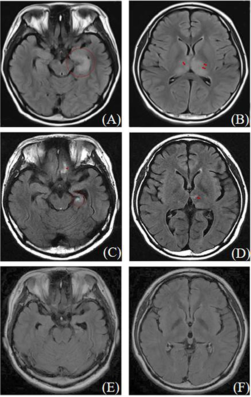 |
Figure 1 Longitudinal brain MRI changes in a patient with GFAP-A treated with tocilizumab. (A and B) Pre-treatment T2 Flair imaging shows bilateral thalamic swelling (red arrow) and abnormal signal lesions in the left hippocampus (red circle). (C and D) After the second tocilizumab treatment, T2 Flair imaging reveals reduced abnormal signal lesions in the bilateral thalamus (red arrow) and left hippocampus (red circle), with abnormal signals in the orbital gyrus (red triangle). (E and F) After the fifth tocilizumab treatment, T2 Flair imaging demonstrates near disappearance of bilateral thalamic abnormal signals, left hippocampal atrophy, and reduced orbital gyrus abnormalities. |
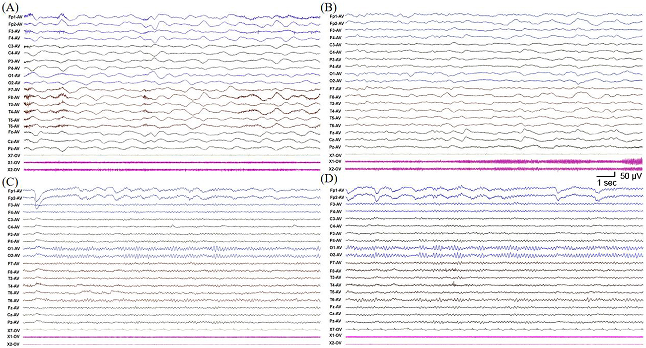 |
Figure 2 Electroencephalography (EEG) in a patient with GFAP-A treated with tocilizumab. (A and B) Diffuse 1–3.5 Hz δ waves with intermittent mixing of continuous 5–7 Hz θ wave activity, and occasional widespread voltage attenuation lasting about 1 s. (C and D) Bilateral occipital 10–11 Hz α rhythm with good regulation and modulation. |
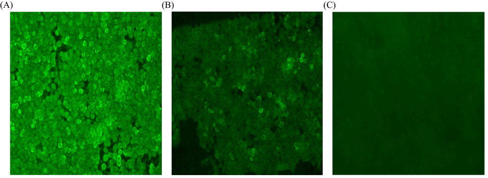 |
Figure 3 Immunofluorescence in serum and CSF in patients with GFAP-A treated with tocilizumab assessed by cell-based assay (CBA). Positive reaction with transfected HEK 293 cells expressing GFAP-α after incubation with the patient’s serum (A) (titer: 1:10) and with CSF (B) (titer: 1:1). (C) Negative control. |
Given the suboptimal response to prior therapies, tocilizumab (8 mg/kg monthly) was initiated on day 57. Pre-treatment IL-6 levels were within normal range (CSF 2.79 pg/mL, serum 4.69 pg/mL). The patient received five cycles of tocilizumab treatment in total. Tocilizumab prompted rapid symptomatic, radiological, and EEG improvement. Following the second dose, brain MRI revealed a reduction of abnormalities in the thalamus and hippocampus (Figure 1C and D); and her EEG normalized. Transient elevations in serum IL-6 were observed after the third and fourth infusions (8.77 pg/mL and 28.4 pg/mL, respectively). No other adverse events, such as infections, gastrointestinal symptoms, or hepatic dysfunction, were attributable to the administration of tocilizumab. After completing five cycles, follow-up MRI demonstrated near-complete resolution of thalamic signals and improvement in the left hippocampus (Figure 1E and F), and her EEG remained normal (Figure 2C and D). Followed up one year after discharge, she was asymptomatic with normal cognition, mood and behavior (MRS 0).
Literature Review
To date, one adult case with refractory GFAP-A treated with tocilizumab has been reported.21 Including the case in this study, a total of two cases were summarized. Anti-GFAP autoantibodies were elevated in the CSF or serum for two cases. Both cases were females and presented with fever, headache, speech reduction, cognitive impairment, and psychiatric disorders. The adult case also exhibited seizures and Graves’ hyperthyroidism. Brain MRI was performed on both patients, with our case demonstrating abnormalities and the other showing normal findings. In addition to anti-GFAP antibodies, the adult patient tested positive for anti-N-methyl-D-aspartate receptor (NMDAR) antibodies. Prior to tocilizumab treatment, both patients had been treated with methylprednisolone, IVIG, and rituximab. Both patients responded well to tocilizumab. After five courses of tocilizumab treatment, both cases demonstrated significant neurological improvement. Despite experiencing a transient rise in IL-6 levels during tocilizumab treatment, our patient—in line with the previously reported case—did not develop any severe treatment-related adverse events.
Discussion
Refractory AE is a challenging disease with significant morbidity and mortality.22,23 Previous studies have demonstrated the efficacy of tocilizumab in several forms of refractory AE, including anti-NMDAR, anti-LGI1, CASPR2, and GAD65-associated encephalitis.24–28 We reported a case of refractory GFAP-A in a patient who was unresponsive to first-line immunotherapies (high-dose corticosteroids and IVIG) and exhibited only a transient benefit from B-cell–depleting therapy with rituximab. Notably, the delay in initiating IV steroids until day 20 is a potential factor that may have contributed to the refractory course. Similarly, a previously reported case of NMDAR encephalitis coexisting with GFAP-A also showed merely a partial response to the same line of conventional immunotherapies.21 Notably, both patients achieved marked clinical improvement after five cycles of tocilizumab, and neither experienced significant side effects from the treatment. This consistent outcome highlights tocilizumab as a safe and effective option for patients with refractory GFAP-A who do not respond to conventional immunotherapies.
Although the precise pathogenic mechanisms of GFAP-A are not yet fully elucidated, emerging evidence indicates a central role of neuroinflammation in the disease process. Histopathological studies in GFAP-A patients have revealed perivascular and parenchymal infiltrates of CD20+, CD3+, and CD8+ lymphocytes, macrophages, granulomatous inflammation, and cytotoxic T-cell response.9,10,29,30 Immunohistochemical analyses further highlight prominent perivascular B cells, diffuse T-cell infiltration, and abundant plasma cells, monocytes, and macrophages in brain tissues.19,31 Importantly, elevated IL-6 levels have been documented in the CSF of patients with GFAP-A compared to controls.11,12 The marked therapeutic response to tocilizumab observed in both our patient and the reported case implies that IL-6 mediated inflammation may represent a critical pathogenic mechanism in certain refractory GFAP-A cases. This mechanism appears to operate independently of, or persist despite, B-cell depletion. Within the CNS, IL-6 functions as a key pro-inflammatory cytokine, promoting B-cell differentiation, plasma cell maturation, T follicular helper cell generation, blood–brain barrier disruption, and T-cell–mediated inflammation. Therefore, IL-6 blockade represents a rational therapeutic strategy for mitigating autoimmune activity in such contexts.13,14
Although corticosteroids are generally effective in GFAP-A, a subset of patients exhibits glucocorticoid resistance and relapsing disease, necessitating more targeted biologic therapies.32–34 While rituximab can help prevent relapses, its efficacy may be limited by incomplete B-cell depletion and its inability to target long-lived plasma cells.35,36 This treatment resistance may occur because the inflammatory storm is driven not only by antibodies but also by a “cytokine storm” mediated by cytokines such as IL-6. Tocilizumab, a potent cytokine inhibitor, can disrupt this cycle, which may explain our patient’s significant response to tocilizumab after multiple ineffective treatments. Notably, the serum and CSF IL-6 levels in our patient were within normal range prior to tocilizumab initiation, indicating that baseline IL-6 levels are not predictive of subsequent outcomes. A transient rise in IL-6 levels was observed in our patient following tocilizumab infusions, consistent with reports of similar, self-limited increases in other treated patients.37,38 This transient rise is a consequence of IL-6 receptor blockade by tocilizumab, which impedes ligand clearance while simultaneously inhibiting downstream inflammatory signaling, ultimately leading to normalized IL-6 levels.39,40 However, persistent elevation of IL-6 after tocilizumab administration has been associated with infections in some reports, highlighting the potential value of dynamically monitoring IL-6 levels to track disease progression and the response to treatment in AE.
In summary, tocilizumab represents a promising treatment option for refractory autoimmune GFAP-A. To date, its efficacy has been supported only by retrospective case reports, and no prospective or controlled studies are available. Future research should prioritize multicenter prospective studies or randomized controlled trials to validate its clinical benefits, evaluate its role in relapse prevention, and optimize treatment protocols.
Data Sharing Statement
The anonymized data are available from the corresponding author upon reasonable request.
Ethics Approval
This study was performed in line with the principles of the Declaration of Helsinki, and was approved by the Institutional Ethics Committee of Xiangya Hospital Central South University. Institutional Ethics Committee of Xiangya Hospital Central South University approved publication of this study when informed consent from legal guardians was obtained.
Consent for Publication
Written informed consent was obtained from the patient and her parents for publication of this case report and any accompanying images.
Acknowledgments
The authors would like to thank the patient and her family for permission to publish his data and thank Hangzhou Hongwang Clinical Laboratory for supporting the detection of the anti-GFAP antibody.
Author Contributions
Xueqin Lin: Conceptualization, Methodology, Writing – original draft, Visualization; Hailan He: Conceptualization, Methodology, Writing – review & editing; Shichen Zhou: Investigation, Writing – review & editing; Yulin Quan: Investigation, Writing – review & editing; Jing Peng: Investigation, Writing – review & editing; Saying Zhu: Visualization, Writing – review & editing; Zhanwei Zhang: Visualization, Writing – review & editing. All authors made a significant contribution to the work reported, whether that is in the conception, study design, execution, acquisition of data, analysis and interpretation, or in all these areas. All authors have drafted or substantially revised the article, have agreed on the journal to which the article will be submitted, have reviewed and agreed on all versions of the article, and agree to take responsibility and be accountable for the contents of the article.
Funding
This work was supported by the National Natural Science Foundation of China (82201316 to HH) and the Natural Science Foundation of Hunan Province (2026JJ50296 to HH).
Disclosure
The authors declare that the research was conducted in the absence of any commercial or financial relationships that could be construed as a potential conflict of interest.
References
1. Fang B, McKeon A, Hinson SR, et al. Autoimmune glial fibrillary acidic protein astrocytopathy: a novel meningoencephalomyelitis. JAMA Neurol. 2016;73(11):1297–9. doi:10.1001/jamaneurol.2016.2549
2. Kunchok A, Zekeridou A, McKeon A. Autoimmune glial fibrillary acidic protein astrocytopathy. Curr Opin Neurol. 2019;32(3):452–458. doi:10.1097/wco.0000000000000676
3. Flanagan EP, Hinson SR, Lennon VA, et al. Glial fibrillary acidic protein immunoglobulin G as biomarker of autoimmune astrocytopathy: analysis of 102 patients. Ann Neurol. 2017;81(2):298–309. doi:10.1002/ana.24881
4. Dubey D, Hinson SR, Jolliffe EA, et al. Autoimmune GFAP astrocytopathy: prospective evaluation of 90 patients in 1 year. J Neuroimmunol. 2018;321:321157. doi:10.1016/j.jneuroim.2018.04.016
5. Gravier-Dumonceau A, Ameli R, Rogemond V, et al. Glial fibrillary acidic protein autoimmunity: a french cohort study. Neurology. 2022;98(6):e653–e668. doi:10.1212/wnl.0000000000013087
6. Li T, Li X, Hong S, et al. Clinical characteristics of autoimmune glial fibrillary acidic protein (GFAP) astrocytopathy in children: a case series of 16 patients. J Neuroimmunol. 2023;382:382578176. doi:10.1016/j.jneuroim.2023.578176
7. Long Y, Liang J, Xu H, et al. Autoimmune glial fibrillary acidic protein astrocytopathy in Chinese patients: a retrospective study. Eur J Neurol. 2018;25(3):477–483. doi:10.1111/ene.13531
8. Ahmed S, K VCC, Kannoth S, et al. Autoimmune GFAP astrocytopathy-beyond the Known Horizon, India’s first multifaceted institutional experience. Ann Neurosci. 2025;32(4):248–258. doi:10.1177/09727531241230213
9. Yuan Z, Li H, Huang L, et al. CD8(+) T-cell predominance in autoimmune glial fibrillary acidic protein astrocytopathy. Eur J Neurol. 2021;28(6):2121–2125. doi:10.1111/ene.14778
10. Sasaki K, Bean A, Shah S, et al. Relapsing-remitting central nervous system autoimmunity mediated by GFAP-specific CD8 T cells. J Immunol. 2014;192(7):3029–3042. doi:10.4049/jimmunol.1302911
11. Kimura A, Takemura M, Yamamoto Y, et al. Cytokines and biological markers in autoimmune GFAP astrocytopathy: the potential role for pathogenesis and therapeutic implications. J Neuroimmunol. 2019;334:334576999. doi:10.1016/j.jneuroim.2019.576999
12. Luo Y, Yan W, Zhou Z, et al. Elevated levels of NLRP3 in cerebrospinal fluid of patients with autoimmune GFAP astrocytopathy. Front Neurol. 2019;10:1019. doi:10.3389/fneur.2019.01019
13. Venkiteshwaran A. Tocilizumab. MAbs. 2009;1(5):432–438. doi:10.4161/mabs.1.5.9497
14. Lee WJ, Lee ST, Moon J, et al. Tocilizumab in autoimmune encephalitis refractory to rituximab: an institutional cohort study. Neurotherapeutics. 2016;13(4):824–832. doi:10.1007/s13311-016-0442-6
15. Randell RL, Adams AV, Van Mater H. Tocilizumab in refractory autoimmune encephalitis: a series of pediatric cases. Pediatr Neurol. 2018;86:66–68. doi:10.1016/j.pediatrneurol.2018.07.016
16. Ruan Z, Tang Y, Gao T, et al. Efficacy and safety of tocilizumab in patients with refractory generalized myasthenia gravis. CNS Neurosci Ther. 2024;30(6):e14793. doi:10.1111/cns.14793
17. Xu X, Wang Y, Sun M, et al. Efficacy and safety of tocilizumab treatment in refractory MOG-IgG related optic neuritis. Ther Adv Neurol Disord. 2024;17:17562864241306685. doi:10.1177/17562864241306685
18. Scott LJ. Tocilizumab: a review in rheumatoid arthritis. Drugs. 2017;77(17):1865–1879. doi:10.1007/s40265-017-0829-7
19. Iorio R, Damato V, Evoli A, et al. Clinical and immunological characteristics of the spectrum of GFAP autoimmunity: a case series of 22 patients. J Neurol Neurosurg Psychiatry. 2018;89(2):138–146. doi:10.1136/jnnp-2017-316583
20. Riley DS, Barber MS, Kienle GS, et al. CARE guidelines for case reports: explanation and elaboration document. J Clin Epidemiol. 2017;89:218–235. doi:10.1016/j.jclinepi.2017.04.026
21. Zhang YX, Cai MT, Li EC, et al. Efficacy of tocilizumab in anti- N -methyl- -asparate receptor encephalitis with Graves’ hyperthyroidism and positive anti-glial fibrillary acidic protein antibodies. CNS Neurosci Ther. 2022;28(11):1898–1900. doi:10.1111/cns.13949
22. Dinoto A, Ferrari S, Mariotto S. Treatment options in refractory autoimmune encephalitis. CNS Drugs. 2022;36(9):919–931. doi:10.1007/s40263-022-00943-z
23. Cheng L, Jia B, Wang C, et al. Immunotherapy for autoimmune encephalitis. Cell Death Discov. 2025;11(1):207. doi:10.1038/s41420-025-02459-z
24. Benucci M, Tramacere L, Infantino M, et al. Efficacy of tocilizumab in limbic encephalitis with anti-CASPR2 antibodies. Case Rep Neurol Med. 2020;2020:5697670. doi:10.1155/2020/5697670
25. Jaafar F, Haddad L, Koleilat N, et al. Super refractory status epilepticus secondary to anti-GAD antibody encephalitis successfully treated with aggressive immunotherapy. Epilepsy Behav Rep. 2020;14(100396). doi:10.1016/j.ebr.2020.100396
26. Jang S, Kim SY, Kim WJ, et al. A case of pediatric anti-leucine-rich glioma inactivated 1 encephalitis with faciobrachial dystonic seizure. Brain Dev. 2023;45(6):348–353. doi:10.1016/j.braindev.2023.02.003
27. Krogias C, Hoepner R, Müller A, et al. Successful treatment of anti-Caspr2 syndrome by interleukin 6 receptor blockade through tocilizumab. JAMA Neurol. 2013;70(8):1056–1059. doi:10.1001/jamaneurol.2013.143
28. Nosadini M, Thomas T, Eyre M, et al. International consensus recommendations for the treatment of pediatric NMDAR antibody encephalitis. Neurol Neuroimmunol Neuroinflamm. 2021;8(5). doi:10.1212/nxi.0000000000001052
29. Guo Y, Endmayr V, Zekeridou A, et al. New insights into neuropathology and pathogenesis of autoimmune glial fibrillary acidic protein meningoencephalomyelitis. Acta Neuropathol. 2024;147(1):31. doi:10.1007/s00401-023-02678-7
30. Gklinos P, Athanasopoulos F, Giatrakou V, et al. Unveiling GFAP astrocytopathy: insights from case studies and a comprehensive review of the literature. Antibodies. 2024;13(4):79. doi:10.3390/antib13040079
31. Yang X, Liang J, Huang Q, et al. Treatment of autoimmune glial fibrillary acidic protein astrocytopathy: follow-up in 7 cases. Neuroimmunomodulation. 2017;24(2):113–119. doi:10.1159/000479948
32. Allen A, Gulhar S, Haidari R, et al. Autoimmune glial fibrillary acidic protein astrocytopathy resulting in treatment-refractory flaccid paralysis. Mult Scler Relat Disord. 2020;39:101924. doi:10.1016/j.msard.2019.101924
33. Cao S, Du J, Pan S, et al. Case Report: ofatumumab treatment in a patient with rituximab-intolerant refractory autoimmune GFAP astrocytopathy. Front Immunol. 2023;14:1164181. doi:10.3389/fimmu.2023.1164181
34. Morishima Y, Hata T, Nakajima S, et al. Case report: atypical case of autoimmune glial fibrillary acidic protein astrocytopathy following COVID-19 vaccination refractory to immunosuppressive treatments. Front Immunol. 2024;15:1361685. doi:10.3389/fimmu.2024.1361685
35. Dale RC, Brilot F, Duffy LV, et al. Utility and safety of rituximab in pediatric autoimmune and inflammatory CNS disease. Neurology. 2014;83(2):142–150. doi:10.1212/wnl.0000000000000570
36. Whittam DH, Tallantyre EC, Jolles S, et al. Rituximab in neurological disease: principles, evidence and practice. Pract Neurol. 2019;19(1):5–20. doi:10.1136/practneurol-2018-001899
37. Jun JS, Lee ST, Kim R, et al. Tocilizumab treatment for new onset refractory status epilepticus. Ann Neurol. 2018;84(6):940–945. doi:10.1002/ana.25374
38. Jaiswal A, Patel S, Khare S, et al. Tocilizumab in a child with acute leukoencephalopathy with restricted diffusion. Ann Indian Acad Neurol. 2023;26(1):95–96. doi:10.4103/aian.aian_795_22
39. Garbers C, Thaiss W, Jones GW, et al. Inhibition of classic signaling is a novel function of soluble glycoprotein 130 (sgp130), which is controlled by the ratio of interleukin 6 and soluble interleukin 6 receptor. J Biol Chem. 2011;286(50):42959–42970. doi:10.1074/jbc.M111.295758
40. Campbell IL, Erta M, Lim SL, et al. Trans-signaling is a dominant mechanism for the pathogenic actions of interleukin-6 in the brain. J Neurosci. 2014;34(7):2503–2513. doi:10.1523/jneurosci.2830-13.2014
 © 2026 The Author(s). This work is published and licensed by Dove Medical Press Limited. The
full terms of this license are available at https://www.dovepress.com/terms
and incorporate the Creative Commons Attribution
- Non Commercial (unported, 4.0) License.
By accessing the work you hereby accept the Terms. Non-commercial uses of the work are permitted
without any further permission from Dove Medical Press Limited, provided the work is properly
attributed. For permission for commercial use of this work, please see paragraphs 4.2 and 5 of our Terms.
© 2026 The Author(s). This work is published and licensed by Dove Medical Press Limited. The
full terms of this license are available at https://www.dovepress.com/terms
and incorporate the Creative Commons Attribution
- Non Commercial (unported, 4.0) License.
By accessing the work you hereby accept the Terms. Non-commercial uses of the work are permitted
without any further permission from Dove Medical Press Limited, provided the work is properly
attributed. For permission for commercial use of this work, please see paragraphs 4.2 and 5 of our Terms.