Back to Journals » Advances in Medical Education and Practice » Volume 16
To Evaluate Interdisciplinary Integration in Medical Education Through a Pulmonary Fibrosis Case Study
Received 20 March 2025
Accepted for publication 28 July 2025
Published 24 August 2025 Volume 2025:16 Pages 1529—1536
DOI https://doi.org/10.2147/AMEP.S529501
Checked for plagiarism Yes
Review by Single anonymous peer review
Peer reviewer comments 5
Editor who approved publication: Dr Md Anwarul Azim Majumder
Xiaorun Zhai, Yunjuan Nie
Department of Basic Medicine, Wuxi School of Medicine, Jiangnan University, Wuxi, Jiangsu, 214122, People’s Republic of China
Correspondence: Yunjuan Nie, Department of Basic Medicine, Wuxi School of Medicine, Jiangnan University, Wuxi, Jiangsu, 214122, People’s Republic of China, Tel +86-13812078105, Email [email protected]
Introduction: This article examines the teaching method of experimental design in the context of Chinese universities, specifically focusing on the exploration of the disease development process. Cultivating innovative and research-oriented medical skills is a crucial objective within medical education, and the design of high-quality experimental courses is a significant aspect of optimizing and reforming medical education.
Methods: Innovative experimental teaching design serves as a novel approach to integrating scientific research into undergraduate instruction. Centered on the exploration of the disease development process and combining the characteristics of clinical diseases, this method integrates experimental techniques from diverse disciplines such as anatomy, pathology, and molecular biology. It achieves a seamless fusion of theoretical knowledge and experimental operations. In this teaching initiative, students are fully immersed in the entire journey, from establishing disease-model mice to collecting and identifying samples at various time points during disease progression. Through hands-on engagement, students not only enhance their experimental skills but also gain a vivid and profound understanding of the dynamic evolution of diseases.
Results: From the perspective of experimental exploration, students’ theoretical understanding is reinforced, and they grasp the occurrence and progression of diseases. This approach provides technical support for scientific research and offers a new line of thinking for the treatment of clinical diseases.
Conclusion: Experimental teaching focused on exploring the disease development process can foster students’ scientific thinking, enhance their practical capabilities, and cultivate research-oriented medical professionals while they learn experimental techniques.
Keywords: disease progression, experimental design, disciplinary integration, experimental teaching
Introduction
The emergence of the new medical construction concept has established novel benchmarks and requisites for the cultivation of medical professionals. The objective of medical education is to foster innovative, research-oriented, and well-rounded talents.1,2 Experimental teaching constitutes a vital part of medical education. In China, traditional medical experimental courses predominantly consist of confirmatory experiments. For instance, anatomy experimental courses mainly emphasize anatomical observation, pathology experimental courses center on the examination of tissue and cell morphology, and molecular biology experimental courses focus on molecular experiments. Additionally, traditional medical laboratory courses are characterized by a lack of innovation, simplistic experimental procedures, feeble connections between experiments, and a tendency to be detached from scientific research and clinical practice. Consequently, the creation of innovative experimental courses that are closely intertwined with scientific research and clinical practice represents a crucial aspect in advancing experimental teaching reform.3,4
The respiratory system ranks among the eight major systems in charge of gas exchange within the human body. The lungs, being a crucial element of the respiratory system, possess a complex structure and fulfill diverse functions, engaging in numerous physiological processes. There exists a wide variety of lung diseases, with idiopathic pulmonary fibrosis standing as a typical chronic progressive lung ailment. In recent years, the incidence rate of pulmonary fibrosis has been on a yearly upward trend. This disease has a protracted course, severe symptoms, and a high mortality rate, and currently, there is no efficacious cure in clinical practice,5–7 which severely impacts the living standards of patients. The prevention and treatment of pulmonary fibrosis currently represent a focal point in scientific research. Previous investigations have indicated that in the early phases of pulmonary fibrosis, there will be a substantial amount of inflammatory infiltration, and suppressing early inflammation can effectively restrain pulmonary fibrosis.8–10 Nevertheless, due to the faint and easily overlooked early symptoms of pulmonary fibrosis, the majority of patients are liable to miss the prime treatment window. Thus, comprehending the development process of pulmonary fibrosis forms the bedrock for exploring novel treatment modalities.
Bleomycin (BLM), being the preferred drug for inducing models of pulmonary fibrosis disease, triggers a pathological process of pulmonary fibrosis in mice that closely resembles that in humans and can effectively mirror the disease development process.11,12 The objective of this experiment is to probe into the development process of idiopathic pulmonary fibrosis and is formulated as a practical, interdisciplinary medical integration experiment. It reinforces theoretical knowledge while linking with clinical characteristics, which not only augments students’ interest in learning but also boosts their practical innovation capabilities.
The One Health approach has emerged as a pivotal strategy for addressing emerging infectious diseases and zoonoses, with significant implications for chronic conditions such as pulmonary fibrosis. However, surveys reveal that a substantial proportion of clinicians remain unfamiliar with One Health principles,13 underscoring a critical gap in medical education. Our experimental design employing bleomycin-induced pulmonary fibrosis in rodent models serves dual purposes: as a research platform investigating interspecies disease mechanisms, and as an educational template for cultivating comprehensive health perspectives in preclinical training.
Experimental Design
Based on the above background, this experiment focuses on students and transforms traditional lecture style experimental teaching into exploratory experimental teaching centered on student operation and supplemented by teachers. Integrating multiple disciplines, students integrate knowledge and techniques from various disciplines while conducting experimental operations. By observing and testing the relevant indicators of bleomycin treatment in mice at different time periods, students can cultivate scientific thinking and improve their scientific research and practical abilities. The experimental mode is shown in Table 1, and the specific implementation method is shown in Table 2.
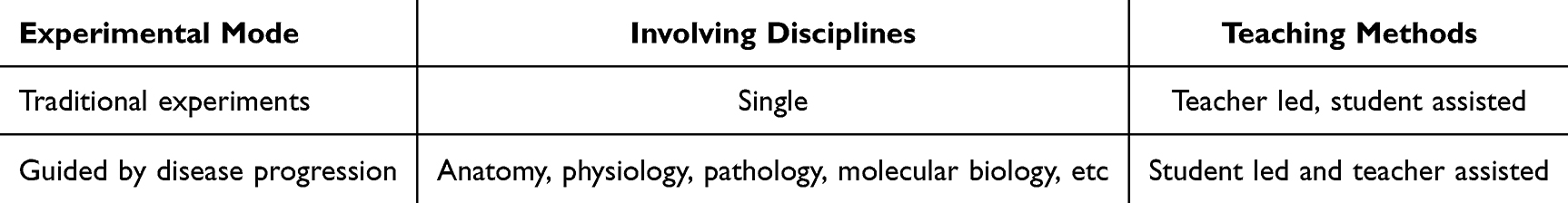 |
Table 1 The Experimental Mode |
 |
Table 2 The Specific Implementation Method |
Experimental Animals
C57BL/6 mice were purchased from Shanghai Slack Laboratory Animal Co., Ltd. The mice were placed in a specific pathogen free environment at the Experimental Animal Center of University. All animal experiments in this study followed the requirements of the “Guidelines for the Care and Use of Laboratory Animals at Jiangnan University” and were approved by the Ethics Committee of Jiangnan University (Approval Number: 20211130 m1720615 [501]).
Experimental Materials
Bleomycin (BIOTANG, USA), hematoxylin eosin staining kit (Nanjing Jiancheng Technology, China), xylene analytical purity (National Pharmaceutical Reagent, China), paraffin sectioning (National Pharmaceutical Reagent, China), mouse intubation and retention needle (Braun Melsingen, Germany), semi-automatic paraffin sectioning machine (Leica, Germany), Nikon microscope (Nikon, Japan), Trizol reagent (Novozymes, China), fluorescence quantitative PCR instrument (Roche, USA).
Experimental Steps and Methods
(1) Establishment of mouse pulmonary fibrosis model. Take out BLM from the refrigerator 30 minutes in advance, reheat at room temperature, invert several times, and mix the reagent well. Take 6–8 week old male wild-type mice and inject pentobarbital sodium (50 mg/kg) intraperitoneally for anesthesia. After deep anesthesia (ensuring smooth breathing, no toe pinching reaction, and no corneal reflex). Use a pipette to aspirate 50 ul of BLM, administer it through non-invasive tracheal intubation, and use physiological saline as a control, this part take 15 minutes.14
(2) Mouse lung tissue was collected and sliced. Anesthetize the mouse abdominal cavity, disinfect the skin with 75% alcohol, and cut open the chest cavity to expose the mouse’s heart and lung tissue. Inject 15 mL of cold PBS into the right atrium, cut the inferior vena cava, and perfuse until the mouse lung tissue turns white. Ligature the junction between the heart and left lung. Inject 4% paraformaldehyde through the trachea, ligate the trachea after infusion into the left lung, cut off the left lung and fix it with 4% paraformaldehyde. After 24 hours of fixation, the lung tissue was dehydrated in ethanol and xylene at different gradients. Embed lung tissue and perform paraffin sectioning. The tissue processing protocol includes: 15-min sampling, automated dehydration, and 30-min sectioning.15
(3) Stained with hematoxylin and eosin. Soak the cut white slices in xylene and ethanol with different gradients to dewax them into water, and stain them in hematoxylin dye solution for 5–10 minutes. Differentiate the color with 1% hydrochloric acid ethanol solution, then turn back to blue with 1% ammonia solution, immerse them in eosin solution for 15 seconds to 1 minute, rinse excess dye with tap water, and then dehydrate the sliced gradient ethanol. Seal the slices with transparent xylene and observe and take photos under a microscope. The complete staining procedure requires 40 minutes to complete.
(4) Fluorescence quantitative PCR. Euthanize mice, disinfect the skin with 75% alcohol, and cut open the chest cavity to expose the heart and lung tissues of the mice. Inject 15 mL of cold PBS into the right atrium, cut the inferior vena cava, and perfuse until the mouse lung tissue turns white. Cut off the lung tissue and place it in Trizol for homogenate extraction of total RNA, followed by reverse transcription. Take a volume of 2 ng of reverse transcribed cDNA according to its concentration as a model. Each group consists of three repeated holes, and outliers are excluded. The entire procedure from RNA extraction to data analysis takes approximately 5 hours (2 hours for total RNA isolation, 2 hours for reverse transcription and quantitative PCR, and 1 hour for result analysis and calculation).
The sequence of gene primers used:
TNF-α (forward, 5′-TTCTCAT TCCTGCTTGTGG-3′,
reverse, 5′-ACTTGGTGGTTTG CTACG-3′);
IL-1β (forward, 5′-CCAGCTTCAAATCTC ACAGCAG-3′;
reverse, 5′-CTTCTTTGGGTATTGCTT GGGATC-3′);
a-SMA (forward 5′-GACGCTGAAGTATCCGATAGAACAC G-3′,
reverse 5′-CACCATCTCCAGAGTCCAGCACA AT-3′);
fibronectin (forward 5′-TCTGGGAAATGGAA AAGGGGAATGG-3′,
reverse 5′-CACTGAAGCAGGT TTCCTCGGTTGT-3′);
and GAPDH (forward 5′-TGCGACTTCAAC AGCAACTC-3′,
reverse 5′-CTTGCTCAGTGTCCTTG CTG-3′)
Data Analysis and Statistics
The experimental data was processed using Graphpad Prism 6.0 software, and the comparison of inter group data was conducted using one-way ANOVA or two-way ANOVA with post hoc Student’s t-test.
Results Detection and Analysis
Observation of Respiratory Function in Mice
The mice in the saline control group had stable breathing and normal chest movements, while the mice in the BLM 7 days group had no significant changes in breathing compared to the saline group. The mice in the 14 days group of BLM began to experience occasional shortness of breath, while the mice in the 21 days group of BLM had shortness of breath, shallow and fast frequency, and greatly reduced chest fluctuations. Starting from day 14, the pulmonary fibrosis process in mice began to worsen.
Comparison of Lung Tissue Section Results of Mice at Different Time Periods
By observing the staining of lung tissue slices from mice at different time periods, it was found that compared with the mice given physiological saline, the lung tissue structure of mice in the 7 days BLM group was slightly damaged, and a large number of inflammatory cells such as neutrophils and lymphocytes appeared. The lung tissue structure of mice in the 14 days group treated with BLM worsened with some collagen deposition, while the normal alveolar structure of mice in the 21 days group disappeared and a large number of fibroblasts gathered, showing typical fibrotic pathological changes (Figure 1).
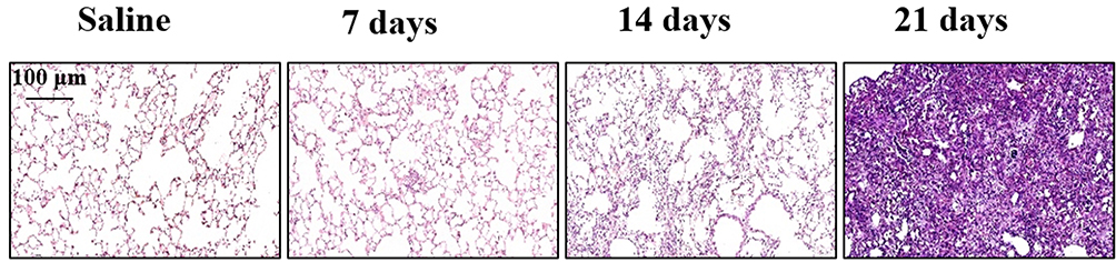 |
Figure 1 Schematic diagram of HE staining of mouse lung tissue at different time periods. (n=6 mice per group). |
Comparison of Lung Tissue Pathological Scores
According to the clinical Ashoft scoring criteria, the degree of pulmonary fibrosis in mice at different time periods was scored, and it was found that the pathological process of pulmonary fibrosis was divided into two parts. In the early stage, inflammatory infiltration was the main factor, and after 14 days, fibrosis was the main factor (Figure 2).
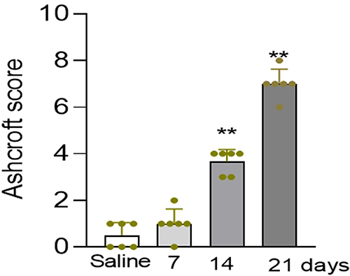 |
Figure 2 Ashcroft pathological scoring chart, compared with the saline group, **P<0.01. (n=6 mice per group). |
Changes in Lung Tissue Expression Factors
Based on the characteristics of the course of pulmonary fibrosis reflected in the graph, this experiment used BLM modeling for 14 days as a node to detect inflammatory cell indicators in lung tissue before 14 days, as well as indicators of pulmonary fibrosis after 14 days. The results showed that compared with the saline group, the mRNA expression of inflammatory cytokines TNF-α and IL-1β in the lungs of mice in the 7 days and 14 days groups increased (Table 3). Similarly, compared with the saline group, the mRNA expression of α-smooth muscle actin (α-SMA) and fibronectin in the lung tissues of mice in the 14 days and 21 days groups increased (Table 4).
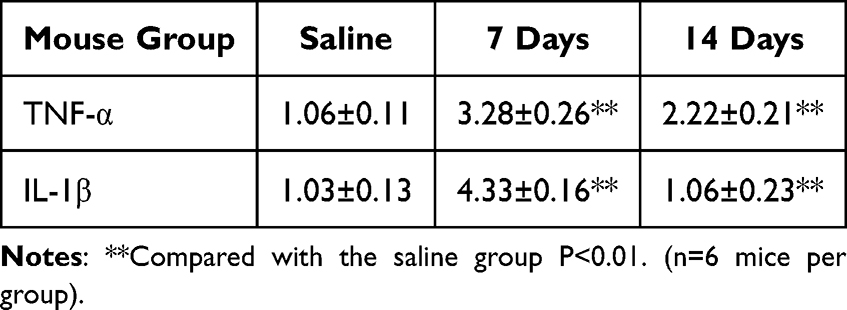 |
Table 3 Expression Levels of Inflammatory Factors in Mouse Lung Tissue |
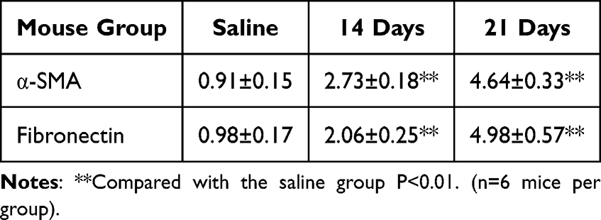 |
Table 4 Expression Levels of Fibrosis Factors in Mouse Lung Tissue |
Discussion and Analysis
This experiment is aimed at exploring the pathological process of pulmonary fibrosis, and is designed to observe the characteristics of lesions in different stages of pulmonary fibrosis disease. By establishing a mouse model of pulmonary fibrosis and detecting changes in mouse lung tissue at different time points, the process characteristics of pulmonary fibrosis lesions can be understood. The following is a schematic diagram of the overall conclusion (Figure 3).
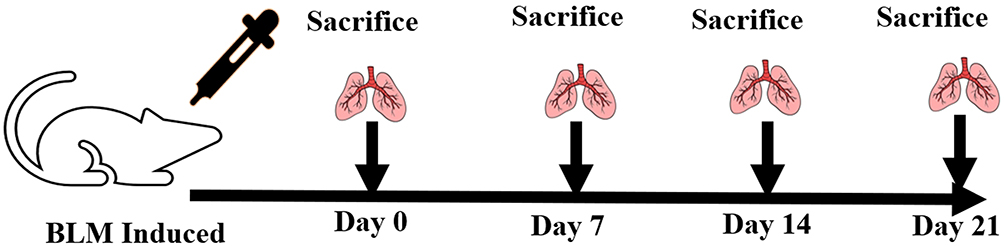 |
Figure 3 Summary of Experimental Design Process. |
In this study, TNF-α and IL-1β were selected as early inflammatory markers due to their significant upregulation during the initial stages of pulmonary fibrosis and their involvement in regulating subsequent fibrotic progression.16 The mRNA results of early detection of inflammatory factors in this experiment showed that the expression of inflammatory factors increased in the 7 days, and decreased in the 14 days. When students discuss, do these results represent improvement in the disease? By observing the slice results, it can be found that this is not the case. Although the expression of inflammatory factors decreases, the severity of the disease still increases. According to literature, macrophages play an important role in the pathological process of pulmonary fibrosis by releasing inflammatory factors and regulating the inflammatory response. Currently, polarized macrophages are classified into two main types: classically activated macrophages and alternatively activated macrophages. Classical activation of macrophages drives inflammatory responses and can also induce tissue damage. Alternating activation of macrophages drives antigen responses and assists in tissue repair.17,18 This theory not only verifies the accuracy of the experimental results, but also further confirms the complexity of the progression of pulmonary fibrosis disease.
BLM is a well-established inducer for establishing animal models of pulmonary fibrosis, but different administration methods may affect modeling efficiency and animal survival rates. To optimize the dosing protocol, we employed a non-invasive intratracheal instillation method for bleomycin delivery. This approach not only offers operational simplicity and minimal invasiveness but also significantly improves the survival rate of experimental mice, thereby providing a more reliable model for subsequent research.19 The survival rate data after BLM administration showed a 21 day survival rate of 92.5% ± 3.1%, which meets the internationally recognized stability standards for BLM models.
This experiment is a design experiment, and the assessment method requires diversified judgment. According to the experimental design project, score students based on their proportion. The specific assessment mode is shown in Table 5.
 |
Table 5 Course Assessment Methods |
Challenges and Solutions
The process of designing and implementing experiments is fraught with numerous challenges. We have enumerated several common ones and put forward targeted solutions.
(1) Challenge: Limited resources and equipment
Some universities may face shortages of experimental resources and advanced equipment, which can affect the implementation of experimental projects.
Solution: Strengthen cooperation and resource sharing among universities. Establish shared experimental platforms or cooperation projects with other institutions or research institutes to make full use of existing resources.
(2) Challenge: Difficulty in integrating multidisciplinary knowledge
Students may have difficulty integrating knowledge from different disciplines due to differences in learning backgrounds and knowledge systems.
Solution: Provide pre-course training and guidance to help students review and understand relevant knowledge points. At the same time, encourage teachers from different disciplines to cooperate in teaching and jointly guide students.
(3) Challenge: Evaluation and assessment of students
It is difficult to comprehensively and objectively evaluate students’ performance in experimental design and research.
Solution: Establish a multi-faceted evaluation system. In addition to evaluating experimental results and reports, also consider students’ participation in the experimental process, teamwork ability, innovation ability, and problem-solving ability. Use a combination of quantitative and qualitative evaluation methods to ensure the fairness and comprehensiveness of the evaluation.
This study has certain limitations regarding its cross-cultural and educational system applicability. The epidemiological characteristics of pulmonary fibrosis (such as etiology distribution and incidence rates) exhibit significant regional variations. For example, clinical profiles differ markedly between areas with high prevalence of pneumoconiosis and those with high rates of idiopathic pulmonary fibrosis, necessitating potential localization adjustments to teaching case designs based on regional disease patterns. Furthermore, substantial differences exist in medical education standards across countries or regions (such as licensing exam content and competency requirements). Such diversity in educational policies may influence the prioritization of pulmonary fibrosis in curricula, thereby potentially limiting the direct applicability of the study’s conclusions in non-native educational contexts.
Conclusion
Design-based experiments represent a novel form of experimental course instruction. In recent times, design-based experimental teaching has been implemented across multiple academic disciplines.20–23 The design of this experimental technique combines research foci, aligns with clinical treatment benchmarks, and takes the exploration of the disease development process as its backdrop. Initiating from experimental operational techniques, it enables students to acquire experimental skills while broadening their understanding of diseases and integrating knowledge elements from anatomy, pathology, molecular biology, and other fields. Through mutual permeation, it nurtures students’ scientific research capabilities and establishes a solid groundwork for their evolution into research-oriented clinical professionals. The post-course survey showed students highly valued this lab course for effectively stimulating their research interest while teaching cutting-edge techniques. Many described it as exceptionally practical for their academic growth.
The teaching method of experimental design guided by exploring the development process of diseases has shown great potential and value in Chinese universities. Although there are some challenges in the implementation process, through continuous improvement and innovation, it can effectively improve the quality of medical education, cultivate students with high-quality research and practical abilities, and contribute to the development of the medical field. Future research and practice should focus on further optimizing the teaching method, strengthening resource support, and improving the evaluation system to promote the wider application and development of this teaching approach.
Disclosure
All authors declare that they have no conflict of interest in this work.
References
1. Feng X, Wang Y, Wei L, Meng K. How to become an excellent pediatric resident: a qualitative comparative study from China. BMC Health Serv Res. 2023;23(1):53. doi:10.1186/s12913-023-09038-x
2. Zhang G, Zhao K, Hong Y, Qiu X, Zhang K, Wei B. SHA-MTL: soft and hard attention multi-task learning for automated breast cancer ultrasound image segmentation and classification. Int J Comput Assist Radiol Surg. 2021;16(10):1719–1725. doi:10.1007/s11548-021-02445-7
3. Li N, Zhang C, Cui J, Wang Q, Li T, Peng G. Using nobel prize case-based learning in medical immunology to cultivate critical thinking dispositions for medical undergraduates. BMC Med Educ. 2024;24(1):1213. doi:10.1186/s12909-024-06155-x
4. Liu C, Ren M, Luo C, et al. The application effect of the segmented teaching method in training medical students on clinical practice skills. BMC Med Educ. 2024;24(1):1090. doi:10.1186/s12909-024-06060-3
5. Yang L, Gilbertsen A, Xia H, et al. Hypoxia enhances IPF mesenchymal progenitor cell fibrogenicity via the lactate/GPR81/HIF1α pathway. JCI Insight. 2023;8(4):e163820. doi:10.1172/jci.insight.163820
6. Lynch DA, Sverzellati N, Travis WD, et al. Diagnostic criteria for idiopathic pulmonary fibrosis: a Fleischner Society White Paper. Lancet Respir Med. 2018;6(2):138–153. doi:10.1016/S2213-2600(17)30433-2
7. Wendisch D, Dietrich O, Mari T, et al. SARS-CoV-2 infection triggers profibrotic macrophage responses and lung fibrosis. Cell. 2021;184(26):6243–6261. doi:10.1016/j.cell.2021.11.033
8. O’Dwyer DN, Ashley SL, Gurczynski SJ, et al. Lung microbiota contribute to pulmonary inflammation and disease progression in pulmonary fibrosis. Am J Respir Crit Care Med. 2019;199(9):1127–1138. doi:10.1164/rccm.201809-1650OC
9. Henrot P, Blervaque L, Dupin I, et al. Cellular interplay in skeletal muscle regeneration and wasting: insights from animal models. J Cachexia Sarcopenia Muscle. 2023;14(2):745–757. doi:10.1002/jcsm.13103
10. Yang G, Yang Y, Liu Y, Liu X. Regulation of alveolar macrophage death in pulmonary fibrosis: a review. Apoptosis. 2023;28(11–12):1505–1519. doi:10.1007/s10495-023-01888-4
11. Ishida Y, Kuninaka Y, Mukaida N, Kondo T. Immune mechanisms of pulmonary fibrosis with bleomycin. Int J Mol Sci. 2023;24(4):3149. doi:10.3390/ijms24043149
12. Gul A, Yang F, Xie C, et al. Pulmonary fibrosis model of mice induced by different administration methods of bleomycin. BMC Pulm Med. 2023;23(1):91. doi:10.1186/s12890-023-02349-z
13. Meurer IR, Silva MR, Roland RK, Corrêa JOA, Coimbra ES. Evaluation of medical professionals’ knowledge about Q fever. SciMed. 2024;34(1):e45474.
14. Chen F, Duan N-F, Zhang X, Zhang W. [Effect of Fuzheng Tongluo Granules on macrophage pyroptosis in rat model with pulmonary fibrosis based on NLRP3/caspase-1/GSDMD pathway]. Zhongguo Zhong Yao Za Zhi. 2024;49(23):6399–6406. Catalan. doi:10.19540/j.cnki.cjcmm.20240904.402
15. Gu Y-Q, Wu J, Wang T, et al. PINK1 deficiency alleviates bleomycin-induced pulmonary fibrosis in mice. Cell Signal. 2025;133:111868. doi:10.1016/j.cellsig.2025.111868
16. Gu T, Zhou Y, Wang Q, Zhu X, Wu C, Dong Z. Downregulation of miR-410-3p via the METRNL-mediated AMPK/SIRT1/NF-κB signaling axis inhibits oxidative stress and inflammation in idiopathic pulmonary fibrosis. Cell Signal. 2025;130:111667. doi:10.1016/j.cellsig.2025.111667
17. Ge Z, Chen Y, Ma L, Hu F, Xie L. Macrophage polarization and its impact on idiopathic pulmonary fibrosis. Front Immunol. 2024;15:1444964. doi:10.3389/fimmu.2024.1444964
18. Zhang R, Jiang Q, Gao S, et al. Favipiravir ameliorates bleomycin-induced pulmonary fibrosis by reprogramming M1/M2 macrophage polarization. Int Immunopharmacol. 2024;131:111774. doi:10.1016/j.intimp.2024.111774
19. Kim JK, Vinarsky V, Wain J, et al. In vivo imaging of tracheal epithelial cells in mice during airway regeneration. Am J Respir Cell Mol Biol. 2012;47(6):864–868. doi:10.1165/rcmb.2012-0164OC
20. Alberti S, Motta P, Ferri P, Bonetti L. The effectiveness of team-based learning in nursing education: a systematic review. Nurse Educ Today. 2021;97:104721. doi:10.1016/j.nedt.2020.104721
21. Goodman BE. An evolution in student-centered teaching. Adv Physiol Educ. 2016;40(3):278–282. doi:10.1152/advan.00056.2016
22. Zhang D, Zhang J, Cao M, Zhu Y, Yang G. Testing the effectiveness of motivation-based teaching in Nursing English course: a quasi-experimental study. Nurse Educ Today. 2023;122:105723.
23. Fortepiani LA, Marsh SA. Innovative techniques for developing an inclusive teaching environment. Adv Physiol Educ. 2023;47(4):904–907. doi:10.1152/advan.00014.2023
 © 2025 The Author(s). This work is published and licensed by Dove Medical Press Limited. The
full terms of this license are available at https://www.dovepress.com/terms
and incorporate the Creative Commons Attribution
- Non Commercial (unported, 4.0) License.
By accessing the work you hereby accept the Terms. Non-commercial uses of the work are permitted
without any further permission from Dove Medical Press Limited, provided the work is properly
attributed. For permission for commercial use of this work, please see paragraphs 4.2 and 5 of our Terms.
© 2025 The Author(s). This work is published and licensed by Dove Medical Press Limited. The
full terms of this license are available at https://www.dovepress.com/terms
and incorporate the Creative Commons Attribution
- Non Commercial (unported, 4.0) License.
By accessing the work you hereby accept the Terms. Non-commercial uses of the work are permitted
without any further permission from Dove Medical Press Limited, provided the work is properly
attributed. For permission for commercial use of this work, please see paragraphs 4.2 and 5 of our Terms.