
Back to Journals » Infection and Drug Resistance » Volume 19
Thrombosis in Severe Influenza A Pneumonia: Distribution and Risk Factors in Hangzhou, China
Authors Ma N, Pan Q, Qi Y, Shao F
Received 7 January 2026
Accepted for publication 27 April 2026
Published 13 May 2026 Volume 2026:19 590329
DOI https://doi.org/10.2147/IDR.S590329
Checked for plagiarism Yes
Review by Single anonymous peer review
Peer reviewer comments 3
Editor who approved publication: Dr Hazrat Bilal
Nannan Ma, Qing Pan, YuanYuan Qi, FengLin Shao
Department of Nursing, The First Affiliated Hospital, Zhejiang University School of Medicine, Hangzhou, Zhejiang, 310003, People’s Republic of China
Correspondence: FengLin Shao, Department of Nursing, The First Affiliated Hospital, Zhejiang University School of Medicine, No. 79, Qingchun Road, Hangzhou, Zhejiang, 310003, People’s Republic of China, Tel +86 18267156779, Email [email protected]
Background: Severe viral pneumonia, including that caused by influenza A, can trigger a systemic inflammatory response that can lead to coagulation abnormalities, which substantially increases the risk of life-threatening thrombotic complications.
Objective: This study aimed to examine the distribution of thrombosis and its risk factors among patients with severe influenza A pneumonia to inform early clinical identification and intervention.
Methods: A retrospective analysis was performed on 175 patients with severe influenza A pneumonia who were admitted to the Department of Respiratory and Critical Care Medicine, The First Affiliated Hospital, Zhejiang University School of Medicine between January 2024 and March 2025. Patients were categorised into a thrombosis group (n = 82) and a control group (n = 93). Clinical data were analysed using univariate and multivariate logistic regression to identify independent risk factors.
Results: The thrombosis group showed significantly higher D-dimer (3.06 [0.95, 12.42] vs 0.90 [0.54, 1.53] μg/mL, p < 0.001) and prolonged prothrombin time (PT) (14.90 [13.56, 18.58] vs 13.70 [13.20, 14.50] s, p < 0.001) than the controls. Multivariate analysis identified elevated D-dimer (odds ratio [OR] = 1.016, p = 0.021) and decreased haemoglobin (OR = 1.026, p = 0.013) as independent risk factors, whereas prolonged PT was independently associated with a lower risk of thrombosis (OR = 0.751, p = 0.010). Venous thrombosis predominated (78.0%), with isolated muscular vein thrombosis (IMVT) being the most common subtype (36.6%).
Conclusion: This study clarified that venous thrombosis, predominantly IMVT and pulmonary embolism, is the primary thrombotic complication in severe influenza A pneumonia. We identified a unique biomarker profile for risk stratification; elevated D-dimer and decreased haemoglobin are independent risk factors, whereas prolonged PT was independently associated with a lower risk of thrombosis. This combination provides a practical tool for early clinical identification of patients at high risk, guiding tailored thromboprophylaxis.
Keywords: influenza A virus, severe pneumonia, venous thrombus, risk factor, D-dimer
Introduction
Influenza A is an acute respiratory infectious disease caused by the influenza A virus (IAV); it is highly infectious and potentially fatal.1 The virus can cause severe pneumonia, acute respiratory distress syndrome and multiple organ dysfunction, resulting in a considerably high mortality rate.2 In patients with chronic diseases that substantially elevate thrombotic risk — including chronic obstructive pulmonary disease, heart failure and cancer — influenza infection exerts a synergistic effect with these pre-existing conditions,3 profoundly aggravating coagulation system dysregulation and vascular endothelial damage, making these patients a high-risk group for thrombotic complications.3,4
Studies have shown that in addition to damaging the respiratory system, influenza A infection can induce systemic inflammatory response and coagulation dysfunction, significantly increasing the risk of thromboembolic events.4 Although COVID-19-related coagulopathy is largely driven by profound direct endothelial damage and microvascular thrombosis, influenza A primarily induces a state of “immunothrombosis”, where the innate immune system and coagulation pathways synergistically drive thrombus formation.5,6 Epidemiological data indicate that thrombotic complications in severe influenza occur in up to 18–30% of critically ill patients.7 This pathological state not only increases mortality but also leads to serious complications, affecting the long-term prognosis of patients. Thrombosis has become an important complication in patients with severe influenza, potentially leading to deep vein thrombosis (DVT), pulmonary embolism (PE), cerebral infarction and myocardial infarction, seriously affecting patients’ quality of life and rehabilitation.8,9 Following IAV infection, the body releases a large number of pro-inflammatory factors, such as interleukin 6 and tumour necrosis factor. These factors can activate vascular endothelial cells and promote the expression of tissue factor, thereby initiating the exogenous coagulation pathway.5 Moreover, the virus directly damages the vascular endothelium, exposes subcutaneous collagen and further promotes platelet activation and aggregation.10 In addition, patients with severe infection often experience venous blood stasis, due to being bedridden, hypoxaemia and haemodynamic instability, further increasing the risk of thrombosis.11
In clinical practice, D-dimer, a fibrin degradation product, is substantially elevated in influenza-related thrombosis and has been widely used as a key marker for predicting thrombotic events.12 Studies have shown that elevated D-dimer levels are significantly associated with the risk of thrombosis.13 In addition, prolongation of prothrombin time (PT) and activated partial thromboplastin time (APTT), as well as thrombocytopenia or platelet dysfunction, indicates a coagulation system disorder.14
Extensive studies on severe respiratory viral infections have demonstrated that abnormalities in coagulation-related biochemical markers, such as D-dimer, are pivotal indicators for predicting coagulopathy and adverse outcomes. These changes provide clinicians with a robust reference to dynamically evaluate disease severity, manage the high risk of influenza-associated thrombosis, and guide early anticoagulant interventions.15,16
Recent literature reviews and cohort studies indicate that, although it is well-established that severe influenza complicated by thromboembolic events leads to prolonged intensive care unit stays and significantly increased mortality, systematic analyses of risk factors and screening for distribution characteristics specifically tailored to severe influenza A pneumonia remain highly scarce. Existing evidence is predominantly limited to small-sample observations or case reports lacking multivariate adjustment.17,18 However, reports on the distribution characteristics of thrombus vary between studies, which may be related to different baseline characteristics, detection methods and anticoagulation strategies in patients.17,18 Some studies have determined venous thrombosis to be the main form of influenza-related thrombosis, with a high incidence of lower extremity DVT and PE, whereas the incidence of arterial thrombosis (such as stroke) is relatively low.19 However, reports on the distribution characteristics of thrombus vary between studies, which may be related to different baseline characteristics, detection methods and anticoagulation strategies in patients. In terms of risk factors, advanced age, elevated inflammatory markers (such as C-reactive protein [CRP] and procalcitonin), increased D-dimer levels and abnormal coagulation function are considered to be high-risk factors for thrombosis.20 However, there remains an explicit knowledge gap due to the distinct scarcity of multivariate risk analyses specifically for patients in the respiratory intermediate care setting. Therefore, the in-depth study of thrombosis characteristics and related risk factors in patients with severe influenza A pneumonia is vital for improving clinical management and reducing the incidence of complications.
This study aims to investigate the distribution characteristics of thrombosis in patients with severe influenza A pneumonia and to analyse the single and multiple risk factors of thrombosis. Through systematic analysis and research, we hope to provide clinicians with more accurate assessment tools to develop individualised anticoagulation programmes to improve the prognosis of patients with severe influenza. In-depth research in this field can provide an important theoretical basis and practical guidance for future influenza management.
Materials and Methods
Study Participants
This study is a retrospective analysis. The data of 175 patients with severe influenza A pneumonia who were admitted to the Department of Respiratory and Critical Care Medicine, The First Affiliated Hospital, Zhejiang University School of Medicine between January 2024 and March 2025 were collected. All patients were diagnosed with IAV infection using real-time fluorescence quantitative polymerase chain reaction (PCR).
The inclusion criteria were as follows: (1) age ≥18 years; (2) meeting the American Society of Infectious Diseases diagnostic criteria21 for severe influenza pneumonia, including influenza-like symptoms (fever ≥38°C with cough or sore throat), chest imaging showing pneumonia and at least one severe standard (oxygenation index [PaO2/FiO2] ≤300 mmHg, need for mechanical ventilation, septic shock or multiple organ failure); and (3) patients must have undergone a complete thrombosis screening programme (including lower extremity venous ultrasound and computed tomography [CT] pulmonary angiography) during their hospitalisation. The exclusion criteria were as follows: (1) pregnant or lactating women, (2) patients diagnosed with venous thromboembolism or who had received anticoagulant therapy prior to admission, (3) presence of other diseases known to cause a hypercoagulable state (such as malignant tumours, active haematologic malignancies or antiphospholipid antibody syndrome), (4) incomplete clinical data or patients lost to follow-up and (5) length of hospital stay <72 hours.
The study was approved by the hospital’s ethics committee and complied with the ethical guidelines of the 1975 Helsinki Declaration.
Grouping and Data Collection
Thrombosis screening, including bilateral lower extremity venous compression ultrasound or selective CT pulmonary angiography, was triggered by clinical suspicion of thrombotic events rather than systematic screening of all patients. If imaging was positive, thrombosis was diagnosed. Based on the thrombosis screening results during hospitalisation, the patients were divided into a thrombosis group (n = 82; new thrombotic events confirmed by imaging) or a control group (n = 93; no thrombotic events detected during hospitalisation). The following information was collected: age, gender, white blood cell count (WBC), neutrophil count (NEUT), lymphocyte count, haemoglobin (Hb) level, platelet count, high-sensitivity CRP level, procalcitonin level, PT, international normalised ratio (INR), APTT, D-dimer level, fibrinogen level, aspartate aminotransferase (AST) level, alanine aminotransferase (GPT) level, albumin to globulin ratio (A/G) and serum creatinine level.
Data collection was performed independently by two trained researchers using standardised common reporting format tables. All imaging results were double-blindly interpreted by two radiologists with the title of deputy director or above (kappa value = 0.86). Standard automatic biochemical analysers were used for laboratory testing with daily quality control calibration, and standard X-ray CT scanners were utilised for imaging.
Statistical Analysis
Statistical analysis was performed using SPSS 26.0 statistical software. The normality test was performed using the Kolmogorov–Smirnov method. Continuous variables that conformed to the normal distribution were expressed as mean ± standard deviation, and an independent sample t-test was used for comparison between groups. Non-normal distribution data were described by the median (interquartile range) (M [Q1, Q3]), and the Mann–Whitney U-test was used for comparison between groups. The categorical variables were expressed as frequency (percentage), and the differences between groups were tested using the chi-square test or Fisher’s exact test. Missing data were handled using multiple imputation. Prior to inclusion in the regression model, multicollinearity was assessed using the variance inflation factor for related parameters, such as PT and INR, to ensure model stability. Prior to inclusion in the regression model, multicollinearity was assessed using the variance inflation factor for related parameters, such as PT and INR, to ensure model stability; all VIF values were below 5.0, confirming no significant multicollinearity. Sample size adequacy was evaluated using the events-per-variable (EPV) criterion: with 82 outcome events and 4 independent predictors in the final model, the EPV was 20.5, substantially exceeding the recommended minimum of 10 EPV, thereby confirming sufficient statistical power for the multivariable logistic regression analysis. Variables with p < 0.05 in the univariate analysis were included in the multivariate logistic regression model (forward stepwise method), and the odds ratio (OR) and 95% confidence interval (CI) were calculated. A p-value of <0.05 was considered statistically significant. The kappa coefficient was used to evaluate the consistency between readers (a kappa value of >0.80 was highly consistent).
Results
Clinical Characteristics
Comparison of baseline characteristics between the two groups showed that there was no significant difference in age (68.30 ± 15.36 vs 66.22 ± 12.89 years, p = 0.329) or gender distribution (male: 68.3% vs 63.4%, p = 0.500) between the thrombus group and the control group. Patients in the thrombosis group had a longer hospital stay (p < 0.001), a higher mortality rate during hospitalisation and a less favourable prognosis (p = 0.018) than patients in the control group. There was no significant difference in the incidence of cardiovascular diseases or metabolic diseases in the thrombosis group (p = 0.16, p = 0.949). The proportion of patients with chronic respiratory diseases and malignant tumours in the thrombosis group was significantly higher than that in the control group (p < 0.001, p = 0.009). Specifically, 28 patients in the thrombosis group and 16 in the control group had malignant tumours (see Table 1).
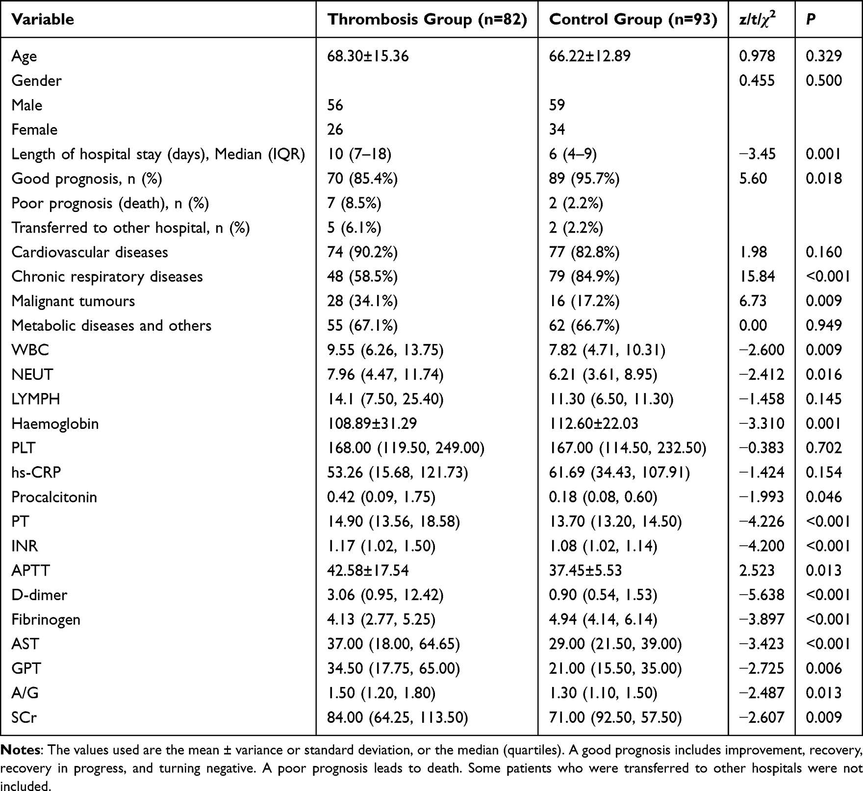 |
Table 1 Univariate Analysis of Thrombosis in Patients with Severe Influenza A Pneumonia |
Univariate Analysis of Thrombosis Events in the Thrombus Group
Analysis of inflammation-related indicators showed that WBC, NEUT and procalcitonin levels in the thrombus group were significantly increased (p < 0.05); that is, patients in the thrombus group showed a more significant inflammatory response than patients in the control group. In terms of coagulation function, in the thrombus group, PT was significantly prolonged (14.90 [13.56, 18.58] vs 13.70 [13.20, 14.50] s, p < 0.001), INR was significantly increased (1.17 [1.02, 1.50] vs 1.08 [1.02, 1.14], p < 0.001), APTT was prolonged (42.58 ± 17.54 vs 37.45 ± 5.53 s, p = 0.013) and D-dimer levels were abnormally increased (3.06 [0.95, 12.42] vs 0.9 [0.54, 1.53] μg/mL, p < 0.001). Notably, patients in the thrombus group exhibited more significant anaemia (Hb = 108.89 ± 31.29 vs 112.60 ± 22.03 g/L, p = 0.001) and abnormal liver function (AST = 37.00 vs 29.00 U/L, p < 0.001; GPT = 34.50 vs 21.00 U/L, p = 0.006). These results suggest that thrombosis in patients with severe influenza A pneumonia is the result of a combination of factors, including pathophysiological processes such as inflammatory response, coagulation disorders and multiple organ dysfunction (see Table 1).
Distribution Characteristics of Thrombotic Events
Thrombosis site analysis (Table 2) classified the primary or most significant thrombotic event per patient. Among the 82 patients with thrombotic events, venous thrombosis was the primary event in 64 cases (78.0%), and arterial thrombosis was the primary event in 18 cases (22.0%). Within the venous category, isolated muscular vein thrombosis (IMVT) was the most common primary event (30 cases), followed by PE (24 cases) and DVT (10 cases). Within the arterial category, cerebral infarction predominated (14 cases), followed by myocardial infarction (4 cases). Furthermore, co-occurrence of venous and arterial events within the same patient was distinctly observed in 9 cases (11.0%).
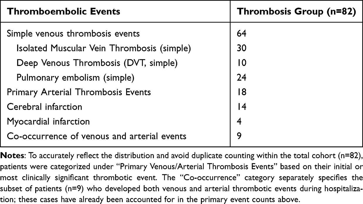 |
Table 2 Distribution of Thrombosis Sites in Patients with Severe Influenza A Pneumonia |
Multiple Logistic Regression
In this study, 175 patients with severe influenza A pneumonia were divided into a thrombosis group (n = 82, venous/arterial thrombosis diagnosed through lower extremity venous ultrasound, CT pulmonary angiography and other imaging) and a control group (n = 93, no thrombotic events detected). The statistically significant predictors selected by univariate analysis were included as independent variables in the multivariate logistic regression model for in-depth analysis. The final multivariate logistic regression model (Table 3) identified two independent risk factors: elevated D-dimer (OR = 1.016, 95% CI: 1.002–1.030, p = 0.021) and decreased Hb (OR = 1.026, 95% CI: 1.006–1.047, p = 0.013). Additionally, the model identified two independent protective factors: prolonged PT (OR = 0.751, 95% CI: 0.604–0.934, p = 0.010) and a decreased A/G ratio (OR = 0.244, 95% CI: 0.061–0.986, p = 0.048). For elevated D-dimer, a 1-unit increase leads to a 1.6% increase in odds of thrombosis. To illustrate these findings visually, a forest plot (Figure 1) was generated to display the ORs and 95% CIs of the independent risk and protective factors.
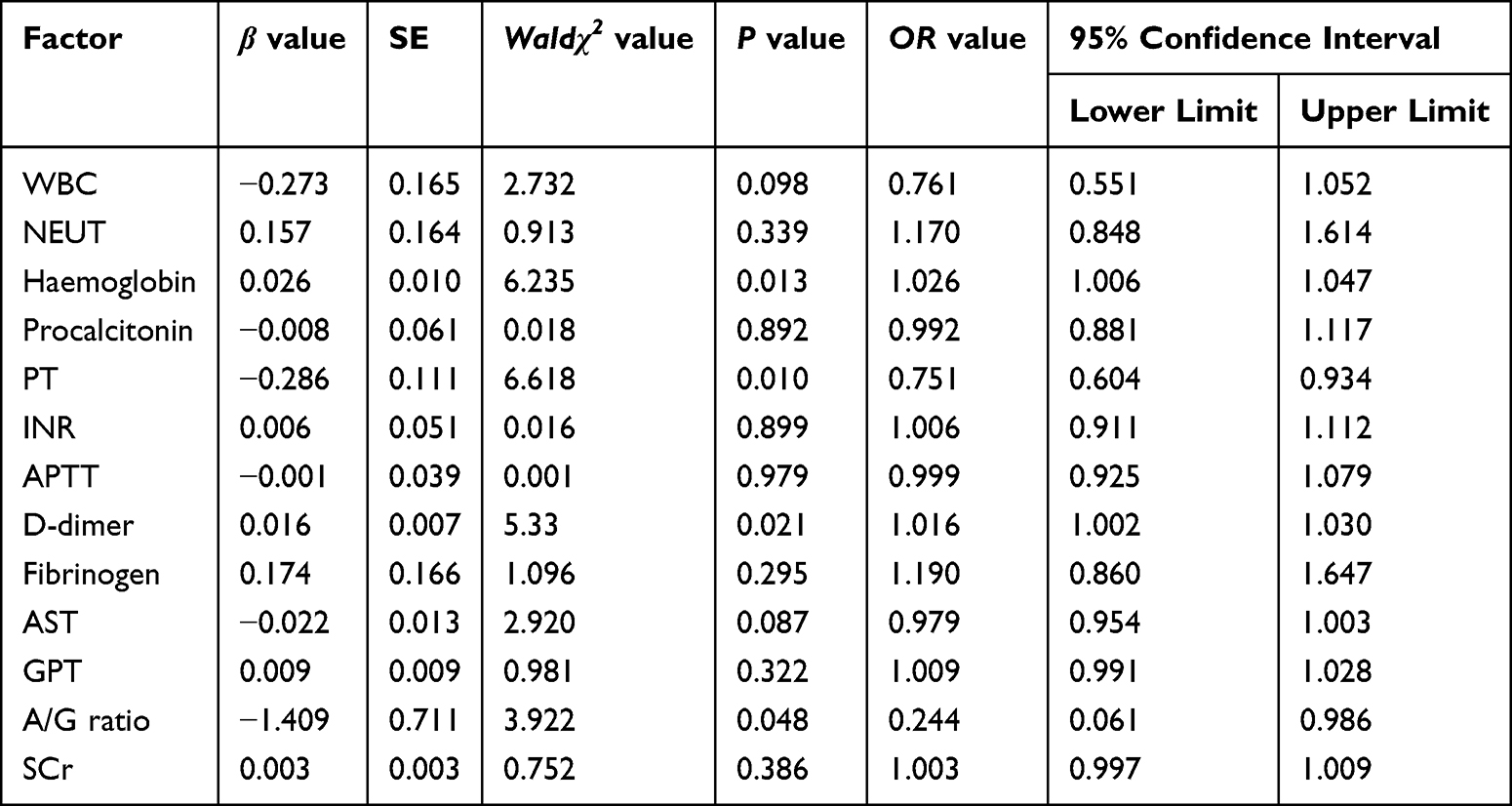 |
Table 3 Multivariate Logistic Analysis of Thrombosis in Patients with Severe Influenza A Pneumonia |
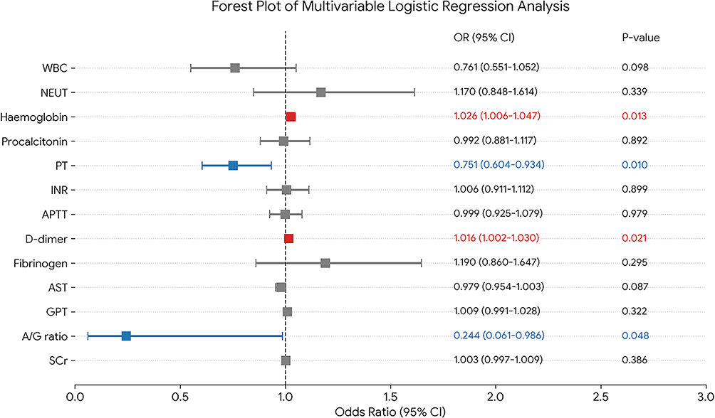 |
Figure 1 Forest plot of independent predictors for thrombosis in patients with severe influenza A pneumonia. |
Notably, although WBC, NEUT and other inflammatory indicators were statistically significant in univariate analysis, they did not show independent predictive value in multivariate analysis (p > 0.05).
Discussion
In this study, the risk factors, distribution characteristics and clinical significance of thrombosis were systematically discussed by retrospective analysis of 175 patients with severe influenza A pneumonia. The results not only verify the previous hypothesis on the mechanism of influenza-related thrombosis but also reveal some new discoveries with important clinical value.
Multivariate logistic regression analysis identified elevated D-dimer, decreased Hb and a decreased A/G ratio as independent risk factors for thrombosis. Prolonged PT was independently associated with a lower risk of thrombosis. This finding echoes the findings of Kollias et al,22 who also observed a significant positive correlation between D-dimer levels and the risk of thrombosis in patients with influenza. The finding that prolonged PT was independently associated with a lower risk of thrombosis warrants an in-depth discussion from a pathophysiological perspective. Traditionally, PT prolongation signifies a dysfunction in the extrinsic coagulation pathway. In the context of a hypercoagulable state triggered by severe infection, a relative impairment in thrombin generation via this pathway may paradoxically attenuate the risk of macroscopic thrombosis, tipping the balance towards a bleeding diathesis rather than a thrombotic one. This protective effect could be mediated through several specific mechanisms. First, severe viral infections, such as influenza, can trigger the production of acquired autoantibodies, such as inhibitors against clotting factors (eg. anti-factor VIII, anti-prothrombin antibodies). These antibodies may prolong the in vitro PT assay while concurrently inhibiting procoagulant activity in vivo, thereby exerting a net protective effect against thrombosis. Second, prolonged PT may reflect a specific consumptive phenotype within the spectrum of virus-associated coagulopathy. In some patients, the initial prothrombotic drive (immunothrombosis) may evolve into a more balanced or even hypocoagulable state due to the consumption of coagulation factors, a process overlapping with low-grade disseminated intravascular coagulation (DIC). Alternatively, it may be a marker of thrombotic microangiopathy, in which endothelial damage and microvascular thrombosis lead to platelet consumption and secondary coagulation factor deficiency, manifesting as prolonged PT. This association suggests that prolonged PT in severe influenza could identify a subset of patients transitioning from a hyper- to a hypocoagulable phase. Furthermore, prolonged PT could act as a marker for consumption coagulopathy (akin to low-grade DIC) or more severe liver dysfunction, both scenarios being associated with a lower risk of large vessel thrombosis.23 This suggests that prolonged PT may indicate a more severe inflammatory response and coagulation system disorders. Possible explanations for this phenomenon include the following: (1) tissue factor overexpression leading to increased thrombin production, (2) a decrease in anticoagulant protein (such as protein C and protein S) consumption and (3) fibrinolytic system inhibition.
In addition, this study found that venous thrombosis accounted for 78.0% of all thrombotic events, of which IMVT (36.6%) and PE (29.3%) were the most common. This distribution is considerably different from that of ordinary hospitalised patients, which is usually dominated by DVT. The possible mechanisms of this difference include the following: (1) the influenza virus directly damages the pulmonary vascular endothelium and increases the risk of PE, (2) microcirculation disorder induced by inflammatory factors is more likely to lead to IMVT and (3) long-term bed rest in critically ill patients leads to blood stasis.24 Arterial thrombosis events accounted for 22.0%, with cerebral infarction predominating (77.8% of arterial cases). This proportion is significantly higher than that of patients with ordinary influenza (usually <5%),25 suggesting that there may be a special mechanism underlying arterial thrombosis in critically ill patients. We speculate that this may be related to the following factors: (1) inflammatory factors disrupt the blood–brain barrier, (2) the virus directly invades the cerebrovascular endothelium and (3) cardiogenic embolism risk increases. Notably, four of the six patients with myocardial infarction had venous thrombosis at the same time, supporting the “venous–arterial thrombosis transformation” hypothesis.26 This phenomenon may be further explained by paradoxical embolism26 or severe inflammation-induced, platelet-mediated mechanisms.27
The results of this study support the role of immunothrombosis theory in severe influenza.24 We found that patients in the thrombus group exhibited a more significant inflammatory response (increased WBC, NEUT and procalcitonin) and coagulation dysfunction (prolonged PT and APTT) than patients in the control group. This vicious cycle of inflammation–coagulation may be achieved in the following ways: the release of neutrophil extracellular traps (NETs), enhanced platelet activation and vascular endothelial injury aggravation.23 This pathophysiological paradigm finds strong parallels in COVID-19, in which a high incidence of thrombotic complications, particularly in critically ill patients, has been extensively documented. Studies on COVID-19-associated coagulopathy have highlighted similar mechanisms, including endothelial dysfunction, a profound inflammatory cytokine storm and NETosis, further solidifying the concept of immunothrombosis as a critical mechanism in severe viral pneumonias beyond influenza.24 It is particularly noteworthy that decreased Hb is associated with an increased risk of thrombosis. This seemingly contradictory phenomenon may be related to the following mechanisms: anaemia leads to tissue hypoxia and induces a hypoxia-inducible factor 1-alpha-mediated procoagulant state, the change in blood viscosity affects haemodynamics and iron metabolism disorder affects platelet function.28 This finding provides a new perspective for understanding the complex mechanism of influenza-associated thrombosis. Although WBC/NEUT was not retained as an independent predictor in multivariate analysis, the interaction between coagulation and inflammatory imbalance suggests that inflammation may indirectly influence thrombosis by promoting a hypercoagulable state.
Based on the results of this study, we recommend the following prevention and treatment strategies for patients with severe influenza A pneumonia: early identification of patients at high risk – while a formal scoring system requires prospective validation, a preliminary risk profile integrating the model coefficients suggests that patients with D-dimer >1000 μg/L, PT prolongation >14 s or Hb <110 g/L should be monitored closely; patients with D-dimer >1000 μg/L, PT prolongation >14 s or Hb <110 g/L should be monitored; individualised anticoagulation regimens – low molecular weight heparin should be considered for prevention, with the dose adjusted according to body weight and renal function; multidisciplinary collaborative management – multidisciplinary teams that include staff from intensive care, haematology and imaging departments should be established; and a dynamic monitoring strategy – D-dimer levels and coagulation function should be monitored every 48 hours until the condition is stable.
Our results introduce several novel and clinically significant insights. First, we identified prolonged PT as an independent protective factor, a counterintuitive finding in the context of a hypercoagulable state, which may signal a distinct coagulopathy phenotype related to factor inhibitors or consumptive processes. Second, our detailed analysis revealed IMVT as the most prevalent form, highlighting a unique thrombotic distribution likely driven by inflammation and microvascular injury. Third, we established an integrated multivariable risk profile combining elevated D‑dimer, decreased Hb and prolonged PT, offering a holistic tool for stratification.
This study has several limitations. First, the retrospective observational design may introduce selection bias, and due to incomplete electronic medical records, certain key covariates (such as the specific timing and dosage of anticoagulation therapy, mechanical ventilation parameters and limb immobilisation duration) could not be adjusted for in the multivariate model. Second, the study did not detect the expression levels of novel biomarkers (such as NETs and thrombomodulin), and the relatively limited sample size resulted in insufficient statistical power for subgroup analysis. Third, screening was based on clinical suspicion, which may introduce detection bias. Furthermore, unmeasured confounders, such as specific disease severity scores and varying anticoagulant use during hospitalisation, could not be adjusted for due to the retrospective nature of the electronic medical records. Additionally, the study failed to evaluate the specific impact of different anticoagulation regimens on patient prognosis. The lack of detection of viral subtypes also limits our study. In the future, we plan to perform the following activities: conduct prospective multicentre studies to validate risk factors and collect ventilation parameters, anticoagulation details and limb activity data to establish a thrombosis registry database; explore the application of novel anticoagulation strategies in patients with influenza; develop artificial intelligence-based risk prediction models; and conduct in-depth research on virus–host interaction mechanisms to refine the predictive framework.
Conclusion
This study clarified the distribution characteristics of venous thrombosis in patients with severe influenza A pneumonia, particularly IMVT and PE, in Hangzhou and identified a potential biomarker profile that may assist in risk stratification. Elevated D-dimer and decreased Hb are independent risk factors for thrombotic events, whereas prolonged PT was independently associated with a lower risk. This unique combination of biomarkers provides a concise and objective tool for early clinical identification of patients at high risk. However, external validation and prospective confirmation in independent cohorts are essential before clinical implementation of this profile or broad recommendation of individualised preventive anticoagulation strategies.
Abbreviations
PT, prothrombin time; IAV, influenza A virus; ARDS, acute respiratory distress syndrome; DVT, deep vein thrombosis; PE, pulmonary embolism; TF, tissue factor; APTT, activated partial thromboplastin time; IDSA, American Society of Infectious Diseases; VTE, Venous thromboembolism; WBC, white blood cell; NEUT, neutrophil; LYMPH, lymphocyte; Hb, haemoglobin; PLT, platelet; hs-CRP, high-sensitivity C-reactive protein; INR, international normalized ratio; SCr, serum creatinine; OR, odds ratio; NETs, neutrophil extracellular traps.
Data Sharing Statement
All data generated or analyzed during this study are included in this published article.
Ethics Approval and Consent to Participate
This study was conducted in accordance with the declaration of Helsinki. This study was conducted with approval from the Ethics Committee of The First Affiliated Hospital, Zhejiang University School of Medicine ([2025B] IITEANo.0642). Written informed consent was obtained from all participants.
Funding
This research did not receive any funding support.
Disclosure
The authors declare that they have no competing interests.
References
1. Lal SK. Influenza A virus: host-virus relationships. Viruses. 2020;12(8):870. doi:10.3390/v12080870
2. Iuliano AD, Roguski KM, Chang HH, et al. Estimates of global seasonal influenza-associated respiratory mortality: a modelling study. Lancet. 2018;391(10127):1285–10. Erratum in: Lancet. 2018;391(10127):1262. doi:10.1016/S0140-6736(17)33293-2
3. Keene S, Abbasizanjani H, Torabi F, et al. CVD-COVID-UK/COVID-IMPACT consortium and the longitudinal health and wellbeing COVID-19 national core study. Risks of major arterial and venous thrombotic diseases after hospitalisation for influenza, pneumonia, and COVID-19: a population-wide cohort in 2.6 million people in wales. Thromb Res. 2025;245:109213. doi:10.1016/j.thromres.2024.109213
4. Levi M, van der Poll T. Coagulation and sepsis. Thromb Res. 2017;149:38–44. doi:10.1016/j.thromres.2016.11.007
5. Engelmann B, Massberg S. Thrombosis as an intravascular effector of innate immunity. Nat Rev Immunol. 2013;13(1):34–45. doi:10.1038/nri3345
6. Mackman N, Antoniak S, Wolberg AS, Kasthuri R, Key NS. Coagulation abnormalities and thrombosis in patients infected with SARS-CoV-2 and other pandemic viruses. Arterioscler Thromb Vasc Biol. 2020;40(9):2033–2044. doi:10.1161/ATVBAHA.120.314514
7. Klok FA, Kruip MJHA, van der Meer NJM, et al. Incidence of thrombotic complications in critically ill ICU patients with COVID-19. Thromb Res. 2020;191:145–147. doi:10.1016/j.thromres.2020.04.013
8. Knight R, Walker V, Ip S, et al. Association of COVID-19 with major arterial and venous thrombotic diseases: a population-wide cohort study of 48 million adults in england and wales. Circulation. 2022;146(12):892–906. doi:10.1161/CIRCULATIONAHA.122.060785
9. Middeldorp S, Coppens M, van Haaps TF, et al. Incidence of venous thromboembolism in hospitalized patients with COVID-19. J Thromb Haemost. 2020;18(8):1995–2002. doi:10.1111/jth.14888
10. Koupenova M, Corkrey HA, Vitseva O, et al. The role of platelets in mediating a response to human influenza infection. Nat Commun. 2019;10(1):1780. doi:10.1038/s41467-019-09607-x
11. Goeijenbier M, van Wissen M, van de Weg C, et al. Review: viral infections and mechanisms of thrombosis and bleeding. J Med Virol. 2012;84(10):1680–1696. doi:10.1002/jmv.23354
12. Wells PS, Anderson DR, Rodger M, et al. Derivation of a simple clinical model to categorize patients probability of pulmonary embolism: increasing the models utility with the SimpliRED D-dimer. Thromb Haemost. 2000;83(3):416–420. doi:10.1055/s-0037-1613830
13. Thachil J, Tang N, Gando S, et al. ISTH interim guidance on recognition and management of coagulopathy in COVID-19. J Thromb Haemost. 2020;18(5):1023–1026. doi:10.1111/jth.14810
14. Levi M, Thachil J, Iba T, Levy JH. Coagulation abnormalities and thrombosis in patients with COVID-19. Lancet Haematol. 2020;7(6):e438–e440. doi:10.1016/S2352-3026(20)30145-9
15. Gorog DA, Storey RF, Gurbel PA, et al. Current and novel biomarkers of thrombotic risk in COVID-19: a consensus statement from the international COVID-19 thrombosis biomarkers colloquium. Nat Rev Cardiol. 2022;19(7):475–495. doi:10.1038/s41569-021-00665-7
16. Qiu X, Liu M, Wang Q, Zhang Y, Kong L, Zhou L. Thrombosis in critically ill influenza patients: incidence and risk factors. Clin Appl Thromb Hemost. 2024;30:10760296241278615. doi:10.1177/10760296241278615
17. Rubino R, Imburgia C, Bonura S, Trizzino M, Iaria C, Cascio A. Thromboembolic events in patients with influenza: a scoping review. Viruses. 2022;14(12):2817. doi:10.3390/v14122817
18. Lee WC, Chang CC, Ho MC, et al. Associations between severe influenza-complicated thromboembolism events, intensive care unit stays and mortality, and associated risk factors: a retrospective cohort study. Influenza Other Respir Viruses. 2024;18(9):e13354. doi:10.1111/irv.13354
19. Al-Samkari H, Karp Leaf RS, Dzik WH, et al. COVID-19 and coagulation: bleeding and thrombotic manifestations of SARS-CoV-2 infection. Blood. 2020;136(4):489–500. doi:10.1182/blood.2020006520
20. Tang N, Li D, Wang X, Sun Z. Abnormal coagulation parameters are associated with poor prognosis in patients with novel coronavirus pneumonia. J Thromb Haemost. 2020;18(4):844–847. doi:10.1111/jth.14768
21. Uyeki TM, Bernstein HH, Bradley JS, et al. Clinical practice guidelines by the infectious diseases society of America: 2018 update on diagnosis, treatment, chemoprophylaxis, and institutional outbreak management of seasonal influenza. Clin Infect Dis. 2019;68(6):e1–e47. Erratum in: Clin Infect Dis. 2019;68(10):1790. doi:10.1093/cid/ciy866
22. Kollias A, Kyriakoulis KG, Dimakakos E, Poulakou G, Stergiou GS, Syrigos K. Thromboembolic risk and anticoagulant therapy in COVID-19 patients: emerging evidence and call for action. Br J Haematol. 2020;189(5):846–847. doi:10.1111/bjh.16727
23. Connors JM, Levy JH. COVID-19 and its implications for thrombosis and anticoagulation. Blood. 2020;135(23):2033–2040. doi:10.1182/blood.2020006000
24. Loo J, Spittle DA, Newnham M. COVID-19, immunothrombosis and venous thromboembolism: biological mechanisms. Thorax. 2021;76(4):412–420. doi:10.1136/thoraxjnl-2020-216243
25. Bilaloglu S, Aphinyanaphongs Y, Jones S, Iturrate E, Hochman J, Berger JS. Thrombosis in hospitalized patients with COVID-19 in a New York City health system. JAMA. 2020;324(8):799–801. doi:10.1001/jama.2020.13372
26. Windecker S, Stortecky S, Meier B. Paradoxical embolism. J Am Coll Cardiol. 2014;64(4):403–415. doi:10.1016/j.jacc.2014.04.063
27. Fuchs TA, Brill A, Wagner DD. Neutrophil extracellular trap (NET) impact on deep vein thrombosis. Arterioscler Thromb Vasc Biol. 2012;32(8):1777–1783. doi:10.1161/ATVBAHA.111.242859
28. Gupta N, Zhao YY, Evans CE. The stimulation of thrombosis by hypoxia. Thromb Res. 2019;181:77–83. doi:10.1016/j.thromres.2019.07.013
 © 2026 The Author(s). This work is published and licensed by Dove Medical Press Limited. The
full terms of this license are available at https://www.dovepress.com/terms
and incorporate the Creative Commons Attribution
- Non Commercial (unported, 4.0) License.
By accessing the work you hereby accept the Terms. Non-commercial uses of the work are permitted
without any further permission from Dove Medical Press Limited, provided the work is properly
attributed. For permission for commercial use of this work, please see paragraphs 4.2 and 5 of our Terms.
© 2026 The Author(s). This work is published and licensed by Dove Medical Press Limited. The
full terms of this license are available at https://www.dovepress.com/terms
and incorporate the Creative Commons Attribution
- Non Commercial (unported, 4.0) License.
By accessing the work you hereby accept the Terms. Non-commercial uses of the work are permitted
without any further permission from Dove Medical Press Limited, provided the work is properly
attributed. For permission for commercial use of this work, please see paragraphs 4.2 and 5 of our Terms.
Recommended articles
Clinical Value of Coagulation Function Indicators in Children with Severe Pneumonia
Song J, Li N, Li R, Xu Y
International Journal of General Medicine 2024, 17:4659-4668
Published Date: 15 October 2024