Back to Journals » Journal of Inflammation Research » Volume 16
The Technical Feasibility of Digital Spatial Profiling in Immune/Inflammation Study of Thrombosis
Received 8 February 2023
Accepted for publication 2 June 2023
Published 8 June 2023 Volume 2023:16 Pages 2431—2436
DOI https://doi.org/10.2147/JIR.S405903
Checked for plagiarism Yes
Review by Single anonymous peer review
Peer reviewer comments 2
Editor who approved publication: Professor Ning Quan
Jianjun Jiang, Yang Liu
Department of General Surgery, Vascular Surgery, Qilu Hospital of Shandong University, Jinan, Shandong, People’s Republic of China
Correspondence: Yang Liu, Department of General Surgery, Vascular Surgery, Qilu Hospital of Shandong University, Jinan, Shandong, 250012, People’s Republic of China, Tel +86 18560088317, Email [email protected]
Background: A comprehensive study of the distribution and role of immune/inflammatory cells in thrombosis is still lacking because traditional pathology techniques cannot accomplish the analysis of numerous protein and genetic data simultaneously. We aimed to evaluate the feasibility of digital spatial profiling (DSP) to study immune/inflammation reaction in thrombosis progression.
Methods and Results: An 82-year-old male patient underwent iliofemoral thrombectomy at our institution. The white, mixed and red thrombi were fixed in formalin, dehydrated in ethanol and embedded in paraffin, which were incubated with morphology-labeled fluorescent antibodies (CD45, SYTO13) and the entire target mixture in GeoMx Whole Transcriptome Atlas panel. DSP system was applied to investigate the regions of interest from fluorescence imaging. Fluorescence imaging showed infiltration of immune/inflammation cells in white, mixed and red thrombosis. Whole genome sequencing revealed 16 genes differentially expressed. Pathway enrichment analysis revealed that these genes were significantly enriched in ligand binding and uptake related signaling pathways of the scavenger receptor. The distribution of immune/inflammation cell subsets was different in white, mixed and red thrombosis. The abundance of endothelial cells, CD8 naive T cells, and macrophages in red thrombosis was significantly higher than in mixed and white thrombosis.
Conclusion: The results showed that DSP can facilitate efficient analysis using very few thrombosis samples and provide valuable new leads, suggesting that DSP may be a viable and important new tool to study thrombosis and inflammation.
Keywords: digital spatial profiling, immune/inflammation cell, arterial thrombosis, whole genome sequencing, scavenger receptor
Background
There has been increasing evidence of the role of systemic inflammation and innate and adaptive immunity in pathological process of cardiovascular and peripheral vascular disease.1 Thrombosis and thrombolysis are considered to be the important pathological process for vascular disease.2 Chilingaryan et al3 performed detailed histological and immunohistochemical analysis of coronary artery thrombi and identified neutrophil extracellular traps in thrombi by anti-citrullinated histone 3 and anti-myeloperoxidase staining. Marx et al4 observed neutrophil, macrophage, and smooth muscle cell accumulation in plaque area by immunofluorescence stainings using Oil-Red-O in ApoE−/− mice. However, these previous studies are limited to the role of single type of cells in thrombosis. A comprehensive study of the distribution and role of immune/inflammatory cells in thrombosis is still lacking because traditional pathology techniques cannot accomplish the analysis of numerous protein and genetic data simultaneously. Digital spatial profiling (DSP) allows in situ expression analysis of a large number of proteins and mRNAs on a single paraffin tissue section with quantitative analysis efficiency and reproducibility that far outperforms traditional pathology techniques.5 Here, we report the first application of DSP to study immune/inflammation reaction in arterial thrombosis.
Material and Method
A large number of arterial thrombi were removed by femoral thrombectomy from one patient. Paraffin sections (FFPE) of thrombus were dewaxed and hydrated. Antigens were high-temperature repaired by using an immunohistochemical pressure cooker and Tris-EDTA buffer. RNA targets were exposed by digestion processing using proteinase K. The sample was fixed with formaldehyde for 5 minutes at room temperature. FFPE were incubated overnight in a light-proof hybridization oven with morphology-labeled fluorescent antibodies (CD45, SYTO13) and the entire target mixture in the GeoMx Whole Transcriptome Atlas panel. Then, FFPE were closed at temperature and protected from light. The scanning of FFPE was carried out by the DSP instrument, and then the regions of interest (ROI) were selected. Oligonucleotide sequences contained in regions of interest (ROI) are collected and used as templates for the downstream next-generation sequencing (NGS) process. NGS results are returned to the DSP data analysis system to identify the segments and complete the one-to-one correspondence between FASTQ results and ROI.
The study was conducted in accordance with the Declaration of Helsinki (as revised in 2013). It was approved by the Ethics Committee of Qilu Hospital of Shandong University, and written consent was obtained from the study participants prior to study commencement.
Instruments
DSP Instrument (NanoString Technologies, Inc.), nCounter (NanoString Technologies, Inc.), Immunohistochemistry Pressure Cooker (BioSB), PCR instrument (Biorad).
Agents
GeoMx DSP Protein Slide Prep Kit for FFPE (Nanostring), GeoMx Morphology Kit for Protein (Nanostring), GeoMx Panel & Seqcode kit (Nanostring). Fluoromount G mounting medium (Northern Biotech), Limonene (Sigma), Citric acid buffer (10x pH6) (Sigma), 16% formaldehyde solution (Thermo).
Experimental Procedure
FFPE were dewaxed and hydrated. Antigens were high-temperature repaired by using an immunohistochemical pressure cooker and Tris-EDTA buffer. RNA targets were exposed by digestion processing using proteinase K. The sample was fixed with formaldehyde for 5 minutes at room temperature. FFPE were incubated overnight in a light-proof hybridization oven using antibodies from whole transcriptome analysis (WTA) panel. Then, FFPE were closed at temperature and protected from light. Nucleus was stained by SYTO13. The scanning of FFPE was carried out by the DSP instrument, and then the regions of interest (ROI) were selected (Supplementary Figure 1). Oligomers from ROIs were disintegrated by UV irradiation, passed through capillaries, pooled into collection plates, and resealed with DAPI fluoromount G mounting medium. Sequencing database was constructed by a three-step process: (1) Collection plates were closed with permeable membranes, dried in a PCR instrument, resuspended in DPEC water, left to stand, and rapidly centrifuged; (2) PCR assays were performed to complete the database construction, where primer plate contained primer sequences specific to each well and master mix contained enzymes and dNTP for PCR. (3) Solidified database samples were obtained after purification using magnetic beads. The purified products were quantified with QFX and the fragment length of the purified products was detected with Qsep100, and the database was then sequenced.
Data Analysis
Segmented data quality control (QC) was the quality control of ROI. If an ROI fails in quality control, this ROI will be deleted by default and not included in the final result analysis. (1) The technical signal control was evaluated for sequencing quality for each ROI (Supplementary Figure 2). It consists of three metrics: Raw Reads, Aligned Reads Percentage, and Sequencing Saturation. Raw Reads means all read sequences of the ROI at the time of sequencing. Sequencing Saturation means sequencing reads of ROI can be sequenced once or more times. Sequencing Saturation is the percentage of reads that have been detected at least 2 times. Percentage more than 50% is recommended. (2) Technical Background QC is the operational control set up by the GeoMx DSP (Supplementary Figure 3). It consists of two metrics: no-template control count (NTC count) and negative probe count. NTC is a negative control group in each WTA experiment to detect template contamination during database construction, which does not contain template. The NTC value is 3000. Negative probe count in each ROI is used to evaluate the technical noise level. The low technology noise needs to evaluate whether the ROI is too small, whether the database construction is successful, and whether the sequencing depth is too low. GeoMx DSP limits the nuclei counts and surface area per ROI and it is not recommended to use low nuclei counts or surface area. (3) Target LOQ QC: LOQ represents the limit value of the target confirmation expression, which is calculated as follows:  . A threshold of 2.5 represents a strict criterion, while a threshold of 2.0 represents a slightly less strict criterion. In this study, the threshold value is 2.5. Supplementary Figure 4 shows the distribution of the ratio of target signal to LOQ in this study, and the y-axis has been log2-transformed. (4) Normalization QC was performed using the Q3 method, which is based on gene expression at the 75th percentile. Housekeeping gene counts distribution in each ROI is shown in Supplementary Figure 5, and the geometric mean of HouseKeeping genes in the data is calculated in log2. Abnormally low/high values in different groups are used with caution.
The analysis for gene expression profiles is displayed in the form of heat maps. Samples located in the same branch of the same cluster share certain similarities, and the matrix heat map is displayed normalized by target, with supervised heat map based on ROI. Statistical analysis by Kruskal–Wallis test was performed for each ROI, and the expression of genes with significant differences in each ROI was expressed as count values and presented in violin plots. Pathway enrichment analysis was performed for differentially expressed genes based on the information available at https://reactome.org/.
Immune/inflammation cell profiling in each ROI was performed based on of gene expression using SpatialDecon tool of Nanostring. SpatialDecon applies a restricted log-normal regression algorithm, which is more compatible with the long trailing feature of gene expression. The predefined robust immune/inflammation cell expression matrix (safeTME) can be used for immune infiltration analysis. The results are displayed as immune/inflammation cell abundance map, hierarchical clustering heat map and cell abundance correlation matrix. Differences of cell abundance in white, mixed and red thrombus are shown as box plots. The percentage of immune/inflammation cells and the differences in white, mixed and red thrombus are shown in Supplementary Figures 6 and 7. The abundance of immune/inflammation cells in each ROI is shown in Supplementary Table 1. A heat map of the correlation between immune/inflammation cell abundance and gene expression was calculated and produced.
Results
Fluorescence imaging showed infiltration of immune/inflammation cells in white, mixed and red thrombosis (Figure 1A). There were no excess endothelial cells from the arterial wall identified in the marginal area of the thrombus. Immune cells in white thrombi accumulate in localized areas, while those in mixed and red thrombi are distributed throughout the area. Whole genome sequencing revealed 16 genes differentially expressed (Figure 1B and Supplementary Figure 8). Among them, DUX4 is related to cell apoptosis and proliferation, USP17L11 and USP17L12 are related to cell apoptosis, HBA1 and HBA2 are related to cell death, and FTH1 is related to immune response and cell proliferation. Pathway enrichment analysis revealed that these genes were significantly enriched in ligand binding and uptake-related signaling pathways of the scavenger receptor.
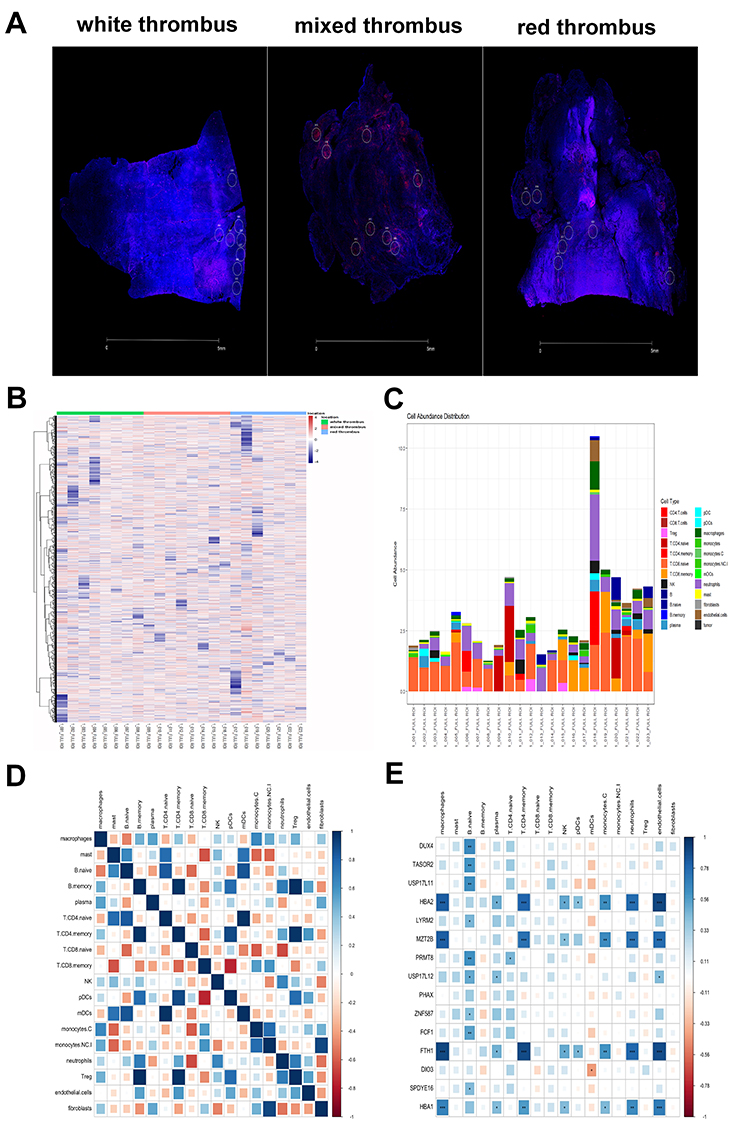 |
Figure 1 (A) Immunofluorescence images of white thrombosis, mixed thrombosis and red thrombosis with CD45-labeled immune cells (red) and SYTO13-labeled nuclei (blue). Regions of interest (ROI) were selected based on morphological marker antibodies. (B) Heat map of expression matrix of whole gene assay results in all regions of interest (ROI). (C) Abundance map of immune/inflammation cell subsets for each ROI. (D) Correlation matrix plot of the abundance of immune/inflammation cell subsets in white, mixed and red thrombosis. (E) Heat map of correlation between immune/inflammation cell abundance and gene expression. *p<0.05; **p<0.01; ***p<0.001. |
The distribution of immune/inflammation cell subsets was different in white, mixed and red thrombosis (Figure 1C). The first ten cells in white thrombosis in descending order of cell abundance were T.CD8.naive, neutrophils, T.CD4.memory, plasma, macrophages, mast cells, pDCs, T.CD8.memory, Treg, and NK, and in mixed thrombosis were T.CD8.naive, neutrophils, T.CD4.naive, T.CD8.memory, macrophages, Treg, NK, B.naive, mast, monocytes.NC, and in red thrombosis were T.CD8.naive, neutrophils, T.CD8.memory, T.CD4.memory, macrophages, T.CD4.naive, endothelial cells, B.naive, plasma, NK. The abundance of endothelial cells, CD8 naive T cells, and macrophages in red thrombosis was significantly higher than in mixed and white thrombosis. Correlation analysis showed a strong positive correlation for immune/inflammation cell subsets in red thrombosis (Figure 1D), a diminished positive correlation in mixed thrombosis (Supplementary Figure 9), and a predominantly negative correlation in white thrombosis (Supplementary Figure 10). Correlation analysis of cell abundance and gene expression showed that USP17L11 and USP17L12 were mainly expressed in B-type naïve cells, plasma and endothelial cells, and HBA1, HBA2 and FTH1 were mainly expressed in macrophages, plasma, CD4 memory T cells, NK, pDCs, monocyte C, neutrophils and endothelial cells (Figure 1E).
Discussion
This study reports the first study of arterial thrombosis and immune/inflammatory cells using DSP technology. The results show that DSP technology can achieve high-throughput sequencing analysis using a small number of samples and can greatly improve the efficiency of the study. The abundance of immune/inflammatory cell subsets was varied at different stages of thrombosis. The differentially expressed genes were associated with apoptosis, cell death, regulation of cell proliferation and immune response, which were significantly enriched in signaling pathways associated with binding and uptake of ligands via scavenger receptors.
Previous studies have verified that neutrophils are involved in thrombosis through extracellular traps.6 In addition, numerous studies have confirmed the predictive role of NLR for diagnosis and prognosis of thrombotic diseases.7 The findings in our study revealed that the highest abundance of immune/inflammatory cell subsets at different stages of thrombosis was lymphocytes and neutrophils. This is in agreement with previous studies and suggests that lymphocytes and neutrophils may play a major role in thrombosis progression. Previous studies have confirmed that activation of endothelial cells exposes vascular hemophilic factor, integrins and other receptors that interact with activated platelets, red blood cells and coagulation factors, ultimately leading to thrombosis.8 In our study, endothelial cells were also significantly more abundant in red thrombi than in other stages of thrombosis, suggesting an important role of endothelial cells in the initial stages of thrombosis. Previous studies have shown that macrophages undergo phenotypic transformation during thrombosis and play a key role in the initiation phase and subsequent lysis of thrombi by stimulating endothelial cell-mediated adhesion molecules, which are mainly involved in the chemotactic aggregation of inflammatory cells.9 Our study also showed that the number of macrophages was significantly higher in red thrombi and gradually decreased with thrombosis progression.
Our study showed there were differential genes in different stages of thrombotic evolution. And among them, DUX4, USP17L11, USP17L12, HBA1, HBA2 and FTH1 were associated with apoptosis, cell death, regulation of cell proliferation, and immune response. Previous studies suggested that platelets have the ability to transfer cytoplasmic RNA during the regulation of vascular homeostasis, with increased expression of HBA1/HBA2 directly related to transport.10 However, the relationship between DUX4, USP17L11, USP17L12, FTH1 and thrombosis has not been verified. These genes deserve further study as suggested by our study. Pathway enrichment analysis revealed significant enrichment of differential genes in the binding and uptake of ligands by scavenger receptor signaling pathway. Scavenger receptors (SR) are an important family of pattern recognition receptors in intrinsic immunity that mediate the major pathway of cellular uptake of oxidatively modified low-density lipoprotein, which is a critical factor in foam cell formation and atheromatous plaque lesion progression. SR ligands existing in the circulation under pathological conditions interact with platelet SR and modulate platelet reactivity, which may lead to acute cardiovascular events.11 Our study suggests that scavenger receptor-related signaling pathways may be an important link in thrombus formation and evolution, which warrants further investigation.
Our study also has some limitations. The patient number is limited, so these preliminary findings need to be validated in more samples. The differential genes and signaling pathways also need to be further investigated in vivo and in vitro experiments to explore the underlying mechanism.
Conclusion
The DSP technique was applied in this study to investigate the distribution difference of immune/inflammation cell subsets during thrombosis progression, as well as to identify potential signaling pathways and differentially expressed genes. The results demonstrated that DSP can facilitate efficient analysis using very few samples and is an important new tool to study thrombosis and inflammation.
Data Sharing Statement
The datasets used and/or analysed during the current study are available from the corresponding author on reasonable request.
Ethical Approval and Consent to Participate
The study was conducted in accordance with the Declaration of Helsinki (as revised in 2013). It was approved by the Ethics Committee of Qilu Hospital of Shandong University, and written consent was obtained from the study participants prior to study commencement.
Funding
This report is supported by the National Natural Science Foundation of China (Grant No.82000451).
Disclosure
The authors declare that the research was conducted in the absence of any commercial or financial relationships that could be construed as a potential conflict of interest.
References
1. Koupenova M, Clancy L, Corkrey HA, Freedman JE. Circulating platelets as mediators of immunity, inflammation, and thrombosis. Circ Res. 2018;122(2):337–351. doi:10.1161/CIRCRESAHA.117.310795
2. Lippi G, Favaloro EJ. Venous and arterial thromboses: two sides of the same coin? Semin Thromb Hemost. 2018;44(3):239–248. doi:10.1055/s-0037-1607202
3. Chilingaryan Z, Deshmukh T, Leung HHL, et al. Erythrocyte interaction with neutrophil extracellular traps in coronary artery thrombosis following myocardial infarction. Pathology. 2022;54(1):87–94. doi:10.1016/j.pathol.2021.05.099
4. Marx C, Novotny J, Salbeck D, et al. Eosinophil-platelet interactions promote atherosclerosis and stabilize thrombosis with eosinophil extracellular traps. Blood. 2019;134(21):1859–1872. doi:10.1182/blood.2019000518
5. Merritt CR, Ong GT, Church SE, et al. Multiplex digital spatial profiling of proteins and RNA in fixed tissue. Nat Biotechnol. 2020;38(5):586–599. doi:10.1038/s41587-020-0472-9
6. Döring Y, Soehnlein O, Weber C. Neutrophil extracellular traps in atherosclerosis and atherothrombosis. Circ Res. 2017;120(4):736–743. doi:10.1161/CIRCRESAHA.116.309692
7. Xue J, Ma D, Jiang J, Liu Y. Diagnostic and prognostic value of immune/inflammation biomarkers for venous thromboembolism: is it reliable for clinical practice? J Inflamm Res. 2021;14:5059–5077. doi:10.2147/JIR.S327014
8. Bochenek ML, Schütz E, Schäfer K. Endothelial cell senescence and thrombosis: ageing clots. Thromb Res. 2016;147:36–45. doi:10.1016/j.thromres.2016.09.019
9. Jiang P, Xue D, Zhang Y, et al. The extrinsic coagulation cascade and tissue factor pathway inhibitor in macrophages: a potential therapeutic opportunity for atherosclerotic thrombosis. Thromb Res. 2014;133(4):657–666. doi:10.1016/j.thromres.2014.01.012
10. Risitano A, Beaulieu LM, Vitseva O, Freedman JE. Platelets and platelet-like particles mediate intercellular RNA transfer. Blood. 2012;119(26):6288–6295. doi:10.1182/blood-2011-12-396440
11. Ma Y, Ashraf MZ, Podrez EA. Scavenger receptor BI modulates platelet reactivity and thrombosis in dyslipidemia. Blood. 2010;116(11):1932–1941. doi:10.1182/blood-2010-02-268508
 © 2023 The Author(s). This work is published and licensed by Dove Medical Press Limited. The
full terms of this license are available at https://www.dovepress.com/terms
and incorporate the Creative Commons Attribution
- Non Commercial (unported, 3.0) License.
By accessing the work you hereby accept the Terms. Non-commercial uses of the work are permitted
without any further permission from Dove Medical Press Limited, provided the work is properly
attributed. For permission for commercial use of this work, please see paragraphs 4.2 and 5 of our Terms.
© 2023 The Author(s). This work is published and licensed by Dove Medical Press Limited. The
full terms of this license are available at https://www.dovepress.com/terms
and incorporate the Creative Commons Attribution
- Non Commercial (unported, 3.0) License.
By accessing the work you hereby accept the Terms. Non-commercial uses of the work are permitted
without any further permission from Dove Medical Press Limited, provided the work is properly
attributed. For permission for commercial use of this work, please see paragraphs 4.2 and 5 of our Terms.