
Back to Journals » ImmunoTargets and Therapy » Volume 14
The Role of Intestinal Microbiota and Immune System Interactions in Autoimmune Diseases
Authors Yurtseven B  , Aydemir E , Ayaz F
Received 20 September 2025
Accepted for publication 19 November 2025
Published 26 November 2025 Volume 2025:14 Pages 1347—1372
DOI https://doi.org/10.2147/ITT.S569016
Checked for plagiarism Yes
Review by Single anonymous peer review
Peer reviewer comments 2
Editor who approved publication: Professor Michael Shurin
Batuhan Yurtseven,1 Esra Aydemir,2 Furkan Ayaz2,3
1Department of Molecular Biology and Genetics, Faculty of Engineering and Natural Sciences, Biruni University, Istanbul, 34010, Türkiye; 2Department of Molecular Biology and Genetics, Faculty of Engineering and Natural Sciences, Istinye University, Istanbul, 34396, Türkiye; 3Odesa National Medical School, Department of Medical Biology and Chemistry, Odesa, Ukraine
Correspondence: Esra Aydemir, Email [email protected] Furkan Ayaz, Email [email protected]
Background: The intricate interplay between the intestinal microbiota and the immune system has emerged as a central theme in understanding autoimmune disease pathogenesis. This review comprehensively explores the role of gut microbiota in shaping immune development, establishing immune tolerance, and contributing to both local and systemic immune regulation.
Methods: This review synthesizes the modulatory effects of microbial metabolites (eg, short-chain fatty acids and indole derivatives) on regulatory T cells (Tregs) and inflammatory pathways. The concept of “dysbiosis” is examined from functional and compositional perspectives, linking microbial imbalances to autoimmune disorders (IBD, MS, RA, and T1D). Microbiota-targeted therapeutic interventions (probiotics, prebiotics, FMT) are also evaluated.
Key Findings: The synthesis of the literature confirms that microbial metabolites have a direct impact on Treg differentiation and inflammatory pathways. Dysbiosis, through functional and compositional disruptions, is strongly associated with the pathogenesis of various autoimmune disorders, including Inflammatory Bowel Disease, Multiple Sclerosis, Rheumatoid Arthritis, and Type 1 Diabetes. Therapeutic interventions such as probiotics, prebiotics, and Fecal Microbiota Transplantation show promising potential in restoring microbial and immune homeostasis.
Conclusion: This review highlights the role of the gut-immune axis in autoimmune diseases. Despite current challenges, such as individual variability and determining causality, future directions toward precision microbiota and immune modulation are promising. This study provides a robust foundation for researchers and clinicians seeking to understand and therapeutically target the gut-immune axis.
Keywords: gut microbiota, immune system, autoimmune diseases, dysbiosis, regulatory T cells, Tregs
Introduction
The goal of this review is to gather and thoroughly examine the ways in which the immune system and microbiome interact. This study aims to provide a comprehensive overview that will guide basic science researchers, clinicians, and other experts interested in the microbiota-immunology relationship.
The immune system’s growth and homeostasis maintenance are just two of the many areas in which the microbiota is essential. It is known that a healthy microbiota balance is important in the control of inflammatory responses by supporting immune tolerance. However, an imbalance in the microbiota known as dysbiosis can lead to immune system dysfunction, playing a significant role in the development of autoimmune diseases.
In this context, first of all, the historical development of microbiota and immune system interactions will be examined and the effects of microbiota on the immune system will be detailed. Then, the molecular and cellular mechanisms of microbiota imbalance in specific autoimmune diseases will be evaluated. In addition, the effects of microbiota-derived metabolites, especially short-chain fatty acids (SCFA) and other immunomodulatory molecules on the immune system will be analyzed. In this review, we will focus on the clinical consequences of these interactions and describe microbiota-targeted therapeutic approaches, prebiotics, probiotics, and treatment methods such as fecal microbiota transplantation. Finally, we will discuss the gaps in current research on microbiota-immune system interactions and offer suggestions for future studies in this area.
The Gut-Immune Axis: A Symbiotic Nexus of Homeostasis
The human gastrointestinal tract represents the most extensive and dynamic interface between the host and the external environment. Far from being a passive digestive organ, it functions as a primary immunological barrier, housing the vast majority of the body’s immune cells. This mucosal surface is co-habited by a dense and complex ecological community of trillions of microorganisms—the gut microbiota.1 The co-evolution of the mammalian immune system with this microbial consortium has resulted in a deeply integrated and symbiotic relationship, often termed the “gut-immune axis”, which is fundamental to systemic immune homeostasis (Figure 1).2
 |
Figure 1 The Gut-Immune Axis: A Symbiotic Nexus of Homeostasis. This dual-panel schematic illustrates the critical balance between the gut microbiota and the host immune system in both (A) Healthy Homeostasis and (B) Dysbiosis & Immune Dysregulation. (A) Healthy Homeostasis (Left Panel): Commensal bacteria reside within the gut lumen, interacting with the intestinal epithelium. This barrier is maintained by a robust Mucus Layer and intact Tight Junctions, preventing uncontrolled microbial translocation. Beneath the epithelium, in the lamina propria, antigen-presenting cells (eg, Dendritic Cells (DCs) and Macrophages) sample microbial signals. Commensal bacteria produce beneficial metabolites like Short-Chain Fatty Acid (SCFAs), which are crucial for the differentiation and expansion of anti-inflammatory Regulatory T cells (Tregs). These Tregs, along with other immune cells (B cells, Th-17 cells), maintain a delicate balance, promoting immune tolerance and suppressing excessive inflammation through cytokines such as IL-10 and TGF-β. (B) Dysbiosis & Immune Dysregulation (Right Panel): In a state of dysbiosis, there is an imbalance in the gut microbiota, often characterized by an increase in Pathobionts and a decrease in beneficial Commensal Bacteria. This can lead to a compromised mucus layer and disrupted tight junctions, resulting in increased intestinal permeability and microbial translocation (leaky gut). This leads to an overproduction of pro-inflammatory cytokines like IL-6 and IL-17. The altered microbial metabolism produces fewer beneficial SCFAs, which in turn reduces Treg induction. This shift favors the differentiation of pro-inflammatory Th17 cells and an overall increase in systemic inflammation. |
This dialogue is bidirectional and essential for immune calibration. The gut-associated lymphoid tissue (GALT), the largest lymphoid organ in the body, is strategically positioned to sample microbial antigens. This constant sampling “educates” the mucosal immune system to maintain a state of tolerance towards commensal organisms while remaining poised to attack pathogens.2 Conversely, the microbiota is indispensable for the maturation and function of the immune system. Germ-free animal models, for example, exhibit profound immunological defects, including underdeveloped lymphoid tissues and impaired immune cell functionality.3 Mechanistically, this cross-talk is mediated not just by cellular contact but by a complex biochemical language of microbial-derived metabolites. Short-chain fatty acids (SCFAs), such as butyrate, propionate, and acetate, are produced by the fermentation of dietary fiber. These metabolites are crucial signaling molecules, most notably by promoting the differentiation and expansion of anti-inflammatory regulatory T cells (Tregs) through mechanisms like histone deacetylase (HDAC) inhibition.4,5 Concurrently, microbial metabolites derived from tryptophan, such as indole, signal through the aryl hydrocarbon receptor (AhR) on immune and epithelial cells. This signaling is critical for maintaining epithelial barrier integrity and regulating the balance between pro-inflammatory Th17 cells and anti-inflammatory Treg cells.6
Therefore, immune tolerance is not a default state but an actively maintained process, critically dependent on a balanced microbial community. The disruption of this symbiosis, termed “dysbiosis”, represents a functional and compositional failure of this ecosystem.7 Dysbiosis is characterized not only by compositional shifts (eg, loss of beneficial taxa or pathobiont blooms) but also by functional alterations (eg, diminished SCFA production, impaired tryptophan metabolism, and compromised barrier integrity).8 This functional impairment is increasingly recognized as a central driver of immune dysregulation.
A breakdown in the gut-immune axis, initiated by dysbiosis, can lead to the erosion of immune tolerance and the promotion of chronic inflammatory pathways that underpin autoimmunity.9 While many reviews have independently described the gut microbiota or the mechanisms of autoimmunity, a critical research gap persists in elucidating the precise causal mechanisms linking functional dysbiosis to the initiation and exacerbation of specific autoimmune diseases. It is increasingly clear that dysbiosis is not merely a consequence of established disease or treatment, but an active contributor to pathogenesis.10 This review is positioned to synthesize these complex interactions, moving beyond basic definitions to explore the functional consequences of a disrupted gut-immune axis. By examining how this loss of homeostasis manifests in prevalent autoimmune conditions, we establish the framework for understanding the significant burden of these disorders in modern medicine.
Prevalence of Autoimmune Diseases and Their Importance in Modern Medicine
Autoimmune diseases (AIDs) are more than 80 chronic and often disabling disorders in which the immune system attacks and damages organs, tissues and cells for no apparent reason. They are usually characterized by impaired immune tolerance, the presence of high titers of autoantibodies and/or the induction of inflammation. As a result, chronic inflammation occurs and thus different diseases develop that target different areas of the body. This impaired immune response is due to the body’s inability to become tolerant to its own antigens.11,12 Autoimmune diseases affect 3–5% of the population worldwide.13 Figure 2 maps the spectrum of autoimmune diseases, distinguishing organ-specific disorders from systemic conditions. The emergence of these diseases is based on the combination of genetic predisposition and environmental factors.14 The pioneering work developed by Macfarlane Burnett approximately 50 years ago created an important understanding of autoimmunity by discovering the mechanisms used by the immune system to respond to its own antigens. Treatment methods for autoimmune diseases have improved significantly to date, and the response to medical treatment of diseases such as rheumatoid arthritis has improved.15 Autoimmune disorders can be organ-specific, such as autoimmune thyroid disease, or manifest in systemic forms with a wider area of effect, as exemplified by systemic lupus erythematosus.16 In addition, great progress has been made in diagnosis and disease classification in recent years with the development of molecular immunology and clinical laboratory tests.15 It has also been found that autoimmune diseases are higher in women and ethnic groups.16 The importance of diseases such as autoimmune diseases in medicine increases with the complexity of their diagnosis and treatment; these diseases, which we mentioned, are triggered by the interaction of genetic and environmental factors, are addressed with a multidisciplinary approach and more effective treatment methods are developed using biotechnological innovations.17 In particular, research on the pathogenesis of diseases such as rheumatic diseases, thyroid disorders and multiple sclerosis aims to increase the quality of life of patients with treatments targeting the immune system.16
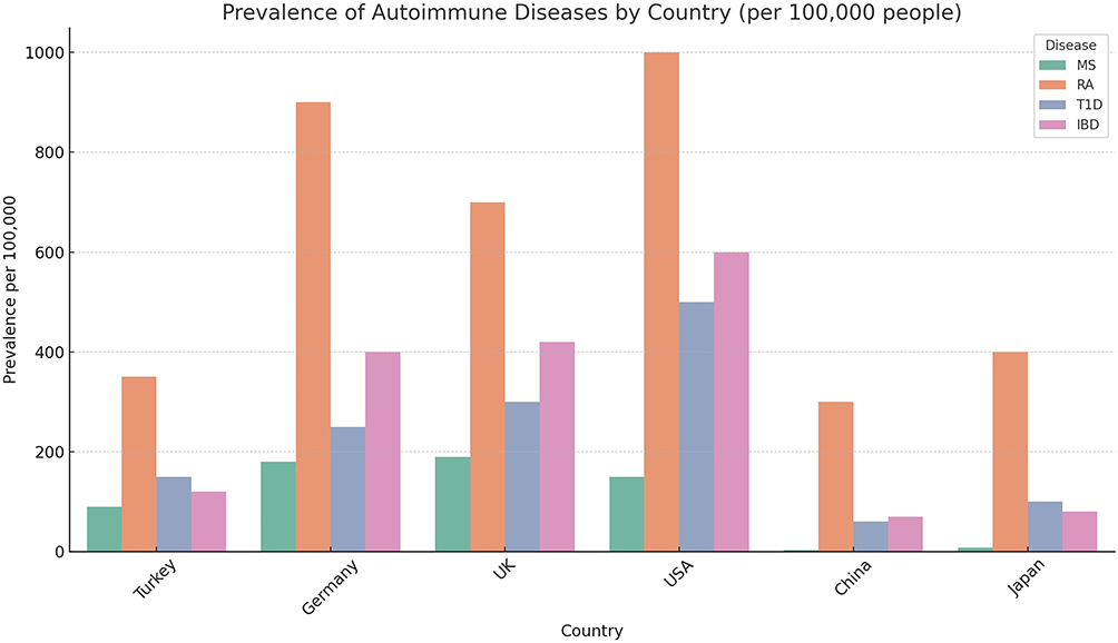 |
Figure 2 Prevalence of Selected Autoimmune Diseases in Specific Countries. This grouped bar chart compares the prevalence (cases per 100,000 people) of four common autoimmune diseases across six different countries: Turkey, Germany, the UK, the USA, China, and Japan. The Y-axis represents the prevalence (ranging from 0 to 1000), while the X-axis denotes the countries. The four bars within each country represent Multiple Sclerosis (MS, green), Rheumatoid Arthritis (RA, Orange), Type 1 Diabetes (T1D, blue), and Inflammatory Bowel Disease (IBD, pink), respectively. The data illustrate the global burden and geographical distribution of autoimmune diseases, highlighting that RA and IBD show high prevalence in Western countries (eg, USA, Germany), while MS prevalence varies across the studied populations. |
Microbiota and Immune Development
Microbiota Shapes the Immune System From Birth
The immune system begins to form its foundations during the prenatal period, and it is increasingly supported by evidence that signals from the maternal microbiota may have an impact on the immune development of the fetus.18 Recent studies have emphasized the role of the maternal microbiota in the development of the fetal immune system. This interaction begins with the integration of environmental and internal factors that contribute to the development of the two main branches of the immune system, the innate and adaptive immune systems, during embryonic organogenesis.19,20 Studies in mice have revealed that various organs and anatomical regions, such as the placenta, fetal liver, thymus, and bone marrow, play important roles in the development of the immune system.19,21 Studies on the effect of the maternal microbiota on the fetus reached a turning point with the detection of the presence of bacterial DNA in fetal tissues during pregnancy.18 According to these results, microbial molecules start to impact the fetal immune system in the uterus throughout pregnancy, and fetal microbial colonization is not limited to the postnatal phase. It has been determined that babies born vaginally are colonized with maternal vaginal and fecal microbiota (rich in immune-educating species like Lactobacillus and Bifidobacterium), while babies born via cesarean section are colonized primarily with microbes from the hospital environment and skin (such as Staphylococcus species) (Ignacio et al, 2024). This fundamental difference in initial microbial contact is thought to critically shape the trajectory of immune tolerance and development. Studies on this first microbial contact help us understand the role of the maternal microbiota in early.19 Studies on this first microbial contact help us understand the role of the maternal microbiota in early life. In the postnatal period, when the newborn immune system encounters both microbes and environmental antigens, the microbiota plays a very important role in regulating these interactions.18,19 Breast milk is the primary vehicle of pre- and postnatal immune transfer and has important effects on the fetal immune system.18,22 Figure 3 shows how successive stages of microbial exposure—prenatal, birth, breastfeeding, and weaning—drive the maturation of mucosal and systemic immunity, highlighting that early-life microbial colonization coincides with critical windows for Treg induction and mucosal immune education. Immunoglobulin G (IgG) and other protective factors in breast milk help direct the immune responses of the newborn, and interactions with the microbiota can have long-term effects on the development and function of immune cells.23 Another important finding associated with the maternal microbiota is how the maternal diet can affect the evolution of microbes and, as a result, the immune system of the newborn.19,21 Maternal diet is a particularly critical modulator; low-fiber diets, for example, have been shown to negatively impact early-life immune cell development by altering the gut microbiome. Conversely, high-fiber diets support immune cell differentiation and strengthen the immune system, primarily through the production of short-chain fatty acids (SCFAs).19 Finally, studies on early microbial colonization show that the development of the microbiota leads to significant changes during the weaning stage, a critical period in the formation of the immune system.18,19 During this period, when microbial diversity increases and microbes are more exposed, the proliferation and differentiation of immune cells accelerate.22,24 Furthermore, gene expression linked to the microbiome influences how the immune system develops and how diseases manifest in later life (Ignacio et al, 2024; Belkaid & Hand, 2014). These results demonstrate that environmental variables, particularly maternal nutrition and microbiota, are critical in immune system development and may influence illness risk in later life. However, understanding exactly how these processes work requires a more in-depth examination of the interaction between the immune system and the microbiome.
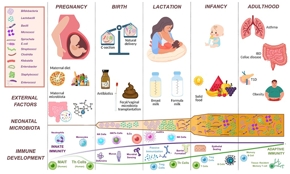 |
Figure 3 Immune System Maturation and Microbial Milestones. This schematic diagram illustrates the critical windows of human immune system development from the prenatal stage to adulthood, and how these processes are influenced by external factors and the neonatal microbiota. The top timeline depicts the stages of Pregnancy, Birth, Lactation, Infancy, and Adulthood. These stages are correlated with the immune development timeline below, which is divided into Innate Immunity and Adaptive Immunity. The figure shows how external factors such as birth mode (Cesarean vs Natural Delivery), nutrition (Breast milk vs Formula milk), and antibiotic use shape the gut microbiota composition (Neonatal Microbiota). This microbial colonization runs parallel to critical immunological events, including the induction of Treg cells, the education of mucosal immunity, and the maturation of adaptive immunity (eg, Memory T cells, B cells). |
Development of Innate Immune Cells
The development of innate immune cells is shaped by the influence of microbes and microbial antigens.19 Monocytes and macrophages, in particular, play an important role in the maturation of the immune system.25 During development, in response to environmental cues from host tissues, local self-renewing populations are established, and these populations are seeded by embryonic liver and yolk sac macrophage precursors.26 For example, intestinal macrophages are continuously renewed by Ly6Chi monocytes derived from the bone marrow, a process driven by postnatal microbial colonization.19
The absence of microbial interactions prevents the maturation of the immune system.27 In germ-free (GF) mice, the absence of microbiota leads to severe defects in the structural and functional development of the immune system.19,21 For example, GF mice exhibit defects in lymphoid tissue development and morphological abnormalities in the intestinal epithelium.21 Interaction with microbiota directly affects structures, particularly gut-associated lymphoid structures (GALTs) and Peyer’s patches.21 Innate lymphoid cells (ILCs) modulate immune responses independently of the microbiota.28 However, the microbiota regulates the activation and functions of ILCs.21 For example, phenotypic activation of natural killer (NK) cells is reduced in GF mice, which can be corrected by colonizing adult mice with microbiota.2,19 However, the formation of these cells in GF mice has been the subject of conflicting findings from various investigations.21,27
The microbiota exerts a significant influence on the expression of cytokines, especially IL-22, and these interactions are essential for the proper functioning of the immune system. These data highlight the critical role of the microbiota in the development of innate immune cells and the maturation of the immune system. The process of microbial colonization is essential for the proper development of the immune system, and mounting evidence suggests a direct link between this colonization and immune functionality.
Immune Tolerance and Microbiota
The microbiota, as a critical ecosystem that supports immune tolerance in the intestinal mucosa and works in a symbiotic relationship with the body’s immune system, plays a vital role in both protecting the body against pathogens and maintaining immune balance.27 Microorganisms in the intestine not only contribute to digestive processes, but also protect the body from autoimmune diseases and excessive inflammation by suppressing excessive reactions of the immune system.29,30 In this process, the interaction between regulatory T cells (Treg) and microbiota is a decisive mechanism. Metabolites like short-chain fatty acids (SCFA) generated by the microbiota inhibit excessive responses of the mucosal immune system by enhancing Treg cell activation, fortifying the intestinal barrier’s integrity, and promoting the establishment of immune tolerance (Izcue et al, 2006). Treg cells ensure a healthy relationship between the immune system and the intestinal microecosystem and maintain the symbiotic balance between these two systems.31,32 This delicate balance between microbiota and immune tolerance serves a unique function in regulating the immune responses of the organism by enabling commensal microorganisms living in the gut to survive without being recognized as pathogens by the immune system.29,33
Characteristics and Basic Functions of Treg Cells
Regulatory T cells (Treg) and conventional T helper cells (Th) are the two subtypes of CD4+ T cells (Corthay, 2009). The immune system is regulated in large part by these cells. While Treg cells reduce excessive immune system responses and help prevent autoimmune diseases, conventional Th cells mediate adaptive immune responses by activating other effector cells like CD8+ cytotoxic T cells, B cells, and macrophages (Corthay, 2009; Vignali et al, 2008). Treg cells are defined as CD4+ T cells that suppress the potentially harmful activities of Th cells, but the identification of these cells is fraught with difficulties because it has been demonstrated that the markers currently used (eg Cluster of Differentiation 25 (CD25), Cytotoxic T-Lymphocyte-Associated protein 4 (CTLA-4), Glucocorticoid-Induced TNFR-related protein (GITR), Lymphocyte-Activation Gene 3 (LAG-3), Cluster of Differentiation 127 (CD127) and Forkhead box P3 (Foxp3)) are not specific to Treg cells but represent general T cell activation markers.34 Treg cell antigen-dependent activation has suggested that the suppressive mechanisms of these cells are somehow linked to antigen specificity.34,35 Treg cells are thought to be self-reactive, yet extensive TCR repertoire analyses point out that this is a rather exceptional feature and not a unique ability of suppressive T cells.34,36 Treg cells recognize antigenic peptides in the context of MHC class II molecules through their somatically rearranged T cell receptors and must be activated via the TCR to perform their suppressive activity.34,37 Although functionally known as cells which only suppress other Th cells, these cells have various subtypes to form an effective immune regulatory mechanism. For instance, the nTregs normally target self-antigens and control the autoimmune inflammation, whereas adaptive or induced Treg cells, on the other hand, give a suppressive response toward foreign antigens or neoantigens.38 The influence of Treg cells does not merely aim at taming the immune responses of the individual but also their limitation prevents the organism from destroying its own cells in certain circumstances.34,39 These cells interact with small proteins, including cytokines, to modulate their activation and, in this way, provide a critical control mechanism against inappropriate immune responses.34,40 Therefore, by reducing immune system overreactions and preserving immunological tolerance, Treg cells are essential for preventing autoimmune illnesses and preserving the equilibrium of immune responses.
Mechanisms of Treg Cells
The regulatory functions of T cells interact particularly with regulatory T cells (Tregs), and these cells suppress immune responses, helping to prevent autoimmune diseases.39 Figure 4 depicts the principal suppressive mechanisms of regulatory T cells, including inhibitory cytokine secretion (IL-10, IL-35, TGF-β), checkpoint interactions (CTLA-4), and metabolic disruption. Inhibitory cytokines such as IL-35, IL-10, and TGF-β released from Treg cells are among the factors that control and balance immune responses.39 These cytokines help regulate immune responses by preventing excessive activation and are essential for preserving immune tolerance.41,42 Interestingly, IL-35 suppresses the immune system and regulates inflammation, and has an effect that provides immune tolerance in particular.39 This effect alleviates intestinal inflammation, prevents the progression of autoimmune diseases, and provides immune balance. The gut environment is crucial in modulating immune responses, with probiotics particularly microbes like Bacteroides fragilis playing a key role in this process.42 The pathogen-associated molecular patterns antigens produced by these probiotics are among the important factors that provide immune tolerance in the intestinal microenvironment.42 PSA promotes the development of Treg cells via dendritic cells and increases IL-10 production, which helps the immune system to mount a tolerogenic response.43 This mechanism demonstrates the effect of the gut microbiota in immune regulation. The effects of the gut microbiome on the immune system are particularly related to the activity of cytokines such as IL-10 and IL-35.39 However, in individuals with IL-10 receptor mutations or IL-10 deficiency, the effect of probiotics such as Lactobacillus species in preventing colitis becomes more pronounced.42 The cross-regulations that different subsets of T cells establish with each other determine how the immune response will be shaped.34 For example, IFN-γ produced by Th1 cells suppresses the activation of Th2 cells, and conversely, IL-4 production by Th2 cells limits the development of Th1 cells.34 These mutual interactions ensure that the immune response remains balanced. In addition, IL-17 production by Th17 cells inhibits the activity of Th1 cells, while IL-21 released from Th2, Th17 and Tfh cells inhibits the functions of Th1 cells.34 All these T cell subgroups have the capacity to produce IL-10, indicating that all CD4+ T cells, not just Tregs, have the ability to suppress the immune response.44 These interactions ensure that the immune system functions harmoniously and flexibly.
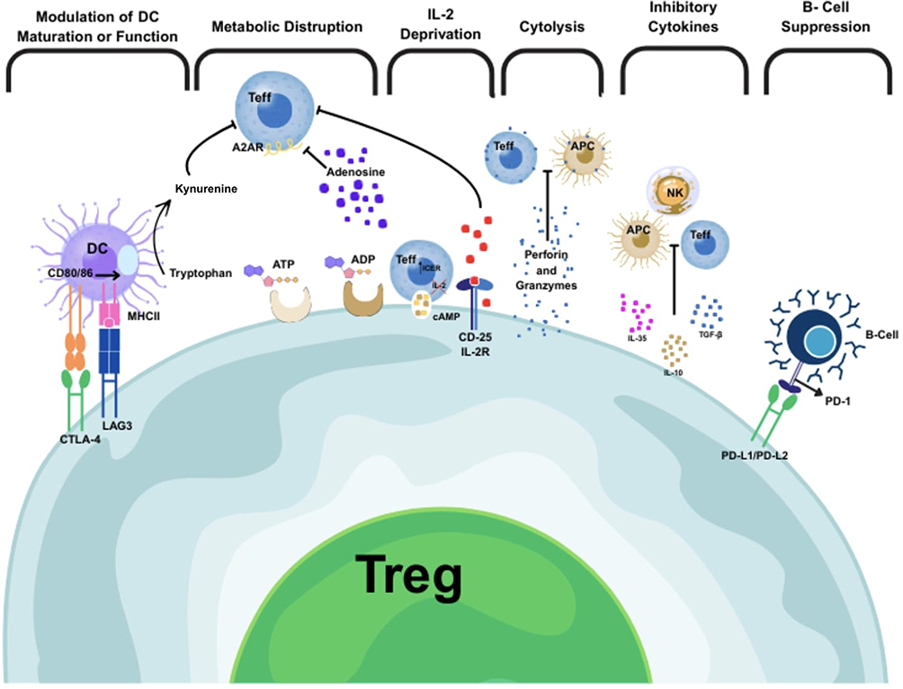 |
Figure 4 Suppressive Mechanisms of Regulatory T (Treg) Cells. This figure illustrates the multifaceted mechanisms employed by Regulatory T (Treg) cells to suppress the immune response. These mechanisms include: Inhibitory Cytokines, wherein Tregs secrete anti-inflammatory cytokines like IL-10, TGF-β, and IL-35 to inhibit the function of other immune cells (eg, M1 Macrophages, T cells); Dendritic Cell (DC) Modulation, where Tregs suppress the antigen-presenting capacity and maturation of DCs via checkpoint molecules like CTLA-4 and LAG3; Metabolic Disruption, in which Tregs outcompete effector T cells by consuming IL-2 via their high-affinity receptors (CD25) (“IL-2 deprivation”) or by converting ATP to immunosuppressive adenosine; Cytolysis (a known mechanism, though not explicitly depicted), allowing Tregs to kill effector T cells or APCs directly; and B-Cell Suppression, where Tregs can directly inhibit B-cell proliferation and antibody production. These pathways, along with others like tryptophan metabolism (Kynurenine production), allow Tregs to maintain immune tolerance. |
Treg cells control immune responses through various pathways, such as inhibitory cytokines such as IL-10 and TGF-β, as well as cellular interactions and metabolic disruptions.42 IL-10, in particular, suppresses allergic responses and inflammation when released from Treg cells, and also plays an important role in the prevention of inflammatory bowel diseases (IBD) and airway hyperreactivity.42 IL-10 also effectively directs the immune response in the tumor microenvironment and stands out as a potential strategy in the fight against cancer. Another important cytokine, TGF-β, is effective in suppressing Th1 cells and also regulates immune responses.34 TGF-β helps the immune system maintain balance with both immunostimulatory and immunosuppressive effects.34,42
All these dynamic interactions indicate complementary mechanisms in the regulation of the immune system. The regulatory role of T cells, especially Treg cells, is of great importance in maintaining immune tolerance and limiting inflammation.45 The suppressive role of IL-35 in the immune system supports immune tolerance while also allowing inflammation to be kept under control.39 This supervisory effect provided by Treg cells with cytokines such as IL-35, IL-10 and TGF-β allows for the precise management of immune responses and sheds light on new treatment approaches in the treatment of diseases such as autoimmune diseases, allergic conditions and cancer.39,46 These effects provided by IL-35 by Treg cells provide a structure that provides balance between different cell types of the immune system and enables therapeutic interventions.
The Importance of Treg Cells in Achieving Immune Tolerance
Regulatory T cells (Tregs) are a special subset of CD4+ T cells that play a critical role in sustaining immune homeostasis and averting autoimmune reactions.45 They perform their role by inhibiting excessive immune activation, preserving peripheral tolerance, and modulating inflammatory reactions to self and non-pathogenic antigens.47
Regulatory T (Treg) cells consist of two subsets: natural Tregs (nTregs), which mature in the thymus, and induced Tregs (iTregs), which mature in peripheral tissues under tolerance-favoring conditions that frequently involve transforming growth factor-β (TGF-β) and interleukin-2 (IL-2).48 Both types perform immunosuppressive roles through numerous mechanisms, including: Secretion of anti-inflammatory cytokines (IL-10, TGF-β, IL-35), cytolysis of effector T cells through granzyme and perforin, metabolic disruption of effector cells through IL-2 consumption, dendritic cell modulation by CTLA-4 and LAG-3 interactions.45 Figure 5 shows how environmental and microbial signals dynamically regulate T cell fate, shaping the Th1/Th2/Th17/Treg landscape implicated in autoimmunity.
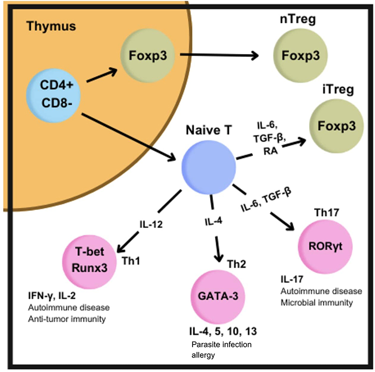 |
Figure 5 T Cell Differentiation Pathways and Autoimmunity. This diagram demonstrates how a Naive T cell differentiates into various T helper (Th) subsets (Th1, Th2, Th17) or a Regulatory T cell (Treg) based on the specific cytokine signals it encounters. The Th1 pathway, associated with anti-tumor immunity and some autoimmune diseases, is triggered by IL-12 and defined by T-bet/Runx3 transcription factors, producing IFN-γ and IL-2. The Th2 pathway, associated with parasitic infections and allergy, is driven by IL-4 and GATA-3, producing IL-4, 5, 10, and 13. The Th17 pathway, linked to microbial immunity and autoimmune disease, is induced by IL-6 and TGF-β and defined by RORγt, producing IL-17. Conversely, iTregs (induced Tregs), essential for tolerance, are generated via TGF-β and IL-2 (or Retinoic Acid) and express Foxp3, similar to nTregs (natural Tregs) which originate from the thymus. A shift in this balance, particularly favoring Th1/Th17, can disrupt homeostasis and drive autoimmunity. |
In the gastrointestinal tract, Treg cells are found in abundance in the lamina propria, especially in the colon, where they are constantly exposed to antigens from dietary products and the resident microbiota. These cells have a fundamental role in inhibiting inflammatory responses to innocuous antigens and ensuring mucosal tolerance.49 Dysregulation of Treg function or frequency has been implicated in numerous autoimmune diseases, including systemic lupus erythematosus (SLE), rheumatoid arthritis (RA), multiple sclerosis (MS), and type 1 diabetes (T1D). Enhancement of Treg induction or function is therefore a principal therapeutic target for restoring immune homeostasis in autoimmune settings.50,51
Commensal Microbiota Tolerance
The intestinal microbiota can survive harmlessly in the body, which allows the immune system to function healthily without being aggressive towards the microbiota.27,29 However, this tolerance is possible when Treg cells are properly active. Treg cells establish a balance between the microbiota and the immune system, and act on the microbiota in a way that does not disrupt this symbiotic relationship.2,29 Treg cells can also defend against harmful pathogens without disrupting the relationship of the microbiota with harmless microorganisms.2,29
Systemic Effects
The effects of Treg cells in the intestine are not limited to local immunity; they also have an important effect on systemic immunity.52 While regulatory responses are created against microorganisms and pathogens in the intestine, these cells also control immune responses in other parts of the body.27 For example, anti-inflammatory cytokines such as IL-10 released from the intestine suppress systemic inflammation and help prevent autoimmune responses. Treg cells also contribute to maintaining balance by preventing the immune system from overreacting in other parts of the body.53 This allows the immune system to be protected from potentially harmful reactions and to maintain its healthy function.
The Role of Metabolites
The gut microbiota is not only a microbial community, but also a biochemical factory in constant interaction with the immune system.54 The metabolites of the gut microbiota, especially SCFA and indole derivatives, are critical regulators of the immune system.55,56 These metabolites regulate immune responses in both the gut and systemic circulation, thereby maintaining immune tolerance and controlling inflammation.
Short Chain Fatty Acids (SCFA)
SCFAs are short-chain fatty acids such as acetate, propionate, and butyrate produced by the fermentation of dietary fibers by the intestinal microbiota.57,58 These components not only serve as an energy source for intestinal epithelial cells, but also play an important role with their multifaceted effects on the immune system. SCFAs act as biochemical mediators that regulate the interaction between the intestinal microbiota and the immune system.59,60 Among these, butyrate is particularly notable for its regulatory activity on the immune system. Through the differentiation and activation of regulatory T cells, butyrate has been shown to enhance immune tolerance by inhibiting the production of pro-inflammatory cytokines and promoting the release of anti-inflammatory cytokines like IL-10 (Duan et al, 2023; Föh et al, 2022). Moreover, butyrate functions as a histone deacetylase (HDAC) inhibitor, playing a role in epigenetic regulation and consequently influencing gene expression in immune cells.61 Through this mechanism, they regulate excessive immune responses and help suppress inflammation.61
Another critical role of SCFAs is the maintenance of intestinal barrier integrity, thus preventing pathogens and toxic molecules from entering the systemic circulation.62 SCFAs improve tight junctions and nourish intestinal epithelial cells. Accordingly, the intestinal barrier acts as a barrier between the inside and the external environment (Parada Venegas et al, 2019). SCFAs also protect the intestinal surface by increasing mucus production.5,63 Moreover, SCFAs act not only on the intestines but also systemically throughout the body.60 SCFAs absorbed into the circulation play important roles in regulating inflammatory processes in peripheral immune organs such as the lymph nodes and spleen.59,63 In addition, SCFAs may help prevent and manage metabolic disorders by regulating energy metabolism. Figure 6 integrates evidence on how diet- and microbiota-derived metabolites, such as SCFAs and indoles, provide mechanistic links between intestinal ecology and systemic immune outcomes by promoting Treg differentiation, strengthening barrier integrity, and modulating epigenetic programs (eg, HDAC inhibition).
 |
Figure 6 Immunomodulatory Effects of Gut-Derived SCFAs and Indole Derivatives on the Immune System. This schematic details how two key classes of metabolites produced by the gut microbiota regulate the immune system. The Left Pathway (SCFAs) shows that Dietary Fibers are fermented by microbiota to produce Short-Chain Fatty Acids (SCFAs). SCFAs then (a) promote Regulatory T cell (Treg) activation, (b) increase the production of anti-inflammatory cytokines (IL-10, TGF-β), (c) provide epigenetic regulation via Histone Deacetylase (HDAC) inhibition, (d) serve as an energy source for intestinal epithelial cells, (e) enhance tight junction integrity, and (f) suppress systemic inflammation. The Right Pathway (Indole Derivatives) shows that dietary Tryptophan is metabolized by microbiota into Indole Derivatives. These molecules (a) activate the Aryl Hydrocarbon Receptor (AhR), (b) regulate the Treg/Th17 balance, (c) support intestinal epithelial cell regeneration, (d) contribute to mucosal immunity development, and (e) exert anti-inflammatory effects. |
In summary, the effects of SCFAs on the immune system are crucial for regulating both local and systemic immunity. Approaches targeting the enhancement of SCFA production by the gut microbiota hold bright prospects for therapeutic applications in a wide range of health conditions, including inflammatory bowel diseases, metabolic disorders, and autoimmune diseases. Therefore, the positive effects of dietary modifications aimed at supporting the gut microbiota and promoting the production of short-chain fatty acids (SCFA) are gaining importance in terms of overall health and immune system function.
Indole Derivatives
The microbiota produces a number of bioactive molecules, referred to as indole derivatives, as a result of the metabolism of dietary tryptophan.64 Indole derivatives interact with aryl hydrocarbon receptors (AhR) on the surface of immune cells, helping to finely tune immune responses.65 AhR activation makes sure that immune cells are balanced, but also takes a front-line position in preventing inflammation.2,65 In particular, the activation of AhR promotes an increase in the number of Treg cells, while ensuring the functioning balance of T helper cell 17 (TH17) cells with pro-inflammatory properties.66 This balance is of great importance for suppressing excessive immune system reactions and preventing autoimmune responses. Also, the derivatives of indole take part in the restoration of intestinal epithelium and promote strengthening of the mucous immune system.67 Epithelial renewal, a promotion process, provides a protective barrier for the intestines, preventing harmful pathogenic and toxic molecules from moving to systemic circulation.68,69 Production of these metabolites is directly related to the general health of the intestinal microbiota and is directly dependent on the amount of tryptophan taken in with the diet. Thus, a diet based on rich tryptophan sources and probiotic strategies that support the intestinal microbiota are of great importance to increase the bioavailability of indole derivatives.69
Microbial Dysbiosis and Autoimmune Diseases
Definition and Types of Dysbiosis
Dysbiosis refers to an imbalance in the microbial communities in the human body, particularly in the gut microbiome, that can lead to adverse health effects. It is marked by alterations in the structure and activity of the microbial community that disrupt normal homeostasis. Scott et al define dysbiosis as an abnormality in the microbial ecosystem that exceeds its capacity to restore and leads to adverse effects on the host.70 This condition can manifest itself in various forms depending on the specific microbial alterations and the health status of the host. Usually brought on by long-term antibiotic usage, dietary modifications, infections, stress, or environmental factors, this phenomena deviates from the symbiotic interaction between the host and the microbiota.71,72
Dysbiosis has been associated with the development of a wide range of conditions, including inflammatory bowel disease, metabolic disorders such as obesity and type 2 diabetes, cardiovascular diseases, and neuropsychiatric disorders like depression and anxiety.30,73
There are several recognized types of dysbiosis, which can be categorized based on their underlying mechanisms and clinical implications. When Dysbiosis Types are examined under 4 main headings;
Loss of Beneficial Microbes
One form of dysbiosis involves the depletion of commensal bacteria that perform essential functions, such as producing short-chain fatty acids (SCFAs) or supporting the immune system. This type often occurs after prolonged antibiotic treatments, which indiscriminately kill both harmful and beneficial microbes. The loss of these beneficial microbes leaves the host susceptible to opportunistic infections, such as Clostridioides difficile, which thrive in the absence of microbial competition and immune modulation.74,75
Pathogenic Microbial Overgrowth
An additional type is distinguished by the proliferation of harmful microorganisms, such as viruses, fungi, or bacteria, which upset the equilibrium of the microbial ecology. For instance, overproliferation of Candida albicans can lead to candidiasis, while excessive growth of bacteria like Escherichia coli or Klebsiella pneumoniae has been linked to intestinal inflammation and systemic infections. In critically ill patients, dysbiosis often involves a shift toward a predominance of harmful microorganisms, compromising the immune response.76
Reduced Microbial Diversity
Reduced microbial diversity is a hallmark of dysbiosis, with significant implications for host health. A diverse microbiota is critical for resilience against environmental stressors and pathogens. A decline in microbial diversity, often associated with low-fiber, high-fat Western diets, impairs essential functions such as nutrient metabolism and pathogen defense.77 This form of dysbiosis is frequently observed in inflammatory bowel diseases (IBD) and other gastrointestinal disorders.78 It is associated with an increase in pathobionts—microbes that can cause disease under certain conditions—exacerbating inflammation and disease progression.79
Functional Dysbiosis
Functional dysbiosis highlights alterations in the metabolic activities, gene expression profiles, or signaling pathways of the microbiota, even when the microbial composition appears unchanged. Such functional imbalances are often observed in conditions like IBD, where health-supporting microbial metabolites are replaced by pro-inflammatory compounds.74 Researchers80 emphasize that understanding dysbiosis requires integrating ecological and immunological perspectives, focusing on microbial functions rather than solely on composition.8 This is supported by Hooks & O’Malley (2017), who further advocate for a functional definition of dysbiosis to establish causal links between microbiota changes and disease.
Disease Associations
Numerous illnesses have been linked to dysbiosis, highlighting its systemic effects. Examples include; Metabolic Disorders, Neurodegenerative Diseases, and Autoimmune Diseases Metabolic Disorders, obesity and diabetes are associated with dysbiosis, which disrupts metabolic pathways and contributes to systemic inflammation. Neurodegenerative Diseases, ın conditions like amyotrophic lateral sclerosis (ALS), dysbiosis may influence disease progression by impairing intestinal barrier integrity and promoting neuroinflammation.81,82 And autoimmune diseases altered microbial composition in autoimmune diseases can trigger inflammatory responses and impair immune tolerance, exacerbating disease severity.83
A Paradigm Shift: Toward Functional Definitions of Dysbiosis
Recent research highlights the need to move beyond traditional compositional definitions of dysbiosis.30,67 Functional assessments that capture microbial activity and interactions with the host offer deeper insights into disease mechanisms and potential interventions. Such approaches could pave the way for targeted therapies aimed at restoring microbial functions rather than merely altering composition.
In conclusion, dysbiosis encompasses diverse mechanisms that disrupt the delicate balance of the microbiota, contributing to various diseases. By integrating ecological, functional, and clinical perspectives, future research can better elucidate the complex relationship between dysbiosis and health, unlocking new opportunities for precision medicine.
Disease-Specific Interactions Between Microbiota and Autoimmune Diseases
Inflammatory Bowel Disease (IBD)
Inflammatory bowel disease (IBD), mainly encompassing Crohn’s disease and ulcerative colitis, refers to a set of chronic inflammatory disorders affecting the gastrointestinal tract. IBD has a strong association with intestinal dysbiosis, such that disrupted microbiota causes imprecise activation of the immune system and sustained intestinal inflammation.84 Figure 7 provides an integrative model of molecular pathways commonly observed across MS, RA, and IBD, highlighting convergent pro-inflammatory signatures and illustrating how microbiota-driven immune dysregulation initiates and sustains autoimmune pathology.
 |
Figure 7 Core Immunological Mechanisms in Autoimmunity. This figure summarizes four fundamental immunological dysfunctions implicated in the pathogenesis of autoimmune diseases. These are: Breakdown of Tolerance, involving defective function of Regulatory T (Treg) cells or the failure of T cells to undergo negative selection (central and peripheral tolerance) against self-antigens; Autoantigen Release, where intracellular self-antigens, normally hidden from the immune system, are exposed due to tissue damage or infection; Epitope Spreading, a process where an immune response initially targeting a single epitope expands over time to recognize other epitopes on the same or different proteins; and Cytokine Dysregulation, characterized by the over-activation of pro-inflammatory T cells (Th1, Th17) and the excessive production of inflammatory cytokines (eg, IFN-γ, IL-17), which is insufficiently controlled by Tregs. |
They have consistently shown IBD patients to have compromised gut microbiota diversity, reducing beneficial commensals such as Faecalibacterium prausnitzii and Roseburia spp. and growing perhaps pathogenic species such as Escherichia coli and Clostridium difficile.85,86 Such change in the microbes is thought to impair the metabolism of anti-inflammatory metabolites such as butyrate, impairing intestinal barrier function and boosting inflammation. From an immunological perspective, IBD is characterized by an overactive mucosal immune response, including overactive Th1 and Th17 cell activation and impaired Treg cell function.87 The IL-23/Th17 pathway, in particular, plays a central role in maintaining chronic intestinal inflammation Innate immune cells’ pattern recognition receptors (PRRs) may be stimulated by microbial antigens like flagellin and lipopolysaccharides, resulting in the release of pro-inflammatory cytokines including IL-1β, TNF-α, and IL-6 that further spread the inflammatory cascade.2,27,67
Animal models such as the IL-10 knockout mouse and the Dextran Sulfate Sodium(DSS)-induced colitis model have confirmed the pathogenic contribution of dysbiosis in IBD. Germ-free mice colonized with microbiota from patients with IBD develop severe colitis, suggesting a causative contribution of disturbed microbiota in disease induction.88 Therapeutically, therapies aimed at restoring microbial homeostasis—probiotics, dietary fiber, or Fecal Microbiota Transplantation (FMT)—have been successfully demonstrated to alleviate symptoms and reduce inflammation in a subgroup of IBD patients. Individual variation attests to the complex nature of host-microbiota interaction and the need for personalized therapies.89
These findings emphasize that in IBD, the intricate interaction between a dysbiotic microbiota and an immune imbalance is the basis of disease pathogenesis. More research into microbial-host-immune interactions will be essential to defining more effective and personalized therapies.
Multiple Sclerosis (MS)
Multiple Sclerosis (MS) is a chronic autoimmune disorder that targets the central nervous system (CNS), leading to demyelination, axonal loss, and progressive neurological disability. Despite the fact that its exact cause is still unknown, mounting data suggests that dysbiosis of the gut microbiota plays a major role in the pathophysiology of MS.90
Several studies have found that MS subjects have a different gut microbiota composition compared to healthy individuals. These consist of reduced anti-inflammatory microbes such as Faecalibacterium prausnitzii and Prevotella spp., and elevated pro-inflammatory microbes such as Akkermansia muciniphila and Methanobrevibacter.91,92 These microbial population changes may worsen immune regulation impairment and create a pro-inflammatory environment in the host. Mechanistically, the gut microbiota influences CNS autoimmunity through several interconnected pathways. One key mechanism is through the regulation of peripheral immune cells that traffic through the blood-brain barrier. For example, a microbiota-influenced imbalance in the Th17:Treg cell ratio has been linked to MS severity.93 Th17 cells, whose formation is promoted by certain microbial signals, produce IL-17—a cytokine involved in neuroinflammation and demyelination. In contrast, SCFAs such as butyrate, produced by commensal bacteria, promote Treg cell differentiation and are neuroprotective.94 Experimental autoimmune encephalomyelitis (EAE), a thoroughly characterized mouse model of MS, has enabled the exploration of microbiota-CNS interactions. It is shown that germ-free (GF) mice or mice treated with antibiotics are resistant to EAE induction. This finding demonstrates that the gut microbiota plays a critical, permissive role in triggering CNS autoimmunity, as the disease does not develop in the absence of microbial signals.93
These findings suggest that microbiota-modulating therapies—probiotic administration, diet, or FMT—may yield new therapeutic avenues for MS. To verify their efficacy and safety, more human clinical trials are required. The linking of gut microbiota to MS is a paramount illustration of the influence of peripheral microbial populations on distant organs like the brain, primarily through immune modulation. Additional knowledge of such interactions can reveal possibilities for personalized microbiota-targeted therapies in neuroinflammatory diseases.
Rheumatoid Arthritis (RA)
Rheumatoid arthritis (RA) is a systemic autoimmune disease characterized by chronic inflammation of the synovial joints, leading to joint damage, pain, and disability. Although the etiology of RA involves complex genetic and environmental interactions, recent studies have underscored the important contribution of gut and oral microbiota in modulating immune responses involved in RA pathogenesis.95
One of the most notable microbial contributors to RA is Porphyromonas gingivalis, a key periodontal pathogen. The bacterium can express peptidylarginine deiminase (PAD), the enzyme that is able to catalyze citrullination—a post-translational modification in the development of anti-citrullinated protein antibodies (ACPAs), which are extremely specific for RA.96 The presence of P. gingivalis in the oral cavity can trigger loss of immune tolerance to citrullinated proteins, promoting systemic autoimmunity. Apart from the oral microbiota, gut dysbiosis has also been observed in RA patients. Several studies have observed overabundance of Prevotella copri in the guts of individuals with early RA, as well as a reduction in beneficial microbes like Bacteroides fragilis.97 P. copri has been associated with enhanced Th17 cell responses and elevated levels of pro-inflammatory cytokines IL-6 and IL-17 that cause joint inflammation.98
The gut microbiota influence the systemic immunity through pattern recognition receptors (PRRs) like toll-like receptors (TLRs) that bind to microbial products like lipopolysaccharide (LPS) to activate inflammatory pathways.99 The pathways lead to dendritic cell and T cell activation and autoimmune induction. Animal models also verified the microbiota–RA connection. Germ-free mice are resistant to collagen-induced arthritis (CIA), a widely used RA model, whereas colonization with specific microbial strains such as Prevotella can induce or exacerbate arthritis symptoms.100 Therapeutic modulation of the microbiota, including probiotics, dietary interventions, and antimicrobial treatments, has shown promise to alter disease course in RA. Certain strains of Lactobacillus, for instance, have been observed to reduce pro-inflammatory cytokine levels and joint swelling in animal models.101
Cumulatively, these results imply that microbiome influences inflammation and immunological response, which in turn influences the onset and course of RA. Manipulation of microbiota-immune system interaction holds promise for novel therapeutic intervention in the treatment of RA.
Type 1 Diabetes (T1D)
Type 1 Diabetes (T1D) is a chronic autoimmune disease hallmarked by the failure of pancreatic β-cell insulin producers, resulting in insulin deficiency and a lifelong reliance on exogenously supplied insulin. Although genetic susceptibility is a significant factor in T1D development, environmental factors, particularly the gut microbiota, have emerged as key contributors to the initiation and progression of the disease.102,103
The gut microbiota contributes to the development of immune tolerance during early life, and disruptions to this microbial balance (dysbiosis) have been linked to a higher risk of developing type 1 diabetes (T1D). Studies of children at genetic risk of T1D have shown that lower microbial diversity, lower abundance of butyrate-producing bacteria (eg, Roseburia, Faecalibacterium), and retarded microbiota maturation are associated with the development of autoimmunity.104 Mechanistically, dysbiosis may promote T1D through increased gut permeability (“leaky gut”) allowing microbial constituents such as lipopolysaccharide (LPS) to translocate into systemic circulation and trigger inflammation. This enhances the activation of dendritic cells and autoreactive T cells targeting pancreatic β-cells.105 Furthermore, reduced synthesis of short-chain fatty acids (SCFAs), especially butyrate, leads to decreased proliferation of regulatory T cells (Tregs), thereby exacerbating the disruption of immune tolerance.5 Studies using animal models have offered strong evidence highlighting the microbiota’s involvement in the development of type 1 diabetes. Non-obese diabetic (NOD) mice raised under germ-free conditions develop T1D more rapidly, while colonization with specific commensals, such as Bacteroides fragilis, has been shown to protect against disease by inducing Treg cells and enhancing gut barrier integrity.106 Furthermore, treatment with SCFA-producing bacteria or SCFA supplementation has delayed T1D onset in preclinical models.107
Therapeutically, microbiota interventions like probiotics, prebiotics, and nutritional interventions to improve microbial diversity and SCFA production have been investigated to prevent or delay T1D. Human trials are in progress, but the complexity of each individual’s microbiome and environmental exposures presents a challenge to the creation of universal therapies. Briefly, the gut microbiota plays a key part in regulating immune responses that induce tolerance or trigger autoimmunity in T1D. Clarification of microbial signatures and immunological pathways involved offers hopeful opportunities for early diagnosis and preventive intervention.
Therapeutic Approaches Targeting the Microbiota
Increasingly compelling evidence indicates that alteration of gut microbiota composition may have valuable therapeutic benefits in the case of autoimmune diseases. Relationships between microbial population interactions and immune homeostasis have placed the gut microbiota in the focus of interest for new therapeutic strategies. Such therapies aim to reconstitute microbial diversity, inhibit inflammatory responses, and reestablish immune tolerance through the administration of living microorganisms, nutritional regimens, or microorganism-derived products. Figure 8 highlights emerging microbiota based interventions and their potential to re-establish immune tolerance and reduce autoimmune disease burden, illustrating how these strategies act at different points along the gut–immune axis to present complementary clinical opportunities.
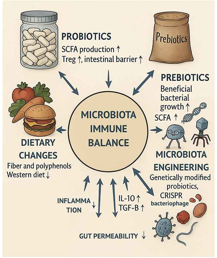 |
Figure 8 Microbiota-Targeted Therapeutic Strategies for Restoring Immune Homeostasis. This diagram centers on “Microbiota Immune Balance” and illustrates various therapeutic interventions designed to restore this homeostasis. These interventions include: Dietary Changes (eg, increasing fiber/polyphenols, reducing Western diet patterns); Probiotics (live beneficial bacteria to boost SCFAs and Tregs); Prebiotics (non-digestible fibers like FOS and inulin that feed beneficial bacteria); and Microbiota Engineering (advanced technologies like genetically modified probiotics, CRISPR, or bacteriophage therapy). All these approaches aim to restore immune homeostasis by increasing anti-inflammatory signals (IL-10, TGF-β), decreasing gut permeability, and reducing overall inflammation. |
Prebiotics and Probiotics
Probiotics are live microorganisms that, when administered in sufficient quantities, provide a health benefit to the host. Probiotics are important for the maintenance of gut homeostasis through the regulation of epithelial barrier integrity, the modulation of immune cell function, and the production of anti-inflammatory metabolites like SCFAs.107 In autoimmune disorders, some probiotic strains—especially Lactobacillus rhamnosus GG, L. casei, and Bifidobacterium infantis—have demonstrated ability to increase the growth of regulatory T cells (Treg), inhibit Th17 differentiation, and dampen the secretion of pro-inflammatory cytokines (eg, IL-17, IL-6, TNF-α) in animal models and early human research.108 In rheumatoid arthritis and inflammatory bowel disease models, probiotic treatment has led to reduced disease severity, improved intestinal histopathology, and normalization of microbiota composition.109 Probiotics are also involved in sustaining the gut barrier through the reinforcement of tight junction proteins such as occludin and claudin, preventing systemic leakage of endotoxins.110
Prebiotics—non-digestible dietary fibers that selectively stimulate the growth and activity of beneficial bacteria—function synergistically with probiotics to shape the microbiota. Inulin, fructooligosaccharides (FOS), and resistant starches enhance the abundance of butyrate-producing bacteria (eg, Roseburia, Faecalibacterium) and increase SCFA levels in the gut. These SCFAs, especially butyrate, promote Treg differentiation through histone deacetylase inhibition and support the generation of anti-inflammatory cytokines such as IL-10 and TGF-β.5 Both probiotics and prebiotics can be combined as synbiotics, which is a promising two-pronged therapeutic strategy for gut-immune axis modulation in autoimmune diseases.
Fecal Microbiota Transplantation (FMT)
The process of reintroducing a diverse and healthy microbial population into a person’s gastrointestinal system by implanting fecal matter from a healthy donor is known as fecal microbiota transplantation, or FMT.111 FMT is mainly indicated for recurrent Clostridioides difficile infection, although interest in using FMT for autoimmune and inflammatory conditions is increasing.
In ulcerative colitis, randomized clinical trials have demonstrated that FMT can induce remission in a substantial proportion of patients, with some experiencing long-term mucosal healing and normalization of inflammatory biomarkers.89 The mode of action is suspected to be via the restoration of microbial diversity, re-establishment of SCFA production, inhibition of pathobionts, and reactivation of anti-inflammatory immune mechanisms.112 In experimental autoimmune encephalomyelitis (EAE), a model for multiple sclerosis in mice, FMT from healthy donors ameliorates disease symptoms and reduces CNS inflammation. However, the harmful significance of dysbiosis is further supported by the fact that microbiota transferred from MS patients worsens the condition.113,114 Similarly, in type 1 diabetes-prone NOD mice, FMT from healthy donors delayed the onset of autoimmune diabetes and reversed intestinal barrier function.115
Despite the promises, FMT is not free of challenges. Variability in donor microbiota, uncertainty regarding long-term risks, and the need for standardization in preparation, delivery, and recipient selection remain significant challenges to its implementation on a wider scale in clinical practice. Next-generation FMT using defined microbial consortia and capsule-based delivery systems may deliver more controlled and scalable solutions.
Nutritional Interventions
Diet is one of the most potent gut microbiota composition modulators. Long-term diets affect microbial community structure, host metabolic output, and inflammatory levels.77 Fiber-, polyphenol-, and fermented food-based diets increase microbial richness and beneficial metabolites such as SCFAs, while Western dietary patterns with their high consumption of saturated fat, sugar, and processed foods are associated with dysbiosis and systemic inflammation.116
In autoimmune diseases, anti-inflammatory diets like the Mediterranean diet have also been shown to be helpful in elevating microbial richness and the abundance of butyrate-producing species like Faecalibacterium prausnitzii and Roseburia spp.117,118 These microbes produce metabolites that enhance epithelial integrity, suppress NF-κB signaling, and balance Treg/Th17.119 Fermented foods such as kefir, kimchi, and sauerkraut also contain live microbes and bioactive molecules that have a role in immune modulation. Elimination diets and allergen-free diets are also being studied to identify dietary etiologies of autoimmunity, especially in conditions such as celiac disease and IBD.120,121 Personalized nutrition, guided by individual microbiome profiles and metabolic responses, represents a sophisticated approach to dietary modification. Utilizing metagenomics in combination with machine learning algorithms, personalized dietary plans can be designed to predict glycemic reactions, inflammatory markers, and short-chain fatty acid generation, ultimately leading to improved therapeutic outcomes.122
Microbial Metabolites and Postbiotics
Postbiotics are bioactive functional substances secreted by the bacteria with physiological impact on the host. They differ from probiotics as they do not necessitate the intake of live organisms, which can be advantageous in immunocompromised hosts.123,124 Key postbiotics are short-chain fatty acids (SCFAs), indole derivatives, and polysaccharide A (PSA).125 Butyrate has been a key immunomodulator that facilitates the expansion of Treg cells, suppresses Th17 responses, and renovates the integrity of the gut epithelial barrier. It also functions as an inhibitor of histone deacetylase (HDAC) and thereby affects gene expression and signaling pathways related to anti-inflammation.126,127
Indole derivatives, which are produced from tryptophan by the gut microbiota, bind to the aryl hydrocarbon receptor (AhR) on immune cells and regulate intestinal immune homeostasis and enhance epithelial healing.65 PSA of Bacteroides fragilis stimulates IL-10 production via dendritic cells and is necessary for mucosal tolerance.128 The therapeutic use of postbiotics is being explored in both oral supplement and pharmaceutical-like formats. These molecules can offer targeted approaches to immune response modulation while maintaining the integrity of the entire microbiome.123
Microbiome Engineering and Precise Modulation
New technologies in synthetic biology, gene editing, and bacteriophage therapy are starting to offer possibilities for targeted microbiota manipulation. Genetically modified probiotics can be designed to deliver immunoregulatory molecules (eg, IL-10, TGF-β) directly to the gut.123 CRISPR-Cas systems can be used to selectively target and remove pathogenic strains or knock out pro-inflammatory genes from resident microbes.129 Phage therapy offers the possibility of specifically targeting specific bacterial species without affecting the integrity of the surrounding microbiota. For example, lytic phages have been used in experimental conditions to reduce the incidence of E. coli and Clostridium species associated with colitis and inflammation.130,131 Also, “designer consortia” of bacteria—synthetic microbiota of defined strains—are being developed to replicate the function of healthy microbiota without the risk of standard FMT. These developing technologies are still in their early stages, but they hold great promise for future treatments that combine the specificity of biologic agents with the complex ecosystem of the microbiome.132
Current Challenges and Future Perspective
Over the last few years, our understanding of the role of the gut microbiota in immune regulation and autoimmune disease pathogenesis has advanced significantly. Despite growing optimism and therapeutic promise, however, numerous issues stand in the way of complete utilization of microbiota based research in routine clinical practice. These obstacles vary from biological complexity to technological limitation, from ethical to regulatory. Mapping these limits is central to establishing future avenues of microbiome-immune system research and therapy.
Interindividual Variability and Complexity of the Human Microbiota
One of the most profound challenges with microbiota therapeutics is high individual-specific heterogeneity of microbial communities.133 Human gut microbiota is shaped by an incredibly large set of factors including genetics, type of birth (cesarean vs vaginal), breastfeeding status, antibiotic exposure, diet, geographic location, and age. This vast variability makes it challenging to have a universal “healthy” microbiota signature that may serve as a target for therapeutic normalization.122,134,135
Furthermore, host genetics and immune responsiveness add to this complexity. For instance, two people with the same microbial profile may have vastly contrasting immune responses depending on their MHC (major histocompatibility complex) variants or cytokine signatures.136 Consequently, the therapeutic results of treatments like probiotics and fecal microbiota transplantation (FMT) vary, with some patients seeing a remission and others experiencing no improvement or even a worsening of their condition.137 To do this, accuracy in microbiome profiling is in desperate need, using metagenomic sequencing and machine learning algorithms to individualize treatment protocols. These techniques can be utilized to inform patient-specific responses and personalize therapies accordingly.
Determining Causality: Association Vs Mechanistic Understanding
A second major challenge is causation separation from correlation in human microbiome research. Many studies have reported significant changes in microbiota composition in autoimmune diseases; however, the question remains in most cases if dysbiosis is a causative agent of disease or a consequence of immunocompromise or use of drugs.10,71,138 For example, individuals with inflammatory bowel disease (IBD) typically present with reduced microbial diversity and increased abundance of pro-inflammatory species such as Escherichia coli. Such changes, however, may result from chronic inflammation, corticosteroid use, or diet alteration, rather than being the underlying cause of disease.139,140 To get around this, researchers apply GF and gnotobiotic animal models mostly, where microbiota from patients are inoculated into mice to determine whether phenotypes of disease occur.141 Although these models, having provided causal proof for some diseases—namely, type 1 diabetes and multiple sclerosis—have shortcomings inherent in them regarding species differences, lack of environmental complexity, and biasing in terms of the number of bacterial taxa versus viruses, archaea, and fungi.142 Future research should employ longitudinal cohort designs, multi-omics integration, and causal inference modeling to unravel these complex associations and determine the functional capacity of individual microbial taxa and metabolites.
Limitations of Current Models and Analytical Technologies
Animal models, as useful as they are for mechanistic study, are not ideal representations of human physiology.143 Mice possess a significantly different immune system than humans, in terms of T cell distribution, cytokine networks, and gut anatomy. Even the most humanized mouse model, one that is colonized with human microbiota, cannot simulate the full range of environmental and epigenetic factors of the human.30 Furthermore, the technology commonly used in microbiome studies is limited. For instance, 16S rRNA sequencing, as a tool for taxonomic identification, is not highly resolved at the species and strain levels and does not provide information on gene expression or metabolic function.144 To fully understand the functional potential of the microbiome, more powerful tools such as: Shotgun metagenomics (to identify genes and pathways), Metatranscriptomics (to measure microbial gene expression), Metabolomics (to quantify microbial metabolites like SCFAs, bile acids, and indoles), and Single-cell sequencing (to quantify heterogeneity in immune and microbial communities) will need to be used. Integration of these data into systems biology workflows will be essential to simulate the microbiota-immune network in an integrated manner.145,146
Safety Issues and Regulatory Gaps in Microbiome Therapies
As microbiota-modulating interventions enter clinical trials and medical practice, biosafety and regulation become increasingly important. FMT, while effective for recurrent Clostridioides difficile infection, has been associated with adverse events including pathogen transmission, long-term microbiota instability, and immune dysregulation.147 Besides, live biotherapeutic products such as engineered probiotics or designer consortia require rigorous testing for toxicity, colonization ability, horizontal gene transfer, and interaction with the immune system of the host. However, current regulatory frameworks (eg, FDA, EMA) are not fully calibrated for the unique nature of microbiota based treatments. Depending on the context, these therapies are often ambiguously treated as drugs, biologics, or transplants.148,149
Therefore, an urgent necessity exists to develop international standards to consolidate donor screening, production methods, strain characterization, quality assessment, and clinical results. Regulatory science will have to be in parallel evolution with microbiome science to make future therapies safer and more effective.
Future Outlook: Precision Microbiota and Immune Modulation
In the years to come, artificial intelligence, multi-omics, and long-term patient monitoring will come together to make precision microbiome medicine a reality. Instead of a “one-size-fits-all” approach, must shortly: predict risk for autoimmune disease based on microbial and immunologic signatures, stratify clinical trial participants based on microbiota subtypes, monitor therapeutic response in real time by employing microbiome biomarkers, and engineer microbiota therapies (eg, CRISPR-edited probiotics, phage therapy, designer SCFA cocktails).150
New tools such as gut organoids, gut-on-chip platforms, and synthetic microbial ecosystems will also serve to bridge the bench-to-bedside gap. Importantly, ethical considerations surrounding privacy, consent, and long-term microbiota manipulation must be addressed appropriately as the field continues to grow.
Lastly, while there are significant challenges ahead, the future of microbiota-directed treatments for autoimmune disease is bright. Interdisciplinary thinking, translational research, and patient-centered design will be required in order to fully capitalize on it.
Conclusion
The sophisticated and dynamic interaction between intestinal microbiota and the host immune system is central to establishing and maintaining health as well as regulating immune tolerance. During the last two decades, growing evidence has proved that dysfunctions of the microbial community so called dysbiosis are highly linked with the initiation and course of several autoimmune disorders, such as inflammatory bowel disease (IBD), multiple sclerosis (MS), rheumatoid arthritis (RA), and type 1 diabetes (T1D).27,104,151
A well-balanced and healthy microbiota plays a pivotal role in shaping immune system development from birth, promoting regulatory T cell (Treg) differentiation, strengthening epithelial barrier integrity, and mitigating chronic inflammation through the production of microbial metabolites including short-chain fatty acids (SCFAs), indole derivatives, and polysaccharide A.65,152,153 Conversely, disturbance of microbiota population can cause atypical Th17 cell activation, increased intestinal permeability, and systemic inflammation and therefore result in immune tolerance interruption and development of autoimmunity.87,113
In disease-specific conditions, a larger number of studies have indicated that particular microbial signatures can promote or alleviate the disease phenotypes. For instance, Prevotella copri overgrowth in RA, Faecalibacterium prausnitzii depletion in IBD, and Akkermansia muciniphila upregulation in MS patients illustrate how compositional shifts are directly affecting immune cell polarization and cytokine patterns.85,92,97
These findings have driven the development of microbiota-centered treatment approaches, including probiotics, prebiotics, fecal microbiota transplantation (FMT), dietary treatment, and new microbial engineering techniques. Early clinical trials, particularly in ulcerative colitis and experimental autoimmune encephalomyelitis (EAE), have been promising, with the reestablishment of microbial balance potentially reducing inflammation and improving disease outcomes.89,154 Moreover, synthetic biology today enables probiotics to be engineered to transfer anti-inflammatory cytokines or block autoimmune reactions on a targeted level.129
Despite this innovation, several significant issues must be resolved. These are such as high inter-personal heterogeneity of the human microbiome, failure to establish causality between dysbiosis and disease, and the lack of harmonized regulatory frameworks for microbiota therapy.155,156 Safety of long-term microbiome manipulation, especially among the susceptible groups, is also a subject of concern. Moreover, the complexity of microbial interactions with host immunity, diet, and genetics necessitates an integrative, systems-level approach to research and therapy.
The future of microbiota-guided therapy of autoimmune diseases lies in precision medicine—patient-specific microbial, immunologic, and genomic data to predict disease risk, individualize interventions, and monitor in real-time treatment response. Leading the way to this future are advances in AI-driven diagnostics, organ-on-chip technology, and multi-omics strategies.157,158
Ultimately, the convergence of immunology, microbiology, genomics, and bioengineering has the potential to revolutionize how we consider and treat autoimmune diseases. Rather than targeting the immune system head-on, we will perhaps be able to regulate the microbial communities that modulate its actions, offering safer, more sustainable, and more efficacious avenues for therapy.
As microbiota-targeted therapies advance from experimental applications to clinical practice, ethical and regulatory considerations are becoming increasingly important. Issues such as informed consent for fecal microbiota transplantation, the long-term ecological impact of microbiome engineering, and the privacy of individual microbiome profiles must be carefully addressed. Moreover, the potential use of genetically modified organisms (GMOs) and synthetic biology in modulating host immunity raises complex questions regarding safety, equity, and accessibility. A robust ethical framework and proactive policymaking are essential to ensure that these novel interventions are developed and implemented responsibly, transparently, and inclusively.
Disclosure
The authors report no conflicts of interest in this work.
References
1. Rooks MG, Garrett WS. Gut microbiota, metabolites and host immunity. Nat Rev Immunol. 2016;16(6):341–352. doi:10.1038/nri.2016.42
2. Zheng D, Liwinski T, Elinav E. Interaction between microbiota and immunity in health and disease. Cell Res. 2020;30(6):492–506. doi:10.1038/s41422-020-0332-7
3. Honda K, Littman DR. The microbiota in adaptive immune homeostasis and disease. Nature. 2016;535(7610):75–84. doi:10.1038/nature18848
4. Marín-Tello C, Jintaridth P, Sanchez F, et al. Epigenetic regulation by metabolites from the gut microbiome. Benef Microbes. 2022;13(6):437–444. doi:10.3920/BM2022.0006
5. Parada Venegas D, De la Fuente MK, Landskron G, et al. Short chain fatty acids (SCFAs)-mediated gut epithelial and immune regulation and its relevance for inflammatory bowel diseases. Front Immunol. 2019;10:277. doi:10.3389/fimmu.2019.00277
6. Lamas B, Natividad JM, Sokol H. Aryl hydrocarbon receptor and intestinal immunity. Mucosal Immunol. 2018;11(4):1024–1038. doi:10.1038/s41385-018-0019-2
7. Levy M, Kolodziejczyk AA, Thaiss CA, Elinav E. Dysbiosis and the immune system. Nat Rev Immunol. 2017;17(4):219–232. doi:10.1038/nri.2017.7
8. Hooks KB, O’Malley MA. Dysbiosis and its discontents. MBio. 2017;8(5). doi:10.1128/mBio.01492-17
9. Ruff WE, Greiling TM, Kriegel MA. Host-microbiota interactions in immune-mediated diseases. Nat Rev Microbiol. 2020;18(9):521–538. doi:10.1038/s41579-020-0367-2
10. De Luca F, Shoenfeld Y. The microbiome in autoimmune diseases. Clin Exp Immunol. 2019;195(1):74–85. doi:10.1111/cei.13158
11. Missoum H, Alami M, Bachir F, et al. Prevalence of autoimmune diseases and clinical significance of autoantibody profile: data from national institute of hygiene in Rabat, Morocco. Hum Immunol. 2019;80(7):523–532. doi:10.1016/j.humimm.2019.02.012
12. Ramos PS, Shedlock AM, Langefeld CD. Genetics of autoimmune diseases: insights from population genetics. J Hum Genet. 2015;60(11):657–664. doi:10.1038/jhg.2015.94
13. Varadé J, Magadán S, González-Fernández Á. Human immunology and immunotherapy: main achievements and challenges. Cell Mol Immunol. 2021;18(4):805–828. doi:10.1038/s41423-020-00530-6
14. Pisetsky DS. Pathogenesis of autoimmune disease. Nat Rev Nephrol. 2023;19(8):509–524. doi:10.1038/s41581-023-00720-1
15. Wang L, Wang F-S, Gershwin ME. Human autoimmune diseases: a comprehensive update. J Intern Med. 2015;278(4):369–395. doi:10.1111/joim.12395
16. Narayanappa Koppala S, Guruprasad V. Overview of autoimmunity: classification, disease mechanisms, and etiology. Turk J Immunol. 2024;93–105. doi:10.4274/tji.galenos.2024.13008
17. Miller FW. The increasing prevalence of autoimmunity and autoimmune diseases: an urgent call to action for improved understanding, diagnosis, treatment, and prevention. Curr Opin Immunol. 2023;80:102266. doi:10.1016/j.coi.2022.102266
18. Kalbermatter C, Fernandez Trigo N, Christensen S, Ganal-Vonarburg SC. Maternal microbiota, early life colonization and breast milk drive immune development in the newborn. Front Immunol. 2021;12:683022. doi:10.3389/fimmu.2021.683022
19. Ignacio A, Czyz S, McCoy KD. Early life microbiome influences on development of the mucosal innate immune system. Semin Immunol. 2024;73:101885. doi:10.1016/j.smim.2024.101885
20. Marshall JS, Warrington R, Watson W, Kim HL. An introduction to immunology and immunopathology. Allergy Asthma Clin Immunol. 2018;14(Suppl 2):1–10. doi:10.1186/s13223-018-0278-1
21. Gensollen T, Iyer SS, Kasper DL, Blumberg RS. How colonization by microbiota in early life shapes the immune system. Science. 2016;352(6285):539–544. doi:10.1126/science.aad9378
22. Jain N. The early life education of the immune system: moms, microbes and (missed) opportunities. Gut Microbes. 2020;12(1):1824564. doi:10.1080/19490976.2020.1824564
23. Macpherson AJ, de Agüero MG, Ganal-Vonarburg SC. How nutrition and the maternal microbiota shape the neonatal immune system. Nat Rev Immunol. 2017;17(8):508–517. doi:10.1038/nri.2017.58
24. Al Nabhani Z, Eberl G. Imprinting of the immune system by the microbiota early in life. Mucosal Immunol. 2020;13(2):183–189. doi:10.1038/s41385-020-0257-y
25. Parihar A, Eubank TD, Doseff AI. Monocytes and macrophages regulate immunity through dynamic networks of survival and cell death. J Innate Immun. 2010;2(3):204–215. doi:10.1159/000296507
26. Lavin Y, Mortha A, Rahman A, Merad M. Regulation of macrophage development and function in peripheral tissues. Nat Rev Immunol. 2015;15(12):731–744. doi:10.1038/nri3920
27. Belkaid Y, Hand TW. Role of the microbiota in immunity and inflammation. Cell. 2014;157(1):121–141. doi:10.1016/j.cell.2014.03.011
28. Abo-Shaban T, Sharna SS, Hosie S, et al. Issues for patchy tissues: defining roles for gut-associated lymphoid tissue in neurodevelopment and disease. J Neural Transm. 2023;130(3):269–280. doi:10.1007/s00702-022-02561-x
29. Wu H-J, Wu E. The role of gut microbiota in immune homeostasis and autoimmunity. Gut Microbes. 2012;3(1):4–14. doi:10.4161/gmic.19320
30. Hou K, Wu Z-X, Chen X-Y, et al. Microbiota in health and diseases. Signal Transduct Target Ther. 2022;7(1):135. doi:10.1038/s41392-022-00974-4
31. Cheng H, Guan X, Chen D, Ma W. The Th17/Treg cell balance: a gut microbiota-modulated story. Microorganisms. 2019;7(12):583. doi:10.3390/microorganisms7120583
32. Ramanan D, Pratama A, Zhu Y, et al. Regulatory T cells in the face of the intestinal microbiota. Nat Rev Immunol. 2023;23(11):749–762. doi:10.1038/s41577-023-00890-w
33. Takiishi T, Fenero CIM, Câmara NOS. Intestinal barrier and gut microbiota: shaping our immune responses throughout life. Tissue Barriers. 2017;5(4):e1373208. doi:10.1080/21688370.2017.1373208
34. Corthay A. How do regulatory T cells work? Scand J Immunol. 2009;70(4):326–336. doi:10.1111/j.1365-3083.2009.02308.x
35. Sakaguchi S, Kawakami R, Mikami N. Treg-based immunotherapy for antigen-specific immune suppression and stable tolerance induction: a perspective. Immunother Adv. 2023;3(1):ltad007. doi:10.1093/immadv/ltad007
36. Nutsch KM, Hsieh C-S. T cell tolerance and immunity to commensal bacteria. Curr Opin Immunol. 2012;24(4):385–391. doi:10.1016/j.coi.2012.04.009
37. Alberts B, Johnson A, Lewis J, Raff M, Roberts K, Walter P. T cells and MHC proteins.In: Molecular biology of the cell.
38. Tanaka A, Sakaguchi S. Regulatory T cells in cancer immunotherapy. Cell Res. 2017;27(1):109–118. doi:10.1038/cr.2016.151
39. Vignali DAA, Collison LW, Workman CJ. How regulatory T cells work. Nat Rev Immunol. 2008;8(7):523–532. doi:10.1038/nri2343
40. Izcue A, Coombes JL, Powrie F. Regulatory T cells suppress systemic and mucosal immune activation to control intestinal inflammation. Immunol Rev. 2006;212:256–271. doi:10.1111/j.0105-2896.2006.00423.x
41. Wu J, Xie A, Chen W. Cytokine regulation of immune tolerance. Burns Trauma. 2014;2(1):11–17. doi:10.4103/2321-3868.124771
42. Liu Y, Tran DQ, Lindsey JW, Rhoads JM. The association of gut microbiota and Treg dysfunction in autoimmune diseases. Adv Exp Med Biol. 2021;1278:191–203. doi:10.1007/978-981-15-6407-9_10
43. Karczewski J, Dobrowolska A, Rychlewska-Hańczewska A, Adamski Z. New insights into the role of T cells in pathogenesis of psoriasis and psoriatic arthritis. Autoimmunity. 2016;49(7):435–450. doi:10.3109/08916934.2016.1166214
44. Taylor A, Verhagen J, Blaser K, Akdis M, Akdis CA. Mechanisms of immune suppression by interleukin-10 and transforming growth factor-beta: the role of T regulatory cells. Immunology. 2006;117(4):433–442. doi:10.1111/j.1365-2567.2006.02321.x
45. Oparaugo NC, Ouyang K, Nguyen NPN, Nelson AM, Agak GW. Human regulatory T cells: understanding the role of tregs in select autoimmune skin diseases and post-transplant nonmelanoma skin cancers. Int J Mol Sci. 2023;24(2):1527. doi:10.3390/ijms24021527
46. Workman CJ, Szymczak-Workman AL, Collison LW, Pillai MR, Vignali DAA. The development and function of regulatory T cells. Cell Mol Life Sci. 2009;66(16):2603–2622. doi:10.1007/s00018-009-0026-2
47. Sakaguchi S, Yamaguchi T, Nomura T, Ono M. Regulatory T cells and immune tolerance. Cell. 2008;133(5):775–787. doi:10.1016/j.cell.2008.05.009
48. Schmitt EG, Williams CB. Generation and function of induced regulatory T cells. Front Immunol. 2013;4:152. doi:10.3389/fimmu.2013.00152
49. Josefowicz SZ, Lu L-F, Rudensky AY. Regulatory T cells: mechanisms of differentiation and function. Annu Rev Immunol. 2012;30:531–564. doi:10.1146/annurev.immunol.25.022106.141623
50. Yu L, Fu Y, Miao R, et al. Regulatory T cells and their derived cell pharmaceuticals as emerging therapeutics against autoimmune diseases. Adv Funct Mater. 2024;34(39). doi:10.1002/adfm.202405133
51. Honing DY, Luiten RM, Matos TR. Regulatory T cell dysfunction in autoimmune diseases. Int J Mol Sci. 2024;25(13):7171. doi:10.3390/ijms25137171
52. Jacobse J, Li J, Rings EHHM, Samsom JN, Goettel JA. Intestinal regulatory T cells as specialized tissue-restricted immune cells in intestinal immune homeostasis and disease. Front Immunol. 2021;12:716499. doi:10.3389/fimmu.2021.716499
53. Zong Y, Deng K, Chong WP. Regulation of Treg cells by cytokine signaling and co-stimulatory molecules. Front Immunol. 2024;15:1387975. doi:10.3389/fimmu.2024.1387975
54. Wiertsema SP, Van Bergenhenegouwen J, Garssen J, Knippels LMJ. The interplay between the gut microbiome and the immune system in the context of infectious diseases throughout life and the role of nutrition in optimizing treatment strategies. Nutrients. 2021;13(3):886. doi:10.3390/nu13030886
55. Wang J, Zhu N, Su X, Gao Y, Yang R. Gut-microbiota-derived metabolites maintain gut and systemic immune homeostasis. Cells. 2023;12(5). doi:10.3390/cells12050793
56. Wang J, He M, Yang M, Ai X. Gut microbiota as a key regulator of intestinal mucosal immunity. Life Sci. 2024;345:122612. doi:10.1016/j.lfs.2024.122612
57. den Besten G, van Eunen K, Groen AK, Venema K, Reijngoud D-J, Bakker BM. The role of short-chain fatty acids in the interplay between diet, gut microbiota, and host energy metabolism. J Lipid Res. 2013;54(9):2325–2340. doi:10.1194/jlr.R036012
58. Choi H, Mook-Jung I. Functional effects of gut microbiota-derived metabolites in Alzheimer’s disease. Curr Opin Neurobiol. 2023;81:102730. doi:10.1016/j.conb.2023.102730
59. Liu X-F, Shao J-H, Liao Y-T, et al. Regulation of short-chain fatty acids in the immune system. Front Immunol. 2023;14:1186892. doi:10.3389/fimmu.2023.1186892
60. Ney L-M, Wipplinger M, Grossmann M, Engert N, Wegner VD, Mosig AS. Short chain fatty acids: key regulators of the local and systemic immune response in inflammatory diseases and infections. Open Biol. 2023;13(3):230014. doi:10.1098/rsob.230014
61. Heidor R, Vargas-Mendez E, Moreno FS, Bishayee A, Bhatia D. Epigenetic aspects of hepatocellular carcinoma chemoprevention. Epigenetics of Cancer Prevention. Elsevier; 2019:231–249. doi:10.1016/B978-0-12-812494-9.00011-1
62. Silva YP, Bernardi A, Frozza RL. The role of short-chain fatty acids from gut microbiota in gut-brain communication. Front Endocrinol. 2020;11:25. doi:10.3389/fendo.2020.00025
63. Lange O, Proczko-Stepaniak M, Mika A. Short-chain fatty acids-a product of the microbiome and its participation in two-way communication on the microbiome-host mammal line. Curr Obes Rep. 2023;12(2):108–126. doi:10.1007/s13679-023-00503-6
64. Hou Y, Li J, Ying S. Tryptophan metabolism and gut microbiota: a novel regulatory axis integrating the microbiome, immunity, and cancer. Metabolites. 2023;13(11):1166. doi:10.3390/metabo13111166
65. Gutiérrez-Vázquez C, Quintana FJ. Regulation of the immune response by the aryl hydrocarbon receptor. Immunity. 2018;48(1):19–33. doi:10.1016/j.immuni.2017.12.012
66. Wang J, Zhao X, Wan YY. Intricacies of TGF-β signaling in Treg and Th17 cell biology. Cell Mol Immunol. 2023;20(9):1002–1022. doi:10.1038/s41423-023-01036-7
67. Zhao M, Chu J, Feng S, et al. Immunological mechanisms of inflammatory diseases caused by gut microbiota dysbiosis: a review. Biomed Pharmacother. 2023;164:114985. doi:10.1016/j.biopha.2023.114985
68. Gieryńska M, Szulc-Dąbrowska L, Struzik J, Mielcarska MB, Gregorczyk-Zboroch KP. Integrity of the intestinal barrier: the involvement of epithelial cells and microbiota-a mutual relationship. Animals. 2022;12(2):145. doi:10.3390/ani12020145
69. Chandrasekaran P, Weiskirchen S, Weiskirchen R. Effects of probiotics on gut microbiota: an overview. Int J Mol Sci. 2024;25(11):6022. doi:10.3390/ijms25116022
70. Scott AJ, Alexander JL, Merrifield CA, et al. International cancer microbiome consortium consensus statement on the role of the human microbiome in carcinogenesis. Gut. 2019;68(9):1624–1632. doi:10.1136/gutjnl-2019-318556
71. Afzaal M, Saeed F, Shah YA, et al. Human gut microbiota in health and disease: unveiling the relationship. Front Microbiol. 2022;13:999001. doi:10.3389/fmicb.2022.999001
72. Patangia DV, Anthony Ryan C, Dempsey E, Paul Ross R, Stanton C. Impact of antibiotics on the human microbiome and consequences for host health. Microbiologyopen. 2022;11(1):e1260. doi:10.1002/mbo3.1260
73. Vamanu E, Rai SN. The link between obesity, microbiota dysbiosis, and neurodegenerative pathogenesis. Diseases. 2021;9(3):45. doi:10.3390/diseases9030045
74. DeGruttola AK, Low D, Mizoguchi A, Mizoguchi E. Current understanding of dysbiosis in disease in human and animal models. Inflamm Bowel Dis. 2016;22(5):1137–1150. doi:10.1097/MIB.0000000000000750
75. Hrncir T. Gut microbiota dysbiosis: triggers, consequences, diagnostic and therapeutic options. Microorganisms. 2022;10(3):578. doi:10.3390/microorganisms10030578
76. Aryasa IA, Widyantara IW, Kusuma Putra IB. Dysbiosis of gut microbiome in neurocritically ill patients in intensive care: systematic review. Int J Sci Healthcare Res. 2023;8(1):294–301. doi:10.52403/ijshr.20230141
77. Conlon MA, Bird AR. The impact of diet and lifestyle on gut microbiota and human health. Nutrients. 2014;7(1):17–44. doi:10.3390/nu7010017
78. Santana PT, Rosas SLB, Ribeiro BE, Marinho Y, de Souza HSP. Dysbiosis in inflammatory bowel disease: pathogenic role and potential therapeutic targets. Int J Mol Sci. 2022;23(7):3464. doi:10.3390/ijms23073464
79. Stange EF. Dysbiosis in inflammatory bowel diseases: egg, not chicken. Front Med. 2024;11:1395861. doi:10.3389/fmed.2024.1395861
80. Tiffany CR, Bäumler AJ. Dysbiosis: from fiction to function. Am J Physiol Gastrointest Liver Physiol. 2019;317(5):G602–G608. doi:10.1152/ajpgi.00230.2019
81. Shivaji S. We are not alone: a case for the human microbiome in extra intestinal diseases. Gut Pathog. 2017;9:13. doi:10.1186/s13099-017-0163-3
82. Wright ML, Fournier C, Houser MC, Tansey M, Glass J, Hertzberg VS. Potential role of the gut microbiome in ALS: a systematic review. Biol Res Nurs. 2018;20(5):513–521. doi:10.1177/1099800418784202
83. Khan MF, Wang H. Environmental exposures and autoimmune diseases: contribution of gut microbiome. Front Immunol. 2019;10:3094. doi:10.3389/fimmu.2019.03094
84. Kostic AD, Xavier RJ, Gevers D. The microbiome in inflammatory bowel disease: current status and the future ahead. Gastroenterology. 2014;146(6):1489–1499. doi:10.1053/j.gastro.2014.02.009
85. Frank DN, St Amand AL, Feldman RA, Boedeker EC, Harpaz N, Pace NR. Molecular-phylogenetic characterization of microbial community imbalances in human inflammatory bowel diseases. Proc Natl Acad Sci USA. 2007;104(34):13780–13785. doi:10.1073/pnas.0706625104
86. Manichanh C, Rigottier-Gois L, Bonnaud E, et al. Reduced diversity of faecal microbiota in Crohn’s disease revealed by a metagenomic approach. Gut. 2006;55(2):205–211. doi:10.1136/gut.2005.073817
87. Neurath MF. Cytokines in inflammatory bowel disease. Nat Rev Immunol. 2014;14(5):329–342. doi:10.1038/nri3661
88. Schaubeck M, Clavel T, Calasan J, et al. Dysbiotic gut microbiota causes transmissible Crohn’s disease-like ileitis independent of failure in antimicrobial defence. Gut. 2016;65(2):225–237. doi:10.1136/gutjnl-2015-309333
89. Paramsothy S, Kamm MA, Kaakoush NO, et al. Multidonor intensive faecal microbiota transplantation for active ulcerative colitis: a randomised placebo-controlled trial. Lancet. 2017;389(10075):1218–1228. doi:10.1016/S0140-6736(17)30182-4
90. Tafti D, Ehsan M, Xixis KL. Multiple sclerosis. In: StatPearls. Treasure Island (FL): StatPearls Publishing; 2025.
91. Miyake S, Kim S, Suda W, et al. Dysbiosis in the gut microbiota of patients with multiple sclerosis, with a striking depletion of species belonging to clostridia XIVa and IV clusters. PLoS One. 2015;10(9):e0137429. doi:10.1371/journal.pone.0137429
92. Jangi S, Gandhi R, Cox LM, et al. Alterations of the human gut microbiome in multiple sclerosis. Nat Commun. 2016;7:12015. doi:10.1038/ncomms12015
93. Cekanaviciute E, Yoo BB, Runia TF, et al. Gut bacteria from multiple sclerosis patients modulate human T cells and exacerbate symptoms in mouse models. Proc Natl Acad Sci USA. 2017;114(40):10713–10718. doi:10.1073/pnas.1711235114
94. Haghikia A, Jörg S, Duscha A, et al. Dietary fatty acids directly impact central nervous system autoimmunity via the small intestine. Immunity. 2015;43(4):817–829. doi:10.1016/j.immuni.2015.09.007
95. Chauhan K, Jandu JS, Brent LH, Al-Dhahir MA. Rheumatoid Arthritis. In: StatPearls. Treasure Island (FL): StatPearls Publishing; 2025.
96. Wegner N, Wait R, Sroka A, et al. Peptidylarginine deiminase from porphyromonas gingivalis citrullinates human fibrinogen and α-enolase: implications for autoimmunity in rheumatoid arthritis. Arthritis Rheum. 2010;62(9):2662–2672. doi:10.1002/art.27552
97. Scher JU, Sczesnak A, Longman RS, et al. Expansion of intestinal prevotella copri correlates with enhanced susceptibility to arthritis. eLife. 2013;2:e01202. doi:10.7554/eLife.01202
98. Maeda Y, Kurakawa T, Umemoto E, et al. Dysbiosis contributes to arthritis development via activation of autoreactive T cells in the intestine. Arthritis Rheumatol. 2016;68(11):2646–2661. doi:10.1002/art.39783
99. Ogrendik M. Rheumatoid arthritis is an autoimmune disease caused by periodontal pathogens. Int J Gen Med. 2013;6:383–386. doi:10.2147/IJGM.S45929
100. Yoshitomi H, Sakaguchi N, Kobayashi K, et al. A role for fungal {beta}-glucans and their receptor Dectin-1 in the induction of autoimmune arthritis in genetically susceptible mice. J Exp Med. 2005;201(6):949–960. doi:10.1084/jem.20041758
101. So J-S, Kwon H-K, Lee C-G, et al. Lactobacillus casei suppresses experimental arthritis by down-regulating T helper 1 effector functions. Mol Immunol. 2008;45(9):2690–2699. doi:10.1016/j.molimm.2007.12.010
102. Herold KC, Delong T, Perdigoto AL, Biru N, Brusko TM, Walker LSK. The immunology of type 1 diabetes. Nat Rev Immunol. 2024;24(6):435–451. doi:10.1038/s41577-023-00985-4
103. Lebastchi J, Herold KC. Immunologic and metabolic biomarkers of β-cell destruction in the diagnosis of type 1 diabetes. Cold Spring Harb Perspect Med. 2012;2(6):a007708. doi:10.1101/cshperspect.a007708
104. Kostic AD, Gevers D, Siljander H, et al. The dynamics of the human infant gut microbiome in development and in progression toward type 1 diabetes. Cell Host Microbe. 2015;17(2):260–273. doi:10.1016/j.chom.2015.01.001
105. Vatanen T, Franzosa EA, Schwager R, et al. The human gut microbiome in early-onset type 1 diabetes from the TEDDY study. Nature. 2018;562(7728):589–594. doi:10.1038/s41586-018-0620-2
106. Zhou W, Sailani MR, Contrepois K, et al. Longitudinal multi-omics of host-microbe dynamics in prediabetes. Nature. 2019;569(7758):663–671. doi:10.1038/s41586-019-1236-x
107. Mariño E, Richards JL, McLeod KH, et al. Gut microbial metabolites limit the frequency of autoimmune T cells and protect against type 1 diabetes. Nat Immunol. 2017;18(5):552–562. doi:10.1038/ni.3713
108. Liu Y, Alookaran JJ, Rhoads JM. Probiotics in autoimmune and inflammatory disorders. Nutrients. 2018;10(10):1537. doi:10.3390/nu10101537
109. Zhao T, Wei Y, Zhu Y, et al. Gut microbiota and rheumatoid arthritis: from pathogenesis to novel therapeutic opportunities. Front Immunol. 2022;13:1007165. doi:10.3389/fimmu.2022.1007165
110. Gou H-Z, Zhang Y-L, Ren L-F, Li Z-J, Zhang L. How do intestinal probiotics restore the intestinal barrier? Front Microbiol. 2022;13:929346. doi:10.3389/fmicb.2022.929346
111. Gupta S, Allen-Vercoe E, Petrof EO. Fecal microbiota transplantation: in perspective. Therap Adv Gastroenterol. 2016;9(2):229–239. doi:10.1177/1756283X15607414
112. Lou X, Xue J, Shao R, et al. Fecal microbiota transplantation and short-chain fatty acids reduce sepsis mortality by remodeling antibiotic-induced gut microbiota disturbances. Front Immunol. 2022;13:1063543. doi:10.3389/fimmu.2022.1063543
113. Berer K, Mues M, Koutrolos M, et al. Commensal microbiota and myelin autoantigen cooperate to trigger autoimmune demyelination. Nature. 2011;479(7374):538–541. doi:10.1038/nature10554
114. Melamed E, Palmer JL, Fonken C. Advantages and limitations of experimental autoimmune encephalomyelitis in breaking down the role of the gut microbiome in multiple sclerosis. Front Mol Neurosci. 2022;15:1019877. doi:10.3389/fnmol.2022.1019877
115. Fernandez Trigo N, Kalbermatter C, Yilmaz B, Ganal-Vonarburg SC. The protective effect of the intestinal microbiota in type-1 diabetes in NOD mice is limited to a time window in early life. Front Endocrinol. 2024;15:1425235. doi:10.3389/fendo.2024.1425235
116. Berding K, Vlckova K, Marx W, et al. Diet and the microbiota-gut-brain axis: sowing the seeds of good mental health. Adv Nutr. 2021;12(4):1239–1285. doi:10.1093/advances/nmaa181
117. Abrignani V, Salvo A, Pacinella G, Tuttolomondo A. The Mediterranean diet, its microbiome connections, and cardiovascular health: a narrative review. Int J Mol Sci. 2024;25(9):4942. doi:10.3390/ijms25094942
118. Florio M, Crudele L, Sallustio F, Moschetta A, Cariello M, Gadaleta RM. Disentangling the nutrition-microbiota liaison in inflammatory bowel disease. Mol Aspects Med. 2025;102:101349. doi:10.1016/j.mam.2025.101349
119. Mohebali N, Weigel M, Hain T, et al. Faecalibacterium prausnitzii, bacteroides faecis and roseburia intestinalis attenuate clinical symptoms of experimental colitis by regulating Treg/Th17 cell balance and intestinal barrier integrity. Biomed Pharmacother. 2023;167:115568. doi:10.1016/j.biopha.2023.115568
120. Dimidi E, Cox SR, Rossi M, Whelan K. Fermented foods: definitions and characteristics, impact on the gut microbiota and effects on gastrointestinal health and disease. Nutrients. 2019;11(8):1806. doi:10.3390/nu11081806
121. Rezac S, Kok CR, Heermann M, Hutkins R. Fermented foods as a dietary source of live organisms. Front Microbiol. 2018;9:1785. doi:10.3389/fmicb.2018.01785
122. Zeevi D, Korem T, Zmora N, et al. Personalized nutrition by prediction of glycemic responses. Cell. 2015;163(5):1079–1094. doi:10.1016/j.cell.2015.11.001
123. Thorakkattu P, Khanashyam AC, Shah K, et al. Postbiotics: current trends in food and pharmaceutical industry. Foods. 2022;11(19):3094. doi:10.3390/foods11193094
124. Wegh CAM, Geerlings SY, Knol J, Roeselers G, Belzer C. Postbiotics and their potential applications in early life nutrition and beyond. Int J Mol Sci. 2019;20(19):4673. doi:10.3390/ijms20194673
125. Ragavan ML, Hemalatha S. The functional roles of short chain fatty acids as postbiotics in human gut: future perspectives. Food Sci Biotechnol. 2024;33(2):275–285. doi:10.1007/s10068-023-01414-x
126. Anshory M, Effendi RMRA, Kalim H, et al. Butyrate properties in immune-related diseases: friend or foe? Fermentation. 2023;9(3):205. doi:10.3390/fermentation9030205
127. Korsten SGPJ, Vromans H, Garssen J, Willemsen LEM. Butyrate protects barrier integrity and suppresses immune activation in a caco-2/PBMC co-culture model while HDAC inhibition mimics butyrate in restoring cytokine-induced barrier disruption. Nutrients. 2023;15(12):2760. doi:10.3390/nu15122760
128. Ramakrishna C, Kujawski M, Chu H, Li L, Mazmanian SK, Cantin EM. Bacteroides fragilis polysaccharide A induces IL-10 secreting B and T cells that prevent viral encephalitis. Nat Commun. 2019;10(1):2153. doi:10.1038/s41467-019-09884-6
129. Mimee M, Citorik RJ, Lu TK. Microbiome therapeutics - Advances and challenges. Adv Drug Deliv Rev. 2016;105(Pt A):44–54. doi:10.1016/j.addr.2016.04.032
130. Federici S, Nobs SP, Elinav E. Phages and their potential to modulate the microbiome and immunity. Cell Mol Immunol. 2021;18(4):889–904. doi:10.1038/s41423-020-00532-4
131. Lin DM, Koskella B, Lin HC. Phage therapy: an alternative to antibiotics in the age of multi-drug resistance. World J Gastrointest Pharmacol Ther. 2017;8(3):162–173. doi:10.4292/wjgpt.v8.i3.162
132. Maqsood Q, Hussain N, Sumrin A, Ali SW, Tariq MR, Mahnoor M. Monitoring and abatement of synthetic pollutants using engineered microbial systems. Discov Life. 2024;54(1):9. doi:10.1007/s11084-024-09652-7
133. Aggarwal N, Kitano S, Puah GRY, Kittelmann S, Hwang IY, Chang MW. Microbiome and human health: current understanding, engineering, and enabling technologies. Chem Rev. 2023;123(1):31–72. doi:10.1021/acs.chemrev.2c00431
134. Thomas S, Izard J, Walsh E, et al. The host microbiome regulates and maintains human health: a primer and perspective for non-microbiologists. Cancer Res. 2017;77(8):1783–1812. doi:10.1158/0008-5472.CAN-16-2929
135. Santoro A, Ostan R, Candela M, et al. Gut microbiota changes in the extreme decades of human life: a focus on centenarians. Cell Mol Life Sci. 2018;75(1):129–148. doi:10.1007/s00018-017-2674-y
136. Charles A, Janeway J, Travers P, Walport M, Shlomchik MJ. The major histocompatibility complex and its functions. In: Immunobiology: The immune system in health and disease.
137. Zikou E, Koliaki C, Makrilakis K. The role of fecal microbiota transplantation (FMT) in the management of metabolic diseases in humans: a narrative review. Biomedicines. 2024;12(8):1871. doi:10.3390/biomedicines12081871
138. Xu H, Liu M, Cao J, et al. The dynamic interplay between the gut microbiota and autoimmune diseases. J Immunol Res. 2019;2019:7546047. doi:10.1155/2019/7546047
139. Schirmer M, Garner A, Vlamakis H, Xavier RJ. Microbial genes and pathways in inflammatory bowel disease. Nat Rev Microbiol. 2019;17(8):497–511. doi:10.1038/s41579-019-0213-6
140. Lane ER, Zisman TL, Suskind DL. The microbiota in inflammatory bowel disease: current and therapeutic insights. J Inflamm Res. 2017;10:63–73. doi:10.2147/JIR.S116088
141. Fiebiger U, Bereswill S, Heimesaat MM. Dissecting the interplay between intestinal microbiota and host immunity in health and disease: lessons learned from germfree and gnotobiotic animal models. Eur J Microbiol Immunol. 2016;6(4):253–271. doi:10.1556/1886.2016.00036
142. Jiminez JA, Uwiera TC, Douglas Inglis G, Uwiera RRE. Animal models to study acute and chronic intestinal inflammation in mammals. Gut Pathog. 2015;7:29. doi:10.1186/s13099-015-0076-y
143. Barré-Sinoussi F, Montagutelli X. Animal models are essential to biological research: issues and perspectives. Future Science OA. 2015;1(4):FSO63. doi:10.4155/fso.15.63
144. Kim N, Ma J, Kim W, Kim J, Belenky P, Lee I. Genome-resolved metagenomics: a game changer for microbiome medicine. Exp Mol Med. 2024;56(7):1501–1512. doi:10.1038/s12276-024-01262-7
145. Aguiar-Pulido V, Huang W, Suarez-Ulloa V, Cickovski T, Mathee K, Narasimhan G. Metagenomics, metatranscriptomics, and metabolomics approaches for microbiome analysis. Evol Bioinform Online. 2016;12(Suppl 1):5–16. doi:10.4137/EBO.S36436
146. Puig-Castellví F, Pacheco-Tapia R, Deslande M, et al. Advances in the integration of metabolomics and metagenomics for human gut microbiome and their clinical applications. TrAC. 2023;167:117248. doi:10.1016/j.trac.2023.117248
147. Yadegar A, Pakpoor S, Ibrahim FF, et al. Beneficial effects of fecal microbiota transplantation in recurrent Clostridioides difficile infection. Cell Host Microbe. 2023;31(5):695–711. doi:10.1016/j.chom.2023.03.019
148. Cordaillat-Simmons M, Rouanet A, Pot B. Live biotherapeutic products: the importance of a defined regulatory framework. Exp Mol Med. 2020;52(9):1397–1406. doi:10.1038/s12276-020-0437-6
149. Heavey MK, Durmusoglu D, Crook N, Anselmo AC. Discovery and delivery strategies for engineered live biotherapeutic products. Trends Biotechnol. 2022;40(3):354–369. doi:10.1016/j.tibtech.2021.08.002
150. Wang RC, Wang Z. Precision medicine: disease subtyping and tailored treatment. Cancers. 2023;15(15). doi:10.3390/cancers15153837
151. Vatanen T, Kostic AD, d’Hennezel E, et al. Variation in microbiome LPS immunogenicity contributes to autoimmunity in humans. Cell. 2016;165(4):842–853. doi:10.1016/j.cell.2016.04.007
152. Furusawa Y, Obata Y, Fukuda S, et al. Commensal microbe-derived butyrate induces the differentiation of colonic regulatory T cells. Nature. 2013;504(7480):446–450. doi:10.1038/nature12721
153. Arpaia N, Campbell C, Fan X, et al. Metabolites produced by commensal bacteria promote peripheral regulatory T-cell generation. Nature. 2013;504(7480):451–455. doi:10.1038/nature12726
154. Burrello C, Garavaglia F, Cribiù FM, et al. Therapeutic faecal microbiota transplantation controls intestinal inflammation through IL10 secretion by immune cells. Nat Commun. 2018;9(1):5184. doi:10.1038/s41467-018-07359-8
155. Lloyd-Price J, Arze C, Ananthakrishnan AN, et al. Multi-omics of the gut microbial ecosystem in inflammatory bowel diseases. Nature. 2019;569(7758):655–662. doi:10.1038/s41586-019-1237-9
156. Gilbert JA, Quinn RA, Debelius J, et al. Microbiome-wide association studies link dynamic microbial consortia to disease. Nature. 2016;535(7610):94–103. doi:10.1038/nature18850
157. Duvallet C, Gibbons SM, Gurry T, Irizarry RA, Alm EJ. Meta-analysis of gut microbiome studies identifies disease-specific and shared responses. Nat Commun. 2017;8(1):1784. doi:10.1038/s41467-017-01973-8
158. Zhernakova A, Kurilshikov A, Bonder MJ, et al. Population-based metagenomics analysis reveals markers for gut microbiome composition and diversity. Science. 2016;352(6285):565–569. doi:10.1126/science.aad3369
 © 2025 The Author(s). This work is published and licensed by Dove Medical Press Limited. The
full terms of this license are available at https://www.dovepress.com/terms
and incorporate the Creative Commons Attribution
- Non Commercial (unported, 4.0) License.
By accessing the work you hereby accept the Terms. Non-commercial uses of the work are permitted
without any further permission from Dove Medical Press Limited, provided the work is properly
attributed. For permission for commercial use of this work, please see paragraphs 4.2 and 5 of our Terms.
© 2025 The Author(s). This work is published and licensed by Dove Medical Press Limited. The
full terms of this license are available at https://www.dovepress.com/terms
and incorporate the Creative Commons Attribution
- Non Commercial (unported, 4.0) License.
By accessing the work you hereby accept the Terms. Non-commercial uses of the work are permitted
without any further permission from Dove Medical Press Limited, provided the work is properly
attributed. For permission for commercial use of this work, please see paragraphs 4.2 and 5 of our Terms.