Back to Journals » Drug Design, Development and Therapy » Volume 17
The First Discovery of a Microstructure in Black Plaster and Its Performance Characterization
Authors Li Y, Zhu T, Liang J, Wu Y, Guan X, Liu T, Lü S, Li Y ![]() , Wang Y
, Wang Y ![]() , Ping Y
, Ping Y ![]()
Received 17 February 2023
Accepted for publication 30 June 2023
Published 28 July 2023 Volume 2023:17 Pages 2223—2237
DOI https://doi.org/10.2147/DDDT.S409064
Checked for plagiarism Yes
Review by Single anonymous peer review
Peer reviewer comments 2
Editor who approved publication: Dr Tuo Deng
Video abstract of "Discovery of a microstructure in black plaster and its characterization" [ID 409064].
Views: 41
Yingpeng Li,1 Ting Zhu,2,3 Jingxian Liang,2 Yuanqun Wu,2 Xiantong Guan,2 Tingting Liu,2 Shaowa Lü,2 Yongji Li,2 Yanhong Wang,2 Yang Ping4
1College of Chinese Medicine, Tianjin University of Traditional Chinese Medicine, Tianjin, 300193, People’s Republic of China; 2Key Laboratory of Basic and Application Research of Beiyao (Heilongjiang University of Chinese Medicine), Ministry of Education, Harbin, 150040, People’s Republic of China; 3Center of Pharmaceutical Engineering and Technology, College of Pharmacy, Harbin University of Commerce, Harbin, 150076, People’s Republic of China; 4College of Pharmacy, Jiamusi University, Jiamusi, Heilongjiang, 154007, People’s Republic of China
Correspondence: Yanhong Wang, Key Laboratory of Basic and Application Research of Beiyao (Heilongjiang University of Chinese Medicine), Ministry of Education, Harbin, 150040, People’s Republic of China, Tel +86 18845587616, Email [email protected] Yang Ping, College of Pharmacy, Jiamusi University, Jiamusi, Heilongjiang, 154007, People’s Republic of China, Tel +86 18245482328, Email [email protected]
Background: Black plaster is one of the classic dosage forms of traditional Chinese medicine for external use and has been widely utilized since the Tang and Song Dynasties. In this paper, we take Goupi Gao as the research object and discuss the scientific characteristics of the black plaster dosage form. Goupi Gao ointment is a plaster for external use of traditional Chinese medicine.
Methods: Methods for the morphological and quantitative characterization of black plaster’s microstructure, based on FESEM-IPP (Field Emission Scanning Electron Microscope IPP Image Processing) technology, were established. According to the actual operating temperature of Goupi Gao, three temperatures were selected: 28°C, 35°C, and 45°C. A UPLC analysis method was applied to the cinnamaldehyde and eugenol in Goupi Gao, and the release behavior of Goupi Gao from three samples at three temperatures was investigated using the paddle over disk method. Preparation of rabbit model of knee osteoarthritis of cold blood stasis type by cold stimulation combined with drug induction.
Results: In terms of morphology, Goupi Gao and the blank black plaster matrix both formed a double continuous phase system with a thicker vegetable oil phase and crossed “branched” soap crystal fibers. Based on the IPP image quantification parameters, the pore area (A) was highly positively correlated with temperature. After the 28 °C treatment, A1 = (216.8± 59.5) μm2; after the 35 °C treatment, A2 = (259.7± 52.8) μm2; after the 45 °C treatment, A3 = (408.0± 57.7) μm2, and there were no significant differences in other pore parameters.
Conclusion: The black plaster matrix’s unique structure makes it highly applicable in numerous medications; it exhibits slow-release and performs well in extreme temperatures, with good adhesion and peeling properties.
Keywords: black plaster, Goupi Gao, microstructure, quantitative characterization, release behavior, pharmacodynamics
Introduction
For thousands of years, black plaster has been utilized as a traditional Chinese medicine dosage form. Black plaster is a dark brown plaster intended for external application to the skin, prepared by refining prepared slices, edible vegetable oil, and red lead oxide. As a skin topical treatment, the black plaster is spread on the backing material. The earliest record of Goupi Gao was in the book “Yang Ke Xuan Cui” in Ming Dynasty.1 There are broad and narrow definitions regarding Goupi Gao. Broadly speaking, Goupi Gao is a synonym for black plaster. The subject of this report refers to the narrow sense of Goupi Gao, which is a specific preparation included in all editions of the Pharmacopoeia of the People’s Republic of China (except the 1953 Edition, which did not contain any black plaster). Goupi Gao is mainly used for dispelling cold, promoting blood circulation, and relieving pain,2,3 which is a classic preparation for treating arthralgia caused by wind and dampness. Goupi Gao is a compound preparation of Traditional Chinese medicine, which is mainly composed of 29 traditional Chinese medicines such as Aconiti Radix, Caryophylli Flos, Cinnamomi Cortex, etc. At present, there are 11 enterprises producing black plaster in China, with 29 national drug standards. The drugs include black plaster, Wang Huihui Goupi Gao, musk Goupi Gao, detoxification cream, and whole chicken detoxification cream. Mainly used for diseases caused by wind, cold, dampness, and qi and blood stasis, such as numbness in limbs, pain in the waist and legs, muscle and vein constriction, or injuries caused by falls, flashy waist, and local swelling and pain. Or abdominal pain caused by cold dampness and stasis, abdominal pain during menstruation, cold dampness and accumulation of lumps.4 The research on black plaster mainly focuses on the improvement of dosage form, content detection, and other aspects, but the structure of black plaster has not been studied deeply. However, the black plaster matrix has high containment, high adhesion, and peeling properties for different drugs, mainly due to its scientific prescription composition, unique preparation technology, and special structure.5 However, due to the rheology, adhesion, and opaque complex physical state of the black plaster matrix, it is more difficult to observe and study the microstructure.6–8 This paper takes Goupi Gao as the research object and discusses the scientific connotation of the black plaster dosage form. The morphological evaluation of the pretreated Goupi Gao showed that there were peculiar microstructures existed in black plaster. Based on the field emission scanning electron microscope and the Image-Pro-Plus software (FESEM-IPP), a digital image quantification approach for the microstructure of Goupi Gao is developed. The relationship between the action of heat and microstructure, the in vitro release of drugs, and the pharmacodynamics of Goupi Gao on rabbit knee osteoarthritis at different temperatures were also explored.
In this study, Goupi Gao was treated with an organic solvent to obtain the microstructure, we believed that confirming the microstructure in Goupi Gao will be a key to clarifying the characteristics of black plaster.
Materials and Methods
Materials
Goupi Gao (Manufacturer 1, 20170703, 20180113, 20181004), Goupi Gao (Manufacturer 2, 170906, 180105, 180304), Goupi Gao (Manufacturer 3, 20180204, 20180602, 20181003), blank black plaster matrix (The First Affiliated Hospital of Heilongjiang University of Traditional Chinese Medicine).
Ethyl acetate (Tianjin Fuyu Fine Chemical Co., Ltd.), Papain (P3250) Sigma-Aldrich China, ELISA Kit Rabbit IL-1β (YX-091201, Shanghai Huiying Biotechnology Co., Ltd.), ELISA Kit Rabbit IL-4 (YX-091204, Shanghai Huiying Biotechnology Co., Ltd.), ELISA Kit Rabbit IL-6 (YX-091206, Shanghai Huiying Biotechnology Co., Ltd.), ELISA Kit Rabbit IL-17 (YX-091217, Shanghai Huiying Biotechnology Co., Ltd.), ELISA Kit Rabbit cAMP (YX-030129, Shanghai Huiying Biotechnology Co., Ltd.), ELISA Kit Rabbit cGMP (YX-030729, Shanghai Huiying Biotechnology Co., Ltd.), ELISA Kit Rabbit CS (YX-031518, Shanghai Huiying Biotechnology Co., Ltd.), ELISA Kit Rabbit TT3 (YX-202003, Shanghai Huiying Biotechnology Co., Ltd.).
Animals
Male New Zealand rabbits (2.5–3.0 kg) were obtained from the laboratory animal center of Heilongjiang University of Traditional Chinese Medicine, Certificate No. SCXK (Hei) 2016–004 (Heilongjiang, China). All rabbits were acclimated to the laboratory for at least one week before the experiment. Before the test, rabbits fasted overnight with free access to drinking water. All animal experiments were approved by the Animal Ethics Committee of the Heilongjiang University of Chinese Medicine (No. 2018102301) and performed according to the Guidelines for the Care and Use of Laboratory Animals.
Establishment of Characterization Method of Black Plaster Microstructure Based on FESEM Technology
Black Plaster Pretreatment Process
Cut the pre-refrigerated Goupi Gao (Manufacturer 1) into several 5mm×5mm square samples. Put the square sample in a 250mL conical flask, and add 50mL of ethyl acetate reagent, so that the Goupi Gao sample is completely immersed in the ethyl acetate reagent. Cover the conical flask mouth with a membrane material (with holes), and place it in a constant temperature water bath shaker (HZS-HA, Harbin donglian company, Harbin, China) at 35°C, the speed at 50 rpm, gently shake for 12 hours, take it out, and let it stand for 12 hours. If the solution is yellow, it must be replaced with fresh ethyl acetate and shaken again until the ethyl acetate shaking solution is visually colorless. Take out the Goupi Gao sample and let it dry naturally in a clean environment. Use conductive glue to fix it on a circular metal plate and spray gold (ɸ (2.5±0.5) cm), and characterize it by the field emission scanning electron microscope (FESEM) (MERLIN, Carl Zeiss, Oberkochen, Germany) for morphology.
Black Plaster Characterization
The samples were treated with ethyl acetate for 1 day, 2 days, 3 days, 5 days, and 7 days, and the samples without ethyl acetate treatment. Fix them on a ɸ (2.5±0.5) cm round metal plate with conductive glue, spray gold, and characterize morphology by FESEM.9,10 The characterization image is shown in Figure 1.
 |
Figure 1 FESEM images of dog skin ointment pretreated with ethyl acetate for different days. ((A) Day 0; (B) day 1; (C) day 2; (D) day 3; (E) day 5; (F) day 7). |
Quantitative Analysis of Black Plaster Microstructure
Using FESEM technology to obtain the structure image of “branched” soap crystal fiber, after selecting the target area, the digital image of this area is quantified, and the parameters of frame structure boundary and pore are calculated.
Digital Image Processing Method
The obtained FESEM image is imported into Image-Pro-Plus (IPP 6.0) software, and the grayscale unit of the conversion program system is the optical density unit.
The obtained FESEM image is imported into Image-Pro-Plus (IPP 6.0) software (Media Cybernetics, Rockville, USA)6 and the grayscale unit of the conversion program system is the optical density unit. In the FESEM image of plaster, the dark area is generally pores, and the light area is the skeleton structure. Select the following pore parameters: length (L), width (W), area (A), radius ratio (RR), fractal dimension (D), measure AOI, and perform statistics on the measured values to get the most value, mean, standard deviation, etc. The result is shown in Figure 2.
 |
Figure 2 Diagram group of digital image quantization steps of soap crystal fiber structure (×200). ((A) Original image; (B) optical density unit conversion diagram; (C) AOI area selection; (D) statistical calculation). |
Evolution Process of Soap Crystal Fiber Morphology Under the Action of Heat
The black plaster needs to be softened by heating before use. When the human body is in contact with external heat, the temperature should be controlled at 42~45°C, which is the most appropriate. Considering the use temperature and safety, in this study, the morphology of the black plaster treated at 28°C, 35°C, and 45°C respectively was investigated.
The Goupi Gao from the three manufacturers and the blank black plaster matrix were pre-processed according to the requirements of item 2.3.1 After 7 days of treatment at 28°C, 35°C, and 45°C respectively, the morphological characteristics of the samples were examined. FESEM-IPP image results are shown in Figures 3–5.
 |
Figure 3 Quantified image of soap crystal fiber structure after heat treatment at 28°C. ((A) Manufacturer 1; (B) manufacturer 2; (C) manufacturer 3; (D) blank black plaster matrix). |
 |
Figure 4 Quantified image of soap crystal fiber structure after heat treatment at 35°C. ((A) Manufacturer 1; (B) manufacturer 2; (C) manufacturer 3; (D) blank black plaster matrix). |
 |
Figure 5 Quantified image of soap crystal fiber structure after heat treatment at 45°C. ((A) Manufacturer 1; (B) manufacturer 2; (C) manufacturer 3; (D) blank black plaster matrix). |
UPLC-PDA Method to Determine the Content of Cinnamaldehyde and Eugenol in Goupi Gao
Chromatographic Conditions
Chromatographic column: ACQUITYUPLC®BEHC 18 (2.1 × 100 mm, 1.7 μm); mobile phase: acetonitrile: 0.1% phosphoric acid = 30: 70; injection volume: 5.0 μL; Flow rate: 0.3 mL/min; Sample chamber: 15 °C; Column thermostat: 35 °C. The detection wavelength of Cinnamaldehyde is 290 nm; the retention time is 4.685 nm; the detection wavelength of eugenol is 280 nm; the retention time is 7.518 nm (ACQUITY UPLC H-Class, Waters, Milford, MA, USA).
Determination of Cinnamaldehyde and Eugenol in Goupi Gao
Take the Goupi Gao from three manufacturers refrigerated at 4 °C in advance, remove the packaging materials, and cut out the square or round pieces with a mass of 15.00g to be tested. According to the determination method of dissolution and release degree (Chinese Pharmacopoeia 2020 Edition Four General Rules 0931 Fourth Method, paddle over disk method). The release degree of Goupi Gao samples was tested by the paddle over disk method. The release medium was 500 mL 30% ethanol-physiological saline solution, the test sample was placed in stainless steel net dish (approximately 15.0 g), and then the net dish was placed in the dissolution cup. The stirring blade was 25 mm, the rotation speed was 100 rpm, water bath temperature was 28°C, 35°C, and 45°C respectively. Take the solution 1.0 mL at 1, 1.5, 2, 2.5, 3, 4, 5, 6, 7, 8, 9, 10, 12, 24, and 36 hours, respectively, and filter as the test solution. The test solution was analyzed according to the UPLC analysis method, and the content of Cinnamaldehyde and eugenol were measured at the wavelengths of 290 nm and 280 nm, respectively. Calculate the cumulative release rate (%) of each sampling point, and draw the release rate curve. The results are shown in Figures 6 and 7.
 |
Figure 6 The in vitro release curve of Goupi Gao from three manufacturers at 28°C. |
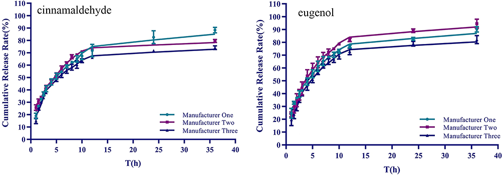 |
Figure 7 The in vitro release curve of Goupi Gao from three manufacturers at 35°C. |
Pharmacodynamics Evaluation of Goupi Gao on the Rabbit Model of Knee Osteoarthritis with Cold Coagulation and Blood Stasis Syndrome
Preparation of Rabbit Model of Knee Osteoarthritis with Cold Coagulation and Blood Stasis Syndrome
The rabbit model of knee osteoarthritis with cold coagulation and blood stasis was made by drug-induced plus cold stimulation.11–13 The model preparation is divided into a pre-test period and a modeling period, each lasting 7 days. During the pre-test period, feed normally every day. During the modeling period, both knee joints of rabbits were depilated and sterilized with 75% ethanol, and the feeding amount was kept unchanged. On day 1, day 4, and day 7, the papain saline solution 0.5mL was injected into the lower part of a patellar bone, once on both sides. After injection, the rabbits were assisted to do flexion and extension of the knee joint of hind limbs, so that the inducer could be fully diffused. After the first injection, the rabbits’ hind limbs were placed in a 0~1°C ice-water mixture once a day, each time for 30 minutes, and the cold coagulation and blood stasis model was reproduced after repeated freezing for 7 days.
Animal Grouping Design
135 rabbits with successful modeling were randomly divided into 15 groups, the high-temperature group of 45 rabbits (There are 9 rabbits in each group of manufacturer 1, manufacturer 2, manufacturer 3, blank black plaster matrix, and model group), another 45 rabbits in the middle-temperature group and last 45 rabbits in the normal temperature group (the specific grouping was the same as above). High-temperature group and middle-temperature group refer to heating the plaster to 45°C and 35°C respectively before application and giving appropriate ambient temperature compensation during application. Another 9 rabbits without modeling were taken as the blank group. Rabbits in the experimental group were depilated around the knee joints and applied Goupi Gao (3×3 cm2) or blank black plaster matrix (approximately 1.25g). Each plaster was applied for 60 hours, and the second plaster was applied at an interval of 12 hours, continuously for 15 days. Rabbits in the blank group were fed daily.
Detection of Inflammatory Factors in Serum
Rabbits in the above groups were taken blood from the auricular vein on day 0, day 1, day 2, day 3, day 4, day 5, day 6, day 7, and day 15, respectively. The contents of IL-1β, IL-4, IL-6, and IL-17 in serum were detected respectively. Meanwhile, the contents of CS and TT3 in serum were detected on day 0, day 1, day 2, day 3, day 4, day 5, day 6, day 9, day 12, and day 15, respectively. The results are shown in Figure 8.
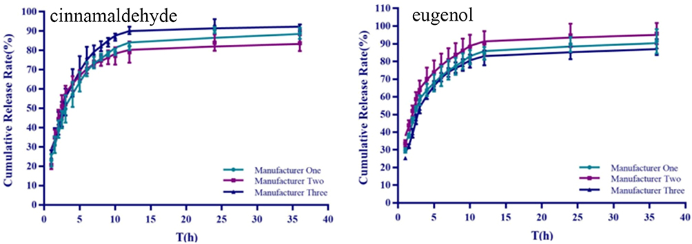 |
Figure 8 The in vitro release curve of Goupi Gao from three manufacturers at 45°C. |
The Imaging Evaluation of Knee Joint in Rabbits
The rabbits in the blank group were inspected by an X-ray machine (VERSA, CAREVET, Germany); Other groups were inspected by X-ray on the 3rd and 7th days after modeling and day 15 after continuous application. Take photos of both knee joints in the lateral position, and rabbits lie on their sides, close to the detector.
Pathomorphological Evaluation of Goupi Gao on Model Rabbits
Rabbits in each group were sacrificed by auricular vein air embolization. Shave the knee joint area and make a longitudinal incision along the middle of the knee joint. Open the joint cavity. The medial malleolus of the knee joint was completely peeled off, fixed, decalcified, dehydrated with graded ethanol, and made transparent with xylene. After that, paraffin-embedded sections were made, and hematoxylin-eosin (HE) staining was performed.14
Statistical Analyses
Each experiment was separately performed at least three times. The data were expressed as the mean±standard deviation, and statistical comparison between different groups was performed by the Dunnett’s C test. The p-value <0.05 indicated statistical significance. Statistical analysis was conducted using SPSS 23.
Results and Discussion
Determination of Plaster Pretreatment Method
According to Figure 1, on the 0th day, the surface of Goupi Gao was smooth and thickened vegetable oil. After being treated with ethyl acetate for 1 day, a small range of thickened oil corrosion plaques appeared on the surface of the plaster. After 2 days, the erosion effect on the plaster surface became obvious gradually, and the area of the plaque gradually expanded. After 3 days, as the ethyl acetate continued to corrode the plaster, the surface showed a “ridges” appearance. After 5 days, several fibrous structures were faintly visible on the plaster surface. After 7 days, the “branches”-like soap crystal fiber structure interwoven with each other.
Therefore, it is speculated that to characterize the microstructure of black plaster, the sample should be treated with ethyl acetate for about 7 days. Due to the different thickening degrees of vegetable oil, the time required for ethyl acetate treatment may be different among samples prepared from different vegetable oils.
The Morphology Analysis of Plaster Microstructure
The image with high magnification can scan a single particle and collect local morphological information, as well as plaster borders and particle contact states. However, the purpose of this paper is to observe the overall appearance of plaster, so the images with medium and low magnification are used for quantitative analysis. Figure 2 shows the FESEM image of the plaster at 200 times magnification. The distance of one pixel in the figure represents 0.6667 μm. (Figures 2A–D illustrate the demonstration process of digital image quantization processing using Figure 3A as an example. The actual color change should occur. Note: The original image in Figure A is black and white.)
Analysis of Microstructure Pore Parameters
The pore parameters of the plaster after heat treatment at 28°C, 35°C, and 45°C were examined, including pore area (A), pore length (L), minor diameter (W), aspect ratio (RR), and pore fractal dimension (D) are shown in Figures 3–5 and Table 1.
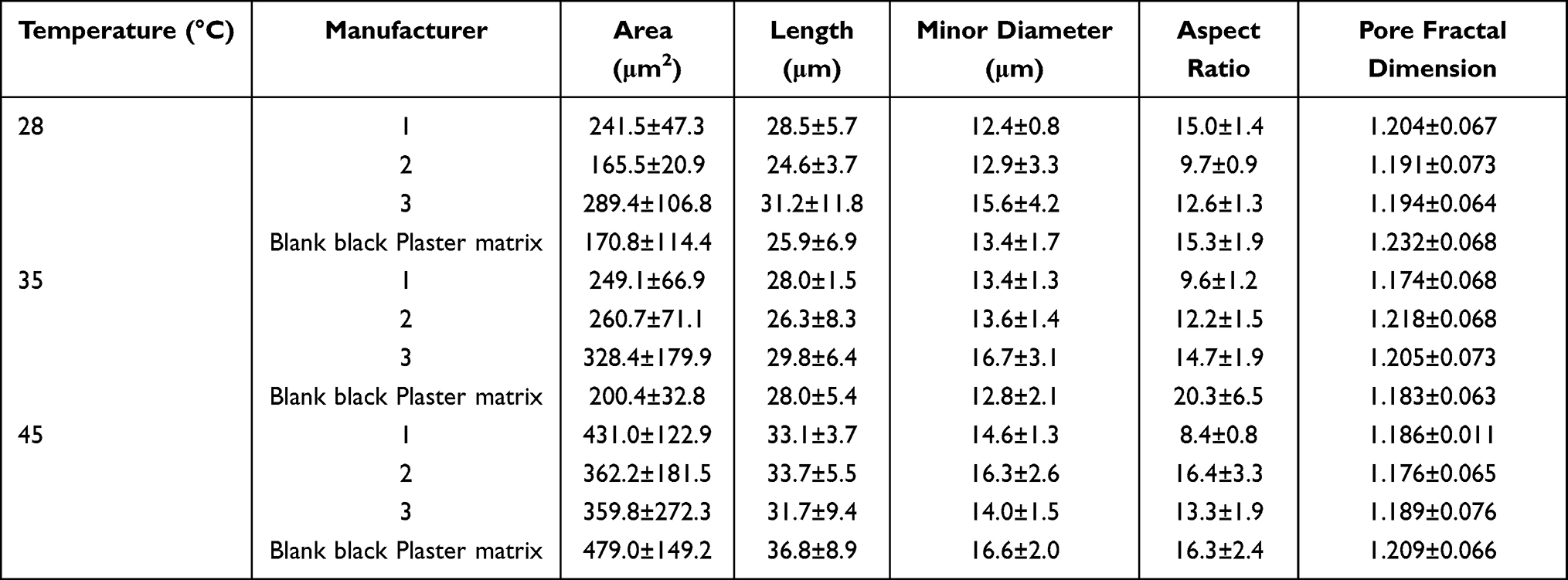 |
Table 1 Microstructure Parameters of Each Group of Samples After Heat Treatment at Different Temperatures (n=5) |
According to the values of the microstructure parameters of the samples under the thermal action of 28 °C, 35 °C, and 45 °C, the unitary linear regression method was used to determine the thermal effect [temperature (independent variable)] and A, L, W, RR, D (dependent variables) are used for regression analysis, the correlation coefficient of the regression equation is calculated, and the correlation between thermal action temperature and plaster microstructure parameters is established. The results are shown in Table 2.
 |
Table 2 Univariate Correlation Between Microstructure Parameters and Temperature of Different Samples |
The corresponding point is 3 between the microstructure parameters of each sample and the temperature, and the degree of statistical freedom is df=n-2=1. Under this degree of freedom, the critical correlation coefficient R is 0.987 (P=0.10). When |R|<R threshold (P=0.10), it shows that the calculated R value does not have statistical significance. The R value with statistical significance, when |R|≥ 0.8, can be regarded as a high degree of correlation between the two variables; when 0.5≤ |R|< 0.8, it can be regarded as a moderate correlation. From Table 2, it can be seen that the pore area (A) of Manufacturer 1 and Manufacturer 2 is highly positively correlated with temperature, that is, the pore space of the plaster increases significantly with the increase of heat application temperature. The other parameters have no statistical significance because the degree of freedom df is relatively small.
Cumulative Release Results
The cumulative release (Q) of three batches of Goupi Gao from three manufacturers (manufacturer 1, 20170703, 20180113, 20181004; manufacturer2, 170906, 180105, 180304; manufacturer 3, 20180204, 20180602, 20181003) was investigated, to evaluate the in vitro release characteristics between different manufacturers and batches. The results are shown in Figures 6–8.
Based on the Origin Pro 9.1 software, the release data of cinnamaldehyde and eugenol in the Goupi Gao mentioned above at 28 °C, 35 °C, 45 °C is performed on the Weibull model and the parameters are calculated. The results are shown in Table 3. Drugs exist on the surface and inside of the matrix. The drugs on the surface are released first when the black plaster is applied to the skin, and the drugs inside the matrix are released after the plaster is softened by heat.
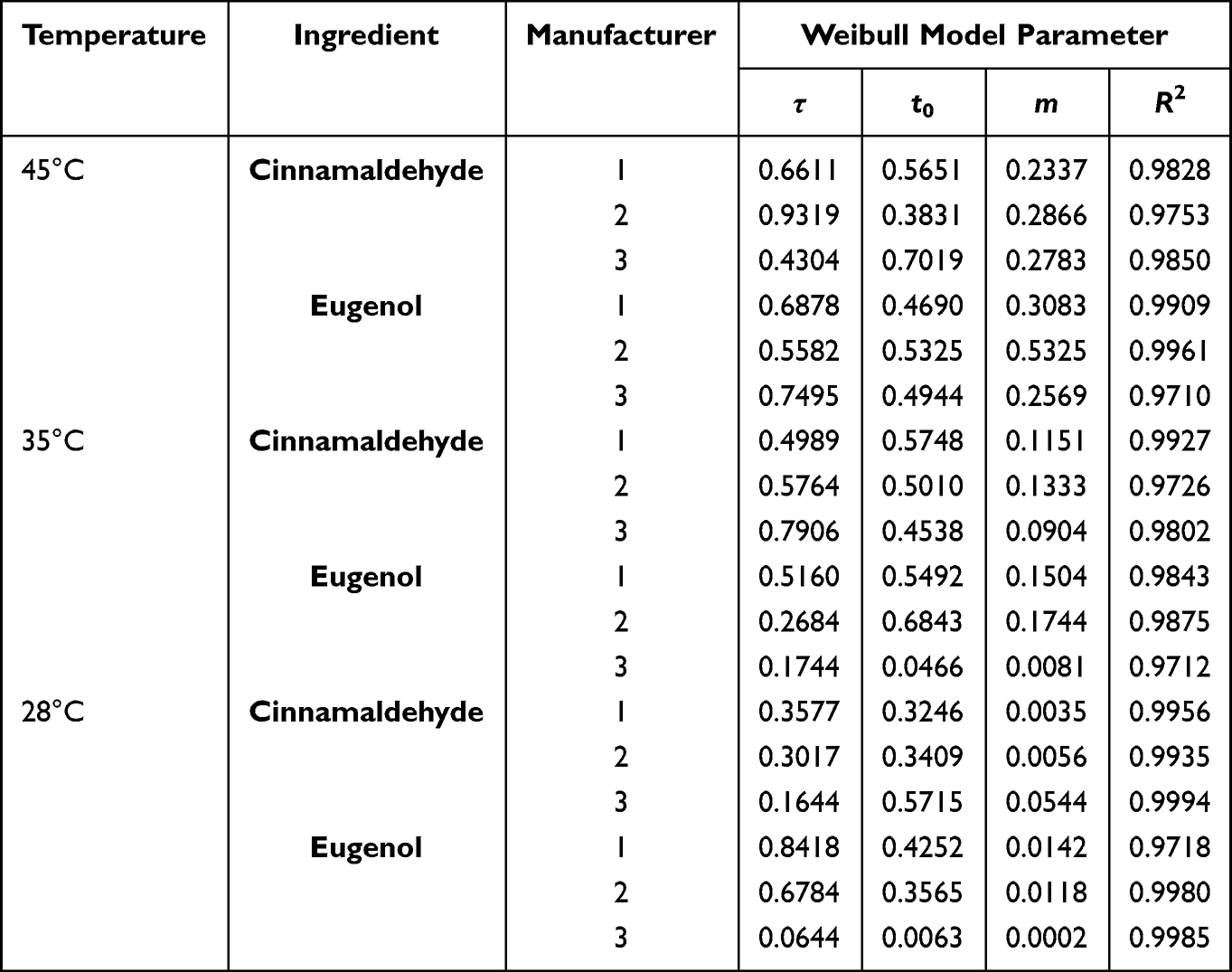 |
Table 3 Characteristic Parameters of in vitro Release of Cinnamaldehyde and Eugenol in Goupi Gao at Different Temperatures |
Inflammatory Factors in Serum
The relevant serum index data (day 0 to day 15) of each group has been subjected to statistical examination and analysis. Dunnett’s C test was used to analyze the variance of Student range pairing. Under the high temperature, medium temperature, and normal temperature conditions, both the administration group and the model group have significant differences; there is a significant difference between the Goupi Gao from the three manufacturers and the blank black plaster matrix group; there is no significant difference between the Goupi Gao from three manufacturers.
Under the conditions of high temperature, medium temperature, and normal temperature, the analysis result of IL-17 and CS content in serum is: ① there are significant differences between the administration group and the model group; ② there are significant differences between the Goupi Gao from three manufacturers and the blank plaster matrix group; ③ there are significant differences between manufacturer 1, manufacturer 2, and manufacturer 3. The changing trend of each index content in serum is shown in Figure 9.
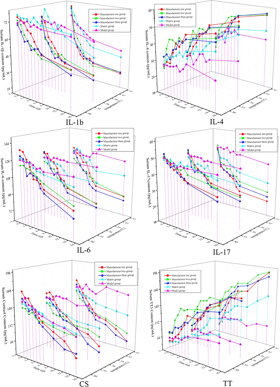 |
Figure 9 Summary chart of the changing trend of each index content in serum. |
Imaging Evaluation of Knee Joint
From the results in Figure 10, compared with the blank group, the model group (modeled for 3 days) had a narrower gap between the knee joints of the rabbits, the epiphyseal line was inclined forward and closed, and the joint surface was not smooth, and partly hardened. In the model group (7 days after modeling), the knee joint surface of rabbits was rough and deformed, accompanied by bone hardening, the joint density increased, the calcification was obvious, and the fluid accumulation in the joint cavity increased. The rabbits in the Goupi Gao normal temperature group had a narrower knee joint space, an anterior epiphyseal line, aberrant tissue proliferation in the joint cavity, and minor improvement in joint calcification as compared to the model. The space between the knee joints was dramatically altered in the high-temperature group, the joint surface tended to be smooth, and fluid collection in the joint cavity was reduced. There was a considerable improvement when compared to the model group. The symptoms of rabbits’ knee joints improved in the middle-temperature group, which was halfway between the normal and high-temperature groups.
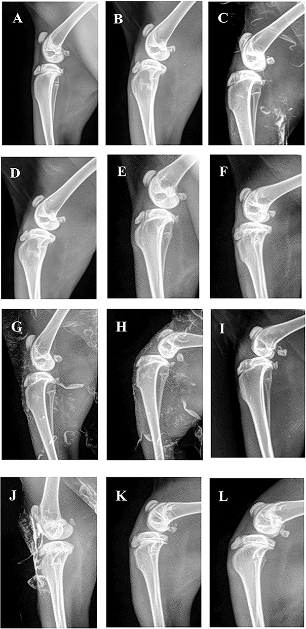 |
Figure 10 The effect of Goupi Gao from three manufacturers on the imaging of rabbit knee joints. (A) Blank group, (B) model group (3 days for modeling), (C) model group (7 days for modeling), (D–F) normal temperature group (Manufacturer 1, Manufacturer 2, Manufacturer 3), (G–I) medium temperature group (grouping as above), (J–L) high-temperature group (grouping as above). |
Pathological Evaluation
The rabbit cells in the blank group were evenly arranged, the cartilage edges were smooth, and there was no hyperemia or edema. After 3 days of modeling, articular cartilage showed different degrees of inflammation abnormalities, with inflammatory cell infiltration and synovial tissue hyperplasia and thickening. After 7 days of modeling, the internal environment of the muscle and tendon cells was out of balance, the cells were swollen and dissolved, the synovial tissues were congested, and edema, capillary hyperplasia, and other pathological states in the later stage of inflammation appeared.
Compared with the model group, after 15 days of application in the normal temperature group of Goupi Gao from three manufacturers, the synovial tissue still showed different degrees of a pathological state, with no obvious improvement in synovial thickening and inflammatory cell infiltration, and muscle-tendon tissue fusion and necrosis. In the middle-temperature group, the inflammation of tissue was improved to some extent, and the number of inflammatory cells decreased, but the connective tissue was still obvious. In the high-temperature group, the cartilage cell proliferation was active, the inflammatory changes were significantly reduced, the proliferation of synovial cells was not obvious, a few inflammatory cells infiltrated, and there was no obvious congestion and edema.
All of the above showed that the Goupi Gao from three manufacturers could significantly improve the inflammatory symptoms and physiological state of knee tissues when the plaster temperature was high. The results are shown in Figure 11.
 |
Figure 11 Goupi Gao on the pathological section of rabbit knee joint. (A) Blank group; (B) model group (3 days for modeling); (C) model group (7 days for modeling); (D–F) normal temperature group (Manufacturer 1, Manufacturer 2, Manufacturer 3); (G–I) medium temperature group (grouping as above); (J–L) high-temperature group (grouping as above). |
Discussion
The microstructure of the paste after washing oil treatment is a three-dimensional network skeleton structure where the soap crystal fibers are highly entangled, so the black plaster is a thermodynamically incompatible heterogeneous blend formed by a polymer/polymer mixture. Polymer system. One phase is the lead-based soap crystal fiber phase, and the other is the thickened vegetable oil phase. The soap crystal fiber phase penetrates through the matrix of the thickened plant oil phase in a network, showing a double continuous phase state.
The motion of the chain segment in the black plaster is highly time-dependent and temperature-dependent. At low temperatures, the thickened vegetable oil phase is in a glassy state. Due to the strong van der Waals effect between the chain segments, it can act as a physical cross-linking point and cross-link with the soap crystal fibers. At this time, the system moves very slowly; When the glass transition temperature of the thickened vegetable oil phase is exceeded, the physical cross-linking point fails and has fluidity, so it exhibits thermoplastic elastomer properties. Therefore, at the working temperature, the soap crystal fiber phase has a toughening effect, maintaining the rigid structure of the structure, and the thickened plant oil phase can increase the impact resistance of the paste.
After thousands of years of inheritance, the black plaster still endures for a long time in the development wave of the contemporary medical era. With today’s rapid advancements in science and technology, however, understanding of the black plaster matrix is limited to the surface of the conventional fat-soluble matrix, and the microstructure and structure characteristics are unknown. This is due to the fact that the black plaster is in a dark semi-solid condition, and many of the fine structures are obscured by the thickened plant oil, making it difficult for researchers to see them and limiting their capacity to delve deep into the black plaster’s body. The problem group found a suitable pre-paste treatment method through many explorations and repeated research, discovered the special microstructure in the black plaster, and used the digital image to quantify the parameters to solve the problem.
Conclusions
The microstructure of black plaster after being treated with ethyl acetate is a three-dimensional network skeleton structure, in which the soap crystal fibers are highly entangled. The black plaster is a thermodynamically incompatible heterogeneous blend formed by a polymer/polymer mixture. One phase is the lead-based soap crystal fiber phase, and the other is the thickened vegetable oil phase. The soap crystal fiber phase penetrates through the matrix of the thickened vegetable oil phase in a network, showing a double continuous phase state.
The motion of the chain segment in the black plaster is highly time-dependent and temperature-dependent. At low temperatures, the thickened vegetable oil phase is in a glassy state. Due to the strong van der Waals effect between the chain segments, it can act as a physical cross-linking point and cross-link with the soap crystal fibers. At this time, the system moves very slowly; When the glass transition temperature of the thickened vegetable oil phase is exceeded, the physical cross-linking point fails and has fluidity, so it exhibits thermoplastic elastomer properties. Therefore, at the working temperature, the soap crystal fiber phase has a toughening effect, keeping the rigidity of the structure, and the thickened vegetable oil phase can increase the impact resistance of the plaster.
After thousands of years of inheritance, the black plaster still endures for a long time in the development wave of the contemporary medical era. With today’s rapid advancements in science and technology, however, understanding of the black plaster matrix is limited to the surface of the conventional fat-soluble matrix, and the microstructure and structure characteristics are unknown. This is because the black plaster is in a dark semi-solid condition, and many of the fine structures are obscured by the thickened vegetable oil, making it difficult for researchers to see them and limiting their capacity to delve deep into the black plaster’s body. The problem group found a suitable pretreatment method through many explorations and repeated research, discovered the special microstructure in the black plaster, and used the digital image to quantify the parameters to solve the problem. Based on FESEM-IPP technology, the microstructure pore parameters can be obtained simply and intuitively, which is beneficial to further study the correlation with other experimental data and explore their internal relations.
Abbreviations
The following abbreviations are used in this manuscript: FESEM, field emission scanning electron microscope; IPP, Image-Pro-Plus; ELISA, Enzyme-linked immunosorbent assay; HE, hematoxylin-eosin.
Author Contributions
All authors made a significant contribution to the work reported, whether that is in the conception, study design, execution, acquisition of data, analysis and interpretation, or in all these areas; took part in drafting, revising or critically reviewing the article; gave final approval of the version to be published; have agreed on the journal to which the article has been submitted; and agree to be accountable for all aspects of the work.
Funding
This work was supported by the National Natural Science Foundation of China (NSFC 82074025), Heilongjiang Touyan Innovation Team Program, Tianjin Research Program of Application Foundation, and Advanced Technology (17JCYBJC41800, 19JCTPJC56900), Tianjin Municipal Bureau of Labor and Social Security (2018018), the Jiamusi University Ph.D. Special Foundation (JMSUBZ2020-14).
Disclosure
The authors report no conflicts of interest in this work.
References
1. Guiqin Z. The Pharmacodynamics and Clinical Observation Research of Traditional External Preparation of Goupi Ointment [Dissertation]. Chengdu University of Traditional Chinese Medicine; 2012.
2. National Pharmacopoeia Committee. Pharmacopoeia of the People’s Republic of China. Beijing: Chemical Industry Press; 2015.
3. Jia X, Yuliang C, Jianhua Q, et al. Theoretical connotation and clinical application of black plaster sticks. J Trad Chin Med. 2021;36:3695–3697.
4. Nan Z, Huan C, Xueyan B, et al. Research progress on traditional Chinese medicine formulations - black plaster. Chin Pharm Std. 2018;19(06):433 436.
5. Chen A, Wang S, Liu H, et al. Recent development of traditional black plaster. J Chin Patent Med. 2014;36:379–382.
6. Padding JT, Mohite LV, Auhl D, et al. Quantitative mesoscale modeling of the oscillatory and transient shear rheology and the extensional rheology of pressure sensitive adhesives. Journal of Soft Matter. 2012;8:7967–7981. doi:10.1039/c2sm07443e
7. Singh I, S A, Park BD. Drug-delivery applications of cellulose nanofibrils. J Nanoarchitectonics Smart Deliver Drug Target. 2016;2016:95–117.
8. Shahidian A, Afshar H, Habibi MR, et al. Therapeutic nanostructures: application of mechanical engineering in drug delivery. J Nanoarchitectonics Smart Deliver Drug Target. 2016;2016:3–34.
9. Zhou Y. Research on the Processing and Quantification Method of the Digital Image of Loess Microstructure [Dissertation] Xi’an University of Science and Technology; 2013.
10. Wang F, Deng N, Fengqing M, et al. Research on 3D fractal of loess large pore morphology based on CT. J Comput Eng. 2014;40:217–220.
11. Chen Z, Lin H, Zhiyi Y, et al. Progress in research on an animal model of knee osteoarthritis. J Arthritis Rheumatol. 2016;5:67–70.
12. Maly R, M AS, Totterman S, et al. Knee adduction moment relates to medial femoral and tibial cartilage morphology in clinical knee osteoarthritis. J Biomech. 2015;48:3495–3501. doi:10.1016/j.jbiomech.2015.04.039
13. Chen D, Peng L, Liao Z, et al. Comparison of papaya protease and stone cream on the establishment of a rabbit knee osteoarthritis model. J Guangdong Med Sci. 2017;38:2114–2118.
14. Aigner T, L CJ, Gerwin N, et al. Histopathology atlas of animal model systems–overview of guiding principles. J Osteoar Cartil. 2010;18(Suppl –S3):1.
 © 2023 The Author(s). This work is published and licensed by Dove Medical Press Limited. The
full terms of this license are available at https://www.dovepress.com/terms
and incorporate the Creative Commons Attribution
- Non Commercial (unported, 3.0) License.
By accessing the work you hereby accept the Terms. Non-commercial uses of the work are permitted
without any further permission from Dove Medical Press Limited, provided the work is properly
attributed. For permission for commercial use of this work, please see paragraphs 4.2 and 5 of our Terms.
© 2023 The Author(s). This work is published and licensed by Dove Medical Press Limited. The
full terms of this license are available at https://www.dovepress.com/terms
and incorporate the Creative Commons Attribution
- Non Commercial (unported, 3.0) License.
By accessing the work you hereby accept the Terms. Non-commercial uses of the work are permitted
without any further permission from Dove Medical Press Limited, provided the work is properly
attributed. For permission for commercial use of this work, please see paragraphs 4.2 and 5 of our Terms.